ICU · cardiovascular
Post-Cardiac Surgery ICU Management — Comprehensive
Also known as Post-cardiac surgery ICU · Post-CABG ICU · Post-valve surgery ICU · Post-cardiopulmonary bypass · Post-CPB management · Cardiac surgical ICU · Post-sternotomy care · Vasoplegic syndrome post-CPB · Low cardiac output syndrome
Post-cardiac surgery ICU management — the systematic care of patients after cardiopulmonary bypass (CPB) surgery (CABG, valve replacement/repair, aortic surgery, congenital repair). The early postoperative period (first 24h) focuses on: (1) haemodynamic stabilisation (vasopressors/inotropes for vasoplegic syndrome [NO-mediated] and low cardiac output syndrome), (2) bleeding and tamponade assessment (chest tube output 200 mL/hr or sudden cessation = tamponade until proven otherwise), (3) rhythm management (new AF in 30% — rate control with amiodarone/beta-blocker, anticoagulate if 48h), (4) ventilatory support and weaning (fast-track: extubate within 4-6h if stable), (5) temperature and metabolic management (rewarm from CPB hypothermia, correct K+/Mg2+/glucose), (6) analgesia (opioid-sparing multimodal). Complications: post-CPB vasoplegic syndrome (30% — refractory hypotension despite vasopressors — methylene blue 1-2 mg/kg IV or vasopressin), post-CPB low cardiac output syndrome (10-20% — from myocardial stunning/ischaemia — IABP/Impella/VA-ECMO), cardiac tamponade (2-5% — atypical presentation post-sternotomy — clinical: equalisation of pressures on PA catheter, echo: organised clot behind RA/RV), postoperative bleeding (5-10% re-exploration rate — chest tube output 200 mL/hr x 3h or 400 mL in 1h), sternal wound infection (1-2% deep — mediastinitis — requires surgical debridement + IV antibiotics), postoperative stroke (2-5% — from atherosclerotic emboli during cannulation/clamping), postoperative AKI (5-10% — from CPB haemolysis + nephrotoxins + low flow). Mortality: 1-3% (elective CABG), 5-15% (emergency/complex).
On this page & tools
Your progress
Saved locally on this device.
Target exams
Red flags
Overview
The cardiac surgery patient is UNIQUE in ICU: they have undergone controlled global ischaemia (cardioplegic arrest on CPB), massive fluid shifts (CPB prime → haemodilution), systemic inflammatory response (CPB activates complement + cytokines), coagulopathy (CPB haemolysis + platelet consumption + heparin), and myocardial stunning (ischaemia-reperfusion). The first 12-24 hours are the most critical — haemodynamic instability, bleeding, and tamponade are the life-threatening complications that demand rapid recognition.[1][2]
The first 6 hours — the critical window
Post-cardiac surgery ICU admission protocol — first 6 hours
-
HANDOVER (from anaesthetist + surgeon + perfusionist): (a) Procedure performed (CABG x N grafts, valve type, aortic cross-clamp time, CPB time). (b) Intraoperative course (difficulty weaning from CPB, arrhythmia, bleeding). (c) Current medications (vasopressors/inotropes, infusions, insulin, antibiotics). (d) Lines and drains (central line, arterial line, PA catheter [if placed], chest tubes [mediastinal + pleural], temporary pacing wires, urinary catheter). (e) ECG, latest ABG, electrolytes, coagulation, haemoglobin [1]
-
INITIAL ASSESSMENT:
- Vital signs: HR, BP (arterial line), SpO2, CVP, temperature, urine output
- ECG: baseline rhythm, ischaemia (ST elevation = graft occlusion)
- Chest X-ray: tube/line position, lung fields, heart size, mediastinal width (widening = bleeding/tamponade)
- PA catheter (if present): cardiac output, SVR, PCWP, mixed venous saturation (SvO2 >65% = adequate perfusion)
- Transthoracic echo (if feasible): LV/RV function, valve function, pericardial effusion [1]
-
HAEMODYNAMIC STABILISATION:
- Target MAP >65 mmHg (some centres target >70 for graft patency)
- Noradrenaline first-line for hypotension (vasoplegia from CPB — most common cause of early hypotension)
- Add vasopressin (0.02-0.04 U/min) if noradrenaline >0.3 mcg/kg/min
- Inotrope (dobutamine 2.5-10 mcg/kg/min or milrinone 0.125-0.5 mcg/kg/min) if cardiac output low (CI <2.2 L/min/m^2) — low cardiac output syndrome
- Methylene blue (1-2 mg/kg IV over 20 min) for refractory vasoplegia (noradrenaline >0.5 mcg/kg/min + vasopressin + normal/high CI + low SVR <800 dynes/s/cm^5)
- IABP if cardiogenic shock (augments coronary perfusion + reduces afterload)
- VA-ECMO for refractory low cardiac output syndrome (bridge to recovery) [1]
-
BLEEDING ASSESSMENT:
- Chest tube output: (a) <100 mL/hr = normal. (b) 100-200 mL/hr = monitor. (c) >200 mL/hr x 3h = concerning. (d) >400 mL in 1h = RE-EXPLORE. (e) SUDDEN STOP in drainage + haemodynamic compromise = TAMPONADE (clot blocking tubes)
- Coagulation: ACT (heparin reversal with protamine — check residual heparin), INR, APTT, fibrinogen, platelets
- Bloods: haemoglobin (target >70-80 g/L in cardiac surgery — higher if active bleeding or ischaemia)
- TEG/ROTEM if available (guides blood product administration — fibrinogen vs platelets vs cryoprecipitate vs FFP)
- Blood product administration: FFP (INR >1.5), cryoprecipitate (fibrinogen <1.5 g/L), platelets (platelets <50) [1]
-
VENTILATION:
- Initial settings: assist-control, Vt 6-8 mL/kg IBW, PEEP 5, FiO2 60% (titrate down), RR 12-14
- Target: SpO2 >95%, PaCO2 35-40 mmHg
- Fast-track weaning: when patient is warm (T >36C), haemodynamically stable, minimal bleeding (<100 mL/hr), awake (GCS >13, following commands), adequate gas exchange (PaO2/FiO2 >200) → extubate within 4-6h
- If unstable (vasopressors escalating, active bleeding, hypothermia, acidosis) → DELAY extubation [1]
-
METABOLIC CORRECTION:
- Rewarm: CPB hypothermia + cold operating room + cold IV fluids → patient arrives at T 34-36C. Use forced-air warmer (Bair Hugger), warmed IV fluids, ambient temperature. Target T >36C (hypothermia worsens coagulopathy + arrhythmia + vasoconstriction)
- Potassium: CPB causes K+ shifts (cardioplegia is high-K+ → initial hyperkalaemia → then K+ falls from diuresis). Target K+ 4.0-4.5 mmol/L (replace: 10-20 mmol KCl via central line)
- Magnesium: depleted from CPB (diuresis + haemodilution). Replace MgSO4 10 mmol IV (also reduces AF risk). Target Mg2+ >0.8 mmol/L
- Glucose: target 8-10 mmol/L (insulin infusion if >10 — tight control has NOT been shown to improve outcomes and increases hypoglycaemia in cardiac surgery)
- Acid-base: metabolic acidosis from CPB (lactate accumulation + renal impairment) → correct with perfusion (not bicarbonate unless pH <7.1)
- Haemoglobin: target >70-80 g/L (>80 if active ischaemia or low cardiac output) [1]
-
ANALGESIA + SEDATION:
- Propofol infusion 10-50 mcg/kg/min (short-acting — allows rapid wake-up for neuro assessment and fast-track extubation)
- Opioid: fentanyl infusion 25-50 mcg/hr or morphine PRN (avoid excessive opioid — delays extubation)
- Paracetamol 1 g IV q6h (baseline analgesia)
- Avoid NSAIDs (first 48h — bleeding risk + AKI risk)
- Regional (paravertebral/serratus plane) — opioid-sparing (some centres) [1]
-
MONITORING FOR COMPLICATIONS:
- Continuous ECG (arrhythmia — AF, VT, heart block)
- Hourly: chest tube output, urine output, haemodynamics, temperature
- 4-hourly: ABG, electrolytes (K+, Mg2+, glucose), haemoglobin, coagulation
- Daily: CXR, troponin (trend — rising troponin = graft occlusion/myocardial ischaemia), creatinine
Post-CPB vasoplegic syndrome — the #1 early challenge
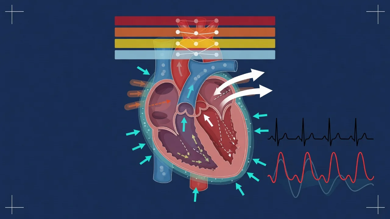
Vasoplegic syndrome is the most common haemodynamic problem in the first 24h post-CPB. It occurs in 8-42% of patients (variable definition) and is associated with increased mortality (15-25% vs 3% without vasoplegia). The mechanism: CPB triggers a systemic inflammatory response → massive NO and cytokine release → loss of vascular tone → distributive shock (resembling septic shock physiology).[1][6]
Post-CPB vasoplegic syndrome vs low cardiac output syndrome — the two post-CPB shock patterns
| Feature | Vasoplegic syndrome (30%) | Low cardiac output syndrome (10-20%) |
|---|---|---|
| Pathophysiology | Distributive shock (NO-mediated vasoplegia) | Cardiogenic shock (myocardial stunning/ischaemia) |
| Cardiac output | NORMAL or HIGH (compensatory) | LOW (CI <2.2 L/min/m^2) |
| SVR | LOW (<800 dynes/s/cm^5) | HIGH (compensatory vasoconstriction) |
| PCWP | Normal or low | Elevated (>18 mmHg) |
| SvO2 | Normal or high | LOW (<65%) |
| Extremities | WARM (vasodilated — "warm shock") | COLD (vasoconstricted — "cold shock") |
| First-line treatment | Noradrenaline (alpha-agonist — restores SVR) | Inotrope (dobutamine/milrinone — increases contractility) |
| Second-line | Vasopressin, methylene blue | IABP, Impella, VA-ECMO |
| Cause | CPB inflammatory response (complement, cytokines, NO) | Myocardial ischaemia (incomplete revascularisation, graft occlusion), prolonged CPB/cross-clamp time, pre-existing LV dysfunction |
Vasoplegic syndrome management — stepwise escalation
- NORADRENALINE — first-line (alpha-1 agonist — restores SVR): 0.05-0.5+ mcg/kg/min. Titrate to MAP >65
- VASOPRESSIN — add if noradrenaline >0.3 mcg/kg/min: 0.02-0.04 U/min (V1 receptor agonist — independent of alpha pathway — restores tone in NO-desensitised vessels)
- METHYLENE BLUE — for refractory vasoplegia (noradrenaline + vasopressin insufficient): 1-2 mg/kg IV over 20 minutes. Mechanism: inhibits soluble guanylate cyclase → blocks NO-cGMP pathway → restores vascular tone. Onset: 30-60 min. Duration: 2-72h. Caution: can cause serotonin syndrome if on SSRIs (methylene blue is a weak MAOI). Can interfere with pulse oximetry (blue dye → falsely low SpO2 for 1-2 min)
- HYDROCORTISONE — 200 mg/day (stress-dose steroids — relative adrenal insufficiency from CPB + inflammation). Controversial — some evidence for benefit in vasoplegia
- ASCRUBILATE/ASCORBIC ACID — vitamin C 1.5 g IV (antioxidant — scavenges reactive oxygen species from CPB). Controversial — some centres use high-dose vitamin C + B + thiamine (HAT therapy) for vasoplegia
- CORRECT ACIDOSIS — if pH <7.1, correct with ventilation optimisation ± bicarbonate (acidosis causes catecholamine resistance — vasopressors don't work in severe acidosis)
- MECHANICAL SUPPORT — if cardiac output also low (mixed shock): IABP or Impella (afterload reduction + coronary perfusion augmentation)
Bleeding and tamponade — the time-critical emergencies
Post-cardiac surgery bleeding — assessment and action thresholds
| Chest tube output | Interpretation | Action |
|---|---|---|
| <50 mL/hr | Normal — drying up | Continue monitoring |
| 50-100 mL/hr | Mild — within normal range | Monitor, check coagulation |
| 100-200 mL/hr | Moderate — monitor closely | Check coagulation (INR, APTT, fibrinogen, platelets, ACT). Correct abnormalities (FFP, cryoprecipitate, platelets, protamine). TEG/ROTEM if available |
| >200 mL/hr x 3 consecutive hours | Significant bleeding | Prepare for possible re-exploration. Correct coagulopathy aggressively. Cross-match blood. Notify surgeon |
| >400 mL/hr in 1 hour | MASSIVE bleeding | URGENT re-exploration. Massive transfusion protocol. Notify surgeon + anaesthetist immediately |
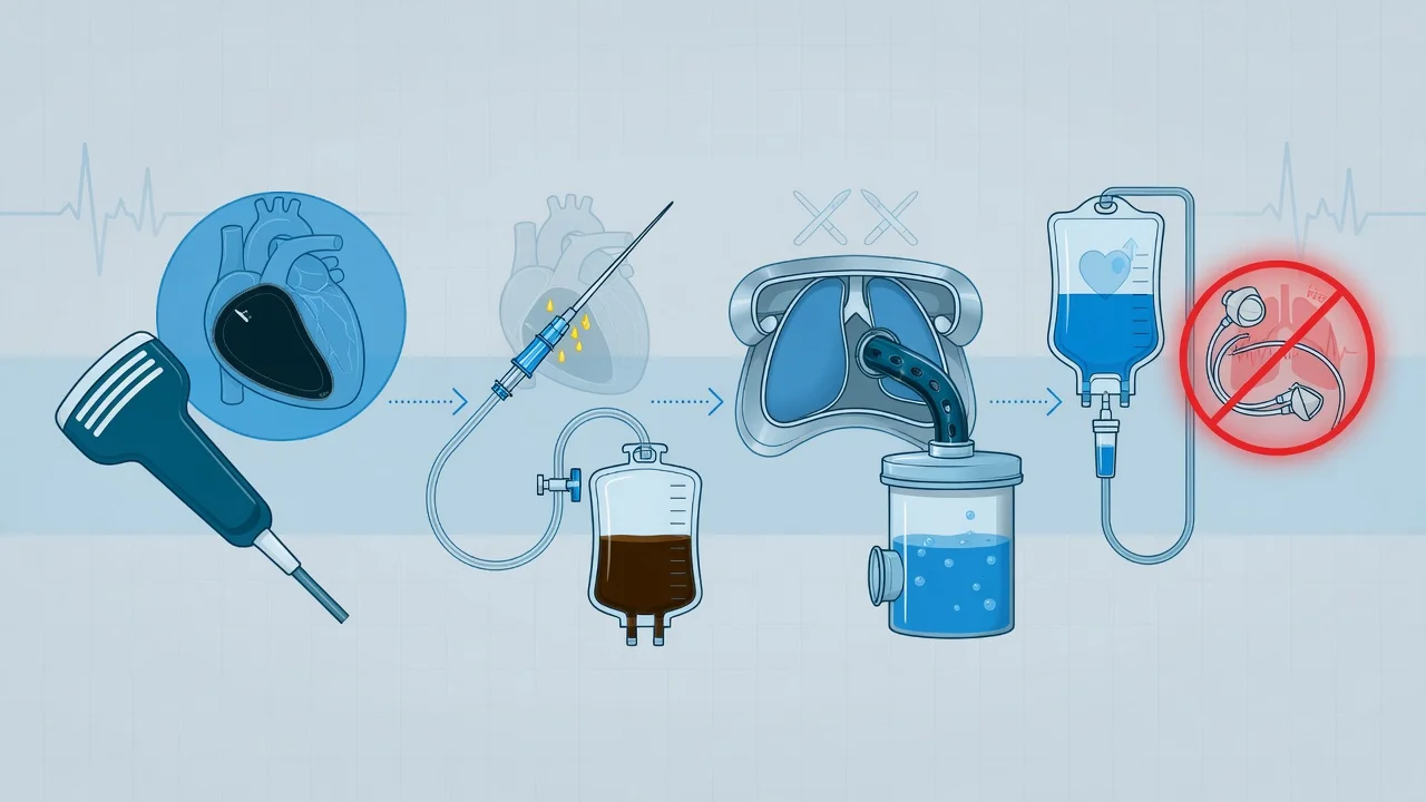
| SUDDEN STOP in drainage | TAMPONADE (organised clot blocking chest tubes) — DANGEROUS — tubes not draining does NOT mean bleeding stopped — blood is accumulating in the mediastinum → compresses the heart | Look for: hypotension, tachycardia, elevated CVP/PCWP (equalisation: RA = PA diast = PCWP within 5 mmHg), pulsus paradoxus, muffled heart sounds. Echo: organised clot posterior to RA/RV. URGENT RE-EXPLORATION |
Post-cardiac surgery arrhythmia — AF is the #1 rhythm problem
Postoperative atrial fibrillation (POAF) occurs in 30% of cardiac surgery patients (highest after CABG + valve combination, lowest after isolated CABG). Peak incidence: day 2-3 postoperatively. Risk factors: age >65, pre-existing AF, valve surgery, prolonged CPB, electrolyte derangement (low K+/Mg2+), beta-blocker withdrawal, pericarditis.[3]
Management:
- Rate control (first priority — if haemodynamically stable): (a) Metoprolol 5 mg IV (if not in heart failure). (b) Amiodarone 300 mg IV over 1 hour, then 900 mg over 24 hours (also helps with rhythm control — preferred in cardiac surgery). (c) Digoxin (if heart failure — less effective for rate control)
- Rhythm control (if haemodynamically unstable — hypotension, ischaemia): (a) Synchronised cardioversion 200J biphasic (if AF >48h, need TOE to exclude LA thrombus — but in acute instability, cardiovert immediately). (b) Amiodarone for pharmacological cardioversion
- Anticoagulation (if AF persists >48h): (a) Heparin infusion (balance with bleeding risk — check chest tube output first). (b) Warfarin for 4-6 weeks (if AF resolves — most POAF resolves by 6 weeks). (c) NOAC (apixaban, rivaroxaban) — increasingly used (no INR monitoring, less bleeding) but evidence in early post-cardiac surgery is limited
- Electrolyte correction (preventative + therapeutic): maintain K+ 4.0-4.5, Mg2+ >0.8 mmol/L (both arrhythmogenic if low)
- Prophylaxis (for all patients): (a) Beta-blocker (metoprolol 25-50 mg BD oral — restart preoperative beta-blocker as soon as possible — strongest evidence for POAF prevention). (b) Amiodarone prophylaxis (controversial — some centres give prophylactic amiodarone for high-risk patients — reduces POAF incidence by 50%). (c) Colchicine (anti-inflammatory — reduces post-pericardiotomy syndrome which triggers AF). (d) Statin (anti-inflammatory + pleiotropic effects) [1]
Other critical complications
Post-cardiac surgery complications — recognition and management
| Complication | Frequency | Recognition | Management |
|---|---|---|---|
| Stroke | 2-5% | New neurological deficit on waking (or delayed from emboli). CT brain. Distinguish from: delayed emergence (anaesthetic/sedative accumulation — should resolve within 4-6h), metabolic encephalopathy | Supportive. If ischaemic stroke <4.5h: thrombolysis controversial post-surgery (bleeding risk). Mechanical thrombectomy if large vessel occlusion. Neuroprotective: maintain MAP >80, SpO2 >95, glucose 8-10, temperature <37.5 |
| AKI | 5-10% (1-2% need RRT) | Rising creatinine, oliguria. Risk factors: pre-existing CKD, prolonged CPB, diabetes, age >70, postoperative bleeding, nephrotoxins (contrast for angiography) | Maintain renal perfusion (MAP >65, cardiac output optimised). Avoid nephrotoxins. CRRT if severe (oliguric + K+ rising + volume overloaded) |
| Sternal wound infection | 1-2% deep (mediastinitis) | Day 5-14: fever + sternal instability + purulent discharge + wound dehiscence. Risk: diabetes, obesity, bilateral IMA harvest, prolonged surgery, re-exploration | SURGICAL DEBRIDEMENT (open or VAC therapy). IV antibiotics (MRSA coverage — vancomycin + gram-negative). Wound VAC. Reconstruction (flap — pectoralis muscle). Mortality 10-20%[4] |
| Phrenic nerve injury | 1-10% (higher with topical cooling/ice slush) | Elevated hemidiaphragm on CXR + respiratory difficulty (especially after extubation — fails weaning). Diagnosed by fluoroscopy or ultrasound (paradoxical diaphragmatic movement) | Usually resolves spontaneously (weeks-months). Diaphragmatic plication if persistent (3-6 months) and impairing ventilation |
| Post-pericardiotomy syndrome | 10-20% | Day 1-12 weeks: fever + pleuritic chest pain + pericardial friction rub + pleural effusion + pericardial effusion on echo. Autoimmune response to pericardial injury | NSAIDs (ibuprofen 600 mg TDS — once bleeding risk resolved). Colchicine 0.5 mg BD x 3 months. Prednisone if refractory. Pericardiocentesis if tamponade |
| Gastrointestinal complications | 1-3% (but high mortality when they occur) | GI bleed (from stress ulcer + anticoagulation), mesenteric ischaemia (from low flow), pancreatitis, cholecystitis, ileus | Maintain perfusion. PPI prophylaxis. Early enteral nutrition. Surgical consultation for mesenteric ischaemia/perforation |
| Delirium | 20-50% | Acute confusion, agitation, fluctuating mental status (especially in elderly, prolonged CPB, pre-existing cognitive impairment) | Haloperidol or quetiapine. Reorientation. Minimise sedatives. Early mobilisation. Family visits. Sleep-wake cycle preservation |
Exam practice — SAQs
SAQ — Low cardiac output syndrome six hours post-CABG
10 minutes · 10 marks
A 68-year-old man with impaired LV function (EF 35%) is six hours post three-vessel on-pump coronary artery bypass grafting. He required two inotropes to separate from cardiopulmonary bypass (cross-clamp time 92 minutes, CPB time 140 minutes). On arrival in ICU he is on noradrenaline 0.25 mcg/kg/min: MAP 58, HR 108 sinus, CVP 16 mmHg, cardiac index 1.9 L/min/m2 via PA catheter, SvO2 58%, cool peripheries, oliguria 0.3 mL/kg/h, lactate 3.2 mmol/L. A 12-lead ECG shows new lateral ST depression and bedside echo shows globally poor LV function with a new lateral wall motion abnormality.
SAQ — Tamponade on day 2 post AVR and CABG with sudden drain cessation
10 minutes · 10 marks
A 65-year-old woman is day 2 post aortic valve replacement and single-vessel coronary artery bypass grafting. Mediastinal drain output was steady at 80-100 mL/hr overnight but ABRUPTLY STOPPED two hours ago. She is now progressively more hypotensive: MAP 62 and falling despite escalating noradrenaline (0.2 mcg/kg/min), HR 124 sinus, CVP rising from 12 to 19 mmHg, cardiac index falling from 2.8 to 1.9 L/min/m2. The PA catheter shows equalisation of diastolic pressures (RA 19, PA diastolic 21, PCWP 22 mmHg). Pulsus paradoxus is 14 mmHg. Cool peripheries, oliguria 0.2 mL/kg/h. Bedside transthoracic echo shows poor windows with a small 1 cm loculated echogenic collection behind the right atrium and a plethoric IVC.
Clinical pearls
Red flags
Prognosis
Cardiac surgery outcomes — what determines survival
| Procedure | Mortality (elective) | Mortality (emergency) | Key prognostic factors |
|---|---|---|---|
| Isolated CABG | 1-2% | 5-8% | EuroSCORE, age, LV function, diabetes, renal function |
| CABG + valve | 3-5% | 8-15% | Complexity, CPB time, re-operation |
| Isolated AVR | 2-3% | 5-10% | Age, LV function, porcelain aorta |
| Isolated MV repair | 1-2% | 5-8% | LV function, pulmonary hypertension |
| Aortic surgery (dissection) | 10-20% | 20-30% | Type A vs B, malperfusion, CPB time |
| Redo surgery | 5-8% | 10-20% | Adhesions, graft patency, LV function |
| With vasoplegia | 15-25% | — | Vasoplegia doubles mortality |
| With AKI requiring RRT | 30-50% | — | AKI requiring RRT is a major mortality predictor |
Key trials and evidence
ROOBY trial — off-pump vs on-pump CABG (PMID 30080455)
Study design
Randomised controlled trial — VA medical centres — 2,203 patients
Population
Veterans requiring CABG (mostly male, higher comorbidity than general population)
Intervention
Off-pump (beating heart, no CPB) vs on-pump (standard CPB) CABG
Primary outcome
Composite of death, non-fatal MI, repeat revascularisation at 1 year: off-pump 9.9% vs on-pump 7.4% (off-pump WORSE, p=0.04)
Long-term (5 years)
Off-pump had HIGHER repeat revascularisation (16.2% vs 11.4%) and no mortality benefit
Key finding
Off-pump had WORSE graft patency and HIGHER incomplete revascularisation
Clinical bottom line
On-pump CABG remains the standard — off-pump has worse outcomes in most patients. Reserve off-pump for high-CPB-risk patients (severe aortic atherosclerosis, severe PVD)
Landoni 2019 — Vasopressors in post-cardiac surgery vasoplegia (PMID 31106420)
Source
Systematic review and meta-analysis — vasopressor choice in post-CPB vasoplegia
Key finding
Noradrenaline is first-line (alpha-agonist — restores SVR). Vasopressin is second-line (V1 receptor — independent pathway)
Key finding
Methylene blue (1-2 mg/kg IV) is effective for refractory vasoplegia — blocks NO-cGMP pathway — onset 30-60 min
Key finding
Hydrocortisone (200 mg/day) may benefit — relative adrenal insufficiency from CPB inflammation
Clinical bottom line
Escalation: noradrenaline → add vasopressin → methylene blue → hydrocortisone. Methylene blue is the rescue therapy for NO-mediated refractory vasoplegia
Examiner densification notes
[1] [1]References
- [1]Lomivorotov VV, et al. Colouterine fistula: A rare complication of colonic diverticulitis Rev Gastroenterol Mex (Engl Ed), 2019.PMID 30661710
- [2]Varghese R, et al. ERRATUM J Forensic Sci, 2017.PMID 29152801
- [3]Aranki SF, et al. Higher risk of preterm birth and low birth weight following oocyte donation: A systematic review and meta-analysis Eur J Obstet Gynecol Reprod Biol, 2017.PMID 28942045
- [4]Engelman R, et al. Amplification-free long-read sequencing of TCF4 expanded trinucleotide repeats in Fuchs Endothelial Corneal Dystrophy PLoS One, 2019.PMID 31276570
- [5]Shroyer AL, et al. Impact Of Medicaid Expansion On Coverage And Treatment Of Low-Income Adults With Substance Use Disorders Health Aff (Millwood), 2018.PMID 30080455
- [6]Landoni G, et al. Bone marrow mesenchymal stem cells modified by angiogenin-1 promotes tissue repair in mice with oxygen-induced retinopathy of prematurity by promoting retinal stem cell proliferation and differentiation J Cell Physiol, 2019.PMID 31106420