ICU · Cardiovascular / vascular surgery
Post-Vascular-Surgery ICU — AAA, Carotid & Peripheral Bypass
Also known as Post-vascular surgery · Post-AAA repair · EVAR · Endovascular aneurysm repair · Carotid endarterectomy · CEA · Peripheral bypass · Endoleak · Spinal cord ischaemia · Reperfusion syndrome · Abdominal compartment syndrome · Hyperperfusion syndrome
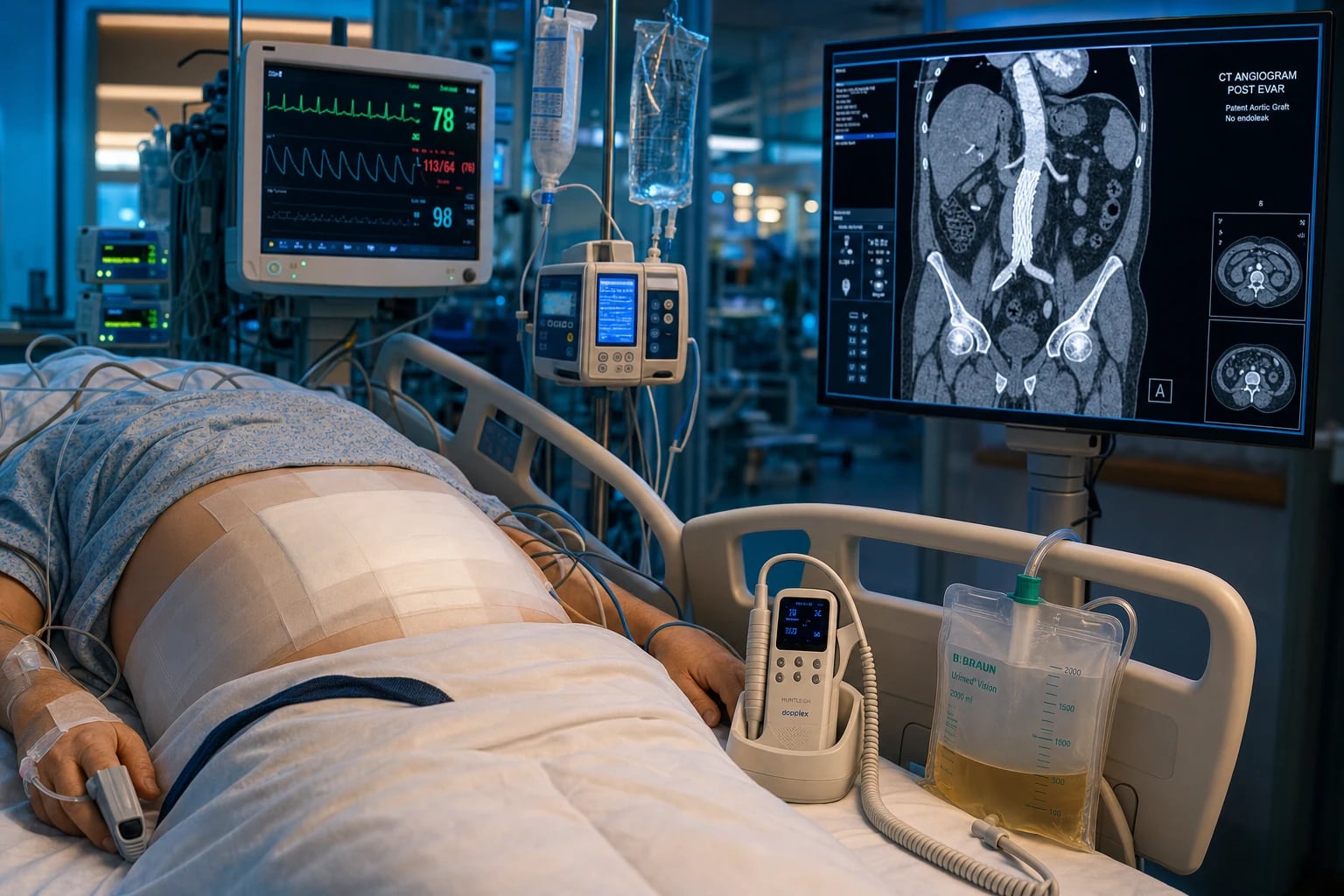
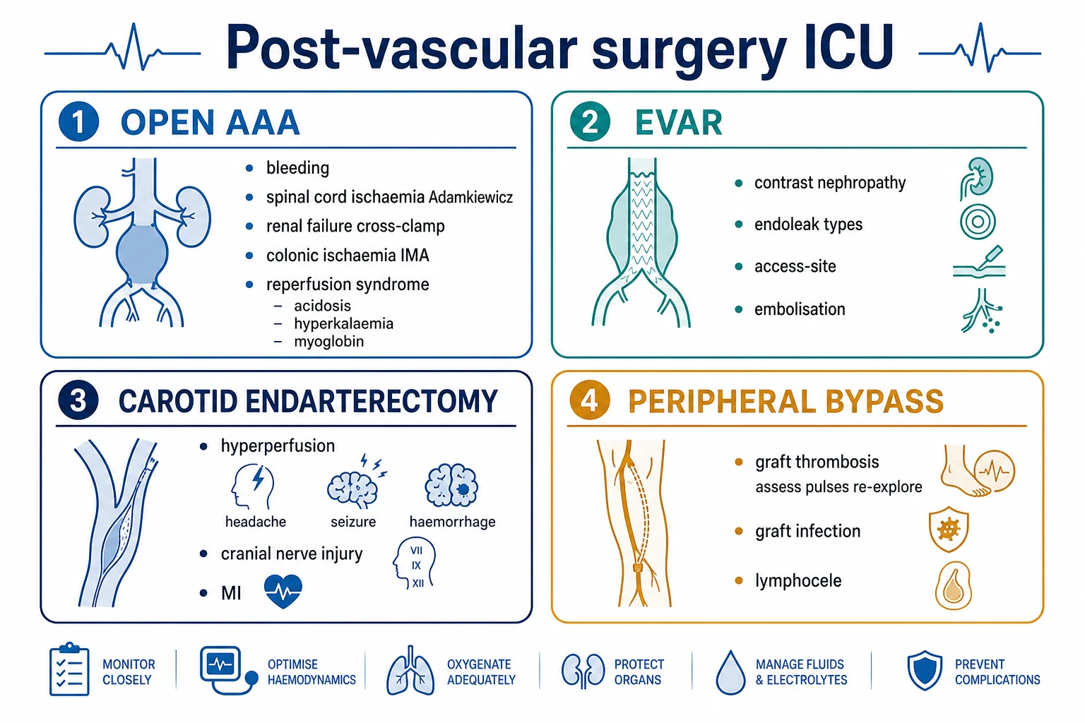
The post-vascular-surgery ICU patient faces surgery-specific complications. After an open AAA repair: bleeding (the graft anastomosis), the spinal cord ischaemia (the cross-clamping of the thoracoabdominal aorta — the Adamkiewicz artery), the renal failure (the cross-clamp and the embolisation), the colonic ischaemia (the IMA ligation), the reperfusion syndrome (acidosis, hyperkalaemia, myoglobin from the lower-limb reperfusion), and the abdominal compartment syndrome. After an EVAR: the contrast-induced nephropathy, the endoleak (Types I to V), and the access-site complications. After a carotid endarterectomy: the hyperperfusion syndrome (headache, seizure, haemorrhage from the restoration of the flow to a chronically underperfused brain), the cranial nerve injury, and the MI (the carotid patient has the coronary disease). After a peripheral bypass: the graft thrombosis (assess the pulses, re-explore), the graft infection (catastrophic), and the reperfusion syndrome.
On this page & tools
Your progress
Saved locally on this device.
Target exams
Overview & definition
The post-vascular-surgery ICU patient faces surgery-specific complications that depend on the procedure. The four main categories are the open AAA repair, the EVAR (endovascular aneurysm repair), the carotid endarterectomy, and the peripheral bypass.[1]
Post-open AAA repair
The major complications:[1]
- Bleeding — from the graft anastomosis (a suture-line bleed); assess the drains, the Hb, the haemodynamics. A high drain output or the haemodynamic instability needs the surgical re-exploration.
- Spinal cord ischaemia — from the cross-clamping of the thoracoabdominal aorta (the artery of Adamkiewicz — the major anterior spinal artery — is compromised). Presents as a paraplegia or a paraparesis (the lower-limb weakness or sensory loss with the preserved arm function). Maintain a high mean arterial pressure (the spinal cord perfusion pressure = the MAP minus the CSF pressure) and consider a CSF drain (to reduce the CSF pressure and improve the cord perfusion).
- Renal failure — from the cross-clamping (the renal ischaemia) and the atheroembolisation (the debris from the aortic wall showering to the kidneys). Monitor the urine output and the creatinine; avoid the nephrotoxins.
- Colonic ischaemia — from the IMA (the inferior mesenteric artery) ligation and the hypoperfusion. Presents with the bloody diarrhoea, the abdominal pain, and the tachycardia on day 1-3. Diagnose with a flexible sigmoidoscopy; treat with the antibiotics and (if severe) the bowel resection.
- The reperfusion syndrome — when the cross-clamp is released, the ischaemic lower-limb metabolites (the lactate, the potassium, the myoglobin) flood the systemic circulation. Presents with a hypotension, an acidosis, a hyperkalaemia, and a myoglobinuric AKI. Treat with the fluid, the bicarbonate for the acidosis, the calcium for the hyperkalaemia, and the renal replacement therapy if needed.[1]
Post-EVAR (endovascular aneurysm repair)
EVAR is less invasive than the open repair, but has its own complications:[1]
- The contrast-induced nephropathy — from the iodinated contrast used during the procedure. Prevent with the pre-procedure hydration and the minimisation of the contrast; monitor the creatinine.
- An endoleak — the persistent perfusion of the aneurysm sac despite the stent graft. Classified:
- Type I — a proximal or distal seal failure (the stent does not seal against the aortic wall). Needs an urgent repair (a cuff, a balloon angioplasty, or a conversion to the open repair).
- Type II — a retrograde flow from a branch vessel (the IMA, a lumbar artery). The commonest; usually observed (it often thromboses spontaneously); intervene if the aneurysm enlarges.
- Type III — a graft defect (a modular disconnection or a fabric tear). Needs a repair.
- Type IV — the graft wall porosity (transient, in the peri-procedural period). Self-limiting.
- Type V — the endotension (the aneurysm enlarges without a demonstrable leak). The cause is unclear; may need a re-intervention.
- Access-site complications — the femoral artery bleeding, a pseudoaneurysm, a dissection, a thrombosis. Assess the groin; an ultrasound or a CT for the diagnosis.
- Lower-limb embolisation — the debris from the aortic wall embolising to the lower limbs (the blue toe syndrome). Assess the pulses and the skin.[1]
Post-carotid endarterectomy (CEA)
- The cerebral hyperperfusion syndrome — the restoration of the normal flow to a chronically underperfused brain (the autoregulation is reset to the high resistance; the sudden normal flow causes a breakthrough). Presents 1-7 days postoperatively with a headache, a seizure, a focal neurological deficit, or an intracerebral haemorrhage. Prevent by controlling the BP postoperatively (target SBP under 140-160).[1]
- A cranial nerve injury — the hypoglossal (the tongue deviation), the vagus/recurrent laryngeal (the hoarseness), the marginal mandibular (the mouth droop) — from the surgical dissection around the carotid sheath. Usually transient.
- An MI — the carotid patient has the extensive atherosclerosis and the coronary disease. Monitor the ECG and the troponin; the perioperative MI is the commonest cause of death after the CEA.
- Restenosis or thrombosis — the carotid occlusion (an acute stroke). Assess the neurological status frequently (the GCS, the motor function).[1]
Post-peripheral bypass
- Graft thrombosis or occlusion — the graft blocks (from the technical failure, the intimal hyperplasia, or the low flow). Presents with a cold, painful, pulseless limb. Assess the pulses and the Doppler signals urgently. A thrombosis needs the immediate surgical re-exploration (a thrombectomy, a revision, or a thrombolysis).
- Graft infection — catastrophic; requires the graft removal and the extra-anatomic bypass (or an in-situ replacement with a biological conduit). Presents with a wound discharge, a pseudoaneurysm, or a sepsis.
- Lymphocele — a collection of lymph at the groin incision; drain if symptomatic.
- The reperfusion syndrome — as above (the acidosis, the hyperkalaemia, the myoglobin from the reperfused limb).[1]
Red flags
The first 6 hours — the post-vascular ICU admission protocol
The post-vascular patient arrives from theatre or the interventional suite with surgery-specific risks. The handover and the first hour set the trajectory for the whole admission.[1][1]
Post-vascular surgery ICU admission protocol — first 6 hours
-
HANDOVER (from the vascular surgeon + anaesthetist): (a) Procedure — open AAA repair (cross-clamp site + time, suprarenal or infrarenal, graft type), EVAR (device, access site, contrast volume, fluoroscopy time), CEA (shunt used? clamps? completion angio?), peripheral bypass (conduit — vein or prosthetic; inflow/outflow vessels). (b) Intraoperative course (difficulty, blood loss, transfusion, haemodynamic instability, vasopressors). (c) Lines and drains (central line, arterial line, urinary catheter, surgical drains, CSF drain if thoracoabdominal, epidural if placed). (d) Current medications (anticoagulation/heparin infusion, antiplatelets, antibiotics, analgesia, vasopressors). (e) Baseline neurology documented IN THEATRE (especially motor and sensory in the legs after aortic surgery; GCS and limb power after CEA) [1]
-
INITIAL ASSESSMENT — ABCDE:
- Airway: usually intubated initially (open AAA, TEVAR); CEA and EVAR often extubated in theatre
- Breathing: SpO2, bilateral air entry, ABG. Watch for pneumothorax after high abdominal closure or supraclavicular access
- Circulation: HR, BP (arterial line), MAP target procedure-dependent (HIGH after aortic/thoracoabdominal for cord perfusion; CONTROLLED after CEA to prevent hyperperfusion), CVP, urine output, capillary refill
- Disability: GCS, pupils, limb power and sensation (feet and legs — for cord ischaemia after aortic surgery; face and limbs — for stroke after CEA)
- Exposure: wounds, the groins (access sites after EVAR), the abdomen (distension after open AAA), all four limbs (pulses, colour, temperature, capillary refill) [1]
-
HAEMODYNAMIC TARGETS — procedure-specific (this is the single most important exam point):
- Open AAA / TEVAR / thoracoabdominal: MAP 80-100 (HIGH — to perfuse the spinal cord; the spinal cord perfusion pressure = MAP minus CSF pressure)
- EVAR (infrarenal): MAP >65 (standard)
- Carotid endarterectomy: SBP 100-140 in the first 24h then <140 for 1-2 weeks (TIGHT CONTROL — labetalol infusion is the agent of choice — too low = watershed stroke; too high = hyperperfusion/haemorrhage)
- Peripheral bypass: MAP >65, optimise cardiac output to keep the graft flowing (low flow = thrombosis) [1]
-
NEUROLOGICAL MONITORING — procedure-specific:
- Aortic surgery (especially thoracoabdominal/TEVAR): hourly lower-limb motor and sensory assessment for the first 48h — any new weakness = spinal cord ischaemia = emergency (raise MAP, drain CSF)
- CEA: hourly GCS, limb power, speech, facial symmetry — any deficit = stroke or hyperperfusion — urgent CT/carotid Doppler
- Document a baseline immediately on arrival and after any sedation has worn off [1]
-
BLEEDING AND COAGULATION: surgical drains, Hb, INR, APTT, platelets, fibrinogen. The vascular patient is on antiplatelets ± heparin ± (pre-warfarinised for some) — bleeding risk is significant. The arterial anastomosis can leak; a high drain output or haemodynamic compromise = surgical re-exploration [1]
-
RENAL PROTECTION: urine output (>0.5 mL/kg/hr), creatinine. The EVAR/TEVAR patient has had iodinated contrast (CIN risk); the open AAA patient has had a suprarenal cross-clamp (renal ischaemia) and atheroembolisation. Maintain euvolaemia, avoid nephrotoxins (NSAIDs, aminoglycosides) [1]
-
TEMPERATURE AND ANALGESIA: rewarm (forced-air warmer); the open AAA patient has a large abdominal incision and will need an epidural or PCA. The epidural provides sympathectomy (improves graft flow) but causes hypotension — manage the vasopressors [1]
-
GLYCAEMIA AND STRESS ULCER PROPHYLAXIS: insulin infusion for glucose <10 mmol/L; PPI for the major-surgery patient
Post-EVAR — the endovascular aneurysm repair patient in ICU
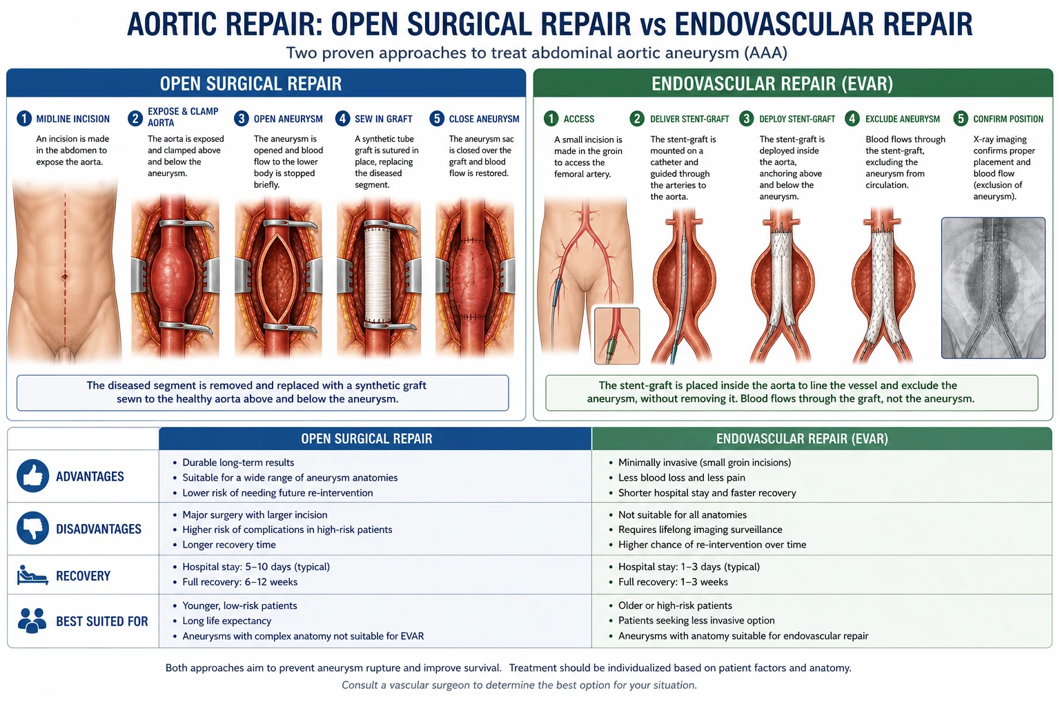
EVAR (endovascular aneurysm repair) is now the dominant strategy for infrarenal AAA. The EVAR-1 and DREAM trials showed a perioperative mortality advantage over open repair (EVAR ~1.5% vs open ~4.5%) but no long-term survival difference, with EVAR requiring more re-interventions (endoleak, graft migration, limb occlusion). The ICU role after EVAR is recognising the device-specific complications — the endoleak, the contrast nephropathy, the access-site complication, and (for TEVAR) the spinal cord ischaemia.[1][1][1]
Endoleak — the persistent perfusion of the aneurysm sac
An endoleak is the persistent perfusion of the aneurysm sac after EVAR — the stent graft has not fully excluded the aneurysm from the circulation. The sac remains pressurised (endotension) and can continue to enlarge and rupture. Endoleak is the MOST COMMON complication of EVAR (15-25% over follow-up). Most are detected on surveillance CT, but a sac enlargement, a new bruit, a back/abdominal pain, or a rupture in the post-EVAR patient is an endoleak until proven otherwise.[1][1]
Endoleak types I-V — classification, mechanism, urgency, and management
| Type | Mechanism | Frequency | Urgency | Management |
|---|---|---|---|---|
| Type I | Seal failure at the proximal (Ia) or distal (Ib) attachment site — the stent does not sit flush against the aortic/iliac wall; high-pressure leak directly into the sac | 5-10% | URGENT — high rupture risk (systemic pressure in the sac) | Repair immediately: balloon angioplasty, aortic cuff, additional stent extension, or conversion to open repair |
| Type II | Retrograde flow from a branch vessel (inferior mesenteric artery, lumbar artery, accessory renal) filling the sac; the branch reverses flow into the sac | 15-25% (commonest) | OBSERVE — most thrombose spontaneously | Surveillance CT. Intervene ONLY if the sac enlarges >5 mm: translumbar embolisation (coil/glue), branch ligation, or laparoscopic IMA clipping |
| Type III | Graft defect — modular disconnection (IIIa), fabric tear (IIIb), or stent fracture; a structural failure of the device | 2-5% | URGENT — systemic pressure communication | Repair: a relining stent graft (endovascular) or conversion to open repair |
| Type IV | Graft wall porosity — transient leakage through the fabric in the peri-procedural period (within 30 days, while the graft seals) from the anticoagulation | Rare | SELF-LIMITING — resolves as the fabric seals and anticoagulation reverses | Observation; correct coagulation |
| Type V | Endotension — the sac enlarges WITHOUT a demonstrable leak on CT angiography; presumed low-flow leak or fluid transudation | 5-10% | INVESTIGATE then treat | MRI, coil embolisation, or explantation if rupture risk. Cause unclear |
Contrast-induced nephropathy (CIN) after EVAR
EVAR requires a substantial volume of iodinated contrast (often 80-150 mL) for the intraoperative angiography. Contrast-induced nephropathy (a creatinine rise >25% or >44 µmol/L within 48-72h) occurs in 8-15% of EVAR patients (higher in CKD, diabetes, urgency). Most cases are transient and recover within 1-2 weeks, but severe cases need renal replacement therapy.[1]
Prevention (do ALL of these for the post-EVAR patient):
- Pre-procedure hydration — isotonic saline 1 mL/kg/hr for 6-12h before and after the procedure (the single most evidence-based prevention)
- Minimise the contrast volume (the surgeon/radiologist should use the minimum for adequate imaging; consider CO2 angiography for the venous phase)
- Hold nephrotoxins — NSAIDs, aminoglycosides, metformin (for 48h post-contrast — also the lactic acidosis risk if AKI develops)
- N-acetylcysteine (NAC) — 1200 mg BD oral for 2 days — cheap, low risk; the evidence is mixed but many centres give it
- Statins — continue the statin (anti-inflammatory and endothelial-protective effect on the kidney)
- Avoid volume depletion — maintain a normal intravascular volume; do not over-diurese [1]
Access-site complications after EVAR
EVAR access is via the femoral arteries (percutaneous or open cut-down) for the device delivery sheaths (often 18-24 French). The access-site complications — bleeding, pseudoaneurysm, dissection, thrombosis, infection, and lymphocele — occur in 2-5% of cases (lower with a closure device and with open cut-down for large sheaths).[1]
EVAR access-site complications — recognition and management
| Complication | Recognition | Management |
|---|---|---|
| Femoral bleeding / haematoma | Groin swelling, bruising, expanding mass, falling Hb, hypotension | Direct pressure. If expanding or haemodynamically significant — surgical exploration. Cross-match. Reverse heparin |
| Pseudoaneurysm | Pulsatile groin mass + systolic bruit +thrill, day 1-7. Ultrasound with colour Doppler confirms (a swirling flow outside the artery, connected by a neck) | Ultrasound-guided thrombin injection (80-90% success, first-line). Surgical repair if fails or infected |
| Arterial thrombosis | Cold, pulseless lower limb (the access-side) | Urgent surgical thrombectomy ± bypass. Do not delay |
| Arterial dissection | Diminished pulses, limb ischaemia, possible extension proximally. CT angiogram | If flow-limiting — stenting or surgical repair. If non-flow-limiting — observation |
| AV fistula | Continuous bruit + thrill over the groin, limb swelling, high-output cardiac failure if large. Doppler confirms | Most close spontaneously. If symptomatic/large — surgical repair or covered stent |
| Lymphocele / lymph leak | Clear drainage from the groin incision + cystic swelling, day 3-14 | Most resolve with conservative care (compression, observation). Surgical ligation if persistent |
| Access-site infection | Erythema, discharge, sepsis, day 5-14. Risk: diabetes, obesity, re-operation | IV antibiotics (MRSA coverage). Surgical debridement. If the graft is exposed — graft excision + extra-anatomic bypass |
Post-implantation syndrome — the self-limiting inflammatory response
Up to 30-50% of EVAR patients develop a post-implantation syndrome — a self-limiting systemic inflammatory response (fever, leucocytosis, raised CRP) from the thrombosis of the excluded aneurysm sac and the endothelial interaction with the graft. It lasts 2-10 days and is benign. The danger is being reassured that fever is "post-implantation syndrome" and missing an access-site infection or colonic ischaemia — investigate any fever with cultures and a CT if it does not settle, if it is high (>39C), or if it appears after day 3. [1]
Post-TEVAR — thoracic endovascular aortic repair
TEVAR (thoracic endovascular aortic repair) is the stent-graft treatment of descending thoracic aortic aneurysms, complicated type B dissection, and traumatic aortic injury. The ICU complications overlap with EVAR but with TWO critical differences: (1) the spinal cord ischaemia risk is much higher (the descending thoracic aorta gives off the intercostal arteries including the artery of Adamkiewicz), and (2) a stroke risk from the wire manipulation in the aortic arch.[1][1]
Spinal cord ischaemia after TEVAR — the feared complication
Spinal cord ischaemia (SCI) after TEVAR occurs in 3-10% (higher with long-segment coverage, previous abdominal aortic surgery, and coverage of the left subclavian artery). The mechanism: the stent graft covers the intercostal arteries supplying the anterior spinal artery (the artery of Adamkiewicz, usually T9-L2). The cord perfusion depends on collateral flow (the subclavian arteries, the hypogastric arteries). The deficit is usually a paraplegia or paraparesis with a sensory level.[1][1]
Prevention and management (the CSF drain is the key intervention):
- Cerebrospinal fluid (CSF) drainage — placed pre-operatively for high-risk TEVAR (long-segment coverage, previous AAA repair). The CSF drain reduces the intrathecal pressure (the spinal cord perfusion pressure = MAP minus CSF pressure). Target CSF pressure <10-15 mmHg. Keep the drain for 48-72h postoperatively
- Maintain a high MAP — target MAP 80-100 mmHg with noradrenaline (a deliberate hypertensive strategy to perfuse the cord)
- Revascularise the left subclavian artery — if the stent covers the subclavian origin, a carotid-subclavian bypass preserves the collateral flow (and reduces the SCI risk)
- Avoid hypotension — any hypotensive episode (bleeding, vasodilation, epidural) can precipitate or worsen the cord ischaemia
- Hourly neurology — lower-limb motor and sensory assessment for the first 48h
- If new weakness occurs: RAISE the MAP (noradrenaline to MAP 100), DRAIN the CSF (lower to <10 mmHg), check the haemoglobin (transfuse to >100 g/L for oxygen carrying), and reconsider the perfusion. A timely response can reverse the deficit [1]
Stroke after TEVAR
Stroke occurs in 2-5% of TEVAR procedures — from embolic shower (atheromatous debris dislodged by the wires in the aortic arch) or from the coverage of the left vertebral artery (with the left subclavian). Higher risk with arch involvement, severe atheroma, and urgent procedures. The stroke is usually embolic (anterior or middle cerebral territory). Management: maintain perfusion (MAP >80), CT brain, and thrombolysis is generally CONTRAINDICATED (recent major vascular procedure/surgery — bleeding risk). [1]
Post-open AAA repair — the open abdominal aortic aneurysm repair
Open AAA repair is the gold standard for the durable exclusion of the aneurysm. The ICU complications reflect the magnitude of the surgery — the aortic cross-clamp, the large retroperitoneal dissection, the blood loss, the haemodynamic swings. The key complications are bleeding, spinal cord ischaemia (more for thoracoabdominal than infrarenal), renal failure, colonic ischaemia, reperfusion syndrome, and abdominal compartment syndrome.[1][1]
Bleeding after open AAA
Bleeding after open AAA comes from: (1) the proximal or distal aortic anastomosis (a suture-line leak), (2) retroperitoneal venous ooze, (3) coagulopathy (dilutional from blood loss + the heparin used intraoperatively + hypothermia + acidosis). The assessment is by the surgical drains (a retroperitoneal drain is usually placed), the haemoglobin trend, the haemodynamics, and the coagulation. A high drain output (>300 mL/hr), a falling haemoglobin, or haemodynamic instability = surgical re-exploration. The coagulopathy is corrected with FFP, cryoprecipitate (fibrinogen <1.5 g/L), platelets, and protamine (if residual heparin — check the ACT). [1]
Graft thrombosis after open AAA
The graft can thrombose (rarely, in <2% of cases) from a technical error (a kink, a twist), a low-flow state (hypotension, low cardiac output), or hypercoagulability. Presents with a loss of the lower-limb pulses, a cool leg, abdominal pain (if mesenteric), or renal failure (if the renal arteries are involved). Diagnose with a CT angiogram. Treat with surgical thrombectomy and graft revision. Anticoagulate post-revision. [1]
Renal failure after open AAA
Renal failure occurs in 5-15% (RRT in 1-3%) from: (a) the suprarenal cross-clamp (renal ischaemia — the clamp above the renals stops the renal perfusion), (b) atheroembolisation (the debris from the diseased aortic wall showering to the kidneys — the "cholesterol embolisation syndrome"), (c) contrast (if any angiography), (d) hypoperfusion (hypotension, bleeding). The atheroembolisation syndrome is irreversible (the emboli lodge in the renal arterioles) and presents with eosinophilia, purple toes, and a slowly progressive renal failure over weeks. Prevention: maintain perfusion (MAP >65, optimise cardiac output), avoid nephrotoxins, adequate hydration. [1]
Colonic ischaemia after open AAA
Colonic ischaemia occurs in 1-7% (sigmoid colon most commonly) from the IMA ligation (the IMA supplies the descending and sigmoid colon — when ligated to free the aneurysm, the colon depends on the marginal artery and the inferior mesenteric/middle colic collateral) and from the perioperative hypoperfusion. Presents day 1-3 with bloody diarrhoea, abdominal pain, fever, tachycardia, and leucocytosis. Diagnose with a flexible sigmoidoscopy (the mucosa is oedematous, haemorrhagic, or ulcerated). Most cases are mild (mucosal ischaemia, resolves with antibiotics, bowel rest, and supportive care). Severe cases (transmural ischaemia, perforation) need a laparotomy and bowel resection with a stoma. Mortality of severe colonic ischaemia is 30-50%. [1]
Abdominal compartment syndrome (ACS) after open AAA
Abdominal compartment syndrome is a recognised complication of the ruptured AAA repair (more than elective). The intra-abdominal pressure (IAP) rises from the oedematous bowel (ischaemia-reperfusion, the massive fluid resuscitation, the retroperitoneal haematoma). When the IAP exceeds 20 mmHg with new-onset organ failure (renal: oliguria; respiratory: high ventilator pressures; cardiovascular: reduced venous return and cardiac output; neurological: raised ICP), it is the abdominal compartment syndrome.[1]
Diagnosis: measure the bladder pressure (a surrogate for the IAP — >20 mmHg with organ failure = ACS). [1]
Management: (1) surgical decompression (open the abdomen — a laparostomy) — the definitive treatment. (2) Medical decompression (neuromuscular blockade, nasogastric and rectal decompression, fluid restriction) is a temporising measure only. (3) After decompression, the open abdomen is managed with a negative-pressure dressing (VAC) and a planned closure once the oedema settles (days 5-14). [1]
Post-carotid endarterectomy (CEA) — the carotid patient in ICU
The carotid endarterectomy patient has THREE unique problems: (1) the hyperperfusion syndrome (the restored flow to a chronically underperfused brain causes a cerebral oedema, a seizure, or a haemorrhage), (2) the cranial nerve injury (from the surgical dissection around the carotid sheath), and (3) the stroke (from a carotid occlusion, an embolus, or a hypoperfusion). The fourth, often forgotten, problem is the myocardial infarction — the carotid patient has the extensive atherosclerosis and the coronary disease is the commonest cause of death.[1][1]
Cerebral hyperperfusion syndrome (CHS)
Cerebral hyperperfusion syndrome occurs in 1-3% of CEA patients. The chronically underperfused brain (proximal carotid stenosis) has reset its autoregulation to a high resistance (cerebral arterioles maximally dilated to maintain flow). The sudden restoration of the normal flow (the endarterectomy) overcomes the autoregulation — a breakthrough — causing a hyperperfusion, a cerebral oedema, and (in the worst case) an intracerebral haemorrhage. Presents day 1-14 (peak day 3-7) with a severe ipsilateral headache, a seizure, a focal deficit (visual, motor), a confusion, or (catastrophically) an intracerebral haemorrhage.[1]
Prevention: tight postoperative BP control (SBP 100-140 in the first 24h, then <140 for 1-2 weeks). The agent of choice is labetalol (a beta-blocker with alpha-blockade — easy to titrate as an infusion). Avoid the pure vasodilators (hydralazine, nifedipine) that may dilate the cerebral vessels and worsen the hyperperfusion. [1]
Management of established CHS: (1) Aggressive BP control (SBP <140, ideally <120 — labetalol infusion, beta-blocker). (2) CT brain (to exclude the haemorrhage). (3) Osmotherapy (mannitol or hypertonic saline) if cerebral oedema. (4) Seizure control (levetiracetam). (5) If intracerebral haemorrhage — neurosurgical consultation, reverse any anticoagulation/antiplatelets (the catastrophic scenario). [1]
Cranial nerve injury after CEA
Cranial nerve injury occurs in 5-10% of CEA patients (mostly transient, permanent in <1%) from the surgical dissection around the carotid sheath. The nerves at risk and their deficits:[1]
Cranial nerve injury after CEA — the nerve, the deficit, the implication
| Nerve | Deficit | Implication |
|---|---|---|
| Hypoglossal (XII) | Tongue deviation TOWARDS the affected side (the lesion) on protrusion; difficulty with speech and swallowing | Usually transient. Bilateral hypoglossal injury (rare) causes airway compromise |
| Vagus / recurrent laryngeal (X) | Hoarseness, vocal cord paralysis (the cord sits in the paramedian position) | If known before surgery (a contralateral vocal cord palsy), CEA is high risk for airway obstruction — check the contralateral cord pre-operatively |
| Marginal mandibular (VII branch) | Droop of the corner of the mouth on the affected side (the lower lip) | Cosmetic; usually transient. Often confused with a stroke — distinguish (the forehead is spared, the upper face is intact) |
| Glossopharyngeal (IX) | Dysphagia, loss of the gag reflex | Aspiration risk; usually transient |
| Superior laryngeal nerve (X branch) | Loss of the high-pitch voice, easy fatigability (the cricothyroid muscle) | Often missed; usually transient |
Acute stroke after CEA
Stroke occurs in 2-5% of CEA procedures (the threshold above which the surgery does more harm than good — CEA is only justified if the perioperative stroke/death rate is <6% for symptomatic and <3% for asymptomatic stenosis). The mechanisms: (a) embolic (the intraoperative dislodgement of the plaque — the most common), (b) carotid thrombosis (an acute occlusion of the repaired artery — a hypercoagulable state or a technical error — a catastrophic ischaemic stroke), (c) hypoperfusion (the cross-clamping of the carotid in a patient with a poor collateral circle — the reason for an intraoperative shunt).[1]
Recognition: any new neurological deficit in the postoperative period (limb weakness, aphasia, visual field defect, decreased GCS). The first action is a CT brain (to distinguish ischaemic from haemorrhagic — a haemorrhage suggests hyperperfusion; an ischaemic suggests embolus or thrombosis) and a carotid Doppler (to check the patency of the repaired artery). A carotid thrombosis is a surgical emergency — re-exploration and thrombectomy. [1]
Post-peripheral bypass — the lower-limb revascularisation patient
Peripheral bypass (the femoropopliteal, the femorodistal, the iliofemoral bypass) restores the flow to a critically ischaemic limb. The bypass can use the autologous vein (the great saphenous — preferred, better patency) or a prosthetic graft (PTFE, Dacron). The ICU complications are the graft thrombosis, the graft infection, the reperfusion syndrome, and the compartment syndrome.[1][1]
Graft thrombosis after peripheral bypass
Graft thrombosis occurs in 5-15% in the first 30 days (higher for prosthetic, lower for vein, higher for distal targets). The causes: (a) a technical error (a kink, a twist, an anastomotic stricture), (b) an intimal hyperplasia (the late cause, month 3-12 — the smooth muscle proliferation at the anastomosis), (c) a low flow (hypotension, low cardiac output, a poor outflow), (d) hypercoagulability. Presents with a cold, painful, pulseless limb (the 6 Ps: Pain, Pallor, Pulselessness, Paresthesia, Paralysis, Perishing-cold).[1]
Management: (1) Assess the pulses and the Doppler signals urgently (a hand-held Doppler at the ankle). (2) A CT or an on-table angiogram if the diagnosis is unclear. (3) Immediate surgical re-exploration — a thrombectomy (open the graft, remove the thrombus with a Fogarty catheter), a revision (correct a technical error), or a thrombolysis (catheter-directed thrombolysis with tPA for a distal clot). (4) Systemic anticoagulation (heparin infusion) to prevent a re-thrombosis. (5) Fasciotomy if there is a concomitant compartment syndrome. [1]
Compartment syndrome after peripheral bypass
Reperfusion of a chronically ischaemic limb causes an ischaemia-reperfusion injury — the muscle and the endothelium swell, the capillaries leak, and the pressure within the osteofascial compartment rises. When the compartment pressure exceeds the perfusion pressure (a compartment pressure within 30 mmHg of the diastolic BP), the tissue ischaemia ensues. The patient develops a severe pain (out of proportion, worse on passive stretch), a tense and tender calf, paraesthesia, and eventually a paralysis.[1]
Diagnosis: clinical (pain out of proportion, pain on passive stretch, tense compartment). Confirm with a compartment pressure measurement (a slit catheter or a needle manometer — a pressure >30 mmHg, or within 30 mmHg of the diastolic, is diagnostic). [1]
Management: an emergency fasciotomy (open all four compartments of the calf — the anterior, the lateral, the superficial posterior, the deep posterior). Leave the wound open with a VAC dressing; close once the swelling settles (days 5-10). Delay risks an irreversible muscle necrosis, a rhabdomyolysis, and a Volkmann's contracture (or an amputation). [1]
Graft infection after peripheral bypass
Graft infection is a catastrophic complication (1-2% incidence). The risk: groin incisions (the perineal flora), prosthetic grafts (much higher than vein), diabetes, and re-operation. Presents week 1 to month 6 (or later) with a wound discharge, a pseudoaneurysm at the anastomosis (a pulsatile mass), a graft-enteric erosion (catastrophic bleeding — the aorto-enteric fistula after an aortic graft), or a sepsis. The organism is usually Staphylococcus aureus (early) or a gram-negative/coagulase-negative staph (late).[1]
Management: (1) Broad-spectrum IV antibiotics (vancomycin for MRSA coverage + a gram-negative agent). (2) CT angiogram to define the extent. (3) Surgical — the graft excision with an extra-anatomic bypass (route the new graft through uninfected tissue — axillo-bifemoral for an aortic graft) OR an in-situ replacement with a biological conduit (a cadaveric homograft, a rifampicin-soaked Dacron). (4) Long-term suppressive antibiotics if complete excision is not possible. [1]
Anticoagulation management in the vascular surgical ICU patient
The vascular patient is the anticoagulation patient — most are on antiplatelets (aspirin, clopidogrel), many on warfarin (atrial fibrillation, a mechanical valve, a venous thromboembolism), and an increasing number on a direct oral anticoagulant (DOAC — apixaban, rivaroxaban, dabigatran). The perioperative management is a high-yield exam topic and a daily ICU decision. The principles are: (1) assess the thrombotic risk (is the indication high-risk — a mechanical mitral valve, a recent VTE — or low-risk — an AF without other risk factors), (2) assess the bleeding risk of the procedure (major — open AAA, peripheral bypass — vs minor — a diagnostic angiogram), (3) decide on the bridging (continue, stop with no bridge, or stop with a heparin bridge).[1][1]
Perioperative anticoagulation — bridging by thrombotic risk and procedure
| Indication | Thrombotic risk | Strategy |
|---|---|---|
| Mechanical mitral valve | HIGH | Bridge: stop warfarin 5 days pre-op, start UFH/LMWH 3 days pre-op, stop LMWH 24h (UFH 4-6h) pre-op, restart warfarin 1-2 days post-op once haemostasis secure |
| Mechanical aortic valve (older generation, other risk factors) | HIGH | Bridge as above |
| Mechanical aortic valve (modern, no other risk factors) | MODERATE | Often NO bridging — stop warfarin, restart early post-op. Individualise |
| VTE within 3 months | HIGH | Bridge (and consider delaying elective surgery) |
| VTE within 3-12 months | MODERATE | Bridge for major surgery |
| VTE >12 months | LOW | Often no bridging |
| AF with high CHA2DS2-VASc (prior stroke/TIA, rheumatic) | HIGH | Bridge |
| AF with low CHA2DS2-VASc | LOW | No bridging (BRIDGE trial — no benefit, more bleeding) |
| Recent coronary stent (<6 months DES, <3 months BMS) | HIGH | Aspirin CONTINUED; clopidogrel stopped 5 days if absolutely necessary — discuss with cardiology; delay surgery if possible |
The BRIDGE trial — bridging is NOT for everyone
The BRIDGE trial (2015, NEJM) randomised patients on warfarin for AF or a mechanical valve to perioperative bridging (LMWH) vs placebo around elective surgery. The findings: (1) bridging did NOT reduce arterial thromboembolism (0.3% vs 0.4%), (2) bridging SIGNIFICANTLY increased major bleeding (3.2% vs 1.3%). The bottom line: bridging is for the HIGH-risk patient (mechanical mitral valve, recent VTE, AF with a prior stroke) — for the low-risk AF patient, stopping warfarin without a bridge is safe and causes less bleeding.[1]
Heparin and DOAC timing in ICU
Anticoagulant pharmacology — onset, reversal, and perioperative timing
| Agent | Half-life | Onset | Reversal | Stop before surgery | Resume after surgery |
|---|---|---|---|---|---|
| Warfarin | 36-42h (slow) | 3-5 days (slow) | Vitamin K (slow), PCC (rapid, 4-factor), FFP | 5 days | 12-24h post-op (when haemostasis secure) |
| Unfractionated heparin (UFH) | 60-90 min | Immediate (IV) | Protamine 1 mg per 100 U heparin | 4-6h | 6-12h post-op |
| Low-molecular-weight heparin (LMWH) | 4-6h | 30-60 min | Protamine (partial — ~60% reversal) | 24h (therapeutic dose) | 24h post-op |
| Apixaban | 12h | 3-4h | Andexanet alfa (factor Xa reversal), PCC (if unavailable) | 48h (major), 24h (minor) | 24-48h post-op |
| Rivaroxaban | 5-13h | 3-4h | Andexanet alfa, PCC | 48h (major), 24h (minor) | 24-48h post-op |
| Dabigatran | 12-17h | 1-2h | Idarucizumab (specific reversal) | 48h (major), 24h (minor) | 24-48h post-op |
| Aspirin | 7-10 days (platelet lifespan) | Irreversible | Platelet transfusion | Often CONTINUED for vascular surgery | Continued |
| Clopidogrel | Irreversible (5-7 days) | Irreversible | Platelet transfusion | 5 days (7 for some) | 24-72h post-op |
VTE prophylaxis in the vascular surgical patient
The vascular patient is at HIGH VTE risk (major surgery, immobility, often a malignancy or a smoking history). Pharmacological prophylaxis (LMWH — enoxaparin 40 mg SC daily) is standard for all major vascular surgery, combined with mechanical prophylaxis (TED stockings, intermittent pneumatic compression). The caveat: the epidural. If an epidural is in situ, the LMWH must be timed (give the LMWH 6-8h after the epidural catheter placement, and remove the catheter 12h after the last LMWH dose — to avoid a spinal haematoma).[1]
Pain management — epidural vs PCA in the vascular patient
The vascular patient has a major incision (a midline laparotomy for the open AAA, a long leg incision for the bypass) and good analgesia is essential — it allows early mobilisation, deep breathing, and prevents the atelectasis and the pneumonia. The two options are the epidural and the patient-controlled analgesia (PCA).[1]
Epidural vs PCA for post-vascular surgery analgesia
| Feature | Epidural | PCA (morphine or fentanyl) |
|---|---|---|
| Mechanism | Local anaesthetic + opioid in the epidural space — blocks the nociceptive transmission | Systemic opioid — binds the central opioid receptors |
| Pain control | SUPERIOR (especially the dynamic pain — coughing, mobilising) | Good (especially at rest) |
| Sympathectomy | YES (a thoracic/lumbar epidural causes a sympathetic block — vasodilation, hypotension) — can be ADVANTAGEOUS for the graft flow (improves the perfusion) but RISKY if the patient is hypovolaemic | NO |
| Effect on the bowel | Improved (the sympathetic block restores the parasympathetic dominance and the gut motility) | Worse (the systemic opioid slows the gut — an ileus) |
| Effect on the respiratory function | Improved (better diaphragmatic function, less atelectasis) | May depress respiration (opioid) |
| Hypotension risk | HIGH (the sympathectomy — needs the vasopressors and the fluid) | LOW |
| Anticoagulation conflict | YES — the LMWH must be timed with the catheter placement/removal (a spinal haematoma risk) | NO |
| Failure / migration risk | 10-20% (the catheter can migrate, the block can be patchy) | LOW (the IV access is reliable) |
| Duration | 2-5 days (then convert to oral) | 2-3 days |
| Best for | The open AAA (a thoracic epidural), the major peripheral bypass — to improve the graft flow and the bowel function | The patient on anticoagulation, the patient with a coagulopathy, the EVAR (less pain) |
SAQ — Post-EVAR: endoleak, access-site complication and contrast AKI
10 minutes · 10 marks
A 74-year-old man is admitted to ICU four hours after an elective endovascular aneurysm repair (EVAR) of a 5.8 cm infrarenal AAA. The procedure used 140 mL of iodinated contrast via a percutaneous right femoral access and an open left femoral cut-down. Past history: CKD (baseline creatinine 135 umol/L), type 2 diabetes, paroxysmal AF on apixaban (withheld 48h pre-op), and a contralateral (left) vocal cord palsy. On assessment: BP 108/64, HR 92 (AF), SpO2 96%, urine output 25 mL/hr, an expanding tense right groin mass with a systolic bruit. CT angiogram performed for a falling haemoglobin (98 to 76 g/L) shows the stent graft in a good position but contrast filling the aneurysm sac at the proximal neck, and the sac now measures 6.1 cm (was 5.8 cm). Creatinine has risen to 198 umol/L.
SAQ — Reperfusion compartment syndrome after femoropopliteal bypass
10 minutes · 10 marks
A 66-year-old male smoker with type 2 diabetes underwent a reversed great saphenous vein femoropopliteal bypass for chronic limb-threatening ischaemia (a gangrenous forefoot ulcer, Rutherford 5). The limb was ischaemic for 16 hours pre-operatively; a Fogarty thrombo-embolectomy restored flow and the bypass was completed. He has a lumbar epidural infusion (ropivacaine 0.2% + fentanyl 2 mcg/mL at 6 mL/hr) running for analgesia. Eight hours post-operatively he complains of severe calf pain (8/10) that is disproportionate to the surgical incision and worse on passive dorsiflexion of the toes. The calf is tense, swollen and tender. Doppler signals are present at the ankle. Sensation is reduced in the first dorsal web space; toe flexion is weak. He has passed 50 mL of dark red-brown urine in the last two hours. Bloods: K+ 6.2 mmol/L, CK 22,000 U/L, venous pH 7.24, lactate 5.2 mmol/L.
Clinical pearls — the high-yield exam points
Prognosis and outcomes
Vascular surgery outcomes — what determines survival
| Procedure | Perioperative mortality | Key complications | Key prognostic factors |
|---|---|---|---|
| Elective open AAA | 4-5% | Bleeding, MI, renal failure, colonic ischaemia, ACS | Age, cardiac function, renal function, COPD |
| Ruptured AAA (open) | 40-50% | Massive transfusion, ACS, multi-organ failure, colonic ischaemia | Pre-op arrest, time to theatre, blood loss |
| Elective EVAR | 1-2% | Endoleak, access-site, CIN | Age, renal function, anatomy (neck length) |
| Ruptured AAA (EVAR) | 20-30% | Endoleak, ACS, colonic ischaemia | Hemodynamic stability, anatomy |
| Elective CEA | 1-2% (stroke/death) | Stroke, MI, hyperperfusion, cranial nerve | Symptomatic status, contralateral occlusion |
| TEVAR | 3-5% | Spinal cord ischaemia (3-10%), stroke, endoleak | Coverage length, prior AAA repair, subclavian coverage |
| Peripheral bypass (elective) | 2-3% | Graft thrombosis, infection, compartment syndrome | Distal target quality, conduit (vein > prosthetic), diabetes |
| Critical limb ischaemia (bypass) | 10-15% 1-year mortality | Major amputation (10-20%), graft failure | Tissue loss, infection, diabetes, dialysis |
Key trials and evidence
EVAR-1 trial — EVAR vs open repair for AAA (Greenhalgh, Lancet 2010)
Study design
Multicentre randomised controlled trial — UK — 1,082 patients with AAA >=5.5 cm fit for open repair
Intervention
EVAR vs open AAA repair
Primary outcome
All-cause mortality at 30 days: EVAR 1.7% vs open 4.7% (EVAR superior, p=0.009)
Long-term (8-12 years)
NO overall survival difference; EVAR had MORE graft-related complications and re-interventions (endoleak, migration, limb occlusion); higher aneurysm-related mortality in later years
Key finding
EVAR has an EARLY survival advantage but requires LIFELONG surveillance and more re-interventions
Clinical bottom line
EVAR is the preferred approach for anatomically suitable AAA — but surveillance is non-negotiable, and the patient must understand the lifelong follow-up
DREAM trial — EVAR vs open repair (Prinssen, NEJM 2004)
Study design
Multicentre randomised controlled trial — Netherlands — 351 patients
Primary outcome
Operative mortality: EVAR 1.2% vs open 4.6% (favouring EVAR)
Long-term
No survival difference at 2+ years; EVAR had more re-interventions
Clinical bottom line
Confirms the EVAR-1 finding — EVAR has an early mortality advantage, equivalent long-term survival, more re-interventions
Coselli CSF drainage trial — spinal cord protection (J Vasc Surg 2002)
Study design
Randomised controlled trial — 145 patients undergoing thoracoabdominal aortic aneurysm repair
Intervention
CSF drainage (target CSF pressure <10 mmHg) vs no drainage
Primary outcome
Paraplegia/paraparesis: CSF drain 2.6% vs no drain 13% (p=0.03 — NINE-FOLD reduction)
Mechanism
Spinal cord perfusion pressure = MAP minus CSF pressure; draining CSF lowers the intrathecal pressure and improves the cord perfusion
Clinical bottom line
CSF drainage is STANDARD OF CARE for high-risk thoracoabdominal and TEVAR procedures — the single most effective intervention to prevent the spinal cord ischaemia
BRIDGE trial — perioperative bridging anticoagulation (Weitz/DOAC, NEJM 2015)
Study design
Multicentre randomised controlled trial — 1,884 patients on warfarin for AF or a mechanical valve, undergoing elective surgery
Intervention
Perioperative bridging (LMWH 100 U/kg BD, last dose 24h pre-op) vs placebo (no bridge)
Primary outcome
Arterial thromboembolism: bridge 0.3% vs no-bridge 0.4% (NO difference)
Safety
Major bleeding: bridge 3.2% vs no-bridge 1.3% (bridging caused MORE bleeding, p=0.005)
Key finding
Bridging does NOT reduce arterial thromboembolism in the low-risk AF patient but DOES increase major bleeding
Clinical bottom line
Reserve bridging for the HIGH-risk patient (mechanical mitral valve, recent VTE, AF with prior stroke). For the low-risk AF patient, stop the warfarin and resume without a bridge
Abou-Chebl — hyperperfusion after carotid stenting (JACC 2004)
Study design
Case series — 450 patients undergoing carotid artery stenting
Key finding
Cerebral hyperperfusion syndrome and intracranial haemorrhage occurred in 1.1% — typically day 1-14, associated with a high post-procedure BP and a severe pre-procedure stenosis
Risk factors
Severe pre-procedure stenosis, hypertension, perioperative BP elevation, contralateral carotid occlusion
Clinical bottom line
Tight post-procedure BP control (SBP <140) is the prevention; the recognition of the early headache and the BP control prevent the catastrophic haemorrhage
CREST trial — carotid stenting vs endarterectomy (NEJM 2010)
Study design
Multicentre randomised controlled trial — 2,502 patients with symptomatic or asymptomatic carotid stenosis
Intervention
Carotid artery stenting (CAS) vs carotid endarterectomy (CEA)
Primary outcome
Composite of stroke, MI, death at 30 days: CAS 5.2% vs CEA 4.5% (no significant difference)
Key findings
CAS had MORE periprocedural stroke (4.1% vs 2.3%); CEA had MORE MI (2.3% vs 1.1%); age effect — stenting better in <70, CEA better in >70
Clinical bottom line
CEA remains the standard for most patients; stenting for selected younger patients or those with high surgical risk (restenosis, neck radiation, contralateral palsy)
BEST-CLI trial — vein vs prosthetic bypass for CLTI (NEJM 2022)
Study design
Multicentre randomised controlled trial — 1,830 patients with chronic limb-threatening ischaemia (CLTI)
Intervention
Surgical bypass with a great saphenous vein vs a prosthetic graft (in patients with a suitable vein)
Primary outcome
Major adverse limb event or death: vein bypass SUPERIOR — the vein conduit significantly outperformed the prosthetic
Key finding
The autologous vein is the preferred conduit for a lower-limb bypass — the prosthetic graft has a higher occlusion and amputation rate
Clinical bottom line
Use the great saphenous vein whenever available for a peripheral bypass; the prosthetic is reserved for the patient without a suitable vein
Mnemonics and rapid recall
[1] [1] [1] [1]References
- [1]Simpson JC, Barrett KE, Tang JE 2025 Update on Vascular Anesthesia. Journal of Cardiothoracic and Vascular Anesthesia, 2026.PMID 42215397
- [2]United Kingdom EVAR Trial Investigators; Greenhalgh RM, Brown LC, Powell JT, et al Endovascular versus open repair of abdominal aortic aneurysm. N Engl J Med, 2010.PMID 20382983
- [3]Schermerhorn ML, O'Malley AJ, Jhaveri A, et al Long-Term Outcomes of Abdominal Aortic Aneurysm in the Medicare Population. N Engl J Med, 2015.PMID 26200979
- [4]Veith FJ, Baum RA, Ohki T, et al Nature and significance of endoleaks and endotension: summary of opinions expressed at an international conference. Journal of Vascular Surgery, 2002.PMID 12021724
- [5]Buth J, Harris PL, van Marrewijk C, et al The significance and management of different types of endoleaks. Seminars in Vascular Surgery, 2003.PMID 12920679
- [6]Weiss SJ, Hogan MS, McGarvey ML, et al Successful treatment of delayed onset paraplegia after suprarenal abdominal aortic aneurysm repair. Anesthesiology, 2002.PMID 12151944
- [7]Coselli JS, LeMaire SA, Conklin LD, et al Cerebrospinal fluid drainage reduces paraplegia after thoracoabdominal aortic aneurysm repair: results of a randomized clinical trial. Journal of Vascular Surgery, 2002.PMID 11932655
- [8]Abou-Chebl A, Yadav JS, Reginelli JP, et al Intracranial hemorrhage and hyperperfusion syndrome following carotid artery stenting: risk factors, prevention, and treatment. Journal of the American College of Cardiology, 2004.PMID 15120817
- [9]Lasic Z, Nikolsky E, Kesanakurthy S, et al Vascular closure devices: a review of their use after invasive procedures. American Journal of Cardiovascular Drugs, 2005.PMID 15901206
- [10]Douketis JD, Spyropoulos AC, Spencer FA, et al Perioperative management of antithrombotic therapy: Antithrombotic Therapy and Prevention of Thrombosis, 9th ed: American College of Chest Physicians Evidence-Based Clinical Practice Guidelines. Chest, 2012.PMID 22315266
- [11]Quinones A, Saric M The cholesterol emboli syndrome in atherosclerosis. Current Atherosclerosis Reports, 2013.PMID 23423524
- [12]Simpson JC, Barrett KE, Tang JE 2025 Update on Vascular Anesthesia. Journal of Cardiothoracic and Vascular Anesthesia, 2026.PMID 42215397
- [13]Hörer TM, Abu-Zidan FM, McGreevy DT, et al Abdominal Compartment Syndrome After Endovascular Repair of Ruptured Abdominal Aortic Aneurysms: A Single-Center Experience of Total Endovascular Care for Ruptured Abdominal Aortic Aneurysms. Journal of Endovascular Therapy, 2026.PMID 40165640
- [14]Kakkos SK, Caprini JA, Geroulakos G, et al Combined intermittent pneumatic leg compression and pharmacological prophylaxis for prevention of venous thromboembolism. Cochrane Database of Systematic Reviews, 2016.PMID 27600864
- [15]Douketis JD, Spyropoulos AC, Kaatz S, et al (BRIDGE) Perioperative Bridging Anticoagulation in Patients with Atrial Fibrillation. N Engl J Med, 2015.PMID 26095867