ICU · Cardiovascular / perioperative
Postoperative Complications in the ICU
Also known as Postoperative complications · Postop complications · Postoperative bleeding · Postoperative respiratory failure · Postoperative delirium · Postoperative AKI · VTE prophylaxis · ERAS · Enhanced recovery after surgery
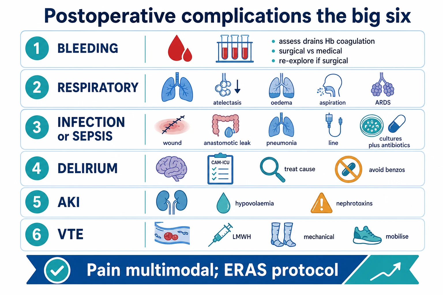
Postoperative complications in the ICU cover six areas. Bleeding (assess the drains, the Hb, the coagulation; distinguish the surgical from the medical bleeding; re-explore the surgical cause). Respiratory failure (atelectasis, oedema, aspiration, ARDS; weigh the early extubation against the prolonged ventilation). Infection or sepsis (a wound infection, an anastomotic leak, a pneumonia, a line infection; cultures and antibiotics). Delirium (common in the elderly; the CAM-ICU assessment; treat the cause — pain, infection, hypoxia, medications; avoid benzodiazepines; haloperidol or quetiapine). Acute kidney injury (from hypovolaemia/hypotension and nephrotoxins; treat the cause). Venous thromboembolism prophylaxis (LMWH, mechanical, early mobilisation). Pain is managed multimodally (paracetamol, regional, opioids sparingly) per the enhanced-recovery-after-surgery (ERAS) protocol.
On this page & tools
Your progress
Saved locally on this device.
Target exams
Overview & definition
The postoperative ICU patient is at risk of six categories of complications: bleeding, respiratory failure, infection/sepsis, delirium, acute kidney injury, and venous thromboembolism. The principles are the early recognition, the treatment of the cause, and the prevention (the enhanced recovery after surgery — ERAS).[1]
1. Postoperative bleeding
Assess:[1]
- The drain output (the volume and the character — bright red = active bleeding; serous = the old blood).
- The haemoglobin (a falling trend; compare with the preoperative and the postoperative levels).
- The coagulation (the INR, the aPTT, the platelet count; correct any coagulopathy — give FFP, platelets, or vitamin K).
- The haemodynamics (a tachycardia and a hypotension suggest an ongoing loss).
Distinguish the surgical from the medical bleeding:[1]
- Surgical bleeding (a bleeding vessel at the operative site) — requires a surgical re-exploration.
- Medical bleeding (a coagulopathy from the dilution, the drugs, or the liver) — correct the coagulopathy (FFP, platelets, cryoprecipitate, vitamin K, tranexamic acid).
If the drain output is high (over 200 mL per hour) or the patient is haemodynamically unstable despite the correction of the coagulopathy, discuss the surgical re-exploration.[1]
2. Postoperative respiratory failure
- Atelectasis (the commonest — from the general anaesthesia and the supine position; the splinting from the pain; the reduced mucociliary clearance). Treat with the physiotherapy, the incentive spirometry, the pain control, and the early mobilisation.
- Pulmonary oedema (from the fluid overload or the cardiac dysfunction). Treat with the diuresis and the oxygen.
- Aspiration (from the reduced consciousness or the ileus). Treat per the aspiration-pneumonitis protocol (supportive; antibiotics only if infected).
- ARDS (from the sepsis, the trauma, or the massive transfusion). Treat with the lung-protective ventilation.[1]
The extubation decision: weigh the early extubation (to reduce the ventilator-associated pneumonia and the ICU stay) against the prolonged ventilation (for the patient with the poor reserve, the ongoing bleeding, the hypothermia, or the acidosis). The ERAS principle favours the early extubation.[1]
3. Postoperative infection and sepsis
- A wound infection — erythema, swelling, pain, a purulent discharge; open and drain; antibiotics.
- An anastomotic leak (after the bowel surgery) — abdominal pain, a fever, a rising lactate, a tachycardia; a CT with contrast; the surgical re-exploration and the diversion.
- A pneumonia (a postoperative or a ventilator-associated pneumonia) — treat per the pneumonia protocol.
- A line infection (the central venous catheter) — remove the line, culture the tip.
- A urinary tract infection (from the catheter).[1]
Send the cultures (blood, wound, urine, sputum) before the antibiotics, and treat empirically with the broad-spectrum antibiotics, de-escalating once the organism is identified.[1]
4. Postoperative delirium
Common in the elderly, the cognitively impaired, and the long-stay patient.[1]
Assess: the CAM-ICU (the Confusion Assessment Method for the ICU) — inattention, an altered level of consciousness, and disorganised thinking.[1]
Treat the cause:[1]
- Pain (undertreated pain is a major cause of the agitation).
- Infection (the occult sepsis).
- Hypoxia and the metabolic disturbance (the hypoglycaemia, the hyponatraemia).
- Medications (the benzodiazepines, the anticholinergics, the opioids — reduce or stop).
- Sleep deprivation, the sensory deprivation (no hearing aids or glasses), the immobility.
Pharmacological management:[1]
- Avoid the benzodiazepines (they worsen the delirium — the dexmedetomidine is preferred for the sedation in the delirious patient).
- Haloperidol or quetiapine for the severe agitation (start low, titrate).
5. Postoperative acute kidney injury
From:[1]
- Hypovolaemia/hypotension (the bleeding, the third-space losses, the vasodilation from the anaesthesia) — the pre-renal AKI. Treat with the fluid resuscitation and the haemodynamic support.
- Nephrotoxic drugs (the NSAIDs, the contrast media, the aminoglycosides, the vancomycin) — stop the nephrotoxins.
- Rhabdomyolysis (from the prolonged surgery, the direct trauma) — the creatine kinase is high; treat with the aggressive fluids.
- The abdominal compartment syndrome (after the abdominal surgery) — the intra-abdominal pressure is high; decompress.
Treat the cause, support the renal function, and start the renal replacement therapy if severe (the fluid overload, the hyperkalaemia, the acidosis).[1]
6. Venous thromboembolism prophylaxis
Every postoperative ICU patient should receive the VTE prophylaxis (unless contraindicated by the active bleeding):[1]
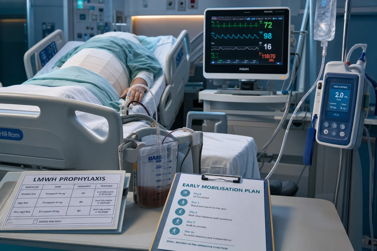
- Pharmacological — the LMWH (enoxaparin 40 mg subcutaneously daily), started within 12-24 hours of the surgery (when the bleeding risk is controlled).
- Mechanical — the intermittent pneumatic compression and the graduated compression stockings.
- Early mobilisation — the most effective prevention.[1]
Pain management — the ERAS principle
The enhanced recovery after surgery (ERAS) protocol uses the multimodal analgesia to reduce the opioid use and to promote the early recovery:[1]
- Paracetamol (the baseline analgesic).
- NSAIDs (caution in the renal impairment and the bleeding risk).
- Regional anaesthesia (an epidural, a nerve block, a local infiltration — the most effective for the postoperative pain).
- Opioids sparingly (the PCA — a patient-controlled analgesia pump; or the scheduled low doses).
- The early mobilisation, the early enteral nutrition, and the fluid restriction (avoid the salt-water overload that delays the gut recovery).[1]
Red flags
7. Risk stratification — who develops complications
Before discussing individual complications, stratify risk — it directs monitoring (telemetry, troponin surveillance, the HDU vs the ICU) and the modifiable prevention. [1]
Perioperative cardiovascular risk stratification — the tools
| Tool | What it scores | High-risk predictors | Use |
|---|---|---|---|
| Revised Cardiac Risk Index (Lee index) | Cardiac death / non-fatal MI after noncardiac surgery | High-risk surgery (intraperitoneal, intrathoracic, suprainguinal vascular), history of IHD, CHF, CVA/TIA, insulin-dependent diabetes, CKD (creatinine >177 µmol/L) | The most validated bedside tool; 0–1 factors = low risk, ≥2 = high |
| NSQIP / MICA (Gupta) | Cardiac arrest or MI | Type of surgery, functional dependence, abnormal CK, ASA class, age | Surgery-specific cardiac risk |
| POSPOM | Operative mortality (UK National) | Surgery-specific category code + age | Population-level 30-day mortality |
| ASA physical status | Overall fitness | Systemic disease severity (ASA III–V) | Crude, ubiquitous; correlates with outcome but not cardiac-specific |
| Functional capacity (METs) | Cardiopulmonary reserve | <4 METs (cannot climb a flight of stairs) = poor; >10 = excellent | If >4 METs and no high-risk features, proceed without further testing |
The key principle from the ACC/AHA guideline[1]: a patient who can exercise to ≥4 METs (climb a flight of stairs, walk up a hill, do heavy housework) without symptoms needs no further cardiac testing before low- or intermediate-risk surgery — "treat the patient, not the number".
8. Cardiac complications — postoperative AF, MI, and heart failure
Cardiac complications are the commonest cause of death after noncardiac surgery. Most events occur in the first 48 hours and many are silent — hence the troponin surveillance in high-risk patients. [1]
Postoperative atrial fibrillation (POAF)
- Cardiac surgery (CABG, valve): occurs in ~30% of patients, peaking on day 2–3 (the post-pericardiotomy syndrome, atrial stretch, the sympathetic surge, and the inflammatory response).
- Noncardiac / thoracic surgery: ~8% after thoracic (especially oesophagectomy and pneumonectomy), lower after general surgery; driven by sympathetic tone, electrolyte shifts, fluid overload, atrial stretch, and right-heart strain.
- Management: rate control first (a beta-blocker — it also treats the sympathetic surge; or a non-dihydropyridine calcium-channel blocker if already beta-blocked). Cardiovert (synchronised DC) if haemodynamically unstable. Anticoagulate by the stroke risk (CHA₂DS₂-VASc), weighing the bleeding risk — the postoperative period is BOTH pro-thrombotic and pro-haemorrhagic. Most POAF is self-limiting within 6–12 weeks. [1]
Myocardial injury after noncardiac surgery (MINS)
- MINS is prognostically important, ischaemic myocardial injury detected ONLY by troponin — without the classic ischaemic features — and it predicts 30-day mortality.[1]
- The VISION study (JAMA 2017) defined MINS by a peak high-sensitivity troponin T ≥0.30 ng/mL within 30 days of surgery and showed a graded, independent association with 30-day mortality.[1]
- The implication for the ICU: troponin surveillance (high-sensitivity troponin at 6–12 h and on day 1–2) in the high-risk patient (age ≥65, prior IHD, vascular surgery, Lee ≥2). Most MINS is Type 2 (supply–demand) rather than Type 1 (plaque rupture) — treat the trigger (tachycardia, hypoxia, hypotension, anaemia), not the cath lab.
Perioperative MI — Type 1 vs Type 2
- Type 1 — plaque rupture/thrombosis: ST-elevation, chest pain; treat by urgent reperfusion (the bleeding risk from recent surgery often precludes fibrinolysis; primary PCI is preferred).
- Type 2 — supply–demand mismatch (tachycardia, hypotension, hypoxaemia, anaemia, spasm): the commonest perioperative mechanism; treat the trigger and the oxygen balance, not the catheter unless the ischaemia is ongoing. [1]
Postoperative heart failure / volume overload
- From third-space fluid mobilisation (the day 2–3 "reabsorption"), iatrogenic fluid overload, diastolic dysfunction in the hypertrophied LV, or new ischaemia.
- Presents as pulmonary oedema on day 2–3 in the elderly — the classic "fluid over the lungs on the day they should be getting better". Treat with oxygen, NIV, an IV loop diuretic; review the fluid balance. [1]
Type 1 vs Type 2 perioperative MI/MINS — the mechanism matters for the treatment
| Feature | Type 1 (plaque rupture) | Type 2 (supply–demand) / MINS |
|---|---|---|
| Mechanism | Atherosclerotic plaque rupture + thrombosis | Imbalance of O₂ supply and demand (tachycardia, hypotension, hypoxaemia, anaemia, fever, pain) |
| ECG | Often ST-elevation or new regional change | Often non-specific, T-wave inversion, or normal |
| Symptoms | Chest pain (if not sedated/analgesed) | Frequently silent — detected on troponin surveillance |
| Frequency postop | The minority | The majority of perioperative events |
| Treatment | Reperfusion (primary PCI preferred; fibrinolysis limited by the surgical bleeding); DAPT with caution | Treat the trigger — control the HR and the pain, correct the hypoxia/anaemia, restore the perfusion; do not reflexively catheterise |
| Troponin | A rise and fall with ischaemic features | A rise (often a single peak); defines MINS when ≥0.30 ng/mL |
Management of postoperative atrial fibrillation — rate vs rhythm vs anticoagulation
| Strategy | When | Agent / action | Pearl |
|---|---|---|---|
| Rate control (first-line) | Stable POAF | IV metoprolol 5 mg q5min (treats the sympathetic surge) or diltiazem if beta-blocked; amiodarone if LV dysfunction or decompensation | Target HR <110 if stable; <80 if unstable or in HF |
| Rhythm control | Instability, HF, failed rate control, first onset <48 h | Synchronised DC cardioversion; or IV amiodarone 300 mg load for pharmacological | New-onset AF <48 h — cardiovert; the postoperative autonomic storm often self-terminates |
| Anticoagulation | CHA₂DS₂-VASc ≥2 (weigh the bleeding) | Heparin/LMWH once the surgical bleeding is controlled; DOAC avoided early | The postoperative state is BOTH pro-thrombotic and pro-haemorrhagic — individualise |
| Treat the triggers | Always | Correct K⁺ (>4.0), Mg²⁺ (>0.8), hypoxia, pain, fluid overload | Most POAF resolves within 6–12 weeks — the anticoagulation may not be lifelong |
Cardiac complications — the landmark trials
POISE (Devereaux, Lancet 2008): 8351 patients, perioperative metoprolol succinate vs placebo started 2–4 h before surgery. Result: metoprolol reduced MI, cardiac revascularisation, and clinically significant atrial fibrillation BUT increased stroke and total mortality (and hypotension/bradycardia). Lesson: perioperative beta-blockade must NOT be started at a high dose immediately before surgery; if used, start days ahead and titrate cautiously; avoid in hypotension/bradycardia.[1] POISE-2 (Devereaux, NEJM 2014): aspirin vs placebo and clonidine vs placebo (2×2 factorial, 10 010 patients). Aspirin did not reduce death or non-fatal MI and increased major bleeding; clonidine did not reduce MI/death and increased clinically important hypotension and bradycardia. Lesson: aspirin is NOT a routine perioperative prophylaxis; stop the clonidine preop.[1] VISION (JAMA 2017): >15 000 patients, high-sensitivity troponin T surveillance. Defined MINS (peak hs-cTnT ≥0.30 ng/mL, ischaemic or not) and showed a graded, independent association with 30-day mortality (≈10% with MINS vs ≈2% without). Lesson: troponin surveillance in the high-risk surgical patient finds the silent, deadly injury.[1]
9. Respiratory complications — the postoperative pulmonary complications (PPCs)
PPCs (atelectasis, pneumonia, respiratory failure, exacerbation of the underlying lung disease) are as common and as deadly as the cardiac complications, and they are largely preventable. [1]
Preventing the postoperative pulmonary complications — the evidence-based bundle
- Preoperative risk stratification and optimisation. Identify the high-risk patient (age >60, COPD, smoker, obesity, OSA, poor functional capacity, upper abdominal/thoracic/aortic surgery, long duration). Optimise the COPD, treat the infection, encourage the smoking cessation (>4 weeks ideal, but even <8 weeks does not increase the PPC risk — do not defer).
- Lung-protective intraoperative ventilation. Tidal volume 6–8 mL/kg predicted body weight, PEEP 5–8 cmH₂O, recruitment manoeuvres. Avoid a high driving pressure.
- Early extubation and early mobilisation. The single most effective preventive measure — mobilise on day 0 if possible.
- Multimodal analgesia to avoid splinting. A regional technique (epidural, paravertebral, erector spinae plane) reduces the splinting and the opioids. Avoid a large systemic opioid load.
- Lung expansion — incentive spirometry, deep breathing, chest physiotherapy. All modestly reduce the atelectasis; the evidence favours the deep breathing, but both are cheap and safe.
- Adequate but not excessive fluid — overload causes the pulmonary interstitial oedema and worsens the gas exchange (see RELIEF).[1]
- VTE prophylaxis and gastric acid suppression where indicated — prevent the PE and the stress ulcer.
The common postoperative respiratory complications — recognise and treat
| Complication | When / why | Hallmark | Treatment |
|---|---|---|---|
| Atelectasis | Day 0–1; anaesthesia, splinting, supine, reduced mucociliary clearance; the commonest PPC | Hypoxaemia, basal crackles, a plateau on the CXR; rapid improvement with deep breathing | Physiotherapy, incentive spirometry, analgesia, mobilisation, CPAP if refractory |
| Pneumonia (HAP/VAP) | Day 3+; aspiration, atelectasis, prolonged intubation | Fever, purulent sputum, a new infiltrate, a rising WCC | Antibiotics per the local protocol; send cultures first; a lung-protective wean |
| Pulmonary embolism | Day 3–7; DVT from immobility, the prothrombotic state | Sudden dyspnoea, pleuritic pain, hypoxaemia, RV strain; often unexpected | Anticoagulation; thrombolysis if massive (see the VTE section) |
| Aspiration pneumonitis / pneumonia | Peri-induction or with ileus/reduced consciousness | Bilateral basal infiltrates, hypoxaemia, often the right lower lobe | Supportive; antibiotics only if a secondary infection; lung-protective ventilation if severe |
| Pulmonary oedema | Day 2–3; fluid overload, negative-pressure (post-obstructive), ischaemia | Pink frothy sputum, crackles, B-lines | Sit up, oxygen, NIV, an IV loop diuretic; treat the trigger |
| ARDS | After sepsis, trauma, massive transfusion, aspiration | Bilateral infiltrates, refractory hypoxaemia, low compliance | Lung-protective ventilation (6 mL/kg, plateau <30), permissive hypercapnia, prone |
10. Postoperative AKI — KDIGO and the preventable causes
Postoperative AKI is common, independently predicts mortality, and is usually multi-factorial and preventable. Apply the KDIGO bundle. [1]
Causes of the postoperative AKI — pre-renal, intrinsic, post-renal
| Category | Postoperative mechanisms | Key tests / clues | Treatment principle |
|---|---|---|---|
| Pre-renal (commonest) | Bleeding, third-space losses, vasodilation from the anaesthesia/neuraxial block, hypotension, low cardiac output | FENa <1%, BUN:Cr >20, muddy-brown casts absent, a rapid response to the fluids | Restore the perfusion — fluids, blood, inotrope/vasopressor; treat the cause |
| Intrinsic (ATN) | Nephrotoxins (NSAIDs, contrast, aminoglycosides, vancomycin), rhabdomyolysis, sepsis, prolonged hypoperfusion, abdominal compartment syndrome | FENa >2%, muddy-brown casts, slow/absent recovery, a high CK in rhabdo | Stop the nephrotoxin; aggressive fluids for rhabdo; treat sepsis; decompress if ACS |
| Post-renal (exclude early) | A blocked catheter, benign prostatic hypertrophy, surgical ureteric injury, retroperitoneal bleed | Bladder scan, renal ultrasound (hydronephrosis) | Relieve the obstruction — catheter, urology |
The KDIGO bundle for preventing the postoperative AKI
- Identify the high-risk patient — age >65, CKD, diabetes, heart failure, emergency surgery, high-risk surgery (cardiac, vascular, hepatobiliary), sepsis, large fluid shifts, iodinated contrast.
- Avoid the nephrotoxins where possible — NSAIDs (afferent arteriolar constriction), aminoglycosides, vancomycin, iodinated contrast; if essential, dose-adjust and monitor the levels.
- Maintain the perfusion — adequate haemodynamics (MAP, cardiac output), treat the hypovolaemia with balanced crystalloid, avoid the prolonged hypotension.
- Use the functional haemodynamic monitoring if unstable — dynamic indices (SVV, PPV, passive leg raise, fluid challenge) to guide the fluid and the vasopressor; avoid both the under- and the over-resuscitation.
- Contrast prophylaxis — isotonic hydration (sodium chloride or bicarbonate) before and after the contrast; minimise the contrast volume; consider an alternative imaging; hold the metformin if AKI.
- Monitor closely — urine output (<0.5 mL/kg/h for >6 h is a KDIGO criterion), serum creatinine trend; do not wait for a creatinine rise (it lags) to act.
- Do NOT use the low-dose dopamine or fenoldopam — proven futile; the renal-dose dopamine myth is dead.
- Renal replacement therapy for the indications — refractory hyperkalaemia, acidosis, fluid overload, uraemic complications.
AKI and fluids — the RELIEF trial
RELIEF (Miller & Myles, NEJM 2018): 3000 patients undergoing major abdominal surgery, a restrictive (target a balanced neutral/slightly negative balance) vs a liberal IV fluid regimen. Result — the restrictive arm was WORSE: more AKI, more surgical-site infection, more delayed gastric emptying. Lesson: the pendulum swung too far toward the "dry surgery"; a balanced, near-neutral strategy — enough to maintain the perfusion without the salt-water overload — is safest. The dogma that aggressive fluid restriction is always better is wrong; the under-resuscitation causes AKI and wound hypoperfusion.[1]
11. Gastrointestinal complications
The common postoperative GI complications
| Complication | Setting / mechanism | Hallmark | Management |
|---|---|---|---|
| Postoperative ileus | After abdominal/pelvic surgery; sympathetic overactivity, opioid use, electrolyte disturbance, immobility | No flatus/stool, distension, absent bowel sounds, nausea | ERAS bundle — minimise opioids, early feeding, mobilise, correct K⁺/Mg²⁺, chew gum; NGT only if vomiting; alvimopan in select centres |
| Anastomotic leak | Day 3–7 after bowel surgery; ischaemia/tension at the join | Fever, tachycardia, abdominal pain, rising lactate, ileus; CT with contrast (extraluminal contrast, free gas) | Surgical re-exploration + diversion; broad-spectrum antibiotics; source control |
| Stress-related mucosal disease / stress ulcer | Critical illness, prolonged ventilation, coagulopathy, shock, major burns, neurotrauma | Overt or occult UGIB on day 3–7 | PPI or H₂RA prophylaxis ONLY in the high-risk (see below) |
| Acute colonic pseudo-obstruction (Ogilvie) | After orthopaedic, pelvic, or retroperitoneal surgery; electrolyte disturbance, opioids | Massive caecal dilatation without mechanical obstruction; perforation risk if caecum >12 cm | Conservative first — decompression, neostigmine; colonoscopic decompression; surgery if ischaemia/perforation |
| Acalculous cholecystitis | Critical illness, prolonged starvation, TPN, sepsis | RUQ pain, fever, gallbladder distension without stones on US | Percutaneous cholecystostomy; antibiotics |
Stress ulcer prophylaxis — who actually benefits
| Indication (give prophylaxis) | Rationale | Drug |
|---|---|---|
| Mechanical ventilation >48 h | The highest single risk factor | IV PPI (e.g. pantoprazole 40 mg daily) or H₂RA |
| Coagulopathy (INR >1.5, platelets <50, on anticoagulants) | The bleeding compounds the risk | PPI |
| Shock, major burns (>30% TBSA), severe neurotrauma (GCS ≤8) | Splanchnic hypoperfusion | PPI |
| Not in a high-risk group? | SUP-ICU (NEJM 2018): pantoprazole did NOT significantly change the 90-day mortality vs placebo in the unselected ICU patient | Do NOT routinely prescribe — adds C. difficile and pneumonia risk |
12. Infectious complications — SSI, line, and urinary
Surgical wound classification and infection risk
| Class | Definition | Example | Infection rate |
|---|---|---|---|
| Clean | Elective, no inflammation, no break in technique, GI/respiratory/GU tract not entered | Hernia repair, joint replacement | ~1–3% |
| Clean-contaminated | GI/respiratory/GU tract entered under controlled conditions, no major spillage | Elective bowel resection, cholecystectomy | ~5–10% |
| Contaminated | Major break in technique, GI spillage, fresh trauma, acute non-purulent inflammation | Appendicectomy (perforated) | ~15–20% |
| Dirty / infected | Purulent inflammation, perforated viscus, old traumatic wound | Faecal peritonitis, abscess drainage | >20% |
The surgical-site infection prevention bundle
- Preoperative antiseptic shower and skin preparation — chlorhexidine-alcohol preferred over povidone-iodine (alcohol-based, allow to dry).
- Appropriate antibiotic prophylaxis — within 60 min of incision (vancomycin/fluoroquinolone within 120 min); re-dose for the long surgery (>2 half-lives) or the major blood loss (>1.5 L); stop within 24 h postop (no benefit to the prolonged courses).
- Normothermia — maintain >36 °C (active warming); hypothermia triples the SSI risk.
- Normoglycaemia — control the perioperative glucose (avoid both the hyper- and the hypoglycaemia); an insulin infusion in the diabetics.
- Adequate but not excessive fluid and oxygen — avoid the tissue hypoperfusion; the wound needs oxygen to heal and to kill bacteria.
- Hair removal only if necessary, with clippers (not razors, which nick the skin).
- Early removal of drains and catheters — each device is a foreign body and an infection highway.
13. Neurological complications — delirium, stroke, and cognitive decline
Postoperative delirium (POD)
POD affects up to 50% of the elderly surgical patients and independently predicts a longer stay, institutionalisation, and mortality. It is hypoactive as often as hyperactive — the quiet, withdrawn patient is missed. [1]
Preventing and managing the postoperative delirium — non-pharmacological FIRST
- Screen for the risk — age >65, cognitive impairment, sensory impairment (hearing/vision), functional dependence, prior delirium, frailty, polypharmacy.
- Multicomponent non-pharmacological prevention (the HELP model) — orientation (clock, calendar, window), sensory re-enablement (hearing aids, glasses), sleep protection (lights off at night, cluster the care), early mobilisation, hydration and nutrition, treat the constipation and the urinary retention.
- Treat the cause (always) — pain (under-treated pain causes the agitation), hypoxia, hypotension, infection (occult sepsis, urine, chest, wound), electrolytes (Na⁺, Ca²⁺, glucose), drugs (benzodiazepines, anticholinergics, opioids — review and stop), urinary retention, constipation, alcohol withdrawal.
- Assess with the CAM-ICU (intubated) or CAM (ward) — inattention is the cardinal feature.
- Pharmacological management ONLY if the patient is a danger to self or others — dexmedetomidine (preferred — does not worsen the delirium), or low-dose haloperidol/quetiapine. AVOID the benzodiazepines except alcohol/benzodiazepine withdrawal.
Postoperative stroke and cognitive decline
- Postoperative stroke is rare overall (~0.1–1%) but catastrophic; the risk is highest after cardiac/carotid surgery and in atrial fibrillation. The mechanisms include the embolism (atheroma from aortic manipulation, paradoxical through a PFO), hypoperfusion, and coagulopathy. Treat as any acute stroke — but weigh the recent surgery against the thrombolysis.
- Postoperative cognitive decline (POCD) — subtle, detectable on neuropsychological testing weeks to months after surgery, more common in the elderly; multifactorial (inflammation, hypotension, drugs, delirium). The link to the anaesthetic agent is unproven (countering the "general anaesthesia causes dementia" lay concern — the evidence is weak). [1]
Delirium vs POCD vs postoperative stroke — distinguish them
| Feature | Postoperative delirium (POD) | Postoperative cognitive decline (POCD) | Postoperative stroke |
|---|---|---|---|
| Onset | Hours–days | Weeks–months | Intraoperative or first 24–48 h |
| Course | Acute, fluctuating, reversible | Subtle, gradual, may persist | Acute focal deficit |
| Hallmark | Inattention, fluctuating consciousness, disorganised thinking (CAM-ICU/CAM) | Decline on neuropsychological testing (memory, executive function) | Focal neurological deficit |
| Associations | Age, cognitive impairment, frailty, polypharmacy, infection, pain | Age, delirium, inflammation, hypotension | AF, carotid/cardiac surgery, aortic atheroma |
| Management | Multicomponent prevention; treat the cause; dexmedetomidine; avoid the benzodiazepines | Prevent the delirium, optimise the perfusion; rehabilitation | Acute stroke pathway; weigh the fibrinolysis against the surgical bleeding |
14. ERAS — Enhanced Recovery After Surgery in detail
ERAS is an evidence-based, multimodal, standardised perioperative care pathway that reduces complications, shortens stay, and accelerates recovery by attenuating the surgical stress response. [1]
Traditional perioperative care vs the ERAS pathway
| Element | Traditional | ERAS |
|---|---|---|
| Preoperative fasting | NPO from midnight | Clear fluids until 2 h, solids until 6 h; a carbohydrate drink 2 h preop |
| Bowel preparation | Routine mechanical prep | Selective; oral antibiotics only for specific colorectal indications |
| Intraoperative fluid | Liberal, saline-heavy | Goal-directed, balanced crystalloid, near-neutral balance |
| Analgesia | Systemic opioids | Multimodal — regional technique, paracetamol, NSAID, opioid-sparing |
| Drains / tubes | Routine NGT, drains | Avoid or remove day 0–1 |
| Feeding | Delayed until flatus | Early oral feeding within 24 h |
| Mobilisation | Bed rest | Early mobilisation day 0 |
| Glycaemia / temperature | Variable | Normoglycaemia, normothermia |
The ERAS pathway — the day-by-day milestones
- Pre-admission (weeks before). Optimise the nutrition, the exercise (prehabilitation), the smoking/alcohol cessation, the anaemia correction, and the counselling. Risk-stratify and plan the postoperative destination.
- Preoperative (day of surgery). No prolonged fasting — clear fluids to 2 h and a carbohydrate drink; NO routine bowel prep or long-acting sedatives; VTE prophylaxis; antibiotic within 60 min of incision.
- Intraoperative. Short-acting anaesthesia, lung-protective ventilation, normothermia, normoglycaemia, goal-directed fluid, regional analgesia, minimise drains and tubes.
- Day 0 (PACU/HDU). Early extubation, regional analgesia running, oral intake started, sit out of bed and mobilise, remove catheter/NGT if safe, VTE prophylaxis.
- Day 1–2. Normal diet, regular oral analgesia, full mobilisation, remove drains/lines, monitor for complications.
- Day 3+ / discharge. Continued mobilisation, a planned discharge with clear written instructions and a structured follow-up. The readmission pathways defined.
15. Postoperative fluid management — the four phases
The postoperative fluid management has four phases (Miller/Myles); the goal is euvolaemia, not a fixed number. [1]
The four phases of the perioperative fluid management
- Phase 1 — Resuscitation (salvage). Treat the hypovolaemia and the shock with boluses of balanced crystalloid or blood; restore the perfusion (lactate, capillary refill, urine output, MAP). Do NOT under-resuscitate — the RELIEF trial showed that the dry patient gets AKI and wound hypoperfusion.[1]
- Phase 2 — Maintenance. Replace the ongoing losses and maintain a near-neutral balance; balanced crystalloid at ~1–2 mL/kg/h, correct the electrolytes; avoid the salt-water overload.
- Phase 3 — Stabilisation/reabsorption (day 2–3). The third-space fluid returns to the intravascular space; reduce or stop the fluids, allow a negative balance, and diurese only if overloaded. This is where most fluid mismanagement happens — the patient "gets wet" on the day they should be drying.
- Phase 4 — Discharge/recovery. The oral intake established; the gut is the safest route. Stop the IV fluids when the patient is eating and drinking.
Balanced crystalloid vs 0.9% saline in the postoperative patient
| Fluid | Composition / effect | Advantage | Caution |
|---|---|---|---|
| Balanced crystalloid (Hartmann's, Plasma-Lyte) | Lactate/acetate-gluconate buffer; chloride closer to plasma | Lower risk of hyperchloraemic metabolic acidosis, AKI, and renal vasoconstriction — preferred for resuscitation (SMART, SALT-ED) | Slightly hypotonic; caution in TBI (Plasma-Lyte may lower the sodium) |
| 0.9% saline | 154 mmol/L Na and Cl | Cheap, compatible with blood, the classic fluid | Hyperchloraemic metabolic acidosis, renal vasoconstriction (tubuloglomerular feedback), AKI with large volumes — limit to <2 L or blood products |
| Colloids (albumin, starches) | Large molecules, oncotic | Theoretical volume-sparing | No mortality benefit; starches increase AKI (CHEST, 6S, CRISTAL) — avoid; albumin only for select resuscitation |
16. Multimodal analgesia in detail
The principle: combine mechanism-different, opioid-sparing analgesics so that each works at a different point in the pain pathway, achieving a better pain control with fewer side effects. [1]
Multimodal analgesia — the agents and their place
| Agent / technique | Mechanism / site | Role | Key cautions |
|---|---|---|---|
| Paracetamol (acetaminophen) | Central (COX, descending serotonergic) — uncertain | Baseline in every regimen; opioid-sparing ~20–30% | Hepatotoxicity in overdose; dose-reduce in low body weight or liver disease |
| NSAIDs (ibuprofen, ketorolac, diclofenac) | Peripheral and central COX inhibition | Potent opioid-sparing; anti-inflammatory | AKI (afferent arteriole), bleeding (platelet), GI ulcer, anastomotic leak association (avoid in colorectal anastomosis), asthma |
| Gabapentinoids (gabapentin, pregabalin) | Voltage-gated calcium channel (α2δ) | Neuropathic and opioid-sparing | Sedation, dizziness, delirium in elderly — dose-reduce; not routine in the older patient |
| Ketamine (subanaesthetic, 0.1–0.3 mg/kg/h) | NMDA receptor antagonist | Opioid-induced hyperalgesia, opioid tolerance, severe acute pain, burn dressing changes | Hallucinations; hypertension |
| Lidocaine infusion (1–2 mg/kg/h) | Sodium channel blockade; anti-inflammatory | Open and laparoscopic abdominal surgery — opioid-sparing, hastens the ileus recovery | Toxicity (perioral tingling, seizures); avoid in heart block; stop on feeding |
| α₂-agonists (dexmedetomidine) | Central α₂A | Sedation without respiratory depression; opioid-sparing; preferred in delirium | Bradycardia, hypotension — titrate |
| Regional (epidural, ESP, rectus sheath, TAP, paravertebral) | Local anaesthetic at nerve plexus | Most effective for the dynamic pain; the cornerstone of opioid-sparing ERAS | Hypotension (sympathectomy with neuraxial), motor block, infection, LAST |
| Opioids (morphine PCA, fentanyl, oxycodone) | µ-receptor | For breakthrough and where regional/adjuncts insufficient — sparingly | Sedation, respiratory depression, ileus, delirium, nausea, dependence |
Regional analgesia techniques — pick the right block
| Technique | Coverage | Best for | Pearl |
|---|---|---|---|
| Thoracic epidural | Dermatomal band; sympathetic block | Thoracic, upper abdominal; multiple rib fractures | Gold standard for thoracoabdominal dynamic pain; causes hypotension (sympathectomy) and may mask the compartment syndrome; contraindicated with anticoagulation |
| Paravertebral block | Unilateral dermatomal | Thoracic surgery (mastectomy, thoracotomy), rib fractures | Unilateral, less hypotension than epidural; equivalent analgesia for thoracic |
| Erector spinae plane (ESP) block | Posterior thoracic/abdominal wall | Rib fractures, mastectomy, abdominal wall | Fascial plane block — safer than neuraxial (no epidural haematoma risk); increasingly used in ERAS |
| Transversus abdominis plane (TAP) block | Anterolateral abdominal wall (T7–L1) | Lower abdominal surgery (caesarean, hernia, appendicectomy) | Somatic wall pain only — does NOT cover visceral pain; pair with paracetamol/NSAID |
| Rectus sheath block | Anterior midline abdominal wall | Midline laparotomy, umbilical surgery | Complements the TAP for the midline incisions |
Building a multimodal analgesia regimen for the postoperative ICU patient
- Establish a regional technique intraoperatively (epidural, ESP, TAP, or rectus sheath) — the cornerstone of opioid-sparing analgesia; continue the infusion/PCA postop.
- Schedule paracetamol 1 g q6h (regular) as the baseline opioid-sparing agent.
- Add an NSAID (e.g. ibuprofen 400 mg q8h or ketorolac) — if the renal function, the bleeding risk, the anastomosis, and the asthma allow; usually for 48–72 h only.
- Consider a ketamine infusion (0.1–0.3 mg/kg/h) for the opioid-tolerant patient or the severe pain; a lidocaine infusion (1–2 mg/kg/h) for the abdominal surgery if no contraindication.
- Use an opioid PCA (morphine or fentanyl) for breakthrough, with the goal of weaning as the regional technique and the adjuncts take effect.
- Avoid the sedatives (benzodiazepines) and minimise the gabapentinoids in the elderly (delirium). Use dexmedetomidine if a sedation is needed in a delirious patient.
- Reassess the pain and the sedation every shift (NRS or CPOT; RASS); transition from IV to oral (paracetamol + NSAID + oral opioid) as the patient tolerates the intake.
17. VTE prophylaxis — risk-stratified and timed
VTE risk and prophylaxis — match the strategy to the risk
| Risk (Caprini/Padua) | Examples | Prophylaxis |
|---|---|---|
| Low | Minor surgery <30 min, fully mobile, no risk factors | Early mobilisation; pharmacological not required |
| Moderate | Most general surgery, age 40–60, minor risk factors | LMWH (e.g. enoxaparin 40 mg SC daily) or mechanical |
| High | Major surgery, age >60, cancer, prior VTE, hip/knee replacement, trauma | LMWH + mechanical (IPC); consider extended prophylaxis (28–35 days) after cancer/hip surgery |
| Very high / bleeding | Active bleeding, neurosurgery, recent spinal anaesthetic | Mechanical (IPC + GCS) until bleeding controlled, then add LMWH |
Additional red flags
[1] [1] [1]The high-yield mnemonic
BIGThe BIG SIX postoperative complications (and the four pillars of prevention)
SAQ — Fellowship exam practice
SAQ — Postoperative respiratory failure after upper abdominal surgery
10 minutes · 10 marks
A 68-year-old man (BMI 32, ex-smoker, eGFR 55) is day 2 following an emergency open Whipple procedure for a periampullary malignancy (operative time 6 h, estimated blood loss 1.2 L, intraoperative fluids 4 L crystalloid and 2 units packed red cells). He is on an ICU patient-controlled analgesia morphine infusion (PCA demands 14/h), a thoracic epidural (running bupivacaine 0.125 percent with fentanyl 2 mcg/mL at 8 mL/h, last bolus 4 h ago), and was extubated 6 hours ago. He is now increasingly tachypnoeic (RR 32), SpO2 89 percent on 6 L Hudson mask, HR 118 sinus, BP 138/82, afebrile, drowsy but rousable. ABG: pH 7.30, PaCO2 50 mmHg, PaO2 56 mmHg, HCO3 24, lactate 1.6. Chest X-ray shows bilateral basal atelectasis with elevation of both hemidiaphragms, no consolidation, no effusion, no pneumothorax. Surgical wound and drains are clean; epidural site is unremarkable; legs are weak but can wiggle toes bilaterally.
SAQ — Postoperative atrial fibrillation after lobectomy with haemodynamic compromise
10 minutes · 10 marks
A 72-year-old man is day 3 following a right upper lobectomy for non-small cell lung cancer (preoperative LVEF 55 percent, normal sinus rhythm on ECG, on atenolol 25 mg daily for hypertension). Surgery was uncomplicated; he is on a thoracic paravertebral block with bupivacaine infusion, paracetamol 1 g q6h, and a small morphine PCA (10 mg over the last 24 h). He suddenly becomes acutely breathless, sweaty, and confused. HR 162 irregularly irregular, BP 82/48 (MAP 59), RR 30, SpO2 88 percent on 4 L nasal cannulae, T 37.4 C. JVP is elevated 6 cm; chest auscultation shows the surgical side clear, the non-operated side with bibasal crepitations. ECG confirms new atrial fibrillation with a rapid ventricular response (no P waves, irregularly irregular narrow-complex tachycardia, rate 158). K+ 4.8 mmol/L, Mg2+ 0.78 mmol/L, Hb 105 g/L, troponin 28 ng/L (normal), lactate 2.4 mmol/L, ABG pH 7.28 PaO2 62 PaCO2 38 HCO3 19.
References
- [1]Kurz V, Kurz M, Fuchs C, et al The surgical safety checklist and patient outcomes after surgery: a prospective observational cohort study, systematic review and meta-analysis. British Journal of Anaesthesia, 2018.PMID 29397122
- [2]Devereaux PJ, Yang H, Yusuf S, et al Effects of extended-release metoprolol succinate in patients undergoing non-cardiac surgery (POISE trial): a randomised controlled trial. Lancet, 2008.PMID 18479744
- [3]Devereaux PJ, Mrkobrada M, Sessler DI, et al Aspirin in patients undergoing noncardiac surgery. N Engl J Med, 2014.PMID 24679062
- [4]Writing Committee for the VISION Study Investigators Association of Postoperative High-Sensitivity Troponin Levels With Myocardial Injury and 30-Day Mortality Among Patients Undergoing Noncardiac Surgery. JAMA, 2017.PMID 28444280
- [5]Myles PS, Bellomo R, Corcoran T, et al (RELIEF) Restrictive versus Liberal Fluid Therapy for Major Abdominal Surgery. N Engl J Med, 2018.PMID 29742967
- [6]Gustafsson UO, Scott MJ, Schwenk W, et al Guidelines for perioperative care in elective colonic surgery: Enhanced Recovery After Surgery (ERAS(®)) Society recommendations. World J Surg, 2013.PMID 23052794
- [7]Lassen K, Soop M, Nygren J, et al Consensus review of optimal perioperative care in colorectal surgery: Enhanced Recovery After Surgery (ERAS) Group recommendations. Arch Surg, 2009.PMID 19841366
- [8]Fleisher LA, Fleischmann KE, Auerbach AD, et al 2014 ACC/AHA guideline on perioperative cardiovascular evaluation and management of patients undergoing noncardiac surgery: a report of the American College of Cardiology/American Heart Association Task Force on practice guidelines. J Am Coll Cardiol, 2014.PMID 25091544