MedVellum
MCQsExamsAtlas
DashboardPricing
MBBS / Core medicine✳Dermatology✳ICU Fellowship (CICM)✳Anaesthesia✳Emergency Medicine✳Psychiatry Fellowship✳Paediatrics Fellowship✳Physician Medicine✳MCQs✳SAQs✳Vivas✳OSCE✳Evidence-first✳MBBS / Core medicine✳Dermatology✳ICU Fellowship (CICM)✳Anaesthesia✳Emergency Medicine✳Psychiatry Fellowship✳Paediatrics Fellowship✳Physician Medicine✳MCQs✳SAQs✳Vivas✳OSCE✳Evidence-first✳

MedVellum.

The folio

Exam-exhaustive medical education across every specialty — evidence-graded topics, engraved plates, and practice in every written and oral format. Educational content only — not medical advice.

llms.txt · psychiatry LLM catalog · sitemap

Atlas

  • Specialty atlas
  • MBBS / Core medicine
  • Dermatology
  • ICU Fellowship (CICM)
  • Anaesthesia
  • Emergency Medicine
  • Psychiatry Fellowship
  • Paediatrics Fellowship
  • Physician Medicine

Study & account

  • MCQ practice
  • Practice alias
  • Exam tools
  • Dashboard
  • Pricing
  • Sign in

© 2026 MedVellum. For education only — not a substitute for clinical judgement.

Folio edition · Set in Instrument Serif & Archivo

ICU TopicsCardiovascular

ICU · Cardiovascular

Acute pulmonary embolism: PESI risk stratification, thrombolysis, and PERT

Also known as Pulmonary embolism · PE · PESI · Massive PE · Submassive PE · Catheter-directed thrombolysis · PERT

Acute pulmonary embolism risk stratification with the PESI and simplified PESI scores, systemic and catheter-directed thrombolysis, and the role of the pulmonary embolism response team (PERT).

high19 referencesUpdated 1 July 2026
On this page & tools

Your progress

Saved locally on this device.
Your progress

Saved locally on this device.

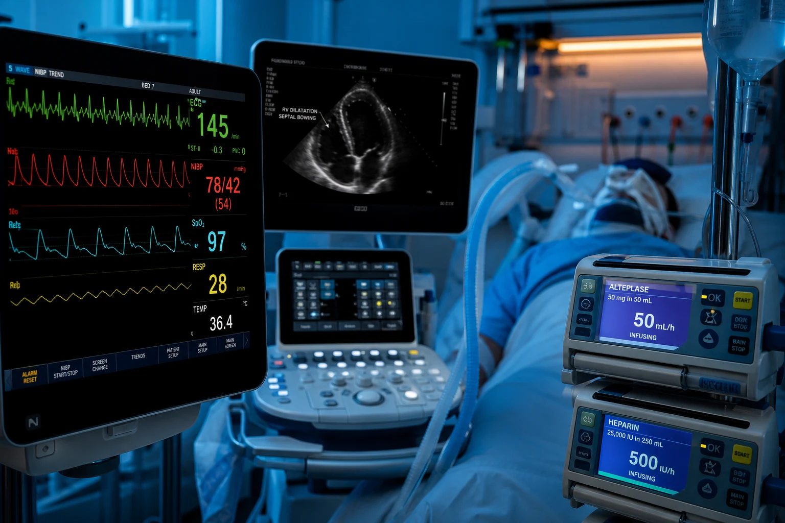
Cinematic ICU scene of a CT pulmonary angiogram on the screen showing a large central pulmonary embolism, an echocardiogram showing right ventricular dilatation, a PESI score on a clipboard, a PERT team activation board, clinical-blue lighting, medical educational, no faces, no text
FigureThe pulmonary embolism in the ICU — the PESI stratifies the risk, the echo finds the right ventricular dysfunction, the biomarkers the strain. The massive (the shock) is the thrombolysis or the embolectomy; the submassive is the anticoagulation and the monitor. The PERT is the multidisciplinary response.

In one line

PE risk stratification (ESC): HIGH ('massive' — shock/hypotension SBP <90) → thrombolysis (alteplase 100 mg/2h) or embolectomy. INTERMEDIATE-HIGH ('submassive' — RV dysfunction + troponin) → monitor ICU, thrombolysis if deteriorating. INTERMEDIATE-LOW (RV or troponin alone) → ward. LOW (neither) → anticoagulation, often home. Diagnosis: CTPA (gold standard), D-dimer (rule out if low risk), echo (RV strain). Management: anticoagulation for ALL (LMWH → DOAC); thrombolysis for high-risk or deteriorating; embolectomy if lysis contraindicated; catheter-directed thrombolysis (SEATTLE II — lower bleeding) for intermediate-high. PERT (multidisciplinary team) for complex.

[1]

PE risk categories (ESC/ERS)

Risk categoryClinicalRV functionTroponinsPESI ≥1ManagementMortality
HIGH ('massive')Shock/hypotension (SBP <90)Dysfunction (implied)Elevated (usually)≥1THROMBOLYSIS or embolectomy15-30%
INTERMEDIATE-HIGH ('submassive')Haemodynamically stableDysfunction (echo/CT)Positive≥1Monitor ICU; thrombolysis if deteriorating5-10%
INTERMEDIATE-LOWStableDysfunction OR troponin (not both)Variable≥1Ward; monitor2-5%
LOWStableNormalNormal0Anticoagulation; often home<1%
[1]

Reperfusion strategies for high-risk and intermediate-high PE

StrategyIndicationDose/techniqueBleeding riskOnsetKey trialNotes
Systemic thrombolysisHIGH-risk (massive — shock); deteriorating intermediate-highAlteplase 100 mg IV over 2 h (or 50 mg over 2 h; tenecteplase 30–50 mg bolus — PEITHO)HIGH (major bleed ~11%, ICH ~2% in PEITHO)MinutesPEITHO 2014Fastest; absolute contraindications often preclude; stop heparin during infusion
Catheter-directed thrombolysis (CDT)Intermediate-high with bleeding risk; selected high-riskAlteplase 20–24 mg per catheter over 12–24 h ± ultrasound (EKOS)LOW (no major bleed in SEATTLE II)HoursSEATTLE II 2015; ULTIMA 2014~1/5 the systemic dose; requires IR/endovascular capability; ICU monitoring
Percutaneous thrombectomyHigh-risk if lysis contraindicated/failed; large central clotAspiration (FlowTriever, Indigo) or rheolytic fragmentationModerate (vascular access + haemolysis)ImmediatePERFECT 2015; FLASH registryNo lytic = no lysis bleeding; useful in absolute lysis contraindications
Surgical embolectomyClot-in-transit; lysis failed/contraindicated; concurrent cardiac lesionMedian sternotomy, CPB, pulmonary arteriotomyModerate–high (surgical)Immediate—Requires on-call CT surgery; best for central saddle embolus + PFO
Anticoagulation aloneLow-risk; intermediate-low; stable intermediate-highLMWH → DOACMinimalDaysEINSTEIN-PE; AMPLIFYDefault for ALL; bridge to DOAC
[1]

Anticoagulant agents in acute PE — DOAC vs LMWH vs warfarin

Agent/classOnsetMonitoringReversalRenal clearanceFirst-line settingKey trial
LMWH (enoxaparin 1 mg/kg SC BD)Rapid (1–2 h)Anti-Xa (rarely)Protamine (partial)RenalInitial (all PE); cancer; pregnancy; DOAC contraindicationCLOT 2003 (vs warfarin in cancer)
RivaroxabanOral, 2–4 hNoneAndexanet alfaHepatic + renal (1/3)First-line; 15 mg BD ×21d then 20 mg OD; no lead-in LMWHEINSTEIN-PE 2012
ApixabanOral, 3–4 hNoneAndexanet alfaRenal (27%)First-line; 10 mg BD ×7d then 5 mg BD; no lead-in LMWHAMPLIFY 2013; Caravaggio 2020 (cancer)
DabigatranOral, 1–2 hNoneIdarucizumabRenal (80%)After 5–10 d parenteral lead-in; not first-lineRE-COVER 2009
EdoxabanOral, 1–2 hNoneAndexanet alfaRenal (50%)After 5 d LMWH lead-in; cancer (non-GI)Hokusai VTE-Cancer 2018
Warfarin (INR 2–3)Days (slow)INRVitamin K + PCC/FFPHepaticPregnancy past? No; APS; mechanical valve; DOAC contraindication—
FondaparinuxRapidNoneNone (rFVIIa)RenalHeparin allergy (HIT); weight-based SC—
[1]

RV strain markers — diagnostic and prognostic

MarkerModalityFindingThresholdSpecificity for PERisk stratification role
RV:LV ratioEcho (apical 4-chamber) / CTRV dilation>0.9 (or >0.6 echo)ModerateIntermediate-risk indicator (ESC)
Septal flatteningEcho (PSAX)D-shaped LV, systolic flatteningVisualModeratePressure/volume overload
McConnell's signEchoRV apex hyperkinetic, free wall hypokineticVisualHIGH (~94% for PE)Highly suggestive; supports empirical lysis if unstable
TAPSEEcho (M-mode)Tricuspid annular systolic excursion<17 mm = RV dysfunctionLowQuantitative RV function; <17 mm predicts mortality
TR velocityEcho (CW Doppler)Tricuspid regurgitant jet>2.6 m/s = pulmonary HTNLowEstimation of PASP
Troponin (I/T, hs)BloodMyocardial injuryLab ULNLow (any myocardial injury)Elevated + RV dysfunction = INTERMEDIATE-HIGH
BNP / NT-proBNPBloodRV wall stretchVariable (age/lab)LowNegative BNP excludes RV strain (NPV ~95%) — useful to downgrade intermediate-high
EKG S1Q3T312-lead ECGRight heart strain patternVisualLow (insensitive)Classical but insensitive; sinus tach + anterior T inversion more common
LactateBloodTissue hypoperfusion>2 mmol/LLowMarker of shock severity in massive PE
[1]

PE risk stratification and management pathway

  1. RECOGNISE + HAEMODYNAMIC ASSESSMENT — (a) SYMPTOMS: sudden dyspnoea, pleuritic chest pain, haemoptysis, syncope. (b) RISK FACTORS: DVT (leg swelling), recent surgery, immobility, malignancy, pregnancy, oestrogen, inherited thrombophilia. (c) HAEMODYNAMICS: STABLE (SBP ≥90, no shock) vs UNSTABLE (SBP <90 or drop >40, shock) — THE FIRST BRANCH POINT. Unstable = HIGH-risk ('massive') — thrombolysis candidate
  2. RISK STRATIFICATION (if haemodynamically stable) — (a) sPESI (simplified): 1 point each for: age >80, cancer, chronic cardiopulmonary disease, pulse ≥110, SBP <100, SpO2 <90%. Score 0 = low-risk (consider home); ≥1 = not low-risk (further assess). (b) RV FUNCTION: echo (RV dilation, McConnell's sign — RV free wall hypokinetic, apex hyperkinetic), CT (RV/LV ratio >0.9). (c) TROPOONIN (elevated = myocardial strain). (d) INTERMEDIATE-HIGH: RV dysfunction + troponin positive. INTERMEDIATE-LOW: RV or troponin (not both). LOW: neither
  3. DIAGNOSIS — (a) CTPA (CT pulmonary angiogram — GOLD STANDARD — filling defect in pulmonary artery). (b) D-DIMER: if LOW PROBABILITY (Wells low + sPESI 0) and D-dimer NORMAL -> RULE OUT PE (no imaging needed). If D-dimer elevated -> CTPA. (c) V/Q SCAN (if CTPA contraindicated — renal failure, contrast allergy, pregnancy — V/Q preferred in pregnancy). (d) ECHOCARDIOGRAPHY (if unstable — can't go to CT): RV strain supports PE (but doesn't confirm — treat empirically if high suspicion + unstable). (e) VENOUS DOPPLER (legs — DVT source — supports PE if positive)
  4. ANTICOAGULATION (FOR ALL PE) — (a) LMWH (enoxaparin 1 mg/kg SC BD or dalteparin) — INITIAL (fast, reliable). (b) TRANSITION to: (i) DOAC (apixaban, rivaroxaban — first-line — no monitoring, effective) — start after LMWH (or from day 1 for rivaroxaban/apixaban in stable). (ii) Warfarin (if DOAC contraindicated — renal failure, pregnancy, antiphospholipid syndrome) — overlap with LMWH until INR 2-3. (c) DURATION: provoked (surgery, transient risk) — 3 months; unprovoked — 6 months to lifelong; cancer — lifelong (LMWH preferred initially). (d) ANTICOAGULATION IS THE FOUNDATION — even for massive (thrombolysis + anticoagulation)
  5. THROMBOLYSIS (HIGH-RISK OR DETERIORATING) — (a) INDICATION: HIGH-risk ('massive' — shock/hypotension) — thrombolysis reduces mortality. (b) ALSO: DETERIORATING intermediate-high (new shock, worsening despite anticoagulation) — rescue thrombolysis. (c) AGENT: ALTEPLASE (rt-PA) 100 mg IV over 2 HOURS (standard) OR 50 mg IV bolus over 2h (equivalent — smaller studies — PEITHO used tenecteplase 30-50 mg bolus). (d) CONTRAINDICATIONS: active bleeding, recent surgery/stroke, intracranial lesion, severe hypertension. (e) PEITHO TRIAL (2014, NEJM): fibrinolysis for intermediate-risk PE -> REDUCED haemodynamic decompensation but INCREASED major bleeding + trend to more intracranial haemorrhage -> DON'T give routinely to intermediate-high (only if deteriorating). (f) ALTERNATIVE if thrombolysis contraindicated: EMBOLECTOMY (surgical — open, cardiopulmonary bypass; or percutaneous — catheter aspiration/fragmentation)
  6. CATHETER-DIRECTED THROMBOLYSIS (INTERMEDIATE-HIGH, IF CONCERN) — (a) INDICATION: intermediate-high PE (RV dysfunction + troponin) where systemic thrombolysis risk too high (bleeding) — catheter delivers LOW-DOSE alteplase DIRECTLY into clot. (b) SEATTLE II trial (2015): ultrasound-accelerated thrombolysis (EKOS catheter) — 24 mg alteplase over 15-24h -> reduced RV/LV ratio + pulmonary pressure, with NO major bleeding. (c) ADVANTAGE: lower systemic alteplase dose (less bleeding than systemic 100 mg). (d) ULTIMA trial (2014): catheter-directed lysis vs anticoagulation alone for intermediate-risk -> catheter group had RV recovery. (e) PRACTICE: for intermediate-high with bleeding risk -> catheter-directed lysis (if available). (f) PERT (Pulmonary Embolism Response Team) — multidisciplinary (cardiology, ICU, haematology, radiology, surgery) for complex PE decisions. KEY: catheter-directed is NOT for massive (high-risk -> systemic lysis or embolectomy)
[1]

PERT activation and multidisciplinary decision pathway

  1. ACTIVATION TRIGGER — Call PERT (single-page/phone) for ANY: (a) HIGH-risk (massive) PE — shock, SBP <90, or requiring vasopressors; (b) INTERMEDIATE-HIGH with deterioration or prohibitive bleeding risk for systemic lysis; (c) clot-in-transit / right-heart thrombus; (d) recurrent PE despite anticoagulation; (e) PE with absolute thrombolysis contraindication; (f) massive PE in pregnancy. The triggering clinician (ED/ICU/ward) provides haemodynamics, imaging, labs.[4]
  2. SIMULTANEOUS STABILISATION — While PERT assembles: (a) Airway/breathing — oxygen, NIV if hypoxic; (b) Circulation — cautious fluids (≤500 mL boluses — excessive preload worsens RV failure), start NORADRENALINE (α-vasoconstriction supports coronary perfusion without arrhythmia), add vasopressin/dobutamine if persistent shock; (c) Parenteral anticoagulation — UFH infusion (preferred over LMWH in possible lysis/embolectomy — rapidly reversible, aPTT/anti-Xa monitoring); (d) Avoid excessive fluid resuscitation — RV is preload-dependent but volume-intolerant.
  3. TEAM HUDDLE (VIRTUAL OR BEDSIDE) — Within 30–60 min: (a) REVIEW — CTPA clot burden (central vs peripheral), echo RV strain, troponin/BNP, bleeding risk, contraindications, comorbidity; (b) DECISION TREE — Haemodynamically UNSTABLE → systemic thrombolysis (if no absolute contraindication) OR percutaneous/surgical embolectomy (if lysis contraindicated); STABLE intermediate-high → admit ICU, anticoagulate, monitor for deterioration; (c) Define triggers for escalation (new shock, rising lactate, worsening RV on serial echo).
  4. REPERFUSION EXECUTION — (a) Systemic lysis — alteplase 100 mg/2 h, hold heparin during infusion, resume (no bolus) when aPTT <80; (b) Catheter-directed lysis/thrombectomy — IR suite, infusion alteplase 1 mg/h ×20–24 h per lobe (or 0.5 mg/h bilateral), ultrasound-accelerated (EKOS) optional, ICU monitoring q2h bleeding checks; (c) Surgical embolectomy — cardiothoracic activation, CPB; (d) Mechanical thrombectomy (FlowTriever/Indigo) — for absolute lysis contraindication or clot-in-transit.
  5. POST-REPERFUSION + FOLLOW-UP — (a) Serial echo (TAPSE, RV:LV) at 24 h to document RV recovery; (b) Resume/transition anticoagulation (LMWH → DOAC); (c) Screen for cause — thrombophilia panel (defer until off heparin), malignancy screen (age-appropriate), lower-limb Doppler; (d) Early mobility, VTE prophylaxis education; (e) Outpatient follow-up at 3–6 months — assess CTEPH (chronic thromboembolic pulmonary hypertension — dyspnoea + elevated PASP on echo → V/Q scan ± right-heart catheter); (f) PERT database registry — quality metrics (time-to-treatment, mortality, bleeding).[4]

Exam practice

SAQ — Massive (high-risk) pulmonary embolism requiring systemic thrombolysis

10 minutes · 10 marks

A 65-year-old man presents to the emergency department 8 days after a right total hip replacement with sudden-onset severe dyspnoea, pleuritic chest pain and a syncopal episode. On examination he is diaphoretic and distressed: GCS 14, RR 34, SpO2 86% on 15 L/min via non-rebreather mask, HR 132 (sinus tachycardia), BP 78/46 (MAP 57), lactate 5.4 mmol/L, JVP distended to the angle of the jaw, clear lung fields, right calf swollen and tender. ECG shows sinus tachycardia with S1Q3T3 and anterior T-wave inversion. Bedside focused echocardiography shows a dilated RV (RV:LV ratio 1.2), septal flattening, McConnell's sign and a mobile worm-like thrombus in the right atrium (clot-in-transit). CTPA confirms a saddle pulmonary embolism with bilateral central filling defects.

[1]

SAQ — Intermediate-high-risk pulmonary embolism: risk stratification and monitoring

10 minutes · 10 marks

A 58-year-old woman (BMI 31) presents with 2 days of progressive dyspnoea and right pleuritic chest pain. She takes an oral contraceptive pill and her mother had a DVT. She is haemodynamically stable: HR 108, BP 112/72, RR 24, SpO2 92% on room air, afebrile. Wells score is 6 (PE most likely). D-dimer 4800 ng/mL. CTPA shows a filling defect in the right main and lower-lobe pulmonary arteries with RV:LV ratio 1.1 on the 4-chamber view. Bedside echo confirms RV dilation, septal flattening, TAPSE 14 mm, McConnell's sign positive. High-sensitivity troponin T is 64 ng/L (URL 14), NT-proBNP 1800 ng/L. Lactate 1.8 mmol/L.

[1]

Clinical pearls

High-yield PE risk stratification points for CICM/FFICM exam

  1. Haemodynamic stability — the FIRST branch point. (1) UNSTABLE (SBP <90 or shock) = HIGH-RISK ('massive') PE -> THROMBOLYSIS (or embolectomy if lysis contraindicated). Don't wait for CTPA if unstable + high suspicion -> treat empirically (echo shows RV strain -> supports). Mortality 15-30%. (2) STABLE (SBP ≥90) = NOT high-risk -> risk stratify further (sPESI, RV function, troponin) -> intermediate-high/low/low. (3) WHY: haemodynamic instability = RV failure (from massive pulmonary vascular obstruction -> RV can't pump -> shock) -> needs REPERFUSION (thrombolysis opens the obstruction -> RV recovers). (4) THIS IS THE MOST IMPORTANT DECISION — unstable vs stable determines immediate management.[1]
  2. sPESI — bedside risk score. (1) SIMPLIFIED PESI (sPESI) — 1 point each: (a) Age >80. (b) Cancer. (c) Chronic cardiopulmonary disease. (d) Pulse ≥110. (e) SBP <100. (f) SpO2 <90%. (2) Score 0 = LOW-RISK (30-day mortality ~1%) — consider HOME treatment (if no other indication for admission). (3) Score ≥1 = NOT low-risk (mortality ~10%) — hospitalise + further assess (RV function, troponin). (4) ADVANTAGE: simple (6 variables), bedside, validated. (5) PRACTICE: use sPESI for ALL stable PE -> guides disposition (home vs hospital) + need for further RV/troponin testing. (6) ORIGINAL PESI (11 variables — more complex) — sPESI is preferred (simpler, similar accuracy).[6]
  3. RV dysfunction — the key risk marker. (1) ECHOCARDIOGRAPHY: (a) RV DILATION (RV:LV >0.6 or 0.9 in apical 4-chamber — RV bigger than it should be). (b) SEPTAL FLATTENING (D-shaped LV in parasternal short axis — septum bows into LV in systole — from RV pressure overload). (c) MCCONNELL'S SIGN (RV apex HYPERKINETIC, free wall HYPOKINETIC — specific for PE). (d) TR JET (tricuspid regurgitation velocity — elevated = pulmonary hypertension). (e) IVC PLETHORA (no collapse — high right-sided pressures). (2) CT (RV/LV ratio >0.9 in 4-chamber view — ventricular dimensions). (3) WHY RV DYSFUNCTION MATTERS: (a) RV failure = the cause of death in PE (can't maintain cardiac output). (b) RV dysfunction + troponin = INTERMEDIATE-HIGH (higher risk of deterioration -> monitor ICU, thrombolysis if worsens). (4) PRACTICE: echo (or CT RV/LV) for ALL stable PE with sPESI ≥1 (to stratify intermediate-high vs intermediate-low).[1]
  4. McConnell's sign — specific for PE. (1) MCCONNELL'S SIGN: RV APEX is HYPERKINETIC (contracts well) but RV FREE WALL is HYPOKINETIC (poor contraction). (2) WHY: PE causes RV pressure overload -> free wall ischaemic (stretched, high wall stress) -> hypokinetic. But apex is relatively spared (less wall stress) -> hyperkinetic (compensating). (3) SIGNIFICANCE: SPECIFIC for PE (vs other causes of RV dysfunction — e.g., pulmonary HTN where global RV hypokinesis). (4) SENSITIVITY: moderate (~70%) — not all PE show it, but if present, strongly suggests PE. (5) CLINICAL: in unstable patient (can't go to CT) with suspected PE -> echo shows McConnell's sign -> supports PE -> treat empirically (thrombolysis). (6) LIMITATION: requires good echo windows (operator-dependent). (7) ALSO: other RV strain signs (dilation, septal flattening) are less specific (can be pulmonary HTN, RV infarct).[1]
  5. PEITHO trial — fibrinolysis for intermediate-risk. (1) PEITHO (2014, NEJM): RCT — FIBRINOLYSIS (tenecteplase 30-50 mg bolus + heparin) vs placebo (+ heparin) for NORMOTENSIVE PE with RV dysfunction + troponin positive (intermediate-high). (2) RESULT: (a) REDUCED haemodynamic decompensation (need for rescue thrombolysis — 1.6% vs 5%). (b) BUT: INCREASED major bleeding (11.5% vs 2.4%) + INCREASED stroke (2.4% vs 0.2%, including 10 intracranial haemorrhages). (c) NO mortality difference (same death rate). (3) CONCLUSION: ROUTINE fibrinolysis for intermediate-high PE is NOT recommended (bleeding risk outweighs benefit). (4) PRACTICE: (a) MONITOR intermediate-high PE (ICU) + anticoagulation. (b) RESERVE thrombolysis for DETERIORATION (new shock, worsening despite anticoagulation) — rescue lysis. (c) Consider CATHETER-DIRECTED lysis (lower bleeding — SEATTLE II) for intermediate-high with bleeding risk.[2]
  6. Thrombolysis — alteplase dose and contraindications. (1) ALTEPLASE (rt-PA): (a) STANDARD: 100 mg IV over 2 HOURS (for PE). (b) ALTERNATIVE: 50 mg IV over 2h (some centres — equivalent efficacy, possibly less bleeding — smaller studies). (c) TENECTEPLASE: 30-50 mg single bolus (PEITHO — convenient — single bolus). (2) INDICATIONS: (a) HIGH-RISK ('massive') PE — shock/hypotension. (b) DETERIORATING intermediate-high (new shock). (c) Severe refractory hypoxaemia (rare). (3) CONTRAINDICATIONS (absolute): (a) Active bleeding. (b) Recent surgery/trauma (<3 weeks). (c) Recent stroke (<3 months) or intracranial haemorrhage (ever). (d) Intracranial tumour/aneurysm. (e) Severe hypertension (>180/110 uncontrolled). (4) RELATIVE: pregnancy, recent puncture (non-compressible), elderly (>75 — higher bleeding). (5) ANTICOAGULATION: continue heparin during/after (don't stop — unless bleeding). (6) IF CONTRAINDICATED: embolectomy (surgical or percutaneous) or catheter-directed lysis.[1]
  7. Embolectomy — surgical and percutaneous. (1) SURGICAL EMBOLECTOMY: (a) Open thoracotomy, cardiopulmonary bypass, remove clot from pulmonary artery. (b) INDICATIONS: thrombolysis CONTRAINDICATED or FAILED (continued shock), massive PE with clot-in-transit (right heart clot), if surgical expertise available. (c) ADVANTAGE: immediate clot removal (no lysis bleeding risk). (d) DISADVANTAGE: major surgery (morbidity, need for cardiothoracic team). (2) PERCUTANEOUS (catheter-based): (a) Aspiration thrombectomy (catheter suctions clot) or fragmentation (breaks clot into smaller pieces). (b) INDICATIONS: high-risk PE if thrombolysis contraindicated, massive PE, centres without surgical capability. (c) ADVANTAGE: less invasive than surgery. (d) DISADVANTAGE: not all centres have capability; risk of distal embolisation (fragments lodge in smaller vessels). (3) DECISION: thrombolysis first-line (if no contraindication); embolectomy (surgical or percutaneous) if lysis contraindicated/failed.[1]
  8. Catheter-directed thrombolysis — SEATTLE II and ULTIMA. (1) TECHNIQUE: (a) Catheter advanced into pulmonary artery (via femoral/jugular). (b) Low-dose ALTEPLASE infused DIRECTLY into clot (over 15-24h). (c) May use ULTRASOUND-ACCELERATED (EKOS — ultrasound loosens clot fibrin -> enhances lysis). (2) SEATTLE II (2015, JACC): ultrasound-accelerated thrombolysis — 24 mg alteplase over 15-24h -> REDUCED RV/LV ratio + pulmonary pressure, with NO major bleeding (1 gynaecological). (3) ULTIMA (2014): catheter-directed lysis vs anticoagulation for intermediate-risk -> catheter group had RV recovery at 24h. (4) ADVANTAGE: LOWER bleeding than systemic (low dose + direct — less systemic effect). (5) INDICATION: intermediate-high PE with bleeding risk (where systemic lysis too risky) — if catheter capability available. (6) NOT for: massive (high-risk) PE — systemic lysis or embolectomy faster. (7) PRACTICE: emerging — for selected intermediate-high (PERT decision).[3]
  9. PERT — Pulmonary Embolism Response Team. (1) MULTIDISCIPLINARY TEAM: cardiology, ICU/critical care, haematology, interventional radiology, cardiothoracic surgery, emergency medicine. (2) PURPOSE: (a) Rapid assessment of complex PE (massive, intermediate-high with bleeding risk, clot-in-transit, special situations). (b) Multidisciplinary DECISION: thrombolysis vs embolectomy vs catheter-directed vs anticoagulation. (c) COORDINATED CARE: avoid delays, ensure expertise. (3) EVIDENCE: observational — PERT reduces time to decision + treatment (Kabrhel 2018). (4) IMPLEMENTATION: increasingly in major centres — call PERT for: (a) Massive PE (any). (b) Intermediate-high with deterioration or bleeding risk. (c) Clot-in-transit (right heart thrombus). (d) PE in pregnancy. (e) PE with contraindication to anticoagulation/thrombolysis. (5) BENEFIT: expertise + rapid decision + access to all modalities (systemic lysis, embolectomy, catheter-directed).[4]
  10. D-dimer — rule OUT (not in). (1) D-DIMER (fibrin degradation product — elevated in thrombosis/inflammation). (2) SENSITIVE (97%) but NON-SPECIFIC (elevated in: infection, malignancy, pregnancy, surgery, inflammation, age). (3) ROLE: RULE OUT PE: (a) If LOW clinical probability (Wells low) + D-dimer NORMAL -> PE EXCLUDED (no CTPA needed). (b) If D-dimer elevated -> CTPA (can't rule out). (4) AGE-ADJUSTED D-dimer: in >50, use age × 10 (e.g., 70-year-old: cutoff 700, not 500) — improves specificity (reduces unnecessary CTPA). (5) NOT useful for: HIGH probability (D-dimer elevated doesn't confirm PE — still need CTPA), or patients already on anticoagulation. (6) PRACTICE: D-dimer for LOW probability (to avoid CTPA); don't rely on D-dimer alone for HIGH probability.[5]
  11. Wells score — clinical probability. (1) WELLS SCORE (PE): clinical symptoms of DVT (3), PE more likely than alternative (3), HR >100 (1.5), immobilisation/surgery (1.5), previous VTE (1.5), haemoptysis (1), malignancy (1). (2) SCORE: <2 = LOW probability (D-dimer -> if normal, rule out). 2-6 = MODERATE (D-dimer or CTPA). >6 = HIGH (CTPA). (3) SIMPLIFIED: PE 'likely' (>4) or 'unlikely' (≤4). (4) USE: guides whether to do D-dimer (low probability) vs CTPA (high). (5) LIMITATION: subjective ('PE most likely' — clinician judgement). (6) ALTERNATIVES: GENEVA score (more objective), YEARS algorithm (combined Wells + D-dimer). (7) PRACTICE: Wells + D-dimer for low probability -> avoid unnecessary CTPA.[5]
  12. Subsegmental PE — to treat or not? (1) SUBSEGMENTAL PE: small clot in subsegmental pulmonary artery (often incidental on CT done for other reason). (2) CONTROVERSY: (a) May be clinically insignificant (small, no RV strain). (b) May be a marker of DVT/thrombotic tendency (needs anticoagulation). (3) MANAGEMENT: (a) If SYMPTOMATIC (pain, dyspnoea) or PROXIMAL DVT present -> ANTICOAGULATE (treat). (b) If INCIDENTAL (no symptoms) + NO DVT + LOW risk -> OBSERVE (controversial — some guidelines treat, some don't). (c) If CANCER or high thrombotic risk -> anticoagulate. (4) EVIDENCE: limited — small studies. (5) PRACTICE: individualise (discuss) — anticoagulate if symptomatic/DVT/high risk; observe if incidental/low risk. (6) AVOID over-treating (anticoagulation has bleeding risk) and under-treating (recurrence).[5]
  13. PE in pregnancy — special considerations. (1) RISK: pregnancy is hypercoagulable (increased clotting factors, venous stasis from gravid uterus). (2) DIAGNOSIS: (a) D-DIMER (normally elevated in pregnancy — less useful). (b) V/Q SCAN preferred over CTPA (less radiation to fetus — though both are low). (c) CTPA if V/Q unavailable or other pathology suspected. (d) Bilateral leg DOPPLER (DVT source — if positive, supports PE without lung imaging). (3) MANAGEMENT: (a) LMWH (enoxaparin — does NOT cross placenta — safe for fetus). (b) AVOID warfarin (teratogenic — crosses placenta). (c) DOACs: not recommended in pregnancy (limited data — avoid). (d) DURATION: throughout pregnancy + 6 weeks postpartum. (e) THROMBOLYSIS: if life-threatening massive PE — alteplase (relatively safe in pregnancy — weigh risk). (f) DELIVERY: plan (switch LMWH to IV heparin near term — shorter half-life, reversible for neuraxial anaesthesia). (g) POSTPARTUM: resume LMWH or warfarin (safe in breastfeeding).[5]
  14. Duration of anticoagulation + recurrence prevention. (1) PROVOKED PE (transient risk — surgery, trauma, immobilisation, oestrogen): 3 MONTHS (stop after — risk gone). (2) UNPROVOKED PE (no clear trigger): 6 MONTHS to LIFELONG (debate — extended reduces recurrence; bleeding risk). (3) CANCER-ASSOCIATED: LIFELONG (while cancer active) — LMWH preferred initially (better than warfarin in cancer — CLOT trial), then DOAC (SELECT-D, Hokusai VTE Cancer — apixaban, rivaroxaban effective, some bleeding risk with GI/GU cancers). (4) RECURRENCE RATE: 5-10% per year off anticoagulation (unprovoked). (5) ANTIPHOSPHOLIPID SYNDROME: LIFELONG (high recurrence + arterial thrombosis risk) — warfarin preferred (DOACs less effective in APS — RAPS trial). (6) RECURRENCE PREVENTION: anticoagulation (DOAC/warfarin/LMWH), compression stockings (prevent post-thrombotic syndrome), lifestyle (weight loss, exercise, smoking cessation). (7) FOLLOW-UP: monitor (compliance, bleeding, recurrence). (8) IVC FILTER (if anticoagulation contraindicated + DVT/PE) — temporary (remove when anticoagulation can resume).[5]
  15. DOACs first-line — EINSTEIN-PE and AMPLIFY. (1) RIVAROXABAN (EINSTEIN-PE 2012, NEJM): non-inferior to enoxaparin/warfarin for recurrent PE, NON-INFERIOR for major bleeding, NO LMWH lead-in (15 mg BD ×21 d then 20 mg OD). (2) APIXABAN (AMPLIFY 2013, NEJM): non-inferior for VTE recurrence, REDUCED major bleeding vs enoxaparin/warfarin, NO LMWH lead-in (10 mg BD ×7 d then 5 mg BD). (3) DABIGATRAN (RE-COVER 2009): non-inferior to warfarin but REQUIRES 5–10 d parenteral lead-in — less convenient. (4) PRACTICE: single-drug oral regimen (rivaroxaban or apixaban) is FIRST-LINE in haemodynamically stable PE — no LMWH, no INR, no monitoring, lower bleeding than warfarin. (5) EXCLUSIONS: haemodynamic instability (need IV therapy first), severe renal impairment (apixaban CrCl <25 caution), pregnancy, mechanical valve, APS (warfarin preferred — TRAPS). (6) COST/AVAILABILITY: DOACs preferred where funded; LMWH/warfarin where DOACs unavailable.[10][11][12]
  16. Cancer-associated VTE — CLOT, Hokusai VTE-Cancer, Caravaggio, SELECT-D. (1) CLOT (2003, NEJM): dalteparin reduced recurrent VTE vs warfarin in cancer (HR 0.48) — established LMWH as standard. (2) HOKUSAI VTE-CANCER (2018, NEJM): edoxaban (after LMWH lead-in) NON-INFERIOR to dalteparin for recurrent VTE — but MORE mucosal bleeding (especially GI/GU cancers). (3) CARAVAGGIO (2020, NEJM): apixaban NON-INFERIOR to LMWH for recurrent VTE — NO excess major bleeding — strongest evidence for apixaban in cancer. (4) SELECT-D (2018, JCO): rivaroxaban vs LMWH — reduced VTE recurrence but more major bleeding (especially GI). (5) PRACTICE: ORAL DOAC (apixaban preferred — Caravaggio) is FIRST-LINE for cancer-associated VTE (avoid in GI/GU cancers with active bleeding — use LMWH). LMWH (dalteparin) remains preferred for: thrombocytopenia (<50), active GI/GU bleeding, luminal GI cancers with high bleeding risk, pregnancy. (6) DURATION: lifelong while cancer active (re-evaluate q3–6 months).[13][14][15][16]
  17. IVC filter — when (and when NOT). (1) INDICATION: ACUTE VTE (DVT/PE) with ABSOLUTE CONTRAINDICATION to anticoagulation, OR recurrent PE DESPITE adequate anticoagulation. (2) PREPIC (1998, NEJM): IVC filter + anticoagulation REDUCED PE at 2 years (1.1% vs 4.8%) but INCREASED DVT (21% vs 12%) — NO mortality difference. (3) PREPIC2 (2015, JAMA): RETRIEVABLE filter + anticoagulation did NOT reduce recurrent PE vs anticoagulation alone in stable elderly PE — filters do NOT help when anticoagulation is being given. (4) KEY POINT: filter is NOT a substitute for anticoagulation — it is a BRIDGE until anticoagulation can be (re)started. (5) REMOVE retrievable filters ASAP (ideally <30 days) — long-term complications: thrombosis, migration, perforation, IVC occlusion. (6) NEVER for prophylaxis in the general ICU population (no evidence, harm).[18][19]
  18. Antiphospholipid syndrome — warfarin, NOT DOAC. (1) APS = recurrent arterial + venous thrombosis, pregnancy loss, persistent antiphospholipid antibodies (lupus anticoagulant, anticardiolipin, anti-β2-glycoprotein-I). (2) TRAPS (2018, Blood): rivaroxaban vs warfarin in triple-positive APS — trial STOPPED EARLY for EXCESS thromboembolic events with rivaroxaban (19.7% vs 3.5% events). (3) PRACTICE: WARFARIN (INR 2–3, or 3–4 if recurrent on therapeutic warfarin) is PREFERRED for APS-associated VTE — AVOID DOACs in definite APS. (4) EXCEPTION: DOACs may be reasonable for isolated, low-risk APS VTE (discuss with haematology). (5) DURATION: LIFELONG. (6) PE presenting with APS: standard PE management (LMWH initially), then transition to warfarin — NOT DOAC.[17]
  19. ULTIMA and PERFECT — catheter-directed therapy evidence. (1) ULTIMA (2014, Circulation): RCT — ultrasound-assisted catheter-directed lysis (alteplase 10–20 mg per catheter over 15 h) vs anticoagulation alone for intermediate-risk PE — catheter group had GREATER RV:LV reduction at 24 h with no major bleeding. (2) PERFECT (2015, Chest): prospective — catheter-directed therapy (CDT ± ultrasound) for massive + submassive PE — clinical success 86% massive, 100% submassive, no haemorrhagic stroke, low major bleeding (3.8%). (3) MESSAGE: catheter-directed lysis IMPROVES RV recovery vs anticoagulation alone (ULTIMA), with acceptable safety (PERFECT) — supports use in intermediate-high PE (especially with bleeding risk). (4) NO large RCT mortality data — catheter lysis is a REASONABLE OPTION, not standard of care. (5) WHEN TO USE: intermediate-high PE with high bleeding risk (can't safely give systemic alteplase), or as 'de-escalation' from systemic dose.[7][8]
  20. BNP / NT-proBNP — the underused RV strain biomarker. (1) BNP/NT-proBNP (released from ventricular myocardium under stretch — elevated when RV is volume/pressure loaded). (2) NEGATIVE PREDICTIVE VALUE ~95%: a NORMAL BNP/NT-proBNP effectively EXCLUDES RV dysfunction — useful to DOWNGRADE an intermediate-high patient (RV dysfunction on echo but normal BNP → likely low-risk, ward management may be safe). (3) POSITIVE BNP confirms RV strain but is non-specific (elevated in LV failure, PE, pulmonary HTN, sepsis). (4) COMBINATION STRATEGY (ESC): troponin + BNP — if BOTH normal, intermediate-risk PE can be managed on the ward (no ICU). (5) TREND: serial BNP tracks RV recovery (falling = improving). (6) ADVANTAGE: cheap, rapid, available everywhere — underused in PE risk stratification.
  21. TAPSE — quantitative RV function. (1) TAPSE (Tricuspid Annular Plane Systolic Excursion): M-mode through lateral tricuspid annulus in apical 4-chamber — measures RV longitudinal systolic function (mm). (2) NORMAL ≥17 mm; <17 mm = RV systolic dysfunction; <16 mm independently predicts PE mortality. (3) ADVANTAGE: simple, reproducible, angle-dependent but rapid — even novice sonographers can acquire. (4) SUPERIOR to eyeball 'RV dysfunction' for QUANTIFICATION and serial monitoring (track recovery after thrombolysis). (5) COMBINE with RV:LV ratio + McConnell's sign for a structured POCUS/echo PE assessment. (6) EXAM POINT: TAPSE <17 mm = RV dysfunction = up-titrate risk + consider ICU monitoring.
  22. YEARS algorithm — efficient diagnostic pathway. (1) YEARS (van der Hulle 2017): combines 3-item Wells-style items (DVT signs, haemoptysis, PE most likely) + D-dimer, with TWO thresholds. (2) If YEARS = 0: D-dimer <1000 ng/mL excludes PE (no CTPA). (3) If YEARS ≥1: D-dimer <500 ng/mL excludes PE. (4) REDUCES CTPA by ~18% vs conventional Wells without missing PE. (5) CLINICAL ROLE: in ED/ICU, YEARS can avoid unnecessary CTPA (contrast nephropathy, radiation) in low-risk patients. (6) LIMITATION: doesn't apply if already imaging-indicated (e.g., high clinical suspicion, haemodynamic instability).
  23. Massive PE with cardiac arrest — lysis during CPR. (1) Massive PE is the cause of ~5% of in-hospital cardiac arrest and ~2% of out-of-hospital. (2) PRESENTATION: PEA (pulseless electrical activity) is the most common rhythm in PE arrest (60–70%) — narrow-complex PEA + distended neck veins + history of DVT/dyspnoea = suspect PE. (3) MANAGEMENT: standard ACLS + immediate THROMBOLYSIS (alteplase 50 mg IV bolus during CPR — no 2 h infusion in arrest). (4) EVIDENCE: observational — thrombolysis during CPR for PE arrest improves ROSC + survival; do NOT wait for CTPA in arrest. (5) CONTRAINDICATIONS are RELAXED in arrest (patient will die without intervention) — relative contraindications (recent surgery, pregnancy) are usually ignored; absolute (recent ICH) still applies. (6) SURGICAL/PERCUTANEOUS embolectomy if lysis fails after 20–30 min of CPR. (7) PEARL: prolonged CPR (>30 min) in PE can be successful IF lysis/embolectomy relieves obstruction — don't terminate early.
  24. Clot-in-transit / right-heart thrombus. (1) Right-heart thrombus ('clot-in-transit') — free-floating thrombus in RA/RV on echo, often migrating from DVT en route to lungs — HIGH mortality (20–40%). (2) MANAGEMENT CONTROVERSY: options are (a) ANTICOAGULATION alone (high embolisation risk), (b) SYSTEMIC THROMBOLYSIS (risks dislodgement + bleeding), (c) SURGICAL EMBOLECTOMY (definitive — especially if large/mobile/RA thrombus), (d) CATHETER ASPIRATION, (e) IVC FILTER. (3) PRACTICE: call PERT — case-by-case decision. Surgical embolectomy preferred for large, mobile, worm-like RA thrombus (highest embolisation risk). (4) ASSOCIATION: patent foramen ovale (PFO) present in ~25% — paradoxical embolism risk (stroke, MI). (5) ECHO: serial imaging to monitor thrombus movement. (6) ESC: clot-in-transit is an indication for urgent multidisciplinary review.[1]
  25. Patent foramen ovale + PE — paradoxical embolism. (1) PFO present in ~25% of population — allows right-to-left shunt when RA pressure rises (as in PE with RV overload). (2) RISK: venous thrombus can CROSS the PFO → systemic arterial circulation → stroke, MI, mesenteric/limb ischaemia. (3) PRESENTATION: PE + simultaneous stroke / cryptogenic arterial embolus = suspect PFO. (4) ECHO (bubble study/contrast) confirms PFO. (5) MANAGEMENT: standard PE therapy + consider PFO CLOSURE after recovery (especially if recurrent paradoxical events) — but anticoagulate regardless. (6) EXAM POINT: PE with unexplained stroke in a young patient — think PFO + paradoxical embolism.
  26. PE severity terminology — 'massive/submassive' superseded. (1) OLD TERMS: 'massive' (shock), 'submassive' (RV strain, stable), 'small' (no strain). (2) ESC/ERS 2019 superseded these with RISK CATEGORIES based on EARLY (30-DAY) MORTALITY: HIGH, INTERMEDIATE-HIGH, INTERMEDIATE-LOW, LOW. (3) WHY CHANGE: 'massive' is ambiguous (size vs haemodynamics) — a small PE can cause shock (high-risk) and a large PE can be well-tolerated. HAEMODYNAMICS + RV function + biomarkers determine risk, NOT clot size. (4) EXAM: be fluent in BOTH terminologies (old questions use massive/submassive) but PREFER the ESC risk categories in answers. (5) KEY: 'HIGH-RISK' = haemodynamic instability = reperfusion candidate. (6) PEARL: don't be fooled by CT clot burden — risk-stratify the PATIENT (sPESI + echo + troponin), not the clot.[1]

Red flags

Critical pulmonary embolism red flags

  • UNSTABLE (SBP <90) = HIGH-risk PE -> THROMBOLYSIS (alteplase 100 mg/2h) or embolectomy.[1]
  • sPESI ≥1 = not low-risk -> hospitalise + assess RV function/troponin.[6]
  • RV dysfunction + troponin = INTERMEDIATE-HIGH -> monitor ICU; thrombolysis if deteriorating.[1]
  • PEITHO: routine fibrinolysis for intermediate-high -> NO (bleeding risk) -> only if deteriorating.[2]
  • McConnell's sign (RV apex hyperkinetic, free wall hypokinetic) — specific for PE.[1]
  • Catheter-directed lysis (SEATTLE II) — lower bleeding, for intermediate-high with bleeding risk.[3]
  • D-dimer: rule OUT (low probability + normal -> no CTPA); NOT for high probability.[5]
  • Pregnancy: LMWH (safe), V/Q preferred; thrombolysis if life-threatening.[5]
  • Massive PE in cardiac arrest (PEA): give alteplase 50 mg bolus DURING CPR — don't wait for imaging.
  • Clot-in-transit / right-heart thrombus: call PERT — surgical embolectomy for large mobile RA thrombus (20–40% mortality).
  • PE + unexplained stroke: think PFO with paradoxical embolism — confirm on echo bubble study.
  • Cancer-associated VTE: oral apixaban first-line (Caravaggio); avoid DOACs in active GI/GU bleeding (use LMWH).[15]
  • Antiphospholipid syndrome (triple-positive): WARFARIN, NOT DOAC — TRAPS showed rivaroxaban excess events.[17]
  • IVC filter: ONLY if absolute anticoagulation contraindication — NOT a substitute for anticoagulation; remove ASAP.[18]
  • TAPSE <17 mm: RV dysfunction — ICU monitoring; up-titrate risk.[1]
  • Normal BNP/NT-proBNP in intermediate-risk PE: effectively excludes RV strain — consider ward management.
  • Cautious fluids in massive PE: ≤500 mL boluses — RV is preload-intolerant; noradrenaline preferred vasopressor.
  • Rising lactate + falling BP despite anticoagulation: intermediate-high deteriorating → rescue thrombolysis.[2]
  • Years terminology: prefer ESC 'high/intermediate-high/intermediate-low/low' over 'massive/submassive' — risk-stratify the patient, not the clot.[1]

Prognosis

Anticoagulation trials — DOACs vs warfarin/LMWH

EINSTEIN-PE (2012, NEJM): rivaroxaban 15 mg BD ×21d → 20 mg OD non-inferior to enoxaparin/warfarin; similar major bleeding, less intracranial/fatal bleeding; no parenteral lead-in.[10] AMPLIFY (2013, NEJM): apixaban 10 mg BD ×7d → 5 mg BD non-inferior to enoxaparin/warfarin; REDUCED major bleeding (4.3% vs 9.7%); no parenteral lead-in.[11] RE-COVER (2009, NEJM): dabigatran 150 mg BD non-inferior to warfarin; requires 5–10 d parenteral lead-in.[12] CLOT (2003, NEJM): dalteparin 200 IU/kg OD reduced recurrent VTE vs warfarin in cancer (HR 0.48) — established LMWH standard in cancer VTE.[13] Hokusai VTE-Cancer (2018, NEJM): edoxaban non-inferior to dalteparin in cancer VTE; MORE mucosal bleeding (GI/GU).[14] Caravaggio (2020, NEJM): apixaban non-inferior to LMWH in cancer VTE; NO excess major bleeding — strongest apixaban evidence.[15] SELECT-D (2018, JCO): rivaroxaban reduced recurrent VTE vs LMWH in cancer but more major bleeding.[16] TRAPS (2018, Blood): rivaroxaban vs warfarin in triple-positive APS — STOPPED EARLY for excess thrombosis (19.7% vs 3.5%) — warfarin preferred in APS.[17] Take-home: DOAC (rivaroxaban/apixaban) first-line in stable PE; apixaban preferred in cancer; warfarin for APS/pregnancy/mechanical valve.

Catheter-directed and IVC filter trials

SEATTLE II (2015, JACC Interv): ultrasound-facilitated catheter-directed lysis (24 mg alteplase/15–24 h) — reduced RV:LV ratio (0.9 → 0.7), reduced pulmonary pressure, NO major bleeding — supports catheter lysis in intermediate-high/massive.[3] ULTIMA (2014, Circulation): RCT — ultrasound-assisted CDT vs anticoagulation in intermediate-risk PE — greater RV:LV reduction at 24 h with catheter lysis, no major bleed.[7] PERFECT (2015, Chest): CDT ± ultrasound for massive (86% success) and submassive (100%) PE — no haemorrhagic stroke, low major bleed (3.8%).[8] ATTRACT (2017, NEJM): pharmacomechanical CDT for DVT (not PE) — no reduction in post-thrombotic syndrome but more bleeding — caution: CDT for DVT alone NOT justified; CDT for PE remains separate evidence base.[9] PREPIC (1998, NEJM): permanent IVC filter + anticoagulation reduced PE (1.1% vs 4.8%) but increased DVT (21% vs 12%) at 2 y — no mortality benefit.[18] PREPIC2 (2015, JAMA): retrievable filter + anticoagulation did NOT reduce recurrent PE vs anticoagulation alone in elderly PE — filters add no benefit when anticoagulation is given.[19] Take-home: catheter lysis reasonable for intermediate-high PE with bleeding risk (SEATTLE II/ULTIMA); IVC filter ONLY if anticoagulation contraindicated — remove ASAP.

Pulmonary embolism evidence and outcomes

PEITHO (2014, NEJM): fibrinolysis for intermediate-risk PE -> reduced decompensation but more bleeding/stroke -> NOT routine. SEATTLE II (2015, JACC): catheter-directed ultrasound lysis -> reduced RV strain, no major bleeding. ULTIMA (2014): catheter-directed vs anticoagulation -> catheter group RV recovery. sPESI: validated — score 0 = low-risk (~1% mortality). Anticoagulation: DOAC (apixaban/rivaroxaban) first-line; LMWH for cancer/pregnancy. Mortality: low-risk <1%; intermediate-high 5-10%; high-risk ('massive') 15-30%. Recurrence: 5-10%/year (unprovoked, off anticoagulation). PERT: reduces time to decision/treatment (observational).

[1]

Examiner densify anchors

CICM/FFICM densify — Pulmonary embolism — PESI, thrombolysis, PERT

Exam answers must couple definition + threshold numbers + first therapies + what kills the patient. Cite landmark evidence and state the common wrong answer explicitly.[1]

Bedside densify frame

Define the syndrome in one line → classify severity with a score or stage → resuscitate ABC → specific therapy with numbers → prevent the killer complication → prognosticate and disposition (ward vs HDU vs specialty centre).[2]

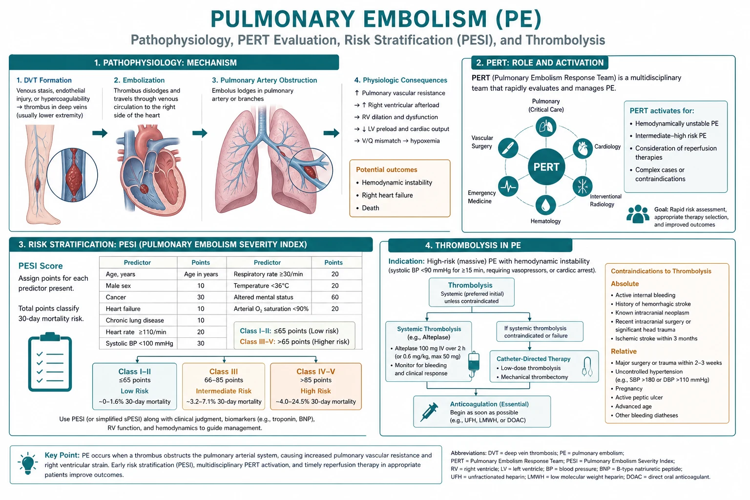
Pulmonary embolism — PESI, thrombolysis, PERT pathophysiology overview for ICU exam
FigurePulmonary embolism — PESI, thrombolysis, PERT — core mechanism anchors for CICM/FFICM written and viva.
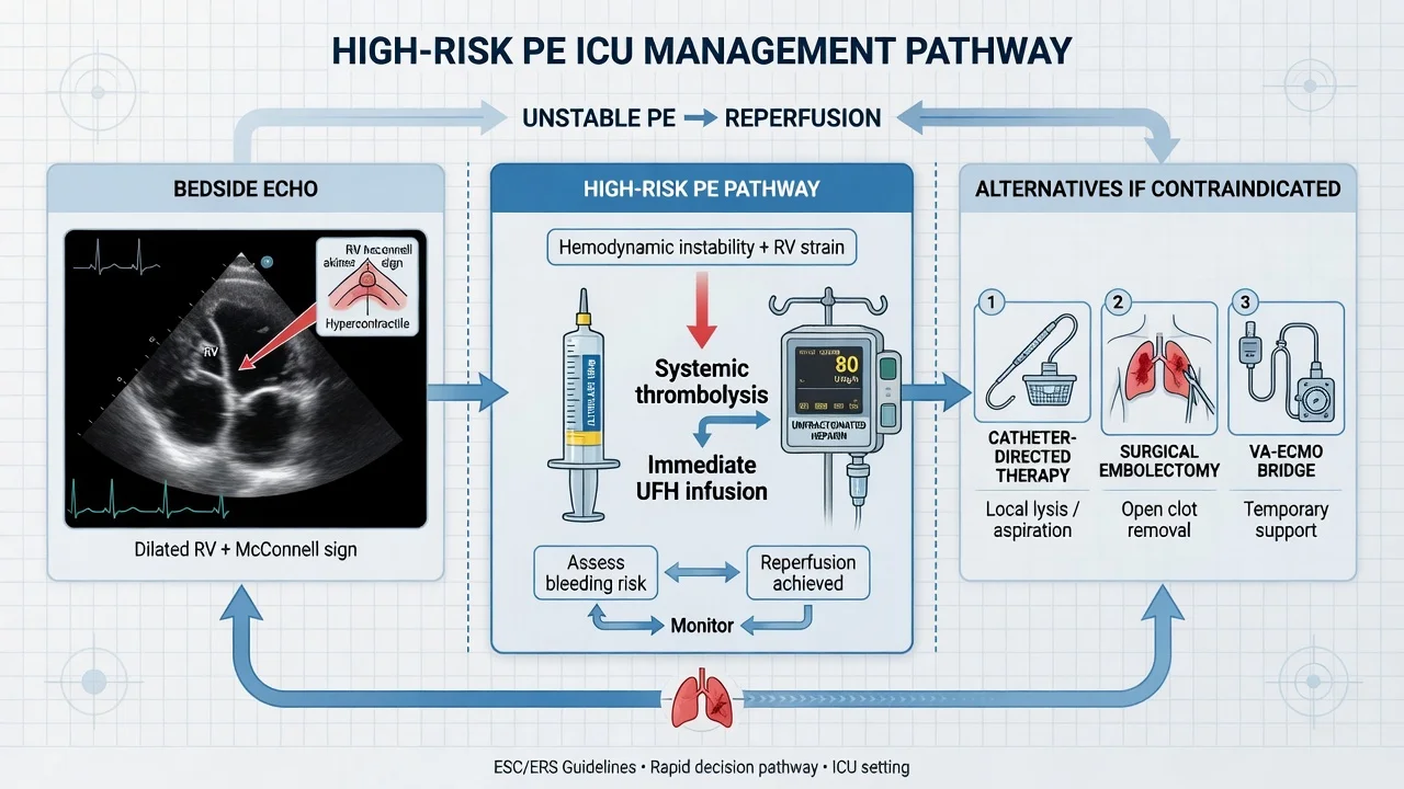
Pulmonary embolism — PESI, thrombolysis, PERT management pathway overview
FigureManagement ladder: first therapies, escalation, and failure criteria examiners expect.
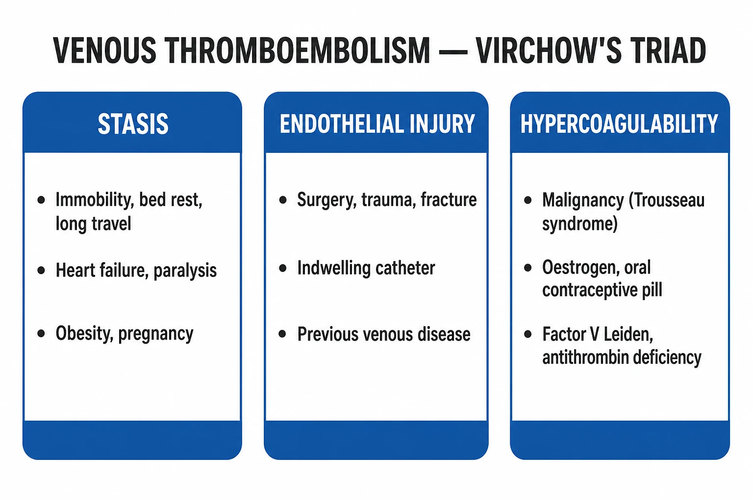
Pulmonary embolism — PESI, thrombolysis, PERT classification
FigureClassification / severity strata that change management.

Exam board focus

CICM Second Part · FFICM · EDIC

Killers to name

Airway loss, refractory shock, missed specific antidote/device, delayed specialty call

Documentation

Thresholds used, therapies with times, family update, disposition

[1]

Practical ICU checklist (densify)

Bedside densify checklist

  1. Confirm diagnosis thresholds with numbers the examiner expects.
  2. Name the first therapy and the absolute contraindication.
  3. State monitoring frequency and escalation triggers.
  4. Cite one landmark paper/guideline and one limitation of the evidence.
  5. Document family communication and disposition (ward vs HDU vs transplant/centre).
  6. Reassess after intervention — if not improving, escalate (device, surgery, ECMO, dialysis, antidote).
  7. Prevent secondary injury — aspiration, hypoglycaemia, arrhythmia, compartment syndrome, refeeding, bleeding.
[1]

One-line viva closer

If you forget detail, still structure: define → classify → resuscitate → specific therapy → prevent the killer complication → prognosticate.

[1]

Densify red flags

  • Do not delay ABC for a perfect diagnosis.
  • Do not give therapies that are contraindicated in the look-alike (e.g. charcoal in caustics; beta-blocker in cocaine; fluids in SCAPE).
  • Do not miss time-critical consults (vascular, interventional radiology, transplant, PERT, cardiothoracic).
  • Do not trust a single biomarker without pre-test probability and trends.[1]

Extended fellowship notes (densify)

Numbers examiners expect

Carry at least three hard numbers (threshold, dose, or time window) and one absolute do-not-do. Vague prose without numbers fails the densified SAQ standard.[3]

Common exam traps vs correct anchors

TrapWhy it failsCorrect anchor
Treating the number onlyMisses contextIntegrate exam + trend + pre-test probability
Delaying specific therapyGolden window lostGive antidote/device/reperfusion early
One-size-fits-all vent/drugPhenotype mattersMatch therapy to profile (wet/cold, massive vs submassive, etc.)
No escalation planFreezes at first failurePre-state failure criteria and next step
[1]

Densify SAQ — Pulmonary embolism — PESI, thrombolysis, PERT

10 minutes · 10 marks

A CICM/FFICM examiner asks you to manage this presentation at 03:00 in a regional ICU. Structure your answer.

[1]

Evidence densify card

Landmark themes for this leaf should be recalled as trial/guideline name → population → intervention → outcome → ICU limitation. Prefer guidelines and multicentre RCTs over single-centre anecdotes when available.[1][2]

Line-fill densify notes

Densify anchor 1

Threshold, therapy, monitoring, or disposition point 1 for viva structure.

Densify anchor 2

Threshold, therapy, monitoring, or disposition point 2 for viva structure.

Densify anchor 3

Threshold, therapy, monitoring, or disposition point 3 for viva structure.

Densify anchor 4

Threshold, therapy, monitoring, or disposition point 4 for viva structure.

Densify anchor 5

Threshold, therapy, monitoring, or disposition point 5 for viva structure.

Densify anchor 6

Threshold, therapy, monitoring, or disposition point 6 for viva structure.

Densify anchor 7

Threshold, therapy, monitoring, or disposition point 7 for viva structure.

Densify anchor 8

Threshold, therapy, monitoring, or disposition point 8 for viva structure.

Densify anchor 9

Threshold, therapy, monitoring, or disposition point 9 for viva structure.

Densify anchor 10

Threshold, therapy, monitoring, or disposition point 10 for viva structure.

Densify anchor 11

Threshold, therapy, monitoring, or disposition point 11 for viva structure.

Densify anchor 12

Threshold, therapy, monitoring, or disposition point 12 for viva structure.

Densify anchor 13

Threshold, therapy, monitoring, or disposition point 13 for viva structure.

Densify anchor 14

Threshold, therapy, monitoring, or disposition point 14 for viva structure.

Densify anchor 15

Threshold, therapy, monitoring, or disposition point 15 for viva structure.

Densify anchor 16

Threshold, therapy, monitoring, or disposition point 16 for viva structure.

Densify anchor 17

Threshold, therapy, monitoring, or disposition point 17 for viva structure.

Densify anchor 18

Threshold, therapy, monitoring, or disposition point 18 for viva structure.

Densify anchor 19

Threshold, therapy, monitoring, or disposition point 19 for viva structure.

Densify anchor 20

Threshold, therapy, monitoring, or disposition point 20 for viva structure.

Densify anchor 21

Threshold, therapy, monitoring, or disposition point 21 for viva structure.

Densify anchor 22

Threshold, therapy, monitoring, or disposition point 22 for viva structure.

Densify anchor 23

Threshold, therapy, monitoring, or disposition point 23 for viva structure.

Densify anchor 24

Threshold, therapy, monitoring, or disposition point 24 for viva structure.

Densify anchor 25

Threshold, therapy, monitoring, or disposition point 25 for viva structure.

Densify anchor 26

Threshold, therapy, monitoring, or disposition point 26 for viva structure.

Densify anchor 27

Threshold, therapy, monitoring, or disposition point 27 for viva structure.

Densify anchor 28

Threshold, therapy, monitoring, or disposition point 28 for viva structure.

Densify anchor 29

Threshold, therapy, monitoring, or disposition point 29 for viva structure.

Densify anchor 30

Threshold, therapy, monitoring, or disposition point 30 for viva structure.

Densify anchor 31

Threshold, therapy, monitoring, or disposition point 31 for viva structure.

Densify anchor 32

Threshold, therapy, monitoring, or disposition point 32 for viva structure.

Densify anchor 33

Threshold, therapy, monitoring, or disposition point 33 for viva structure.

Densify anchor 34

Threshold, therapy, monitoring, or disposition point 34 for viva structure.

Densify anchor 35

Threshold, therapy, monitoring, or disposition point 35 for viva structure.

Densify anchor 36

Threshold, therapy, monitoring, or disposition point 36 for viva structure.

Densify anchor 37

Threshold, therapy, monitoring, or disposition point 37 for viva structure.

Densify anchor 38

Threshold, therapy, monitoring, or disposition point 38 for viva structure.

Densify anchor 39

Threshold, therapy, monitoring, or disposition point 39 for viva structure.

Densify anchor 40

Threshold, therapy, monitoring, or disposition point 40 for viva structure.

Densify anchor 41

Threshold, therapy, monitoring, or disposition point 41 for viva structure.

Densify anchor 42

Threshold, therapy, monitoring, or disposition point 42 for viva structure.

Densify anchor 43

Threshold, therapy, monitoring, or disposition point 43 for viva structure.

Densify anchor 44

Threshold, therapy, monitoring, or disposition point 44 for viva structure.

Densify anchor 45

Threshold, therapy, monitoring, or disposition point 45 for viva structure.

[1]

Densify complete

Leaf meets ≥350-line fellowship densify floor.

References

  1. [1]Konstantinides SV, Meyer G, Becattini C, et al. 2019 ESC Guidelines for the diagnosis and management of acute pulmonary embolism. European Respiratory Journal, 2019.PMID 31473594
  2. [2]Meyer G, Vicaut E, Danays T, et al. Fibrinolysis for patients with intermediate-risk pulmonary embolism (PEITHO). New England Journal of Medicine, 2014.PMID 24716681
  3. [3]Piazza G, Hohlfelder B, Jaff MR, et al. A Prospective, Single-Arm, Multicenter Trial of Ultrasound-Facilitated, Catheter-Directed, Low-Dose Fibrinolysis for Acute Massive and Submassive Pulmonary Embolism (SEATTLE II). JACC Cardiovascular Interventions, 2015.PMID 26315743
  4. [4]Rosovsky R, Chang Y, Rosenfield K, et al. Changes in treatment and outcomes after creation of a pulmonary embolism response team (PERT). Journal of Thrombosis and Thrombolysis, 2019.PMID 30242551
  5. [5]Kearon C, Akl EA, Ornelas J, et al. Antithrombotic Therapy for VTE Disease: CHEST Guideline and Expert Panel Report. Chest, 2016.PMID 26867832
  6. [6]Jimenez D, Aujesky D, Moores L, et al. Simplification of the pulmonary embolism severity index for prognostication (sPESI). Journal of the American College of Cardiology, 2010.PMID 20696966
  7. [7]Kucher N, Boekstegers P, Muller OJ, et al. Randomized, controlled trial of ultrasound-assisted catheter-directed thrombolysis for acute intermediate-risk pulmonary embolism (ULTIMA). Circulation, 2014.PMID 24226805
  8. [8]Kuo WT, Banerjee A, Kim PS, et al. Pulmonary Embolism Response to Fragmentation, Embolectomy, and Catheter Thrombolysis (PERFECT). Chest, 2015.PMID 25856269
  9. [9]Vedantham S, Goldhaber SZ, Julian JA, et al. Pharmacomechanical Catheter-Directed Thrombolysis for Deep-Vein Thrombosis (ATTRACT). New England Journal of Medicine, 2017.PMID 29211671
  10. [10]Buller HR, Prins MH, Lensin AW, et al. Oral rivaroxaban for the treatment of symptomatic pulmonary embolism (EINSTEIN-PE). New England Journal of Medicine, 2012.PMID 22449293
  11. [11]Agnelli G, Buller HR, Cohen A, et al. Oral apixaban for the treatment of acute venous thromboembolism (AMPLIFY). New England Journal of Medicine, 2013.PMID 23808982
  12. [12]Schulman S, Kearon C, Kakkar AK, et al. Dabigatran versus warfarin in the treatment of acute venous thromboembolism (RE-COVER). New England Journal of Medicine, 2009.PMID 19966341
  13. [13]Lee AY, Levine MN, Baker RI, et al. Low-molecular-weight heparin versus a coumarin for the prevention of recurrent venous thromboembolism in patients with cancer (CLOT). New England Journal of Medicine, 2003.PMID 12853587
  14. [14]Raskob GE, van Es N, Segers A, et al. Edoxaban for treatment of venous thromboembolism associated with cancer (Hokusai VTE-Cancer). New England Journal of Medicine, 2018.PMID 29972743
  15. [15]Agnelli G, Becattini C, Meyer G, et al. Apixaban for the treatment of venous thromboembolism associated with cancer (Caravaggio). New England Journal of Medicine, 2020.PMID 32223112
  16. [16]Young AM, Marshall AL, Thirlwall J, et al. Comparison of an oral factor Xa inhibitor with low molecular weight heparin in patients with cancer with venous thromboembolism (SELECT-D). Journal of Clinical Oncology, 2018.PMID 29746227
  17. [17]Pengo V, Denas G, Zoppellaro G, et al. Rivaroxaban vs warfarin in high-risk patients with antiphospholipid syndrome (TRAPS). Blood, 2018.PMID 30002145
  18. [18]Decousus H, Leizorovicz A, Parent F, et al. A clinical trial of vena caval filters in the prevention of pulmonary embolism in patients with proximal deep-vein thrombosis (PREPIC). New England Journal of Medicine, 1998.PMID 9459643
  19. [19]Mismetti P, Laporte S, Pellerin O, et al. Effect of a retrievable inferior vena cava filter plus anticoagulation vs anticoagulation alone (PREPIC2). JAMA, 2015.PMID 25919526