ICU · Cardiovascular
Pulmonary hypertension and right heart failure
Also known as Pulmonary arterial hypertension (PAH) · Right ventricular failure · Cor pulmonale · WHO Group 1-5 classification · Pulmonary vascular disease · Pre-capillary PH · Post-capillary PH · Isolated post-capillary PH · CTEPH · BMPR2 mutation · Inhaled nitric oxide · Prostacyclin therapy
Pulmonary hypertension (PH) = mean pulmonary artery pressure (mPAP) 20 mmHg (ESC/ERS 2022). Five WHO groups: Group 1 (pulmonary arterial hypertension, PAH — idiopathic, heritable [BMPR2], connective tissue disease [SSc], congenital heart disease, portal hypertension, drugs), Group 2 (left heart disease — the commonest cause overall, HFpEF/HFrEF/valvular), Group 3 (lung disease/hypoxia — COPD, ILD, OSA), Group 4 (chronic thromboembolic PH, CTEPH — potentially curable by pulmonary endarterectomy), Group 5 (multifactorial — haematological, systemic, metabolic). Haemodynamic phenotyping by right heart catheterisation: pre-capillary (mPAP 20, wedge <15, PVR 2-3 WU), isolated post-capillary (mPAP 20, wedge 15, PVR <2-3 WU), combined pre/post-capillary (wedge 15 AND PVR 2-3 WU). PATHOPHYSIOLOGY of PAH: a triad of pulmonary vascular remodelling — (1) endothelial dysfunction (reduced NO and prostacyclin, excess endothelin-1), (2) smooth muscle proliferation and medial hypertrophy, (3) in situ thrombosis (procoagulant state) — producing a fixed, obliterative pulmonary vasculopathy and raised PVR. RV FAILURE CASCADE (the main cause of death): chronic pressure overload - RV hypertrophy - RV dilatation - tricuspid regurgitation - septal shift into the LV - reduced LV filling - low cardiac output - cardiogenic shock. RV is uniquely afterload-sensitive (thin-walled, crescent-shaped, adapted to a low-pressure, low-resistance circuit). ICU PRESENTATION: exertional syncope (a red flag of low fixed output), hypoxaemia (right-to-left shunt via PFO, V/Q mismatch), and overt right heart failure (raised JVP, peripheral oedema, ascites, hepatomegaly, oliguria). MANAGEMENT in the ICU: (1) Support the failing RV — maintain preload (cautious 250 mL bolus only if hypovolaemic; the RV is volume-intolerant), reduce afterload (inhaled nitric oxide 5-20 ppm, inhaled/IV prostacyclin, milrinone), support contractility (milrinone PREFERRED — inotropy + pulmonary vasodilation; dobutamine), maintain systemic BP with noradrenaline (preserve RV coronary perfusion — MAP must exceed RV systolic pressure or the RV ischaemias). (2) Optimise the gas exchange and ventilation — avoid hypoxia, hypercapnia, and acidosis (all cause pulmonary vasoconstriction); minimise PEEP and intrathoracic pressure. (3) PAH-specific therapy for confirmed Group 1 PAH — endothelin receptor antagonists (bosentan, macitentan), PDE5 inhibitors (sildenafil, tadalafil), prostacyclin analogues (epoprostenol, iloprost, treprostinil), soluble guanylate cyclase stimulators (riociguat); oral combination therapy is now standard (AMBITION). (4) Treat the underlying group — oxygen for Group 3, CTEPH surgery for Group 4, left heart disease for Group 2 (PAH drugs are HARMFUL in post-capillary PH). (5) Mechanical support (VA-ECMO) for refractory RV failure. Avoid systemic vasodilators, excessive fluids, and high intrathoracic pressure.
On this page & tools
Your progress
Saved locally on this device.
8 MCQs with explanations
Target exams
Red flags
Overview and definition
Pulmonary hypertension (PH) is a pathophysiological and haemodynamic state defined at right heart catheterisation as a mean pulmonary artery pressure (mPAP) >20 mmHg (the 2022 ESC/ERS and 6th World Symposium definition lowered the threshold from the prior >25 mmHg, recognising that mPAP 21-24 mmHg is already abnormal and prognostically adverse).[1][10] Pulmonary arterial hypertension (PAH, WHO Group 1) is a more specific haemodynamic diagnosis: mPAP >20 mmHg, pulmonary artery wedge pressure (PAWP) <15 mmHg, and pulmonary vascular resistance (PVR) >2 Wood units (WU; 2-3 in older definitions). The distinction matters because Group 1 PAH responds to a specific class of pulmonary vasodilator therapy that is ineffective or actively harmful in the other groups.
PH is not a single disease but the final common pathway of many insults — a pan-syndrome unified by raised pulmonary vascular pressure. Five clinical groups differ in cause, pathophysiology, natural history, and (critically) treatment, so classification always precedes management. The single most important practical split is pre-capillary vs post-capillary, determined by the wedge pressure at right heart catheterisation: pre-capillary disease (Groups 1, 3, 4, 5) arises within the pulmonary vasculature and is the target of pulmonary vasodilators, while post-capillary disease (Group 2) is a passive consequence of left heart disease, where the pulmonary pressure is raised simply because the left atrial pressure is raised — and where pulmonary vasodilators, by increasing flow into an already-congested left heart, cause pulmonary oedema.[1]
The clinical importance of PH to the intensivist is the right ventricle. The RV is the most common and most lethal point of failure in the pulmonary hypertension patient: chronic pressure overload drives RV hypertrophy and then dilatation, tricuspid regurgitation, septal shift, reduced LV filling, and a low fixed cardiac output culminating in cardiogenic shock. RV failure is the leading cause of death in PH. Because the RV is uniquely afterload-sensitive — a thin-walled, crescent-shaped chamber adapted to a low-resistance circuit — anything that raises PVR acutely (hypoxia, hypercapnia, acidosis, high intrathoracic pressure, tachyarrhythmia, excess fluid) can tip a compensated PH patient into catastrophic decompensation within hours. ICU management therefore has two arms: treat the underlying group, and protect the failing RV.[2][12]
PH at a glance
WHO clinical classification — the five groups
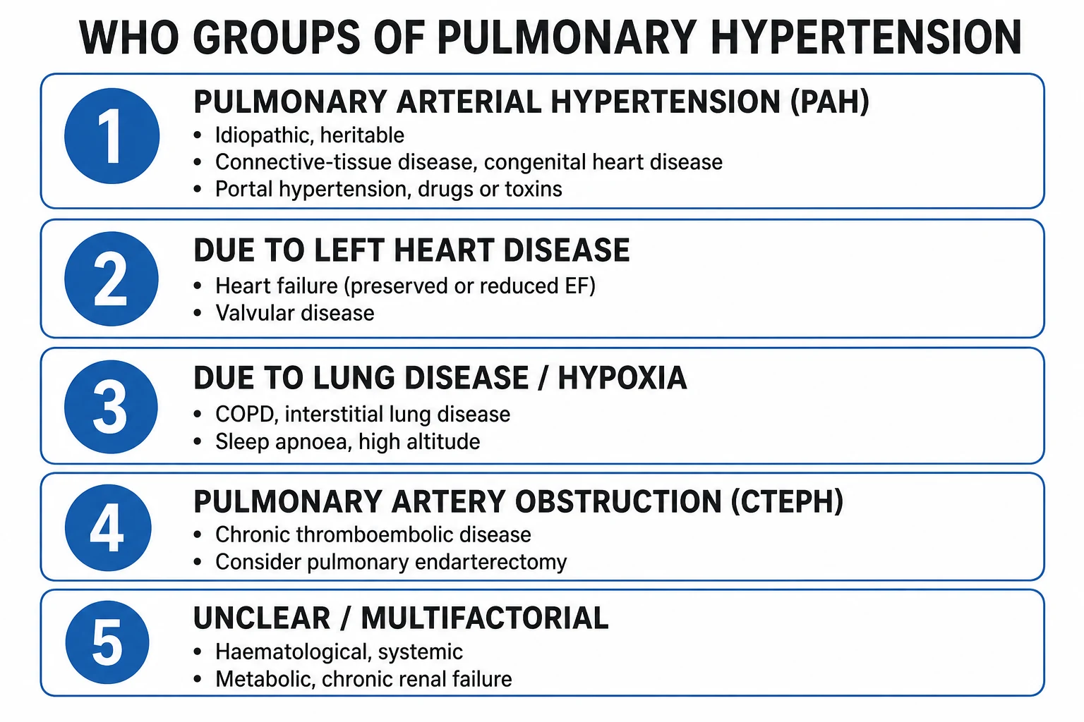
The World Health Organisation (now ESC/ERS) classification groups PH by shared pathology, natural history, and treatment response. It was updated at the 6th World Symposium (Nice 2018) and codified in the 2022 ESC/ERS Guidelines.[1][10]
WHO Group 1 — pulmonary arterial hypertension (PAH): pre-capillary, vasoreactive in a minority
| Sub-group | Key causes / associations | Notes |
|---|---|---|
| Idiopathic PAH (IPAH) | No cause identified; sporadic | Mean age ~50; female > male (4:1); poor prognosis without therapy |
| Heritable PAH | BMPR2 mutation (~70% of familial); ACVRL1/ALK1, ENG, SMAD9, KCNK3 | Autosomal dominant, incomplete penetrance, earlier onset, worse prognosis |
| Drug/toxin-induced | Anorexigens (fenfluramine, dexfenfluramine), benfluorex, toxic rapeseed oil, methamphetamines, dasatinib | Consider in any new PAH diagnosis |
| Associated PAH (APAH) | Connective tissue disease (systemic sclerosis — commonest and worst), congenital heart disease (Eisenmenger, unrepaired shunts), portal hypertension (portopulmonary HTN), HIV, schistosomiasis | CTD-PAH is the largest subgroup in registries |
| PAH long-term responders | Positive vasoreactivity test sustained on calcium channel blockers | A minority of IPAH/HPAH/DPAH only |
| Pulmonary veno-occlusive disease / pulmonary capillary haemangiomatosis (PVOD/PCH) | Idiopathic, heritable (EIF2AK4), drug, CTD | Pulmonary oedema with PAH drugs; refer for transplant |
WHO Groups 2-5 — the non-PAH groups (and why PAH drugs do NOT apply)
| Group | Definition | Causes | Definitive / first-line therapy |
|---|---|---|---|
| Group 2 — PH due to left heart disease (post-capillary) | mPAP >20, PAWP >15 mmHg (passive back-transmission) | HFrEF, HFpEF (commonest overall cause of PH), valvular disease (mitral), aortic stenosis, restrictive cardiomyopathy | Treat the LEFT heart — GDMT, valve repair, etc. PAH drugs harmful (cause pulmonary oedema) |
| Group 3 — PH due to lung disease and/or hypoxia | Pre-capillary, driven by hypoxic vasoconstriction and vascular loss | COPD (commonest), interstitial lung disease, combined pulmonary fibrosis-emphysema, OSA, chronic high-altitude exposure, developmental lung disease | Oxygen (correct hypoxia), treat the lung disease; PAH drugs not routinely indicated |
| Group 4 — PH due to pulmonary artery obstruction (CTEPH) | Pre-capillary; organised thromboembolic material obliterating pulmonary arteries | Chronic (often recurrent) pulmonary embolism; may have no overt PE history | Pulmonary endarterectomy (PEA) — potentially curable; riociguat or balloon pulmonary angioplasty if inoperable |
| Group 5 — PH with unclear/multifactorial mechanism | Mixed mechanisms | Haematological (myeloproliferative, splenectomy), systemic (sarcoidosis, vasculitis), metabolic (glycogen storage, thyroid), chronic renal failure on dialysis, fibrosing mediastinitis | Treat the underlying condition; transplant evaluation in selected cases |
Why classification matters at the bedside. A raised mPAP is the finding, not the diagnosis. The intensivist who treats a Group 2 (HFpEF) patient with sildenafil or bosentan, or a Group 4 (CTEPH) patient with oxygen alone, will harm the patient — the first by flooding an already-congested left heart, the second by missing a curative operation. The single most important question on encountering a new PH patient is: what group, and is it pre- or post-capillary? The answer redirects the entire management plan.[1]
Haemodynamic definitions and phenotyping — pre-capillary vs post-capillary
Right heart catheterisation (RHC) is the gold standard and is mandatory before committing to PAH-specific therapy. It distinguishes pre- from post-capillary disease, quantifies severity, and guides therapy. The 6th WSPH (2018) and ESC/ERS 2022 definitions:[1][10]
Haemodynamic phenotypes of PH (by right heart catheterisation)
| Phenotype | mPAP | PAWP (wedge) | PVR | Clinical correlate / therapy |
|---|---|---|---|---|
| Pre-capillary PH | >20 mmHg | <15 mmHg | >2-3 WU | Disease in the pulmonary vessels — Groups 1, 3, 4, 5. Target of pulmonary vasodilators (if Group 1) |
| Isolated post-capillary PH (IpcPH) | >20 mmHg | >15 mmHg | <2-3 WU | Passive back-transmission from left heart disease (Group 2). Treat the LEFT heart; NO PAH drugs |
| Combined post- and pre-capillary PH (CpcPH) | >20 mmHg | >15 mmHg | >2-3 WU | Left heart disease PLUS fixed vascular remodelling. Treat left heart; PAH drugs used cautiously in trials |
| Exercise PH | mPAP/CO slope >3 mmHg/L/min | variable | — | Early disease; predicts progression to resting PH |
The wedge pressure is the pivotal measurement. It estimates left atrial (and thus left ventricular end-diastolic) pressure. A wedge <15 mmHg places the disease in the pulmonary vasculature (pre-capillary) and opens the door to PAH-specific therapy; a wedge >15 mmHg redirects the investigation and treatment to the left heart. Pitfalls: an over-wedged catheter (balloon pushed too distal) gives a falsely high wedge; under-wedging gives falsely low. In the ventilated critically ill patient, measure the wedge at end-expiration (or use the average of three cardiac cycles) and interpret alongside the clinical picture. If the wedge is equivocal, a fluid challenge or direct left heart catheterisation may be required. The diastolic pressure gradient (DPG = PADP - PAWP) and the mPAP/CO slope on exercise refine the pre/post-capillary split further, particularly when combined disease is suspected.[1][10]
Pathophysiology of PAH — the vascular remodelling triad
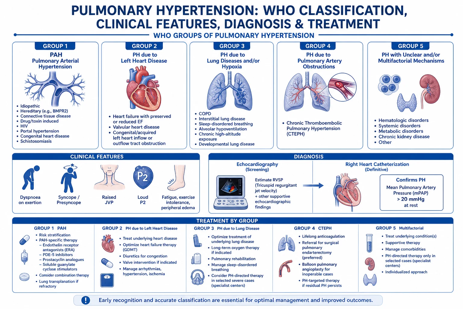
Pulmonary arterial hypertension (Group 1) is a proliferative obliterative vasculopathy of the small pulmonary arteries (typically <500 µm). Three pathological processes act in concert, each of which is the target of a PAH drug class:[1][12]
1. Endothelial dysfunction. The healthy pulmonary endothelium maintains a vasodilator, anti-proliferative, antithrombotic surface. In PAH, this balance is disrupted: there is reduced bioavailability of nitric oxide (NO) and prostacyclin (PGI2) (the two endogenous vasodilators) and an excess of endothelin-1 (ET-1) and thromboxane (vasoconstrictors and mitogens). The genetic lesion best characterised is a loss-of-function mutation in BMPR2 (bone morphogenetic protein receptor type 2), present in ~70% of familial and ~20% of idiopathic PAH cases — it removes an anti-proliferative brake on vascular smooth muscle, permitting unchecked growth. [1]
2. Smooth muscle proliferation and medial hypertrophy. Pulmonary artery smooth muscle cells proliferate and migrate into the intima, the tunica media hypertrophies, and the vessel wall thickens — narrowing the lumen and raising PVR. Pulmonary arterial remodelling produces the characteristic plexiform lesion (a disorganized, endothelial-cell-lined, glomeruloid proliferation) in the severest forms. The proliferative phenotype is driven by ET-1, serotonin, and loss of BMPR2/Smad signalling — a quasi-neoplastic, apoptosis-resistant vascular expansion. [1]
3. In situ thrombosis. The PAH vasculature is prothrombotic — endothelial injury, reduced fibrinolysis, and platelet activation produce microscopic in situ thrombosis that further obliterates the lumen. This is the rationale for the observed (though now-debated) survival benefit of anticoagulation in idiopathic PAH, and for the routine avoidance of pro-thrombotic triggers. [1]
Net effect — a fixed, obliterative pulmonary vasculopathy, raised PVR, and a chronic pressure load on the RV. The lesion is fixed (not purely vasoconstrictive) in most patients — which is why only the minority with a positive vasoreactivity test (a sustained fall in mPAP with an acute pulmonary vasodilator at RHC) respond to calcium channel blockers, and why the modern drugs target the remodelling pathways (endothelin, NO, prostacyclin) rather than simply dilating the vessel.[1]
The PAH drug classes map onto the pathophysiology
| Pathophysiological pathway | Endogenous imbalance | Drug class | Prototype |
|---|---|---|---|
| Endothelin-1 excess (vasoconstrictor + mitogen) | ET-1 up | Endothelin receptor antagonist (ERA) | Bosentan, macitentan, ambrisentan |
| Reduced NO / cGMP (vasodilator + anti-proliferative) | NO/cGMP down | Phosphodiesterase-5 (PDE5) inhibitor | Sildenafil, tadalafil |
| Reduced NO / cGMP | NO/cGMP down | Soluble guanylate cyclase (sGC) stimulator | Riociguat |
| Reduced prostacyclin (vasodilator + anti-proliferative + anti-platelet) | PGI2 down | Prostacyclin analogues / IP receptor agonist | Epoprostenol (IV), iloprost (inhaled), treprostinil, selexipag (oral) |
The right ventricular failure cascade
The right ventricle is the linchpin of pulmonary hypertension. Unlike the LV — a thick-walled, conical, high-pressure pump — the RV is thin-walled, crescent-shaped, and adapted to pumping against a low-resistance, high-capacitance circuit. It is exquisitely afterload-sensitive: a small rise in PVR causes a disproportionate fall in RV stroke volume. This is why the RV, not the pulmonary pressure itself, is the determinant of symptoms and survival, and why RV failure is the common end-game.[2][12]
The cascade (pressure overload -> dilatation -> TR -> low CO): [1]
-
Chronic pressure overload -> concentric hypertrophy. The RV initially adapts to raised PVR by hypertrophying (concentric, wall thickening), maintaining wall stress and output. This is the compensated phase — the patient may be asymptomatic or have only exertional dyspnoea. The RV hypertrophy, however, raises myocardial oxygen demand and reduces coronary flow reserve, sowing the seeds of later failure. [1]
-
Transition to dilatation. When the load exceeds the RV's adaptive capacity, the ventricle dilates — the thin RV free wall is pulled outward, wall stress rises, contractile efficiency falls, and the ejection fraction drops. This is the inflection point: the patient develops exertional presyncope/syncope and signs of right heart failure. RV dilatation is irreversible without afterload reduction. [1]
-
Tricuspid regurgitation (TR). RV dilatation stretches the tricuspid annulus, pulling the leaflets apart and producing functional (secondary) tricuspid regurgitation. The TR worsens venous return to the RV and reduces forward output — a self-reinforcing cycle. [1]
-
Septal shift and LV compression. As the RV dilates in the confined pericardium, the interventricular septum is pushed into the LV during diastole (the D-shaped septum on echo), reducing LV filling, stroke volume, and cardiac output. Pericardial constraint raises the pericardial pressure and further limits both ventricles. [1]
-
RV ischaemia and the coronary perfusion trap. Unlike the LV (perfused in diastole), the RV is perfused throughout the cardiac cycle because RV wall pressure is normally low. But in PH the RV systolic pressure is high — if it exceeds the systemic (aortic) diastolic pressure, RV myocardial perfusion is compromised, producing RV ischaemia, further dysfunction, and a downward spiral. This is the rationale for maintaining systemic BP with noradrenaline. [1]
-
Low cardiac output, shock, and death. The end-state is a low fixed output (cardiac index <2 L/min/m²), rising central venous pressure, falling systemic pressure, oliguria, hepatic congestion, and cardiogenic shock. Once the RV is dilated and TR is severe, the cycle is vicious and hard to reverse — hence the premium on protecting the RV and treating the trigger early. [1]
The RV failure cascade — from pressure overload to cardiogenic shock
1. Chronic pressure overload (raised PVR)
Pulmonary vascular disease raises PVR; the RV faces a high afterload. Initially compensated by concentric hypertrophy (wall thickening preserves wall stress and output). Patient asymptomatic or exertional dyspnoea only. RV hypertrophy raises O2 demand and reduces coronary flow reserve — seeds later failure.
2. Transition to RV dilatation
When load exceeds adaptive capacity, the thin RV free wall dilates outward. Wall stress rises, contractile efficiency falls, RV stroke volume drops. The inflection point — patient develops exertional presyncope/syncope, early right heart failure. RV dilatation is largely irreversible without aggressive afterload reduction.
3. Functional tricuspid regurgitation
RV dilatation stretches the tricuspid annulus, separating the leaflets — secondary (functional) TR. Regurgitant volume increases RV preload, reduces forward stroke volume, and raises systemic venous pressure (raised JVP, hepatic congestion, peripheral oedema, ascites). A self-reinforcing cycle.
4. Septal shift and LV compression
RV dilatation in the confined pericardium pushes the interventricular septum toward the LV in diastole — the D-shaped septum on echo (paradoxical septal motion). LV filling and stroke volume fall. Raised pericardial pressure further limits both ventricles (ventricular interdependence).
5. RV ischaemia (coronary perfusion trap)
The RV is normally perfused throughout the cardiac cycle. In PH, RV systolic pressure is high; if it exceeds systemic diastolic pressure (aortic), RV subendocardial perfusion fails. RV ischaemia worsens dysfunction in a vicious cycle. Rationale for noradrenaline: raise systemic BP above RV pressure to restore RV perfusion.
6. Low cardiac output, shock, death
End-state: low fixed cardiac index (<2 L/min/m²), high CVP, falling systemic pressure, oliguria, hepatorenal dysfunction, cardiogenic shock. Mortality 40-60% once overt shock develops. The premium is on PREVENTING the cascade by protecting the RV and treating the trigger early.
Why the RV is uniquely vulnerable — RV vs LV
| Feature | Left ventricle | Right ventricle |
|---|---|---|
| Wall | Thick, conical | Thin, crescent-shaped |
| Native load | High-pressure systemic circuit | Low-pressure, low-resistance pulmonary circuit |
| Afterload sensitivity | Moderate | Extreme — small rise in PVR → large fall in RVSV |
| Coronary perfusion | Diastole only (high intracavitary pressure) | Throughout the cardiac cycle (low RV pressure) |
| Response to chronic load | Hypertrophies well | Hypertrophies, then dilates (poor adaptation) |
| Clinical marker of failure | Pulmonary oedema | Raised JVP, peripheral oedema, hepatomegaly, low CO |
ICU presentation of PH and the failing RV
The critically ill PH patient presents in one of several ways — the intensivist must recognise each, because the management differs.[2][12]
1. Syncope and presyncope (the cardinal red flag). Exertional syncope in a PH patient is the single most ominous symptom — it reflects a critically low fixed cardiac output that cannot rise with exertion, producing cerebral hypoperfusion. Syncope heralds imminent sudden death (often from arrhythmia or a pulmonary hypertensive crisis). A PH patient who syncope (or with new exertional presyncope) needs admission and aggressive stabilisation — it is a marker of end-stage RV decompensation. [1]
2. Hypoxaemia. Mechanisms: (a) low cardiac output with a high systemic oxygen extraction raises the mixed venous saturation; (b) right-to-left shunting through a patent foramen ovale (PFO) — raised right atrial pressure pops open a probe-patent foramen, producing profound refractory hypoxaemia; (c) V/Q mismatch from the pulmonary vascular disease itself; (d) reduced mixed venous oxygen from a low output state. Hypoxaemia worsens pulmonary vasoconstriction, raising PVR, worsening the RV — a vicious cycle. Supplemental oxygen is supportive in ALL groups (target SpO2 92-96%). [1]
3. Right heart failure (the congestion picture). Raised JVP, peripheral and sacral oedema, ascites, hepatomegaly (with a transaminitis and raised bilirubin from hepatic congestion — "cardiac cirrhosis"), a pulsatile liver, a right ventricular heave, a loud pulmonary component of the second heart sound (P2) with a parasternal heave, a tricuspid regurgitation murmur, an S3 or S4, and features of low output (oliguria, cool peripheries, narrow pulse pressure, altered mentation). Atrial arrhythmia (AF or flutter) is common and poorly tolerated — loss of the atrial kick can precipitate acute decompensation. [1]
4. The pulmonary hypertensive crisis. An acute rise in PVR (from hypoxia, acidosis, pain, catecholamine surge, or pulmonary embolism) produces abrupt RV failure with rapidly falling cardiac output, hypotension, hypoxaemia, and cardiac arrest. This is a high-mortality emergency — treat with inhaled pulmonary vasodilators (NO, prostacyclin), systemic vasopressors (noradrenaline), inotropes (milrinone), correction of the trigger, and VA-ECMO for refractory cases. [1]
5. The incidental finding. Some patients are admitted to ICU for an unrelated problem (sepsis, surgery) and are found to have severe PH on echo — these patients tolerate the ICU insults (positive pressure ventilation, hypoxia, fluid loading, sepsis) very poorly because their RV has no reserve. A pre-existing PH diagnosis on the chart should heighten vigilance and modify management (conservative fluids, early vasopressor/inotrope, lung-protective ventilation, PA-catheter monitoring). [1]
Diagnostic workup in the ICU
The diagnosis of PH and its classification requires a structured workup. In the ICU much of this is concurrent with stabilisation.[1]
- Echocardiography (the first test). Estimates pulmonary artery systolic pressure (PASP) from the tricuspid regurgitant jet velocity (TRV) using the modified Bernoulli equation (PASP = 4 × TRV² + RA pressure). Identifies RV dilatation and dysfunction (reduced TAPSE <17 mm, RV fractional area change <35%), the D-shaped septum (flattened interventricular septum = RV pressure overload), tricuspid regurgitation, pericardial effusion (a poor prognostic sign in PAH), and left heart disease (to distinguish Group 2). Echo estimates are a SCREEN; the diagnosis and treatment decision require RHC.
- Right heart catheterisation (the definitive test). Measures mPAP, PAWP, PVR, and cardiac output; confirms pre- vs post-capillary; allows the vasoreactivity test (inhaled NO, IV epoprostenol, or IV adenosine — a positive response is a fall in mPAP >10 mmHg to <40 mmHg with normal/high cardiac output; identifies CCB responders in IPAH/HPAH/DPAH only). Mandatory before PAH-specific therapy.
- Ventilation-perfusion (V/Q) scan — the screening test for CTEPH (Group 4); a normal/near-normal V/Q excludes CTEPH. More sensitive than CT pulmonary angiography for chronic disease. If abnormal, confirm with CT pulmonary angiography and refer for pulmonary endarterectomy assessment.
- Pulmonary function tests and arterial blood gas — quantify underlying lung disease (Group 3); the ABG documents hypoxaemia/hypercapnia and acid-base status. DLCO is disproportionately reduced in PAH.
- High-resolution CT chest and CT pulmonary angiography — parenchymal lung disease, CTEPH, PVOD/PCH (centrilobular ground-glass nodules, septal lines, mediastinal lymphadenopathy).
- Bloods — BNP/NT-proBNP (diagnostic and prognostic), HIV, connective tissue disease screen (ANA, anti-centromere, anti-Scl-70, anti-U1-RNP — for systemic sclerosis), liver function and portal hypertension assessment, schistosomiasis serology in endemic areas, thyroid function, and a thrombophilia screen.
- Six-minute walk test (6MWT) — functional capacity and prognosis (distance, desaturation, Borg dyspnoea score). The most widely used functional endpoint in PAH trials.
- Cardiopulmonary exercise testing — where available, for the early/subtle case. [1]
ICU management of acute RV failure in PH
The principles are universal: protect the RV by optimising its four determinants of stroke volume — preload, afterload, contractility, and heart rate/rhythm — while maintaining systemic blood pressure (RV coronary perfusion) and gas exchange. Then treat the underlying group.[2][12]
Stepwise ICU management of acute RV failure in pulmonary hypertension
1. Identify and treat the precipitant
PH patients decompensate for a reason — find it. Common triggers: hypoxia/hypercapnia/acidosis (all raise PVR), tachyarrhythmia (AF/flutter — loss of atrial kick), sepsis, pulmonary embolism, excessive fluid loading, anaemia, pneumonia, non-adherence with PAH drugs, anaesthesia/surgery, and high intrathoracic pressure (PEEP). Reverse the trigger or the RV will not recover.
2. Optimise RV preload (cautiously)
The RV is preload-dependent BUT volume-intolerant. A cautious 250 mL crystalloid bolus ONLY if hypovolaemic (flat IVC, low CVP). AVOID excessive fluid — RV overdistension worsens tricuspid regurgitation, pushes the septum into the LV, and reduces cardiac output. Guide with echo (RV size, IVC, septal motion) and CVP/PA catheter. Target CVP 8-12 mmHg initially; a rising CVP on fluid = the RV is failing, STOP fluids. Diurese the congested, volume-overloaded patient (IV furosemide).
3. Reduce RV afterload (reduce PVR) — the central goal
Reduce pulmonary vascular resistance: (1) INHALED NITRIC OXIDE (5-20 ppm) — selective pulmonary vasodilator, no systemic hypotension; the agent of choice in the ventilated patient. (2) INHALED or IV PROSTACYCLIN (epoprostenol, iloprost, treprostinil) — inhaled preferred (selective); IV epoprostenol is powerful but causes systemic hypotension. (3) MILRINONE (PDE3 inhibitor) — inotrope AND pulmonary vasodilator. (4) Continue/optimal PAH-specific oral therapy (PDE5, ERA, sGC) in the chronic Group 1 patient. (5) Optimise ventilation — see step 6.
4. Support RV contractility
Inotropes for the failing, low-output RV: MILRINONE (PDE3 inhibitor) — PREFERRED for RV failure (inotropy + pulmonary vasodilation, less tachyarrhythmia, works in beta-blocked patients; caution — systemic vasodilation/hypotension). DOBUTAMINE (beta-1 agonist — inotropy + chronotropy; faster onset, titratable; caution — tachyarrhythmia, hypotension from beta-2 vasodilation, increases PVR at high dose). COMBINATION milrinone + low-dose dobutamine if severe. Avoid high-dose adrenaline (raises PVR). Levosimendan (calcium sensitizer) is an option in refractory cases.
5. Maintain systemic blood pressure (preserve RV coronary perfusion)
The RV is perfused throughout the cardiac cycle, but only if systemic diastolic pressure exceeds RV pressure. If systemic BP < RV systolic pressure -> RV ischaemia -> worsening failure -> death. NORADRENALINE (alpha-1 + modest beta-1) is first-line to maintain MAP >65 mmHg (ideally >70 for RV perfusion) — it raises systemic vascular resistance without the pulmonary vasoconstriction seen with high-dose adrenaline. VASOPRESSIN is an alternative (may cause less pulmonary vasoconstriction than noradrenaline; useful in septic PH). Avoid pure alpha-agonists in excess (phenylephrine raises PVR).
6. Optimise ventilation and gas exchange (the RV-friendly lung)
Every facet of ventilation affects PVR. AVOID: hypoxia (hypoxic pulmonary vasoconstriction), hypercapnia (pulmonary vasoconstriction), acidosis (pulmonary vasoconstriction), HIGH PEEP (compresses intra-alveolar vessels, raises PVR), high intrathoracic pressure (reduces RV venous return). USE: lung-protective ventilation (low tidal volume 6 mL/kg, plateau pressure <30), the LOWEST PEEP compatible with oxygenation, target PaCO2 35-40 mmHg and pH >7.35. Permissive hypercapnia is NOT acceptable in PH — it raises PVR. Consider prone positioning for oxygenation (improves V/Q, may reduce PVR).
7. Maintain sinus rhythm / treat arrhythmia
Atrial arrhythmia (AF/flutter) is common and devastating in PH — loss of atrial kick drops RV filling and cardiac output acutely. Maintain sinus rhythm; if AF occurs, cardiovert early (chemical or electrical). Rate-control alone is often inadequate. Avoid negative inotropes (CCBs, high-dose beta-blockers) in the decompensated patient. Amiodarone is the usual antiarrhythmic.
8. Mechanical support for refractory RV failure
If medical therapy fails (rising lactate, worsening end-organ dysfunction, inability to maintain cardiac output), escalate to mechanical support as a BRIDGE to recovery, transplant, or decision: VA-ECMO (veno-arterial — supports both RV and systemic circulation; the modality of choice for refractory RV failure). Pulmonary artery catheter for monitoring (PAP, PVR, cardiac output, mixed venous saturation). An RV assist device (PROTEK Duo) or an atrial septostomy (palliative — decompresses the RA at the cost of shunting and hypoxaemia) in selected centres. Define futility criteria EARLY — bridge to recovery, transplant evaluation, or palliation.
Supportive management — oxygen, diuretics, and what to avoid
Across all groups, a set of supportive measures apply; the group-specific therapy layers on top.[1]
- Supplemental oxygen — for all groups; correct hypoxaemia to reduce hypoxic pulmonary vasoconstriction (target SpO2 92-96%; PaO2 >60 mmHg). Especially important in Group 3 (lung disease/hypoxia) where oxygen is the primary therapy. Long-term oxygen does NOT improve survival in isolated PAH (unlike COPD), but is given symptomatically and for hypoxaemia.
- Diuretics — for right heart failure and volume overload; IV furosemide to relieve systemic congestion (oedema, ascites, raised JVP) and reduce RV preload. The RV is volume-intolerant — decongestion improves RV mechanics. Monitor for hypokalaemia, hyponatraemia, prerenal azotaemia, and over-diuresis (which drops preload and cardiac output).
- Anticoagulation — historically recommended for idiopathic and heritable PAH (in situ thrombosis rationale); the benefit is now debated (COMPERA showed no clear survival benefit). Anticoagulate all PH patients with atrial fibrillation, a history of venous thromboembolism, or CTEPH. Warfarin was traditionally used; DOACs are increasingly used but data in PAH are limited.
- Digoxin — may improve RV contractility and rate-control AF; limited evidence; not routine.
- Calcium channel blockers (CCBs) — ONLY for the minority of idiopathic/heritable/drug-PAH patients with a positive vasoreactivity test; high-dose nifedipine, diltiazem, or amlodipine. Never use empirically — they can cause catastrophic collapse in the non-responder.
- Vaccination — influenza and pneumococcal (reduce respiratory infections that trigger decompensation).
- Pregnancy avoidance — pregnancy is contraindicated in PAH (high maternal mortality from volume and haemodynamic load); reliable contraception is essential. [1]
What to AVOID in PH/RV failure: [1]
Avoid in PH / RV failure — and why
| Avoid | Mechanism of harm |
|---|---|
| Systemic vasodilators (nitroglycerin, hydralazine, high-dose CCBs) | Systemic hypotension → fall in RV coronary perfusion pressure → RV ischaemia; no selective pulmonary effect |
| Excessive fluid | RV overdistension → worsening TR, septal shift, reduced LV filling, low CO |
| Hypoxia, hypercapnia, acidosis | Pulmonary vasoconstriction → raised PVR → worse RV failure |
| High PEEP / high intrathoracic pressure | Compresses alveolar vessels → raised PVR; reduces RV venous return |
| Pure alpha-agonists (phenylephrine) in excess | Raises PVR (less pulmonary selectivity than noradrenaline) |
| High-dose adrenaline | Raises PVR; tachyarrhythmia |
| Negative inotropes (non-DHP CCBs, high-dose beta-blockers) | Depress RV contractility |
| PAH drugs in post-capillary (Group 2) PH | Increase flow into a congested left heart → pulmonary oedema |
| Decongestion to a dry, cold state | Over-diuresis drops RV preload and cardiac output |
| Pregnancy | High maternal mortality from haemodynamic load |
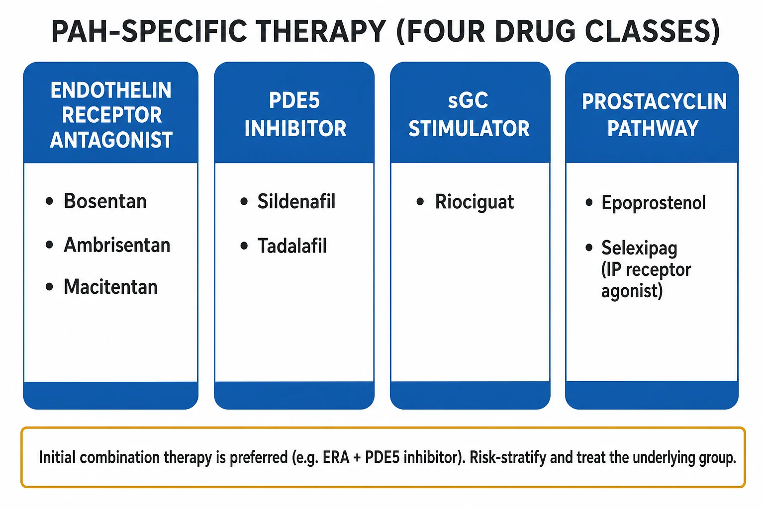
PAH-specific therapy — the four drug classes (Group 1 PAH)
PAH-specific therapy targets the three pathophysiological pathways (endothelin, NO/cGMP, prostacyclin). It is indicated for WHO Group 1 PAH (idiopathic, heritable, drug-induced, and associated PAH) — and is NOT indicated (and is harmful) as primary therapy for post-capillary Group 2 PH. It is used cautiously in selected Group 4 (CTEPH) cases (riociguat is approved for inoperable/persistent CTEPH).[1]
The four PAH drug classes — mechanism, drugs, evidence, cautions
| Class | Mechanism | Prototype(s) | Key trial | Route / cautions |
|---|---|---|---|---|
| Endothelin receptor antagonists (ERA) | Block ET-A (and ET-B) receptors → vasodilation + anti-proliferation | Bosentan (dual A/B), macitentan, ambrisentan (selective A) | BREATHE-1 (bosentan), SERAPHIN (macitentan) | Oral. Bosentan: hepatotoxicity — monthly LFTs. Bosentan/macitentan: teratogenic (contraception). Ambrisentan: less hepatotoxic, peripheral oedema |
| Phosphodiesterase-5 (PDE5) inhibitors | Inhibit cGMP breakdown → potentiate NO → vasodilation + anti-proliferation | Sildenafil, tadalafil | SUPER-1 (sildenafil) | Oral. Headache, flushing, visual disturbance, hypotension. NEVER with nitrates or riociguat (profound hypotension) |
| Soluble guanylate cyclase (sGC) stimulators | Stimulate sGC directly AND sensitise it to endogenous NO → ↑ cGMP → vasodilation | Riociguat | PATENT-1 (PAH), CHEST-1 (CTEPH) | Oral. Unique — works even with low endogenous NO. Hypotension. NEVER with PDE5 inhibitors (profound hypotension) |
| Prostacyclin analogues / IP agonists | Replace deficient PGI2 → vasodilation + anti-proliferation + anti-platelet | Epoprostenol (IV), iloprost (inhaled), treprostinil (IV/SC/inhaled/oral), selexipag (oral IP agonist) | Barst 1996 (IV epoprostenol — only drug with survival benefit in IPAH), AIR (inhaled iloprost), GRIPHON (selexipag) | IV epoprostenol requires a permanent central line and continuous pump — abrupt cessation is fatal (rebound PH). Flushing, jaw pain, diarrhoea, hypotension. Selexipag is oral — better tolerated |
Initial therapy — combination is now standard. The AMBITION trial (Galiè, NEJM 2015) showed that initial oral combination with ambrisentan (ERA) PLUS tadalafil (PDE5) reduced the risk of treatment failure (the composite primary endpoint) by 50% versus either drug alone in treatment-naïve PAH.[7] Contemporary practice therefore starts most Group 1 PAH patients on dual oral therapy (ERA + PDE5) and escalates (adding riociguat, selexipag, or parenteral prostacyclin) based on risk stratification and response. The goal is a low-risk profile (WHO functional class I-II, 6MWT >440 m, RVSP, normal BNP, normal cardiac index, low PVR).[1]
Risk stratification and escalation. PAH is risk-stratified at baseline and on therapy (ESC/ERS four-stratum model; REVEAL and French registries) using WHO functional class, 6MWT, BNP/NT-proBNP, RA area, pericardial effusion, cardiac index, and PVR. High-risk or worsening patients escalate to parenteral prostacyclin (IV epoprostenol/treprostinil) and are evaluated for lung transplant — the definitive therapy for refractory disease. Atrial septostomy and Potts shunt are palliative. Lung transplant is the rescue therapy for end-stage PAH refractory to maximal medical therapy.[1][11]
Comparison tables
Inhaled vs systemic pulmonary vasodilators in acute RV failure
| Agent | Route | Pulmonary selectivity | Systemic BP effect | Role in acute RV failure |
|---|---|---|---|---|
| Inhaled nitric oxide (iNO) | Inhaled (ventilator) | HIGH — only ventilated alveoli | Minimal (no systemic hypotension) | First-line in ventilated patient; rapid onset/offset; monitor methaemoglobin; rebound PH on withdrawal |
| Inhaled epoprostenol / iloprost | Inhaled | HIGH | Minimal | Alternative to iNO; cheaper; nebuliser; iloprost longer action (requires intermittent dosing) |
| IV epoprostenol | IV | LOW (non-selective) | Marked hypotension | Powerful but systemic vasodilation limits use; for severe cases with vasopressor support |
| Sildenafil | Oral/IV | Moderate | Mild | Slower onset; useful adjunct; mainstay of chronic therapy; never with nitrates/iNO caution |
| Milrinone | IV | Moderate (inodilator) | Hypotension | Inotrope + pulmonary vasodilation; preferred inotrope for RV failure |
| Riociguat | Oral | Moderate | Hypotension | Chronic; never with PDE5 inhibitors |
Inotropes and vasopressors in PH with RV failure
| Agent | Class / mechanism | Effect on PVR | Role | Key cautions |
|---|---|---|---|---|
| Noradrenaline | Alpha-1 + modest beta-1 agonist | Neutral/slightly up | First-line vasopressor — restore systemic BP and RV coronary perfusion | Excess raises LV afterload; titrate to MAP |
| Vasopressin | V1 agonist | Down (may lower PVR) | Alternative vasopressor; useful in sepsis-associated PH | Splanchnic/ischaemic effects |
| Milrinone | PDE3 inhibitor (inodilator) | DOWN (pulmonary vasodilation) | PREFERRED inotrope for RV failure — inotropy + pulmonary vasodilation | Systemic vasodilation/hypotension; long half-life (~2 h); thrombocytopenia |
| Dobutamine | Beta-1 (and beta-2) agonist | Up at high dose | Inotrope for low-output RV | Tachyarrhythmia; hypotension (beta-2); increases O2 demand |
| Dopamine | Dose-dependent | Up at high dose | Largely superseded | More arrhythmia than noradrenaline (SOAP-II analogue) |
| Levosimendan | Calcium sensitiser | Down | Refractory RV failure | Hypotension; tachyphylaxis |
Mechanical support for refractory RV failure
| Modality | Support | Haemodynamic effect | Role | Complications |
|---|---|---|---|---|
| VA-ECMO | Veno-arterial | Supports RV and systemic circulation; full cardiopulmonary bypass | Bridge to recovery / decision / transplant in refractory RV failure | Limb ischaemia, bleeding, haemolysis, thrombosis, infection; LV distension (consider Impella) |
| PROTEK Duo (RVAD) | Right atrium to pulmonary artery | RV unloading; supports RV only | Selected centres; RV-specific support | Vascular, bleeding, infection |
| Atrial septostomy | Palliative RA decompression | Creates a right-to-left shunt; lowers RA pressure at the cost of systemic hypoxaemia | Palliative / bridge in refractory right heart failure; selected cases | Worsening hypoxaemia; reserved for severe refractory cases |
| Lung transplant (definitive) | Bilateral lung or heart-lung | Removes the diseased pulmonary vasculature | Rescue for end-stage PAH refractory to maximal medical therapy | Chronic immunosuppression; rejection; chronic rejection (CLAD) |
Exam-style short-answer questions
SAQ — Pulmonary hypertensive crisis with acute RV failure
10 minutes · 10 marks
A 42-year-old woman with known idiopathic pulmonary arterial hypertension on sildenafil and macitentan presents to the ED with syncope. She is hypotensive (BP 78/50), SpO2 84% on room air, heart rate 130 in atrial flutter, with cold peripheries and a raised JVP. Bedside echocardiography shows a severely dilated and hypokinetic right ventricle with a D-shaped septum. Arterial blood gas shows pH 7.18, PaCO2 52, lactate 4.8.
SAQ — ICU management of decompensated pulmonary hypertension
10 minutes · 10 marks
A 68-year-old man with severe mitral regurgitation and chronic breathlessness is admitted to ICU with pulmonary oedema and shock. Bedside echocardiography shows a dilated left atrium, estimated pulmonary artery systolic pressure 75 mmHg, and a dilated dysfunctional right ventricle. The referring team asks whether to start sildenafil and an endothelin receptor antagonist for the pulmonary hypertension.
Clinical pearls
Red flags
Prognosis and landmark trials
PH — the landmark trials that define modern therapy
BREATHE-1 (Rubin, NEJM 2002): bosentan (dual ERA) vs placebo in 213 PAH patients — improved 6MWT and time to clinical worsening; established the ERA class. Hepatotoxicity is the signature adverse effect (monthly LFTs).[3] SUPER-1 (Galiè, NEJM 2005): sildenafil (PDE5 inhibitor) vs placebo in 278 PAH patients — improved 6MWT and functional class; established the PDE5 class.[4] PATENT-1 (Ghofrani, NEJM 2013): riociguat (sGC stimulator) vs placebo — improved 6MWT, PVR, and functional class in PAH. Unique mechanism — works with low endogenous NO. Never combined with PDE5 inhibitors (hypotension).[1] SERAPHIN (Pulido, NEJM 2013): macitentan (ERA) vs placebo in 742 PAH patients — reduced morbidity AND mortality (hard endpoint); established macitentan as a preferred ERA (less hepatotoxic than bosentan).[5] GRIPHON (Sitbon, NEJM 2015): selexipag (oral IP-receptor agonist) vs placebo in 1156 PAH patients — reduced the composite morbidity/mortality endpoint; the oral prostacyclin-pathway option with better tolerability than parenteral agents.[6] AMBITION (Galiè, NEJM 2015): initial ambrisentan + tadalafil combination vs either monotherapy — reduced treatment failure by 50%; established initial oral combination therapy as the standard of care for treatment-naïve Group 1 PAH.[7] Barst (NEJM 1996): IV epoprostenol vs conventional therapy in 81 primary PAH patients — the only PAH drug with a proven SURVIVAL benefit; established parenteral prostacyclin for severe disease (continuous infusion, central line).[8] AIR (Olschewski, NEJM 2002): inhaled iloprost vs placebo — improved 6MWT and functional class; established the inhaled prostacyclin approach (selective, fewer systemic effects).[9] REVEAL registry (Frost, Chest 2011): the large US observational PAH registry — defined the modern phenotype (older, more comorbid than historic series), generated the REVEAL risk score (predicts survival), and set contemporary benchmarks.[11] ESC/ERS 2022 Guidelines (Galiè/Humbert): the current authoritative framework for the diagnosis, classification, risk stratification, and stepped therapy of all five PH groups.[1]
Outcomes. Untreated idiopathic PAH has a median survival of ~2.8 years. Modern combination therapy has transformed the prognosis: contemporary registry survival is ~85% at 1 year and ~60-70% at 5 years for treated Group 1 PAH, with continued improvement as combination therapy and earlier referral take hold. The determinants of survival are the RV (right heart function on echo, cardiac index, right atrial pressure) and the functional class (WHO I-II vs III-IV) and exercise capacity (6MWT) and biomarkers (BNP/NT-proBNP). Once the patient develops RV failure — raised JVP, ascites, low cardiac index, syncope — the prognosis worsens sharply. Acute RV failure in the ICU carries 40-60% mortality even with full intensive care, which is why prevention (protect the RV, treat the trigger early) is paramount. Lung transplant remains the rescue therapy for end-stage disease. The strongest modifiable protections against death and decompensation are: optimal group-specific therapy, sustained low-risk profile, avoidance of hypoxia/hypercapnia/acidosis/excess fluid, maintenance of sinus rhythm, and early transplant referral for the deteriorating patient.[1][2][11]
References
- [1]Galie N, Humbert M, Vachiery JL, et al. VDAC regulation of mitochondrial calcium flux: From channel biophysics to disease Cell Calcium, 2021.PMID 33529977
- [2]Harjola VP, Mebazaa A, Celutkiene J, et al. Notum palmitoleoyl-protein carboxylesterase regulates Fas cell surface death receptor-mediated apoptosis via the Wnt signaling pathway in colon adenocarcinoma Bioengineered, 2021.PMID 34402722
- [3]Rubin LJ, Badesch DB, Barst RJ, et al. Effect of intragastric barostat bag on proximal and distal gastric accommodation in response to liquid meal Am J Physiol Gastrointest Liver Physiol, 2002.PMID 12181183
- [4]Galie N, Ghofrani HA, Torbicki A, et al. Randomized trial of lifestyle modification and pharmacotherapy for obesity N Engl J Med, 2005.PMID 16291981
- [5]Pulido T, Adzerikho I, Channick RN, et al. The endolysosomal cysteine cathepsins L and K are involved in macrophage-mediated clearance of Staphylococcus aureus and the concomitant cytokine induction FASEB J, 2014.PMID 24036885
- [6]Sitbon O, Channick R, Chin KM, et al. Radical aminooxygenation of alkenes with N-fluoro-benzenesulfonimide (NFSI) and TEMPONa Chem Commun (Camb), 2015.PMID 25716908
- [7]Galie N, Barbera JA, Frost AE, et al. Intratympanic steroid injection as a salvage treatment for sudden sensorineural hearing loss J Laryngol Otol, 2014.PMID 25399754
- [8]Barst RJ, Rubin LJ, Long WA, et al. A comparison of continuous intravenous epoprostenol (prostacyclin) with conventional therapy for primary pulmonary hypertension N Engl J Med, 1996.PMID 8532025
- [9]Olschewski H, Simonneau G, Galie N, et al. High-dose ACE inhibition: can it improve renoprotection? Am J Kidney Dis, 2002.PMID 12200823
- [10]Simonneau G, Montani D, Celermajer DS, et al. Heads-up Cataract Surgery: Complication Rates, Surgical Duration, and Comparison With Traditional Microscopes J Refract Surg, 2019.PMID 31059581
- [11]Frost AE, Badesch DB, Barst RJ, et al. How types of premises modulate the typicality effect in category-based induction: diverging evidence from the P2, P3, and LPC effects Sci Rep, 2016.PMID 27982022
- [12]Price LC, Wort SJ, Finney SJ, Marino PS, Brett SJ Severity of acne and its impact on quality of life Skinmed, 2013.PMID 23930353