ICU · Cardiovascular
Sepsis-induced myocardial dysfunction (septic cardiomyopathy)
Also known as Septic cardiomyopathy · Sepsis-induced cardiomyopathy · SICM · Septic myocardial depression · Myocardial dysfunction in sepsis
Sepsis-induced cardiomyopathy (SICM): reversible, biventricular myocardial dysfunction arising during septic shock and NOT explained by ischaemia, infarction, or pre-existing structural heart disease. Presents: NEW LV systolic dysfunction (reduced EF), RV dysfunction, and diastolic dysfunction superimposed on a vasoplegic (distributive) shock state. Mechanism: circulating myocardial depressant substances (TNF-α, IL-1β, IL-6), nitric oxide/iNOS overproduction, mitochondrial dysfunction with impaired oxidative phosphorylation, beta-adrenergic receptor downregulation/desensitisation, and coronary microcirculatory dysfunction. Diagnosis: echocardiography (reduced EF, LV dilatation, RV dysfunction, abnormal global longitudinal strain, diastolic impairment) supported by elevated troponin and BNP/NT-proBNP; cardiac MRI for atypical/persistent cases. Treatment: treat sepsis (source control, antibiotics), fluid resuscitation guided by fluid-responsiveness testing, vasopressors (noradrenaline first-line), inotropes if low cardiac output (dobutamine, milrinone; levosimendan NOT routinely recommended — LeoPARDS). REVERSIBLE — EF usually recovers within 7-10 days in survivors.
On this page & tools
Your progress
Saved locally on this device.
Target exams
Red flags
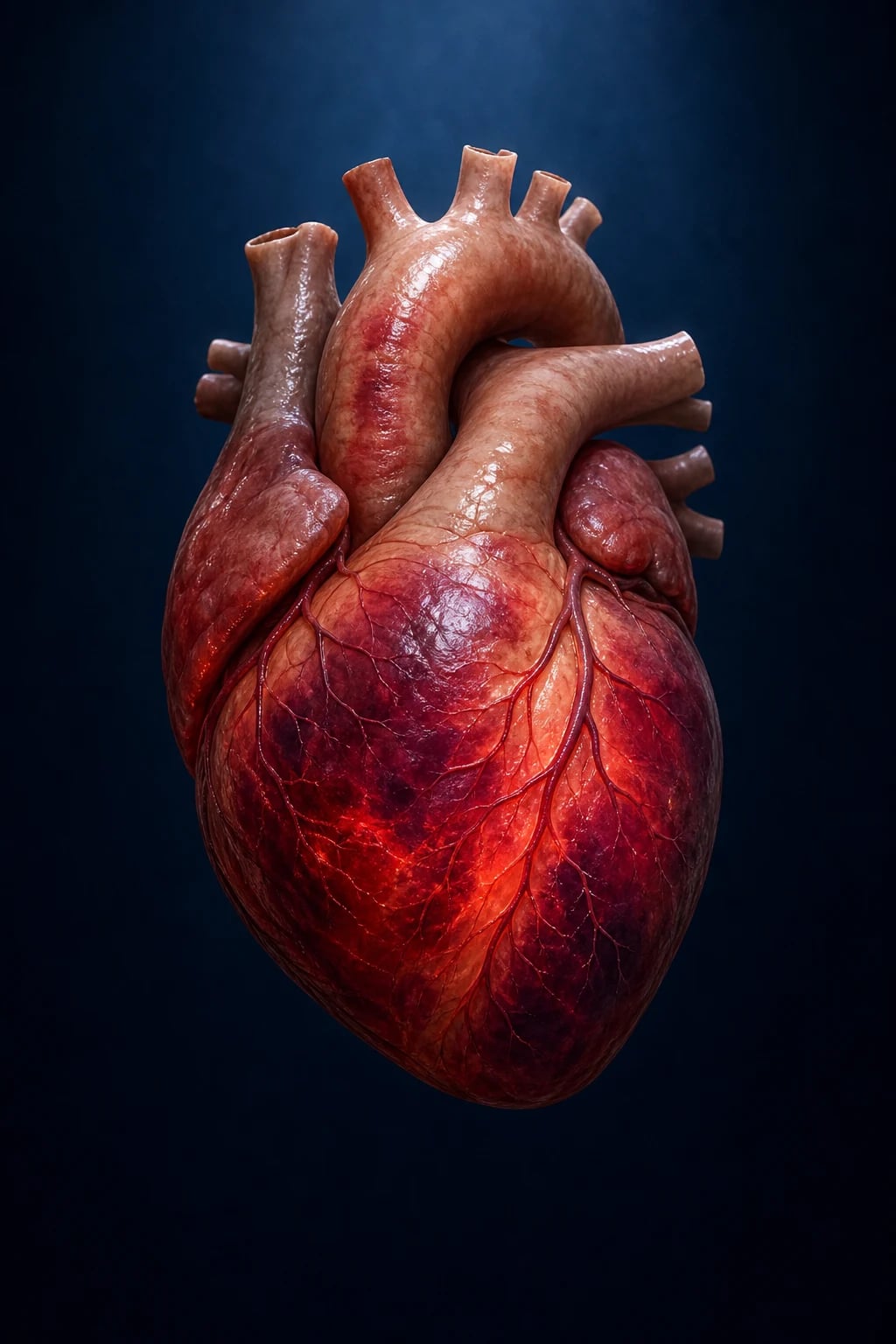
Diagnosis and management of sepsis-induced cardiomyopathy
- Recognise — septic shock with cardiac dysfunction (new LV dysfunction, elevated troponin, BNP)
- Echocardiography — assess: LV systolic function (EF), LV dilatation, RV function, diastolic function, cardiac output, global longitudinal strain. Compare to baseline (if available)
- Distinguish from cardiogenic shock — SICM: WARM shock (vasoplegic), high lactate (sepsis), source of infection, REVERSIBLE. Cardiogenic: COLD shock, elevated filling pressures, ischaemic cause
- Treat sepsis — antibiotics within 1h, source control, fluid resuscitation (30 mL/kg crystalloid — but titrate to fluid responsiveness, avoid overload)
- Vasopressors — noradrenaline FIRST-LINE (alpha vasoconstriction + modest beta). Target MAP ≥65. Add vasopressin if escalating noradrenaline requirement
- Inotropes if low cardiac output — dobutamine (beta-1, increases contractility) or milrinone (PDE inhibitor, reduces afterload, pulmonary vasodilator). Use if: low cardiac output despite adequate volume + vasopressor, ongoing hypoperfusion (lactate, oliguria, mottled skin). Levosimendan NOT routinely recommended (LeoPARDS — no outcome benefit)
- Monitor — echocardiography (serial — assess recovery), troponin/BNP trend, lactate clearance, urine output, clinical perfusion
- Expect recovery — SICM typically REVERSES within 7-10 days (if sepsis controlled). Persistent dysfunction: consider alternative diagnosis (ischaemia, myocarditis, takotsubo, pre-existing cardiomyopathy)
Exam practice
SAQ — Septic cardiomyopathy with EF 25 percent and ongoing hypoperfusion
10 minutes · 10 marks
A 64-year-old man is admitted to ICU with septic shock from a perforated sigmoid diverticulum. He has received 30 mL/kg crystalloid and is on noradrenaline 0.35 mcg/kg/min (MAP 62, target 65). BP 92/56, HR 128 sinus, SpO2 94% on FiO2 0.5, temp 38.8°C, lactate 4.2 mmol/L, urine output 15 mL/hr, cool mottled peripheries. A passive leg raise produces no rise in cardiac output. Bedside echocardiography shows LV ejection fraction 25%, global hypokinesis with LV dilatation, TAPSE 14 mm, E/e' 16, IVC 2.4 cm with <10% collapsibility. High-sensitivity troponin T 480 ng/L, NT-proBNP 6800. ECG shows sinus tachycardia with no ST changes.
SAQ — Sepsis-induced cardiomyopathy versus pre-existing cardiomyopathy
10 minutes · 10 marks
A 72-year-old man is admitted to ICU with community-acquired pneumonia and septic shock (noradrenaline 0.4 mcg/kg/min, MAP 60, lactate 5.1 mmol/L, WCC 28). Bedside echocardiography shows LVEF 28% with global hypokinesis and a dilated LV (LVEDD 6.2 cm), E/e' 15, TAPSE 16 mm, no regional wall motion abnormality. Troponin T 320 ng/L, NT-proBNP 4200. He is unsure of his cardiac history; his wife recalls he was told his heart was 'a bit weak' three years ago but no records are available. You are asked whether this is sepsis-induced cardiomyopathy or pre-existing disease.
Clinical pearls
Red flags
Prognosis
Outcomes of sepsis-induced cardiomyopathy (Parker/Parrillo 1990, Hasegawa 2023 meta-analysis)
Classic study (Parrillo/Parker, Annals of Internal Medicine 1990) + modern systematic review and meta-analysis (Hasegawa et al, J Intensive Care Med 2023):[5][8]
- Prevalence in septic shock: 40-60% (echocardiographic evidence of LV systolic dysfunction); diastolic dysfunction present in up to 70%
- Mortality WITH SICM: significantly higher than septic shock without SICM (pooled OR ~1.9 in the Hasegawa 2023 meta-analysis)
- EF recovery: 60-80% of survivors recover EF within 7-10 days
- Persistent LV dysfunction: 20-40% (may have pre-existing cardiomyopathy or permanent damage)
- RV dysfunction: present in 30-50% of septic shock patients (often coexists with LV, worsens prognosis)
- Troponin elevation: 50-80% of septic shock patients (correlates with severity, not necessarily ischaemia)[12]
KEY: SICM is a MARKER of severity (sicker patients), not necessarily the CAUSE of death. Treat the sepsis, support the heart, expect recovery.
Pathophysiology — deep dive
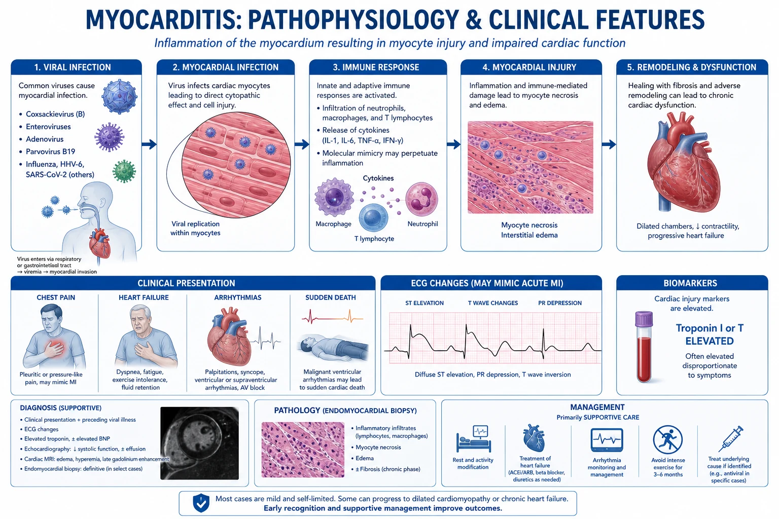
SICM is a functional, cytokine-mediated myocardial injury — not an ischaemic or structurally destructive one. The depression is mediated by soluble factors and reversible cellular dysfunction (mitochondrial, receptor, microvascular) rather than myocyte necrosis, which is why EF recovers when the septic source is controlled. Five overlapping mechanisms account for most of the contractile deficit.[1][6]
1. Circulating myocardial depressant substances
The seminal observation: serum from septic shock patients with reduced EF, when applied to isolated rat myocytes in vitro, depresses the extent and velocity of shortening; serum from septic patients without cardiac depression, or from non-septic critically ill controls, does not. The depressant activity is titratable, heat-labile, and trackable with the patient's clinical course — appearing within 24-48 h of shock onset and disappearing as the patient recovers, paralleling EF normalisation. This is the mechanistic basis for the reversibility of SICM.[3][5]
Fractionation and neutralisation identified the depressants as pro-inflammatory cytokines:[7]
- Tumour necrosis factor-α (TNF-α / cachectin) — the first identified; directly reduces myocyte contractility within minutes via sphingomyelinase → ceramide → negative inotropy, and via nitric oxide synthase induction. Plasma TNF correlates with the degree of myocardial depression.
- Interleukin-1β (IL-1β) — synergistic with TNF-α; depresses contractility more slowly (hours) via autocrine NO and β-receptor uncoupling.
- Interleukin-6 (IL-6) — later peak (sustained phase); reduces contractility via NO/superoxide and downregulates β-adrenergic signalling.
Kumar et al (J Exp Med 1996) proved causality: immunoneutralisation of TNF-α and IL-1β abolished the in vitro depressant effect of septic human serum on myocytes, establishing these two cytokines as the principal mediators.[7]
2. Nitric oxide (NO) pathway
NO is the downstream effector of much of the cytokine-mediated depression. Inducible NO synthase (iNOS / NOS2) is upregulated in cardiac myocytes and the vascular endothelium by TNF-α, IL-1β and endotoxin, producing sustained, high-output NO. NO activates soluble guanylate cyclase → ↑cGMP → ↓myofilament calcium responsiveness → negative inotropy. NO also reacts with superoxide to form peroxynitrite (ONOO⁻), which directly damages contractile proteins, mitochondrial enzymes and membrane lipids. The net effect: reduced contractility, impaired relaxation, and mitochondrial damage.[16]
3. Mitochondrial dysfunction
In sepsis, myocardial oxygen extraction is preserved but oxygen utilisation is impaired — there is pathological oxygen supply-demand uncoupling at the cellular level. Mitochondria in septic myocardium show:[1][6]
- Complex I/II/III dysfunction → reduced oxidative phosphorylation and ATP synthesis
- Mitochondrial permeability transition pore (mPTP) opening → cytochrome c release, apoptosis
- Oxidative/nitrosative stress (superoxide, peroxynitrite) damaging the electron transport chain
- Mitochondrial autophagy (mitophagy) failure → accumulation of damaged organelles
The result is a state of cellular energetic failure ("cytopathic hypoxia") — the myocyte cannot generate ATP even in the presence of adequate oxygen, mimicking hibernation. This is central to the reversible, non-ischaemic nature of SICM: the cell is alive but functionally dormant. [1]
4. Beta-adrenergic receptor downregulation / desensitisation
Sepsis induces β-adrenergic receptor desensitisation in the myocardium: receptor density falls, receptor-G-protein-adenylate cyclase coupling is uncoupled, and downstream cAMP generation is blunted. Mechanisms include elevated circulating catecholamines (agonist-induced desensitisation), cytokine-mediated (TNF/IL-1) receptor phosphorylation, and increased G-protein-coupled receptor kinase (GRK) activity. Clinically this manifests as blunted contractile reserve and reduced responsiveness to exogenous catecholamines — one rationale for phosphodiesterase inhibitors (milrinone), which act downstream of the receptor, and for the calcium-sensitiser levosimendan.[6]
5. Coronary microcirculatory dysfunction
Although global coronary blood flow is usually maintained or increased in septic shock, there is microcirculatory maldistribution: capillary leak, endothelial activation, leucocyte plugging, microthrombi and altered arteriolar tone create patchy hypoperfusion despite adequate macrovascular flow. Coupled with the increased oxygen demand of tachycardia and high inotrope use, this produces regional supply-demand mismatch (a substrate for type 2 MI and troponin leak).[1]
Mechanisms of myocardial depression in sepsis — at a glance
| Mechanism | Key mediator(s) | Cellular effect | Clinical correlate |
|---|---|---|---|
| Circulating depressant substances | TNF-α, IL-1β, IL-6 | ↓ Myofilament shortening (ceramide, sphingomyelinase) | Global hypokinesis; reversible as cytokines clear |
| Nitric oxide / iNOS | NO, cGMP, peroxynitrite | ↓ Calcium responsiveness; mitochondrial damage | Negative inotropy; impaired relaxation |
| Mitochondrial dysfunction | Electron transport chain ↓, mPTP opening | ↓ ATP synthesis ("cytopathic hypoxia") | Hibernating, viable but functionally dormant myocyte |
| β-receptor desensitisation | GRK, receptor phosphorylation | ↓ cAMP, blunted adrenergic signalling | Reduced catecholamine responsiveness |
| Microcirculatory dysfunction | Endothelial activation, capillary leak | Patchy hypoperfusion, regional ischaemia | Troponin leak (type 2 MI substrate) |
Diagnosis
SICM is a clinical-echocardiographic diagnosis made in a patient with septic shock who develops new biventricular dysfunction not explained by ischaemia or pre-existing disease. There is no single pathognomonic test — the diagnosis integrates (1) the clinical context (septic shock), (2) echocardiographic findings, (3) cardiac biomarkers, and (4) exclusion of competing diagnoses. [1]
Echocardiography — the cornerstone
Bedside echocardiography (focus TTE/FOCUS or comprehensive TTE) is first-line and should be performed early (within 6 h of shock recognition) and repeated serially (every 24-48 h) to track recovery.[4]
Findings supporting SICM:
- Reduced LV ejection fraction (EF) — classically <50%, often 30-40% in active SICM. May be profoundly depressed in severe cases.
- LV dilatation — increased LVEDD/LVEDV; the heart dilates to preserve stroke volume via Starling's law (adaptive).
- Global hypokinesis — diffuse, not regional. Regional wall motion abnormality → suspect ischaemia.
- RV dysfunction — reduced TAPSE (<17 mm), RV dilatation, tricuspid annular plane systolic excursion, tricuspid regurgitation, paradoxical septal motion. Present in 30-50%.[17]
- Diastolic dysfunction — impaired relaxation (Grade I-III). Assess E/A ratio, E/e' ratio (E/e' >14 suggests elevated LV filling pressure), left atrial volume index, deceleration time. Diastolic dysfunction is independently associated with mortality.
- Hyperdynamic / normal EF in early sepsis — does NOT exclude SICM; diastolic dysfunction or reduced strain may be the only early clue.
Echocardiographic parameters in SICM — what to measure and why
| Parameter | Abnormal in SICM | Cut-off / finding | Significance |
|---|---|---|---|
| LV ejection fraction (LVEF) | Systolic dysfunction | <50% (often 30-40%) | Hallmark; recoverable in 7-10 days |
| Global longitudinal strain (GLS) | Sub-endocardial dysfunction (precedes EF fall) | Less negative than −18% (−16% to −18%) | Most sensitive marker; detects dysfunction with "normal" EF |
| LVEDD / LVEDV | LV dilatation | LVEDD >5.8 cm (M, simplified) | Adaptive Starling compensation |
| RV function (TAPSE, FAC, S') | RV dysfunction | TAPSE <17 mm | Biventricular disease; worse prognosis |
| E/e' ratio | Diastolic dysfunction / ↑ filling pressure | >14 suggests elevated LV filling pressure | Independent mortality predictor |
| IVC size/collapsibility | Volume status & responsiveness | Collapsible (>50%) → fluid responsive | Guide fluid vs inotrope decision |
| Regional wall motion | Should be ABSENT in SICM | RWM abnormality → suspect ischaemia/takotsubo | Key differentiator |
Global longitudinal strain (GLS) — the sensitive early marker
Speckle-tracking global longitudinal strain detects subclinical myocardial dysfunction before EF falls, because longitudinal sub-endocardial fibres are the most vulnerable to cytokine/ischaemic injury. GLS is reduced (less negative) in SICM even when EF reads "normal", making it the most sensitive echocardiographic marker and a powerful prognostic indicator — worsening GLS correlates with mortality even after EF correction. GLS normalisation lags clinical recovery, so follow trends rather than single values.[4][15]
Cardiac biomarkers
- Troponin (hs-cTnT/cTnI) — elevated in 50-80% of septic shock patients; reflects cytokine-mediated myocyte injury, microvascular dysfunction, type 2 MI and direct oxidative damage (not plaque rupture). A meta-analysis (Sheyin 2015) showed troponin elevation confers significantly higher mortality (OR ~2) in sepsis.[12] Use trends, not a single value, and interpret in context — do not reflexively coronary-angiogram an isolated troponin rise.
- BNP / NT-proBNP — elevated in SICM from ventricular wall stress; correlates with the severity of dysfunction and prognosis. Rising BNP with falling EF suggests worsening SICM; falling BNP parallels recovery.[14] Useful to distinguish cardiogenic from septic pulmonary oedema and to follow response to therapy.
Cardiac MRI — for atypical or persistent cases
Cardiac MRI is reserved for cases where the diagnosis is unclear or EF fails to recover by day 10-14, to distinguish SICM from myocarditis (oedema, late gadolinium enhancement in a non-coronary pattern), ischaemia (subendocardial/transmural LGE in a coronary distribution), or takotsubo (apical ballooning, typical oedema). In SICM itself, MRI typically shows little or no late gadolinium enhancement (consistent with functional, non-necrotic injury), though small focal oedema may be seen — a useful negative finding that supports the diagnosis.[15]
Imaging modalities in suspected SICM
| Modality | Role | Strengths | Limitations |
|---|---|---|---|
| Bedside TTE / FOCUS | First-line, serial | Immediate, repeatable, no transport; assesses EF, RV, IVC, effusion | Operator-dependent; visual EF less precise |
| Comprehensive TTE + GLS | Definitive echo assessment | Speckle-tracking GLS detects subclinical dysfunction; diastolic assessment | Needs experienced sonographer/vendor |
| Transoesophageal echo (TOE) | If TTE poor windows / valvular query | High image quality; intraoperative | Semi-invasive; not first-line |
| Cardiac MRI | Persistent/atypical dysfunction | Tissue characterisation (oedema, LGE); excludes myocarditis/ischaemia | Transport of critically ill; not acute |
| Coronary angiography | If ischaemia suspected (regional WMA, type 1 MI) | Defines coronary anatomy; therapeutic (PCI) | Invasive; not for isolated troponin rise |
Diagnostic workup of suspected sepsis-induced cardiomyopathy
- Suspect — septic shock with new cardiac dysfunction, rising lactate despite resuscitation, hypoxaemia, oliguria, cool peripheries OR unexpectedly low SvO₂
- Early bedside TTE (≤6 h) — EF, LV size, RV function (TAPSE), diastolic (E/e'), IVC, pericardial effusion, regional wall motion
- Biomarkers — troponin (baseline + trend), BNP/NT-proBNP, lactate, venous/arterial blood gas
- Compare to baseline — any prior echo, ECG, troponin (pre-existing cardiomyopathy vs new)
- Classify haemodynamics — fluid-responsive vs not; warm (vasoplegic) vs cold (low output) shock phenotype
- If EF depressed and fluid-resuscitated but still hypoperfused → add inotrope (dobutamine first; milrinone if RV dysfunction/vasoconstriction)
- Serial echocardiography every 24-48 h — track EF/GLS recovery; daily biomarker trend
- If EF NOT recovering by day 10-14 → cardiac MRI ± coronary angiography to exclude ischaemia, myocarditis, takotsubo, pre-existing cardiomyopathy
Differentiating SICM from pre-existing cardiomyopathy and mimics
The single most important diagnostic decision is whether the cardiac dysfunction is new and sepsis-related (SICM, reversible) versus pre-existing (unmasked by sepsis) or an alternative acute cardiomyopathy (ischaemic, myocarditis, takotsubo) — because the management, prognosis and counselling differ fundamentally. [1]
Key differentiators:
- History — any prior echo, heart failure admission, IHD, valve disease, cardiotoxic chemotherapy, familial cardiomyopathy. A normal prior EF strongly supports SICM.
- ECG — Q waves, old infarct, persistent ST changes suggest chronic/pre-existing disease. Diffuse non-specific changes common in both sepsis and myocarditis.
- Echo pattern — SICM: global hypokinesis ± dilatation, normal valve structure, little/no LVH. Pre-existing: regional WMA (post-MI), marked LVH (hypertensive/HFpEF), restrictive/hypertrophic phenotypes, valvular disease.
- Wall motion — regional → ischaemia/takotsubo; global → SICM.
- Takotsubo — apical ballooning with basal hyperkinesis, often triggered by sepsis; can coexist with SICM and is itself reversible.
- Biomarker trajectory — SICM: troponin mild-moderate, rises then falls with recovery. Acute MI: steep rise, territory echo. Myocarditis: troponin often markedly elevated, ECG changes, viral prodrome.
- Coronary angiography — the gold-standard discriminator when ischaemia is plausible (regional WMA, type 1 MI features).
- Cardiac MRI — tissue characterisation to distinguish myocarditis (oedema + mid-wall/subepicardial LGE) from SICM (minimal LGE) and ischaemia (subendocardial LGE).[15]
SICM vs cardiogenic shock vs takotsubo vs myocarditis vs pre-existing cardiomyopathy
| Feature | SICM | Cardiogenic shock (AMI) | Takotsubo | Acute myocarditis | Pre-existing cardiomyopathy |
|---|---|---|---|---|---|
| Context | Septic shock | Ischaemic event | Emotional/physical stress (incl. sepsis) | Viral prodrome | Chronic HF history |
| SVR | Low (vasoplegic, warm) | High (cold) | Variable | Variable | Variable |
| EF | Reduced, recoverable | Reduced, regional WMA | Reduced, apical ballooning | Reduced, global | Chronically reduced |
| Wall motion | Global hypokinesis | Regional (coronary) | Apical ballooning, basal hyperkinesis | Global, often patchy | Regional or global |
| LGE (MRI) | Minimal/absent | Subendocardial/transmural | Apical/mid-cavity oedema | Mid-wall/subepicardial + oedema | Variable (often established) |
| Troponin | Mild-moderate, trend | Marked, rise-fall | Moderate | Often markedly high | Variable |
| Reversibility | Yes, 7-10 days | Depends on revascularisation | Yes, days-weeks | Variable | Usually no |
| First treatment | Noradrenaline + treat sepsis + inotrope | Revascularise + MCS | Supportive, treat trigger | Immunosuppression (selected) | Guideline HF therapy |
Reversibility and natural history
The natural history of SICM is one of the most exam-friendly and clinically reassuring aspects of the condition. The dysfunction is acquired rapidly (within 24-48 h of shock onset), is maximal at days 2-3, and recovers over 7-10 days in survivors as the septic source is controlled and the cytokine load clears.[1][5]
Timeline:
- Onset (Day 0-2): EF falls within hours-days of shock; troponin/BNP rise; LV dilates; RV dysfunction may appear.
- Nadir (Day 2-4): maximal depression; inotrope requirement greatest; highest mortality risk.
- Recovery (Day 4-10): EF climbs back towards baseline as cytokines clear and mitochondrial function recovers; inotropes wean; biomarkers fall.
- Resolution (Day 7-10+): 60-80% of survivors regain a normal (or near-normal) EF. A minority (20-40%) have persistent dysfunction — usually pre-existing cardiomyopathy unmasked by sepsis, or (rarely) permanent sepsis-induced injury. [1]
Caveat: recovery presumes adequate source control and antimicrobial therapy. If the septic drive persists (undrained collection, resistant organism), SICM persists and may progress. [1]
Reversibility data — Parker/Parrillo 1990 and modern series
- Parker/Parrillo (Ann Intern Med 1990): in survivors of septic shock, depressed EF recovered to normal within 7-10 days; non-survivors had a paradoxically higher early EF (less depression) — the so-called "reversal" finding, since explained by inability to mount the adaptive LV dilatation.[5]
- Modern echo series: EF recovery in 60-80% of survivors by day 7-10; persistent LV dysfunction in 20-40%.
- Diastolic dysfunction (E/e') recovery often lags systolic recovery by days — do not be alarmed by persistent diastolic impairment if EF is normalising.
- GLS recovery lags EF recovery — follow GLS trends for completeness.
Management
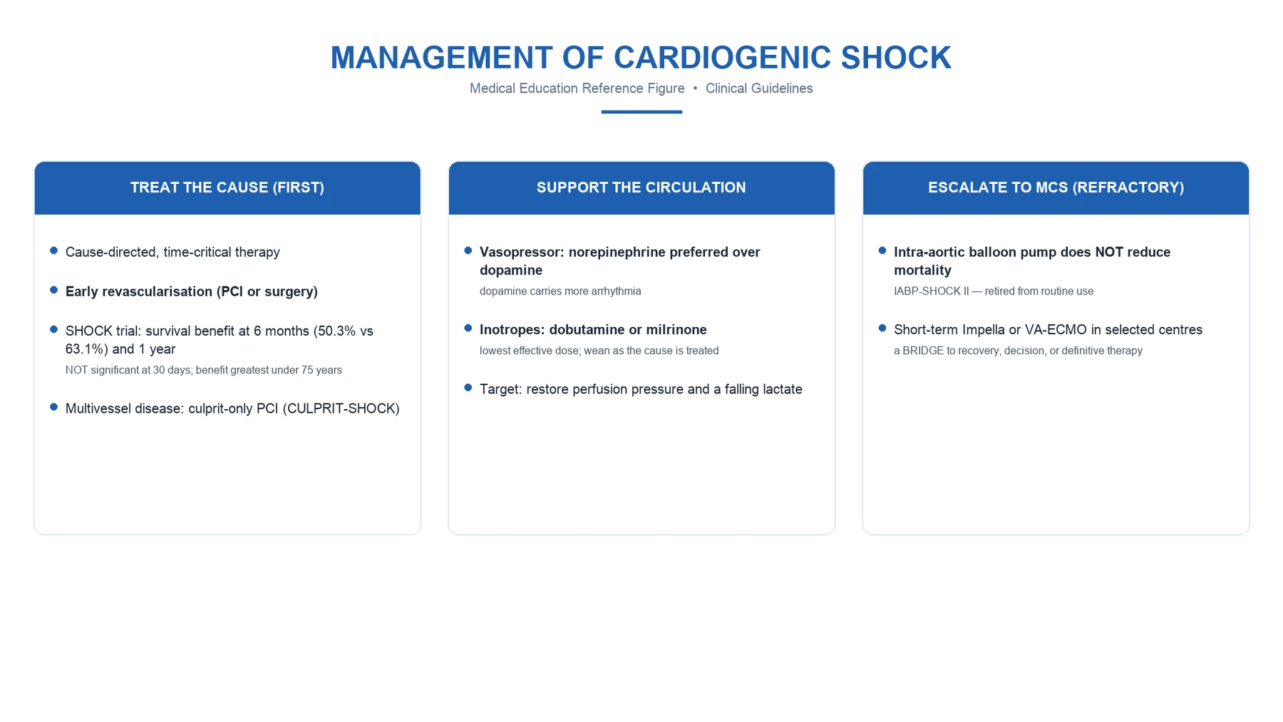
The principles of SICM management mirror those of septic shock, with cardiac-specific layers added once volume status is addressed. Treat the sepsis first — source control and antibiotics are the definitive therapy for the cardiac depression.[11]
Step 1 — Resuscitate the sepsis (Surviving Sepsis Campaign 2021)
- Antibiotics within 1 hour of recognition; broad-spectrum, source-directed.
- Source control as soon as practical (drainage, debridement, device removal).
- Early crystalloid — at least 30 mL/kg in the first 3 h for septic shock with hypoperfusion, but titrated dynamically to dynamic fluid-responsiveness metrics. The 2021 SSC weakened the fixed 30 mL/kg recommendation precisely because of harm from injudicious fluid in non-responders.[11]
Step 2 — Assess fluid responsiveness BEFORE giving more fluid
Giving inotropes to a hypovolaemic patient worsens outcomes; giving more fluid to a non-responder causes oedema, worsens gas exchange, raises intra-abdominal pressure, and (in SICM) precipitates pulmonary oedema and RV overload. Test responsiveness with:[13]
- Passive leg raise (PLR) — the best bedside test (sensitivity/specificity ~85%); starts at 45° semi-recumbent → flat + legs elevated 45° for 60-90 s; a ≥10% rise in cardiac output/stroke volume (or pulse pressure/variations) = responder. Monnet's meta-analysis confirms PLR outperforms static indices.[13]
- IVC collapsibility (>50% collapse in spontaneously breathing; distensibility >18% in ventilated) — quick, qualitative.
- Stroke volume variation / pulse pressure variation — in fully ventilated, deeply sedated patients with closed chest.
- Fluid challenge — 250-500 mL bolus with real-time CO measurement.
- Echocardiographic SV assessment (LVOT VTI) before/after PLR.
Decision: responder → give fluid (small aliquots, recheck). Non-responder or hypoperfusion persists after adequate fluid → move to vasopressors ± inotropes. [1]
Tests of fluid responsiveness in septic shock
| Test | How performed | Threshold for "responder" | Pitfalls |
|---|---|---|---|
| Passive leg raise (PLR) | Semi-recumbent 45° → legs up 45°, trunk flat, 60-90 s | ΔSV or ΔCO ≥10% | Start from 45° (not flat); auto-transfuse ~300 mL |
| IVC collapsibility (spontaneous) | M-mode subxiphoid | >50% collapse | Unreliable if mechanically ventilated or high PEEP |
| IVC distensibility (ventilated) | M-mode | >18% distensibility | Needs fully controlled ventilation, closed chest |
| SVV / PPV | Arterial/pulse-contour | >12-13% | Only valid in deep sedation, controlled ventilation, regular rhythm |
| Mini fluid challenge | 50-250 mL bolus + real-time CO/VTI | ΔSV ≥10% | Needs CO monitor/echo; transient |
Step 3 — Vasopressors (noradrenaline first-line)
Target MAP ≥65 mmHg (lower acceptable if chronic HTN controlled; individualise).[11]
- Noradrenaline (norepinephrine) FIRST-LINE — α-1 vasoconstriction restores SVR + modest β-1 inotropy. Even with SICM it is preferred: it improves coronary perfusion pressure and supports the depressed myocardium without the arrhythmia burden of dopamine.
- Vasopressin (0.01-0.04 U/min) — add as second agent to reduce noradrenaline dose (catecholamine-sparing); fixed low dose. Not a first-line solo agent.
- Adrenaline (epinephrine) — alternative/add-on; more arrhythmias, ↑lactate (β-2 glycolysis), but potent inotrope + vasoconstrictor.
- Dopamine — AVOID unless specifically bradycardic shock. SOAP II (De Backer, NEJM 2010): dopamine vs noradrenaline showed no mortality difference overall but significantly more arrhythmias with dopamine (mostly atrial fibrillation), and a subgroup signal of worse outcome in cardiogenic shock.[9]
Step 4 — Inotropes (when low cardiac output persists)
Add an inotrope when there is ongoing hypoperfusion (rising lactate, oliguria, mottled skin, low SvO₂, cold peripheries) despite adequate fluid resuscitation and adequate MAP, and/or echo shows reduced EF with low cardiac output.[6][11]
- Dobutamine — β-1 agonist (with some β-2). Increases contractility and CO, modest vasodilation (can drop SVR/MAP — usually co-administered with noradrenaline). First-line inotrope in SICM with low output. Start 2.5-5 µg/kg/min, titrate.
- Milrinone — PDE-3 inhibitor; ↑cAMP independent of β-receptor (useful given β-desensitisation in sepsis); inotrope + lusitrope + vasodilator, including pulmonary vasodilation → preferred when RV dysfunction or pulmonary hypertension coexists. Longer half-life (~2.5 h) — harder to titrate acutely; can cause hypotension (often need noradrenaline concurrently).
- Levosimendan — calcium sensitiser; improves contractility without rising intracellular calcium (theoretically less arrhythmia/O₂ demand). LeoPARDS trial (Antcliffe, ICM 2019) found NO benefit on SOFA-derived organ dysfunction or mortality in septic shock with biochemical cardiac dysfunction; not routinely recommended.[10]
Inotropes in sepsis-induced cardiomyopathy
| Agent | Mechanism | Haemodynamic effect | Best use in SICM | Key cautions |
|---|---|---|---|---|
| Dobutamine | β-1 (±β-2) agonist | ↑CO, ↓SVR (modest), ↑HR | First-line; LV low output | Tachyarrhythmia; ↑myocardial O₂ demand; can drop MAP |
| Milrinone | PDE-3 inhibitor → ↑cAMP | ↑CO, ↓SVR + ↓PVR | RV dysfunction / pulmonary HTN; β-desensitised patient | Long t½; hypotension; often need noradrenaline |
| Levosimendan | Calcium sensitiser | ↑CO, vasodilation | NOT routine (LeoPARDS negative) | Hypotension; cost; no outcome benefit |
| Adrenaline | α/β agonist | ↑CO + ↑SVR (high dose) | Refactory shock with low CO | ↑Lactate (β-2), arrhythmias, ↑O₂ demand |
| Dopamine | Dose-dependent D/α/β | Variable | Only if bradycardic shock | SOAP II: more arrhythmias; AVOID generally |
Step 5 — Avoid excessive fluid (a recurring, lethal error)
Fluid overload worsens outcomes in septic shock and is doubly harmful in SICM: it precipitates pulmonary oedema, worsens gas exchange, raises intra-abdominal pressure (renal impairment), and overloads a depressed RV (TR, low LV preload from septal shift). Strategies:[11][13]
- Deresuscitate — once stable, achieve negative fluid balance with diuretics (furosemide) or RRT if needed.
- Late shock = less fluid, more vasopressor/inotrope. The classic "give fluid, give more fluid" reflex kills in SICM.
- Monitor cumulative fluid balance daily; target even-to-negative balance after the first 24-48 h.
Step 6 — Mechanical circulatory support (rare, refractory cases)
In the small subset with refractory SICM (biventricular failure, persistent low output despite maximal inotrope + vasopressor), short-term mechanical support — IABP (limited in vasoplegia), VA-ECMO, or Impella — may bridge to recovery. This is an MDT decision and a bridge-to-recovery/decision, not destination therapy; outcome is dictated by the underlying sepsis.[6]
Haemodynamic management algorithm for SICM
- Resuscitate sepsis — antibiotics <1 h, source control, initial 30 mL/kg crystalloid (titrate)
- Assess volume status + fluid responsiveness — PLR, IVC, SVV/PPV, echo VTI. If responsive → 250-500 mL aliquots, reassess
- Start vasopressor — noradrenaline first-line; target MAP ≥65; add vasopressin 0.03 U/min if escalating
- Reassess perfusion — lactate trend, SvO₂, urine output, skin temp, echo EF/CO
- If hypoperfusion persists + reduced EF/low CO → add dobutamine 2.5-5 µg/kg/min (titrate to CO/SvO₂ 65-70%); ensure adequate MAP with noradrenaline
- If RV dysfunction / pulmonary HTN → add/switch to milrinone (with noradrenaline for MAP)
- Avoid dopamine (SOAP II: more arrhythmias); do NOT use levosimendan routinely (LeoPARDS negative)
- Stop fluids once euvolaemic — deresuscitate with diuretics; track cumulative balance
- Serial echo + biomarkers — expect EF recovery by day 7-10; if not, reassess diagnosis
- Refractory low output despite maximal therapy → MDT for short-term MCS (VA-ECMO/Impella) as bridge to recovery
Clinical significance — does myocardial depression drive mortality?
The relationship between SICM and mortality is subtle and exam-critical. Patients with SICM have higher mortality than those without (Hasegawa 2023 meta-analysis pooled OR ~1.9), but the cardiac dysfunction is largely a marker of illness severity (a greater inflammatory burden) rather than the proximate cause of death.[1][8]
Why SICM correlates with mortality:
- A larger cytokine load produces both greater myocardial depression AND greater multi-organ failure — SICM flags the sicker patient.
- SICM reduces the compensatory rise in cardiac output needed to meet the high septic oxygen demand, worsening tissue hypoxia and lactate.
- RV dysfunction (a component of SICM) independently predicts mortality through hypoxaemia and low output.
- Severe SICM drives escalating catecholamine use → arrhythmias, myocardial O₂ demand, microvascular insult. [1]
Why SICM itself is not usually the cause of death:
- EF recovers in survivors — death from "SICM" alone is rare.
- Most deaths are from multi-organ failure secondary to uncontrolled sepsis, with the recovering heart as a bystander.
- The Parker "reversal" paradox: non-survivors had less early EF depression (could not dilate adaptively). [1]
Practical implication: treat the sepsis, support the heart (fluids, vasopressors, inotropes), expect recovery — but do not attribute death to "cardiac failure" if the EF is recovering. Persistent search for and control of the septic source is the highest-yield intervention. [1]
Evidence base — landmark trials and meta-analyses
- Parrillo/Parker 1990 (Ann Intern Med) — established the reversible, dilated, depressant-substance phenotype of human septic shock cardiomyopathy.[5]
- Parrillo 1985 (J Clin Invest) — proved a circulating myocardial depressant substance in septic serum (in vitro myocyte depression).[3]
- Kumar 1996 (J Exp Med) — TNF-α and IL-1β are the principal depressant cytokines (immunoneutralisation abolishes the effect).[7]
- De Backer 2010 SOAP II (NEJM) — dopamine vs noradrenaline: no overall mortality difference but more arrhythmias with dopamine → noradrenaline first-line.[9]
- Antcliffe 2019 LeoPARDS (Intensive Care Med) — levosimendan in septic shock with biochemical cardiac dysfunction: no benefit on organ dysfunction/mortality → not routine.[10]
- Hasegawa 2023 (J Intensive Care Med) — systematic review and meta-analysis of SICM prevalence and prognosis: SICM common and associated with higher mortality (OR ~1.9).[8]
- Monnet 2016 (Intensive Care Med) — passive leg raising meta-analysis: best bedside fluid-responsiveness test.[13]
- Sheyin 2015 (Heart Lung) — troponin elevation in sepsis meta-analysis: prognostic (OR ~2 for mortality).[12]
- Evans 2021 (Crit Care Med) — Surviving Sepsis Campaign guidelines: noradrenaline first-line, dopamine to be avoided, inotrope if low CO persisting after adequate fluid + vasopressor.[11]
References
- [1]Hollenberg SM, Singer M Pathophysiology of sepsis-induced cardiomyopathy Nat Rev Cardiol, 2021.PMID 33473203
- [2]Hunter JD, Doddi M Sepsis and the heart Br J Anaesth, 2010.PMID 19939836
- [3]Parrillo JE, Burch C, Shelhamer JH, et al A circulating myocardial depressant substance in humans with septic shock. Septic shock patients with a reduced ejection fraction have a circulating factor that depresses in vitro myocardial cell performance J Clin Invest, 1985.PMID 4056039
- [4]Sato R, Nasu M A review of sepsis-induced cardiomyopathy J Intensive Care, 2015.PMID 26566443
- [5]Parrillo JE, Parker MM, Natanson C, et al Septic shock in humans. Advances in the understanding of pathogenesis, cardiovascular dysfunction, and therapy Ann Intern Med, 1990.PMID 2197912
- [6]Kakihana Y, Ito T, Nakahara M, et al Sepsis-induced myocardial dysfunction: pathophysiology and management J Intensive Care, 2016.PMID 27011791
- [7]Kumar A, Thota V, Dee L, et al Tumor necrosis factor alpha and interleukin 1beta are responsible for in vitro myocardial cell depression induced by human septic shock serum J Exp Med, 1996.PMID 8642298
- [8]Hasegawa D, Ishisaka Y, Sato R, et al Prevalence and Prognosis of Sepsis-Induced Cardiomyopathy: A Systematic Review and Meta-Analysis J Intensive Care Med, 2023.PMID 37272081
- [9]De Backer D, Biston P, Devriendt J, et al (SOAP II) Comparison of dopamine and norepinephrine in the treatment of shock N Engl J Med, 2010.PMID 20200382
- [10]Antcliffe DB, Santhakumaran S, Whitehouse T, et al (LeoPARDS) Levosimendan in septic shock in patients with biochemical evidence of cardiac dysfunction: a subgroup analysis of the LeoPARDS randomised trial Intensive Care Med, 2019.PMID 31428804
- [11]Evans L, Rhodes A, Alhazzani W, et al Executive Summary: Surviving Sepsis Campaign: International Guidelines for the Management of Sepsis and Septic Shock 2021 Crit Care Med, 2021.PMID 34643578
- [12]Sheyin O, Davies O, Duan W, Perez X The prognostic significance of troponin elevation in patients with sepsis: a meta-analysis Heart Lung, 2015.PMID 25453390
- [13]Monnet X, Marik P, Teboul JL Passive leg raising for predicting fluid responsiveness: a systematic review and meta-analysis Intensive Care Med, 2016.PMID 26825952
- [14]Pandompatam G, Kashani K The role of natriuretic peptides in the management, outcomes and prognosis of sepsis and septic shock Rev Bras Ter Intensiva, 2019.PMID 31618357
- [15]Muehlberg F, Blaszczyk E, Besse L, et al Characterization of critically ill patients with septic shock and sepsis-associated cardiomyopathy using cardiovascular MRI ESC Heart Fail, 2022.PMID 35587684
- [16]Hare JM, Colucci WS Role of nitric oxide in the regulation of myocardial function Prog Cardiovasc Dis, 1995.PMID 7568904
- [17]Parker MM, McCarthy KE, Ognibene FP, et al Right ventricular dysfunction and dilatation, similar to left ventricular changes, characterize the cardiac depression of septic shock in humans Chest, 1990.PMID 2295231