ICU · Monitoring / ultrasound
Shock Echocardiography — RUSH & FALLS Protocols
Also known as RUSH protocol · Rapid Ultrasound in Shock · FALLS protocol · Fluid Administration Limited by Lung Sonography · Shock ultrasound · Pump tank pipes · Bedside shock protocol
The RUSH protocol (Rapid Ultrasound in Shock) is a systematic bedside ultrasound exam for the shocked patient that integrates cardiac, lung, abdominal, and vascular ultrasound to determine the type of shock. It examines three things — the PUMP (the heart: LV function, RV dilatation, effusion), the TANK (the volume: IVC, lungs, FAST), and the PIPES (the vessels: aorta, DVT, pneumothorax). The findings map to the four shock types: hypovolaemic (hyperdynamic LV, empty IVC), cardiogenic (hypokinetic LV, B-lines), obstructive (RV dilated or effusion, DVT or pneumothorax), and distributive (hyperdynamic, empty IVC, dry lungs). The FALLS protocol (Lichtenstein) uses lung ultrasound to guide fluid resuscitation — give fluid until A-lines convert to B-lines (the tank is full), then stop.
On this page & tools
Your progress
Saved locally on this device.
Target exams
Red flags
Overview & definition
The RUSH protocol (Rapid Ultrasound in Shock) integrates focused cardiac ultrasound (FOCUS), lung ultrasound, abdominal ultrasound (FAST), and vascular ultrasound into a single, systematic bedside exam for the shocked patient. It examines three things — the pump (the heart), the tank (the volume and the container), and the pipes (the great vessels) — and maps the findings to the four types of shock.[1]
The FALLS protocol (Fluid Administration Limited by Lung Sonography, Lichtenstein) uses lung ultrasound to guide fluid resuscitation: give fluid until the A-lines convert to B-lines (the lung is full — the PAWP-equivalent has reached its threshold), then stop.[1]
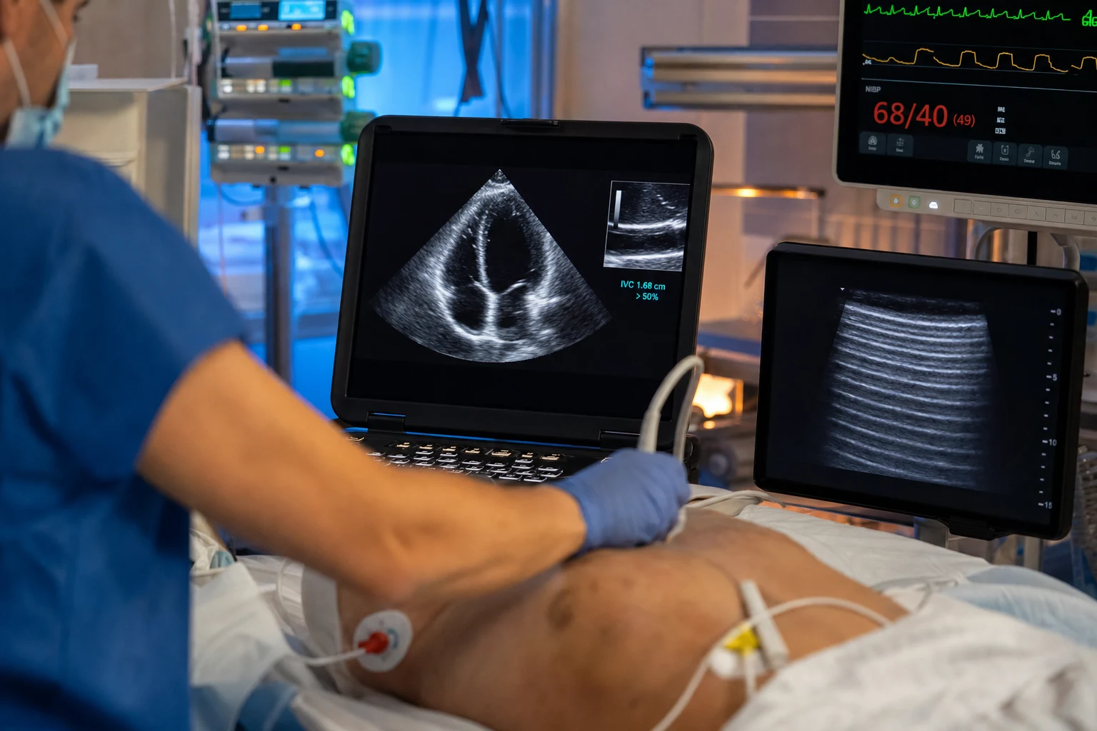
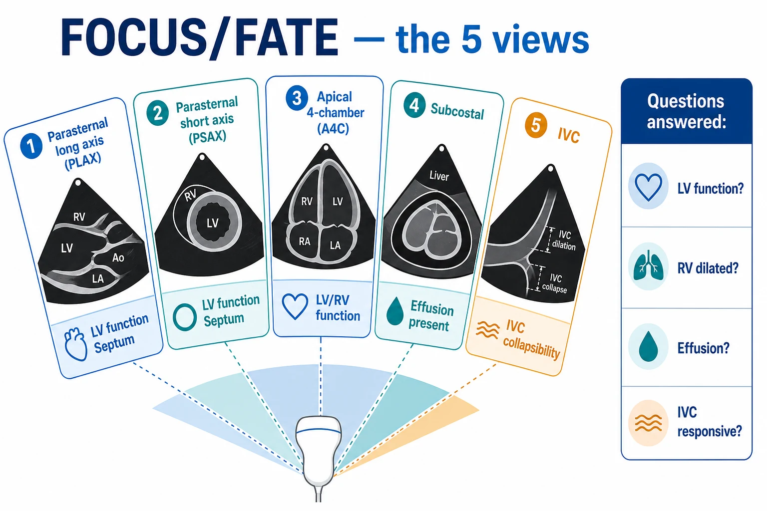
The RUSH protocol — pump, tank, pipes
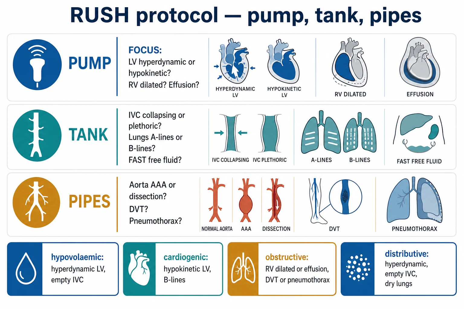
Step 1 — The PUMP (cardiac)
Focused cardiac ultrasound (FOCUS/FATE — see the dedicated topic):[1]
- Is the LV hyperdynamic or hypokinetic? — hyperdynamic (small, kissing) suggests hypovolaemia or distributive shock; hypokinetic (dilated, poorly contracting) suggests cardiogenic shock.
- Is the RV dilated? — RV dilatation with septal bowing (the D-shaped septum) suggests acute cor pulmonale — a pulmonary embolism, ARDS, or RV infarct.
- Is there a pericardial effusion? — an echo-free space with RA/RV collapse suggests tamponade.
- Are there gross valve abnormalities?[1]
Step 2 — The TANK (volume and the container)
- The IVC — collapsing (under 1.5 cm with over 50 per cent collapse) suggests a low volume (hypovolaemia or distributive — fluid-responsive); plethoric (over 2.5 cm with under 20 per cent collapse) suggests a high right-sided pressure (cardiogenic, obstructive).[1]
- The internal jugular veins — collapsing (low volume) or distended (high volume).[1]
- The lungs — A-lines (dry — no oedema) or B-lines (wet — interstitial oedema, the tank is full, the LV is failing).[1]
- The abdomen (FAST) — free fluid (blood — the tank is leaking; ascites — the tank is third-spacing).[1]
- The pleural spaces — bilateral effusions (heart failure — the tank is overloaded).[1]
Step 3 — The PIPES (the great vessels)
- The aorta — an abdominal aortic aneurysm (over 3 cm) or an aortic dissection (an intimal flap, a widened aorta).[1]
- The lower-limb veins — a DVT (a non-compressible vein — the source of a PE that is causing obstructive shock).[1]
- The pneumothorax scan — absent lung sliding (a tension pneumothorax compressing the great vessels — obstructive shock).[1]
The RUSH findings in the four types of shock
| Shock type | Pump | Tank | Pipes |
|---|---|---|---|
| Hypovolaemic | Hyperdynamic LV | Empty IVC (collapsing); dry lungs (A-lines); FAST free fluid if haemorrhage | Normal |
| Cardiogenic | Hypokinetic LV (dilated); RV may be involved | Plethoric IVC; B-lines (oedema); bilateral effusions | Normal |
| Obstructive | RV dilated (PE); pericardial effusion (tamponade); hyperdynamic if early | Collapsing IVC (PE) or plethoric (tamponade) | DVT (PE source); pneumothorax; AAA |
| Distributive | Hyperdynamic LV (early); may become hypokinetic if late (septic cardiomyopathy) | Empty IVC (early; collapsing); dry lungs (A-lines); may become wet if overloaded | Normal |
The FALLS protocol
The FALLS protocol (Lichtenstein) uses lung ultrasound to guide fluid resuscitation in shock:[1]
- The principle: the patient with shock and A-lines (dry lungs) is underfilled — give fluid.
- Monitor the lungs continuously during the fluid resuscitation.
- When the A-lines convert to B-lines, the lung has reached its interstitial threshold (the equivalent of a PAWP of about 18 mmHg) — the tank is full. This is the FALLS endpoint — stop the fluid; further fluid will cause pulmonary oedema.
- If the shock persists despite reaching the FALLS endpoint (B-lines present, lung full, still hypotensive), the diagnosis shifts: the patient who has been adequately filled but remains shocked has cardiogenic shock (the LV is failing — confirmed on the cardiac ultrasound).[1]
Red flags
The RUSH protocol step-by-step — the HI-MAP sequence
The pump-tank-pipes framework tells you what to look for; the HI-MAP sequence tells you the order in which to move the probe so the exam is fast, reproducible and complete. Heart first (the killing diagnoses — tamponade, massive PE, pump failure), then the IVC and Morison's pouch (the tank), then the aorta (a surgical cause), then the pleura (a tension pneumothorax). The whole exam takes 3–5 minutes at the bedside.[1][5]
The RUSH / HI-MAP bedside sequence (run in this order)
H — Heart (subcostal 4-chamber first)
Probe under the xiphisternum, marker towards the patient's left shoulder. First view in any shocked patient because it needs no repositioning and shows effusion + right heart. Then parasternal long/short and apical 4-chamber if windows allow. Answer: LV hyperdynamic vs hypokinetic? RV dilated with D-shaped septum? Effusion with chamber collapse? Gross valve lesion?
I — Inferior vena cava (subxiphoid, longitudinal)
From the subcostal cardiac view, fan right to find the IVC entering the right atrium. Measure 1–2 cm caudal to the hepatic vein confluence, in M-mode. Small (<1.5 cm) + collapsing >50% = empty (hypovolaemic/distributive). Large (>2.5 cm) + <20% collapse = full (cardiogenic/obstructive).
M — Morison's pouch and the peritoneum (FAST)
Curvilinear probe in the right mid-axillary line (Morison's pouch = hepatorenal recess), left upper quadrant (splenorenal), suprapubic (pouch of Douglas / retrovesical), and the subxiphoid for the pericardium. Anechoic free fluid = blood (trauma, ruptured AAA/ectopic) or ascites (third-spacing).
A — Aorta (longitudinal and transverse)
From the xiphoid process, sweep distally in transverse to the bifurcation. Measure anteroposterior diameter outer-wall to outer-wall. >3 cm = aneurysm; >5.5 cm (male) = repair threshold. Look for an intimal flap (dissection) and for a peri-aortic haematoma. A leaking AAA + shock = vascular emergency.
P — Pneumothorax (anterior chest, bilateral)
Linear probe on the anterior chest wall, 2nd–3rd intercostal space, mid-clavicular line, both sides. Look for lung sliding (shimmering pleural line). Absent sliding = PTX; confirm with a lung point (the boundary between sliding and no sliding) if the patient is stable. M-mode: seashore (normal) vs barcode/stratosphere (PTX).
H — Heart: the five cardiac questions
The focused cardiac exam answers five binary questions. Move through them deliberately — do not skip to a favourite view.[1][5]
- LV contractility — hyperdynamic or hypokinetic? EPSS (E-point to septal separation) on parasternal long axis: a normal LV has EPSS <7 mm; a dilated failing LV has EPSS >10 mm. Visual estimate of ejection fraction: hyperdynamic (EF >70%, kissing walls, small cavity) = empty tank (hypovolaemic/distributive); hypokinetic (EF <40%, thin poorly thickening walls) = failing pump (cardiogenic).
- RV size and function — is there acute cor pulmonale? The normal RV is two-thirds the area of the LV in the apical four-chamber view. RV:LV area ratio >1 (RV bigger than LV) = acute RV dilatation. Septal bowing into the LV in systole (the D-shaped septum on parasternal short axis) = RV pressure overload. McConnell's sign (RV free wall hypokinetic, apex hyperkinetic) is moderately specific for PE.
- Pericardial effusion — is there tamponade? An echo-free (black) stripe around the heart. Tamponade physiology (not just fluid) = diastolic collapse of the RV free wall (or RA collapse for >one-third of the cardiac cycle), a plethoric non-collapsing IVC, and a swinging heart. A small circumferential effusion (<1 cm) rarely tamponades; a large (>2 cm) or loculated/posterior effusion can.
- Valves — any gross lesion? A flail mitral leaflet with prolapse into the LA (acute severe MR), a thickened immobile aortic valve (critical AS), or a vegetation (endocarditis) — all can precipitate or mimic shock.
- The IVC at the RA junction — the bridge to the next step (the IVC measurement). [1]
I — Inferior vena cava: size, collapsibility and fluid responsiveness
The IVC is the single most useful "tank" measurement because its size and respiratory variation estimate right atrial pressure and, in the right patient, fluid responsiveness.[1][7]
Small + collapsible (empty)
Diameter <1.5 cm · collapse >50%
- Estimated RAP <5 mmHg
- Hypovolaemic or distributive shock
- Likely fluid-responsive — give a challenge
- Combine with a passive leg raise for confirmation
Intermediate / equivocal
Diameter 1.5–2.5 cm · collapse 20–50%
- Estimated RAP 5–10 mmHg
- Indeterminate — do NOT rely on the IVC alone
- Use a dynamic test (passive leg raise, fluid challenge)
- Most "grey zone" patients are here
Plethoric + fixed (full)
Diameter >2.5 cm · collapse <20%
- Estimated RAP >10 mmHg
- Cardiogenic or obstructive shock (tamponade, PE, severe TR/PS)
- Further fluid unlikely to help — risks pulmonary oedema
- Look for B-lines on lung ultrasound to confirm overload
Measurement technique and the common errors:
- Where to measure: in the longitudinal plane, 1–2 cm caudal to where the hepatic veins drain into the IVC (just below the diaphragm). Measure anteroposterior diameter in M-mode perpendicular to the vessel wall.
- Spontaneously breathing patient: the IVC collapses with inspiration (negative intrathoracic pressure pulls blood into the RA). Use the collapsibility index: (max − min)/max.
- Mechanically ventilated patient: the IVC distends with inspiration (positive intrathoracic pressure pushes blood towards the RA). Use the distensibility index: (max − min)/min. A distensibility >18% predicts fluid responsiveness in a fully sedated, ventilated patient with a regular rhythm and a tidal volume of 8 mL/kg.[7]
- When the IVC is unreliable: low tidal volume ventilation, spontaneous breathing efforts through the tube, cardiac arrhythmia (AF), high intra-abdominal pressure, and right heart failure with a tricuspid regurgitant jet. In these settings use a passive leg raise + a real-time cardiac-output measure instead.
M — Morison's pouch and the FAST: free fluid in the tank
The extended FAST looks for free (anechoic, black) fluid in four dependent spaces. In the shocked patient, the finding is haemorrhage until proven otherwise.[1]
| View | Probe position | What it finds |
|---|---|---|
| Morison's pouch | Right mid-axillary line, coronal | Blood between liver and kidney (the most sensitive abdominal view) |
| Splenorenal recess | Left posterior-axillary line, coronal | Blood around the spleen (often missed) |
| Suprapubic (pelvis) | Suprapubic, transverse and sagittal | Blood in the pouch of Douglas / retrovesical space (the most dependent peritoneal space in supine) |
| Subxiphoid pericardial | Subxiphoid, fan up towards the left shoulder | Haemopericardium (tamponade) |
- Free fluid = blood in trauma (the "tank is leaking") — but also consider a ruptured AAA (a pulsatile periaortic mass), a ruptured ectopic pregnancy, or a spontaneous splenic/liver bleed in the anticoagulated patient.
- Free fluid = ascites/bowel content in the non-traumatic patient — third-spacing (sepsis, cirrhosis, pancreatitis) or a perforated viscus. A periumbilical rounded fluid collection with "bullet sign" suggests intraperitoneal free fluid.
- The FAST does not see retroperitoneal blood (a retroperitoneal bleed from a ruptured AAA or a pelvic fracture can be missed — look at the aorta and the pelvis separately). [1]
A — Aorta: aneurysm and dissection
From the xiphisternum, scan distally in the transverse plane to the bifurcation (around the umbilicus), then return in the longitudinal plane.[1][1]
- Aneurysm: an anteroposterior diameter >3 cm (outer wall to outer wall) at any point. The repair thresholds are 5.5 cm (men) and 5.0 cm (women); a rupture presents as shock, back/abdominal pain and a pulsatile mass — confirm with the FAST showing retroperitoneal/intraperitoneal blood and proceed directly to theatre (do not delay for CT if unstable).
- Dissection: an intimal flap (a thin mobile line within the aortic lumen) with a true and false lumen, a widened aortic root (>3.8 cm at the sinuses) on the parasternal long axis, and sometimes a pericardial effusion (haemopericardium from a retrograde dissection into the pericardium — a pre-tamponade state). Type A involves the ascending aorta (surgical emergency); Type B does not (often medical/TEVAR).
- Measure correctly: always outer wall to outer wall, not lumen to lumen (the thrombus is part of the aneurysm). [1]
P — Pneumothorax and the pleura
Use a linear probe (high frequency) on the anterior chest in the supine patient (air rises anteriorly). The RUSH pneumothorax scan asks one question: is there lung sliding?[3][4]
[1]Lung ultrasound artefacts — A-lines and B-lines
Lung ultrasound reads artefacts, not the lung itself. Two artefacts carry the whole protocol:[3][1][4]
| Artefact | Appearance | Meaning |
|---|---|---|
| A-lines | Horizontal repeating bright lines below the pleural line, equidistant | Air-filled lung; the pleura is aerated — the lung is "dry" (no interstitial oedema) |
| B-lines (comet-tails) | Vertical bright streaks arising from the pleural line, moving with sliding, erasing A-lines, fanning to the bottom of the screen | Interlobular septa thickened by fluid — interstitial syndrome (oedema, fibrosis, inflammation) |
- Normal lung: A-lines + lung sliding everywhere.
- B-lines: 3 or more B-lines in a single intercostal space, bilateral, indicate an interstitial syndrome. The threshold used in the FALLS protocol: A-lines converting to B-lines corresponds to a pulmonary artery occlusion pressure (PAOP) of approximately 18 mmHg — the lung is "full".[1]
- Differential of B-lines: pulmonary oedema (cardiogenic), ARDS, interstitial lung disease/fibrosis, pneumonia. B-lines are NOT specific for heart failure — they mean "wet interstitium", and the cause is read from the clinical context and the cardiac ultrasound.
The BLUE protocol profiles — reading the lung
The BLUE protocol (Bedside Lung Ultrasound in Emergency, Lichtenstein 2008) standardised lung ultrasound into reproducible scan points and "profiles" that map to a diagnosis with ~90% accuracy in acute respiratory failure.[3]
The scan uses the anterior chest divided into upper and lower BLUE points on each side (4 points), plus the posterolateral alveolar and/or pleural syndrome (PLAPS) point laterally. [1]
| Profile | Anterior lung findings | Venous (DVT) scan | Most likely diagnosis |
|---|---|---|---|
| A-profile | A-lines + lung sliding (dry, sliding) | Positive DVT | Pulmonary embolism |
| B-profile | Diffuse anterior B-lines + sliding | — | Pulmonary oedema |
| A/B-profile | One lung A-lines, other lung B-lines | — | Pneumonia |
| C-profile | Anterior alveolar consolidation | — | Pneumonia |
| A'-profile (A-prime) | A-lines, absent sliding | Lung point present | Pneumothorax |
| A-no-V-PLAPS | A-lines + sliding, negative DVT, PLAPS present | Negative | Pneumonia / exacerbation of COPD |
- The critical BLUE rule: an A-profile (dry sliding lungs) in a breathless patient mandates a leg-vein (DVT) scan — if a DVT is found, the diagnosis is pulmonary embolism (until proven otherwise). This single step is why the RUSH combines the cardiac and the leg-vein views. [1]
The FALLS protocol — the decision flow
The FALLS protocol uses lung ultrasound as a surrogate for the pulmonary artery occlusion pressure to titrate fluid in undifferentiated shock. The logic is simple and powerful: a dry lung (A-lines) means the left atrial pressure is low (the patient is not in cardiogenic shock and can take fluid); a wet lung (B-lines) means the left atrial pressure has reached ~18 mmHg (the tank is full).[2][1]
The FALLS protocol — step by step
STEP 1 — Screen for obstructive shock FIRST
Before any fluid: subcostal cardiac view (tamponade? effusion with chamber collapse?), anterior lung sliding bilaterally (tension pneumothorax? absent sliding on one side), and the leg veins (DVT → massive PE). Treat these immediately (decompress, pericardiocentese, lyse). FALLS is for the patient WITHOUT an obvious obstructive cause.
STEP 2 — Read the lung profile
If A-profile (A-lines + sliding): the left heart pressure is LOW — the shock is NOT cardiogenic, and the lung can receive fluid. Proceed to STEP 3. If B-profile at baseline: the lung is already wet — the shock is likely cardiogenic (or ARDS); do NOT follow FALLS, start inotropes / treat the cause.
STEP 3 — Give fluid, watch the lung
Give 500 mL crystalloid boluses, scanning the anterior lung after each. Continue while the lungs remain dry (A-lines). The patient is being filled safely — the lung ultrasound is the safety brake.
STEP 4 — The FALLS endpoint (A → B)
When A-lines convert to NEW diffuse anterior B-lines, the interstitial threshold (PAOP ~18 mmHg) has been reached. STOP the fluid — the tank is full. Further fluid causes pulmonary oedema.
STEP 5 — If shock persists at the endpoint, it is cardiogenic
A patient who is now fully filled (B-lines, PAOP-equivalent ~18 mmHg) but remains shocked has a failing pump — by definition cardiogenic shock. Switch to inotropes (dobutamine/milrinone), vasopressors for MAP, and consider mechanical circulatory support. The echo will confirm a hypokinetic LV.
A-profile → fluid
Dry sliding lungs · low left-heart pressure
- Not cardiogenic by definition
- Hypovolaemic or distributive shock
- Give fluid — watch for the A→B conversion
- FALLS says: keep filling while dry
B-profile → no more fluid
Wet lungs · left-heart pressure ~18 mmHg
- The tank is full — the FALLS endpoint
- If baseline B-profile: cardiogenic or ARDS
- If new B-profile during filling: stop fluid
- Persistent shock now = cardiogenic
POCUS patterns by shock type — putting it together
Each shock type has a signature combination of pump, tank and pipe findings. Memorising the four patterns allows a diagnosis in minutes.[1][1][6]
Hypovolaemic
Hyperdynamic LV · empty IVC · A-lines
- Pump: small kissing hyperdynamic LV
- Tank: collapsing IVC (<1.5 cm, >50%); dry lungs; FAST positive if haemorrhage
- Pipes: normal (or ruptured AAA / bleeding source on FAST)
- Action: fluid + blood; find and stop the bleed
Cardiogenic
Hypokinetic LV · plethoric IVC · B-lines
- Pump: dilated hypokinetic LV (or RV failure)
- Tank: plethoric fixed IVC; diffuse B-lines; bilateral effusions
- Pipes: normal
- Action: inotrope; no fluid; consider MCS
Obstructive
RV dilated / effusion / absent sliding
- Pump: RV dilated + D-septum (PE); pericardial effusion + RA/RV collapse (tamponade)
- Tank: IVC varies (collapsing in PE, plethoric in tamponade)
- Pipes: DVT (PE); absent lung sliding (tension PTX); AAA/dissection
- Action: lyse PE; pericardiocentese; decompress PTX
Distributive
Hyperdynamic LV · empty IVC · A-lines
- Pump: hyperdynamic LV early; may become hypokinetic (septic cardiomyopathy)
- Tank: collapsing IVC; dry lungs early; may overload to B-lines if over-filled
- Pipes: normal
- Action: fluid early; vasopressors; source control
The three "dry lung, empty IVC" mimics that are NOT fluid-responsive
The pattern of an A-profile with a collapsing IVC is usually hypovolaemic or distributive shock — give fluid. But three killers hide in this pattern, and each is obstructive:[3]
| Cause | The hidden finding | Why fluid is dangerous |
|---|---|---|
| Massive PE | RV dilatation + D-shaped septum + a DVT on the leg scan | The RV is already failing; fluid distends it further and worsens shock |
| Tension pneumothorax | Absent lung sliding on one side ± a lung point; the mediastinum is shifted | Fluid does nothing; decompression is the only treatment |
| Anaphylaxis | Hyperdynamic LV, dry lungs, empty IVC — but with bronchospasm, urticaria, hypotension | Fluid is appropriate (distributive), but adrenaline is the definitive treatment |
The lesson: in every "empty tank" patient, look at the heart and the lung sliding before the fluid. The heart view distinguishes PE and tamponade; the lung sliding view distinguishes the tension pneumothorax. [1]
The RUSH exam in cardiac arrest — the SHoC, FEER and CAUSE protocols
In cardiac arrest the same integrated scan is performed during the rhythm check (the 10-second pulse check window), never interrupting compressions for more than 10 seconds. The goal is to find the reversible causes.[1][5]
- Subcostal view during compressions — the only view feasible without pausing.
- The four reversible causes ultrasound finds: tamponade (effusion + chamber collapse), massive PE (RV dilatation), severe hypovolaemia (small kissing LV, empty IVC), and tension pneumothorax (absent sliding).
- PEA with a small hyperdynamic heart and an empty IVC = profound hypovolaemia — give fluid/blood.
- PEA with a dilated RV and a DVT = massive PE — consider thrombolysis during CPR.
- Asystole/PEA with an effusion = tamponade — consider emergency pericardiocentesis or a resuscitative thoracotomy. [1]
Limitations and pitfalls of the RUSH exam
The RUSH exam is powerful but has well-recognised limitations — knowing them is an exam differentiator.[5]
- Operator dependent: accuracy depends on training and image acquisition; a subcostal window may be unobtainable in the obese or post-laparotomy patient.
- The IVC is unreliable in the spontaneously breathing patient with respiratory distress (the pressures are exaggerated), in low-tidal-volume ventilation, in arrhythmia, and in right-heart failure with severe tricuspid regurgitation (the regurgitant jet distends the IVC regardless of volume).
- B-lines are not specific for heart failure — ARDS, pulmonary fibrosis and pneumonia also cause B-lines. The cardiac ultrasound distinguishes cardiogenic B-lines (hypokinetic LV, plethoric IVC) from non-cardiogenic (normal heart, non-homogeneous distribution).
- A small effusion is not tamponade — tamponade is a physiological diagnosis (chamber collapse + a plethoric IVC + a swinging heart), not a size diagnosis.
- RV dilatation is not always PE — ARDS, RV infarct, chronic pulmonary hypertension and severe tricuspid regurgitation all dilate the RV. The DVT scan and the acute-onset history discriminate.
- Lung sliding is absent in apnoea, main-stem intubation and pleural adhesions — do not over-call a pneumothorax in a stable patient without a lung point.
- FAST is blind to retroperitoneal blood and to solid-organ injury without free fluid — a negative FAST does not exclude intra-abdominal bleeding. [1]
Evidence and landmark trials
BLUE protocol
Chest 2008
260 ICU pts with acute respiratory failure — standardised lung ultrasound profiles vs final diagnosis
Key finding
90.5% diagnostic accuracy; A-profile + DVT = PE
Practice change
Lung ultrasound became a first-line tool in undifferentiated dyspnoea
FALLS-protocol
Expert Rev Respir Med 2012
Conceptual/observational — lung ultrasound to titrate fluid in shock using the A→B transition
Key finding
A→B transition marks PAOP ~18 mmHg (the interstitial threshold)
Practice change
Provided a lung-based, PA-catheter-free endpoint for fluid resuscitation
RUSH exam (Perera)
Emerg Med Clin North Am 2010
Structured pump-tank-pipes ultrasound protocol for undifferentiated shock
Key finding
Allows shock-type classification within minutes at the bedside
Practice change
Adopted into emergency and critical care shock bundles worldwide
Jones 2004 (CCM)
Crit Care Med 2004
RCT — immediate vs delayed goal-directed ultrasound in nontraumatic hypotension
Key finding
Immediate US shortened time to diagnosis of the cause of shock
Practice change
Supported early point-of-care ultrasound in undifferentiated hypotension
Clinical pearls
The pump-tank-pipes and the HI-MAP order — two ways to remember the same exam
Additional red flags
[1] [1] [1] [1] [1]Exam practice
SAQ — Undifferentiated shock and the RUSH exam
10 minutes · 10 marks
A 68-year-old man is brought to the emergency department with confusion and hypotension. BP 78/45 (MAP 56), HR 124, SpO2 92% on room air, lactate 4.8 mmol/L. He is cool peripherally with a capillary refill of 5 seconds. You perform a bedside RUSH examination.
Sample Viva 1 — The FALLS protocol
Examiner: "Describe the FALLS protocol and its rationale." [1]
Expected response: "FALLS — Fluid Administration Limited by Lung Sonography, described by Lichtenstein in 2012 — uses lung ultrasound to titrate fluid in undifferentiated shock. The rationale is that the lung ultrasound artefacts track the left atrial pressure: dry lungs with A-lines mean a low left-heart pressure and no cardiogenic shock, so the patient can receive fluid; the appearance of diffuse B-lines marks the interstitial threshold, equivalent to a pulmonary artery occlusion pressure of about 18 mmHg — that is the FALLS endpoint, and further fluid causes pulmonary oedema. The protocol proceeds in steps: first, screen for and treat the obstructive causes of shock — tamponade, tension pneumothorax and massive PE — because FALLS does not detect them and giving fluid would be harmful. Then, if the lung shows an A-profile, give fluid in 500 mL boluses while watching the anterior lung. Continue until A-lines convert to B-lines, then stop. If the patient remains shocked at that point, the shock is, by definition, cardiogenic — switch to inotropes and treat the failing pump." [1]
Sample Viva 2 — Reading the IVC
Examiner: "How do you assess the inferior vena cava at the bedside, and how does its interpretation differ between the spontaneously breathing and the ventilated patient?" [1]
Expected response: "I image the IVC from the subxiphoid approach in the longitudinal plane, identifying it as it enters the right atrium, and I measure the diameter 1–2 cm below the hepatic vein confluence in M-mode. In the spontaneously breathing patient the negative intrathoracic pressure of inspiration pulls blood into the right atrium, so the IVC collapses — I report the collapsibility index, (max − min)/max, and a small (<1.5 cm) IVC collapsing by more than 50% suggests a low right atrial pressure and a fluid-responsive state. In the mechanically ventilated patient the positive intrathoracic pressure of inspiration pushes blood towards the heart, so the IVC distends — I report the distensibility index, (max − min)/min, and a distensibility greater than 18% in a sedated, passively ventilated patient with a regular rhythm and a tidal volume of about 8 mL/kg predicts fluid responsiveness. I would also mention the limitations: the IVC is unreliable in low-tidal-volume ventilation, arrhythmia, high intra-abdominal pressure, right-heart failure with tricuspid regurgitation, and that in the equivocal 'grey zone' I would use a passive leg raise with a real-time cardiac output measure rather than the IVC alone." [1]
References
- [1]Perera P, Mailhot T, Riley D, Mandavia D The RUSH exam: Rapid Ultrasound in SHock in the evaluation of the critically lll Emerg Med Clin North Am, 2010.PMID 19945597
- [2]Lichtenstein DA Fluid administration limited by lung sonography: the place of lung ultrasound in assessment of acute circulatory failure (the FALLS-protocol) Expert Rev Respir Med, 2012.PMID 22455488
- [3]Lichtenstein DA, Meziere GA Information technology for health in developing countries Chest, 2007.PMID 17998362
- [4]Volpicelli G, Elbarbary M, Blaivas M, et al. Generation of a convalescent model of virulent Francisella tularensis infection for assessment of host requirements for survival of tularemia PLoS One, 2012.PMID 22428026
- [5]Levitov A, Frankel HL, Blaivas M, et al. Reply: understanding the anatomic basis for obstructive sleep apnea syndrome in adolescents: how to proceed? Am J Respir Crit Care Med, 2015.PMID 26426792
- [6]Jones AE, Tayal VS, Sullivan DM, Kline JA Factors associated with nurse assessment of the quality of dying and death in the intensive care unit Crit Care Med, 2004.PMID 15286539
- [7]Barbier C, Loubieres Y, Schmit C, et al. Online automated detection of cerebral embolic signals using a wavelet-based system Ultrasound Med Biol, 2004.PMID 15183231