ICU · Cardiovascular / valvular
Valvular Disease in the ICU
Also known as Valvular heart disease · Aortic stenosis · Aortic regurgitation · Mitral regurgitation · Mitral stenosis · TAVI · TAVR · Prosthetic valve · Valve thrombosis · Balloon mitral valvuloplasty · BMV · MitraClip · Papillary muscle rupture
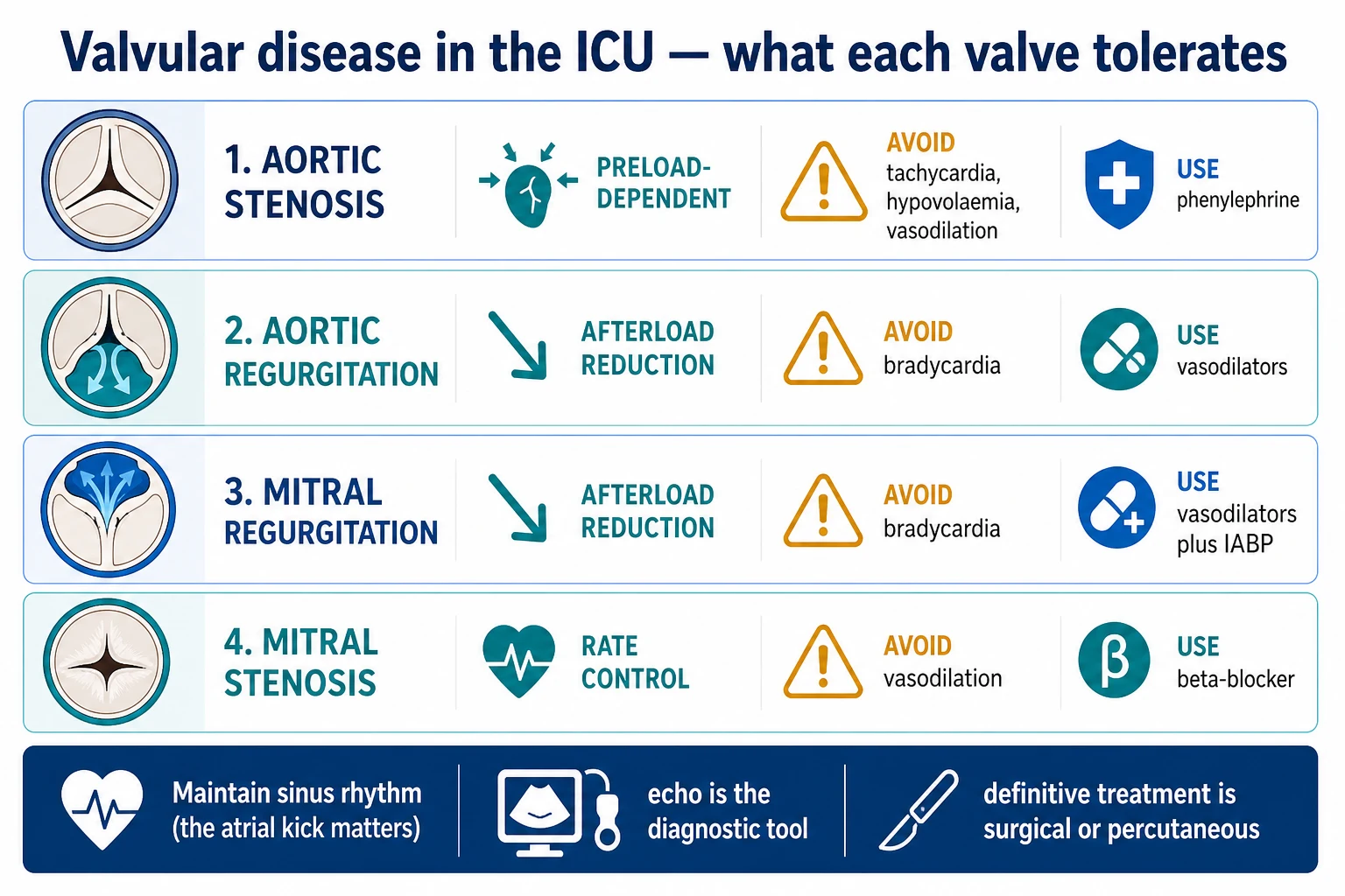
Severe valvular disease in the ICU has valve-specific haemodynamic implications that determine what the patient can and cannot tolerate. Severe aortic stenosis (a stiff, hypertrophied LV) is preload-dependent and vasodilator-intolerant — avoid tachycardia, hypovolaemia, and vasodilation; use phenylephrine. Severe aortic and mitral regurgitation (volume overload) benefit from afterload reduction and a higher heart rate — use vasodilators and avoid bradycardia. Severe mitral stenosis (an obstructed inflow) needs rate control and avoids vasodilation. Maintain sinus rhythm in all (the atrial kick matters for the stiff or overloaded LV). Echocardiography is the diagnostic tool; the definitive treatment is surgical or percutaneous (AVR, TAVI/TAVR, MV repair, balloon mitral valvuloplasty). Prosthetic valves require lifelong anticoagulation (mechanical) and carry risks of thrombosis and infective endocarditis.
On this page & tools
Your progress
Saved locally on this device.
Target exams
Overview & definition
Severe valvular disease in the ICU has valve-specific haemodynamic implications — each valve lesion changes what the patient can and cannot tolerate, which determines the choice of vasoactive drugs, fluids, and rhythm management. Understanding these principles is essential for managing the valvular ICU patient.[1]
The unifying principle is that the heart in severe valvular disease operates on a narrow, lesion-specific haemodynamic margin. A drug, a fluid bolus, a new arrhythmia, or an anaesthetic that is well tolerated in a normal patient can precipitate cardiovascular collapse in the valvular patient. The intensivist's job is therefore less to "treat the valve" (that is surgical or percutaneous) and more to preserve the haemodynamic conditions each lesion needs while the definitive procedure is arranged. Echocardiography is the pivotal diagnostic tool — it identifies the lesion, grades its severity, and defines the compensatory state of the ventricle.[1][2]
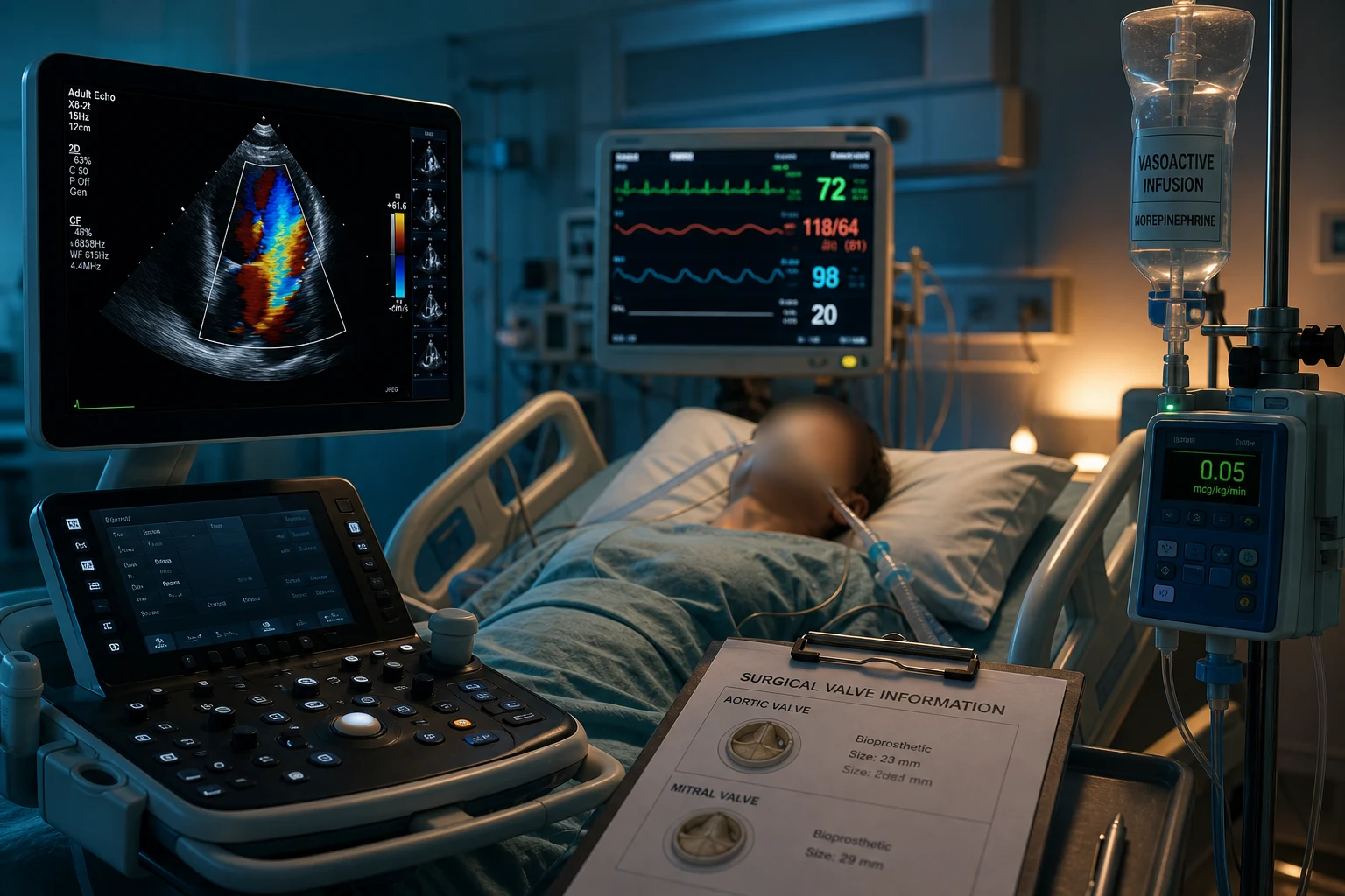
Severe aortic stenosis (AS)
The fixed outflow obstruction produces a hypertrophied, stiff LV (diastolic dysfunction).[1]
Cannot tolerate:[1]
- Tachycardia — the stiff LV needs a long diastolic filling time; a fast rate reduces the preload and the coronary perfusion.
- Hypovolaemia — the stiff LV is preload-dependent; a small preload drop causes a large CO fall.
- Vasodilation — the SVR is already high (to maintain the pressure across the stenotic valve); further vasodilation drops the BP catastrophically.
- Atrial fibrillation — the loss of the atrial kick (which contributes up to 30-40 per cent of the filling in a stiff LV) causes an abrupt CO drop.
Management: maintain sinus rhythm (cardiovert the AF), adequate preload (cautious fluid), a pure alpha-vasopressor (phenylephrine — raises the SVR without a tachycardia), and cautious beta-blockade for rate control.[1]
Definitive: aortic valve replacement (AVR) or transcatheter aortic valve implantation (TAVI / TAVR).[1]
Severe AS — the diagnostic numbers and the symptomatic triad
Severe AS is defined echocardiographically, and the numbers are examinable to the decimal.[1][2]
Aortic valve area
The orifice
- Severe AS: an indexed aortic valve area under 0.6 cm²/m², or an absolute area under 1.0 cm².
- Critical AS: an area under 0.6 cm². The smaller the area, the higher the gradient for any given flow.
- Area is flow-dependent — a low-output state underestimates the gradient and overestimates the area (the low-flow low-gradient paradox).
Mean gradient
The pressure drop
- Severe AS: a mean gradient over 40 mmHg (or over 50 mmHg by some older criteria).
- Directly proportional to flow and inversely proportional to valve area — so it falls in low-output states.
- A mean gradient over 40 mmHg with a normal LV output is unambiguous severe AS.
Peak jet velocity
Continuous-wave Doppler
- Severe AS: a peak aortic jet velocity over 4.0 m/s.
- The velocity is the single best predictor of symptom onset and outcome in AS — the higher it is, the worse the prognosis without AVR.
Dimensionless index
Flow-independent check
- The ratio of the LVOT velocity-time integral to the aortic jet VTI — under 0.25 is severe.
- Useful when the orifice area is hard to measure (heavy calcification, odd geometry) because it is relatively flow-independent.
The symptomatic triad of severe AS — angina, syncope, and heart failure — is the classical presentation, and each carries a sobering median survival without AVR: angina around 5 years, syncope around 3 years, and heart failure around 2 years. Once symptoms appear, the mortality without AVR exceeds 25 per cent per year. Any of the three is an indication for intervention.[1]
[1] [1]Severe aortic regurgitation (AR)
The regurgitant volume overloads the LV (dilated, volume-overloaded).[1]
Needs:[1]
- A high heart rate — a faster rate shortens the diastole and the time for the regurgitation.
- Afterload reduction — vasodilators (GTN, nitroprusside) lower the aortic diastolic pressure and reduce the regurgitant fraction, improving the forward output.
- Inotropy — to support the volume-overloaded LV.
Cannot tolerate: bradycardia (a long diastole = more regurgitation).[1]
Severe mitral regurgitation (MR)
The regurgitant volume reduces the forward output; the LV ejects into the low-resistance left atrium (the path of least resistance).[1]
Needs:[1]
- Afterload reduction — vasodilators reduce the aortic pressure and the regurgitant fraction, improving the forward output. The IABP (which reduces the afterload in systole) is particularly effective for acute severe MR.
- A higher heart rate — shortens the systole and the regurgitation time.
Cannot tolerate: bradycardia (a long systole = more regurgitation) and afterload increase (raises the regurgitant fraction).[1]
Severe mitral stenosis (MS)
The obstructed inflow raises the left atrial pressure → pulmonary oedema and atrial fibrillation.[1]
Needs:[1]
- A low heart rate — a slower rate lengthens the diastolic filling time across the stenotic valve, increasing the LV filling and the CO.
- Adequate preload — the LV is underfilled; it needs the available inflow.
- Sinus rhythm — the atrial kick contributes to the filling across the stenotic valve.
Cannot tolerate: tachycardia (shortens the filling time; the LA pressure rises → pulmonary oedema) and vasodilation (the LV is underfilled; the SVR is needed).[1]
Management: rate control (beta-blocker), diurese if overloaded, avoid vasodilators. Definitive: balloon mitral valvuloplasty (BMV) or MV replacement.[1]
Prosthetic valves
- Mechanical valves — lifelong anticoagulation with warfarin (the INR target depends on the valve position — typically 2.5-3.5). Thrombosis risk if the INR is subtherapeutic.[1]
- Bioprosthetic valves — anticoagulation for 3-6 months, then aspirin. Less durable but no lifelong anticoagulation.[1]
Valve thrombosis: an obstructed valve (acute dyspnoea, a muffled or absent valve click on auscultation, an embolic event). Diagnose with an echocardiogram (a reduced leaflet mobility, a thrombus). Treat with surgery or thrombolysis (for a mechanical valve thrombosis — though the thrombolysis carries an embolic risk).[1]
Infective endocarditis: positive blood cultures, a vegetation on echo, the Duke criteria. Antibiotic prophylaxis before procedures for high-risk patients (a prosthetic valve, a previous IE, some congenital heart disease).[1]
[1]Echocardiography — the pivotal investigation
Transthoracic echo (TTE) is the first and often the only investigation needed in the valvular ICU patient. It answers the four questions that drive every subsequent decision: which valve, how severe, what is the ventricle doing about it, and are there complications. Transoesophageal echo (TOE) is reserved for the prosthetic valve (shadowing on TTE), the suspected vegetation or abscess, the detail of the mitral apparatus (repairable or not), and the intraoperative or periarrest setting. Echocardiography should be performed before any vasoactive drug is committed to writing — choosing a vasodilator for a hypotensive patient who turns out to have severe AS is a preventable disaster.[1][2]
TTE — 2D and Doppler
The workhorse
- Defines the valve morphology (bicuspid, rheumatic, calcific, flail leaflet, vegetation), the chamber sizes, and the LV ejection fraction.
- Doppler quantifies severity: peak velocity and mean gradient (AS), pressure half-time and area by planimetry (MS), regurgitant volume and fraction and the effective regurgitant orifice area (AR and MR).
- Colour-flow mapping locates the regurgitant jet and its direction (an eccentric posteriorly-directed jet in MR = an anterior leaflet problem, and vice versa).
TOE
When TTE cannot see
- Mandatory for a suspected prosthetic valve thrombosis or endocarditis (TTE is blinded by the metal), for grading MR before a repair decision, and for the intraoperative assessment of the repair.
- Better visualisation of the mitral subvalvular apparatus (the chords and papillary muscles), the aortic root, and a paravalvular abscess.
- Semi-invasive — performed in the intubated or sedated ICU patient, or under sedation in the awake patient with a protected airway.
Stress echo
For the low-flow low-gradient puzzle
- Low-dose dobutamine stress echo distinguishes true-severe from pseudo-severe AS in the low-flow low-gradient state (a low gradient with a low flow that rises with dobutamine).
- A contractile reserve (a 20 per cent rise in stroke volume with dobutamine) predicts a better outcome after AVR.
- The examinable point: dobutamine raises the gradient and lowers the calculated area in true-severe AS, but raises the area in pseudo-severe AS.
Acute decompensated severe AS in the ICU
The patient with known or previously undiagnosed severe AS who decompensates (pulmonary oedema, cardiogenic shock, a new fast AF) is one of the most fragile patients in the unit. The decompensation is usually triggered by one of the four things the stenotic LV cannot tolerate — a tachyarrhythmia, a vasodilator, hypovolaemia, or the vasodilation of sepsis — and the resuscitation must reverse the trigger while preserving the haemodynamic conditions the valve needs.[1][1]
Acute decompensated AS — the resuscitation pathway
Recognise the valve and the trigger
A bedside echo confirms the heavily calcified aortic valve, the small hypertrophied LV with preserved or hyperdynamic function, and the trigger (AF, vasodilation, hypovolaemia, sepsis). The gradient and the valve area confirm severity. Identify the trigger — because the resuscitation is largely trigger-reversal.
Restore sinus rhythm if AF is the trigger
AF with a rapid ventricular response is the commonest cause of acute decompensation in AS — the loss of the atrial kick plus the short diastole cuts the preload catastrophically. Chemical (amiodarone) or synchronised electrical cardioversion early. Do not let the AF run.
Phenylephrine for the vasodilated / shocked patient
IV phenylephrine 100–200 mcg boluses titrated to an SBP over 90 mmHg, then an infusion at 0.5–5 mcg/kg/min. Pure alpha-1 — restores the SVR and the coronary perfusion pressure without any tachycardia. Avoid noradrenaline if it drives a tachycardia; avoid adrenaline entirely (inotropy and tachycardia worsen the subvalvular gradient in HCM-like physiology).
Cautious preload, never vasodilate
Cautious 250 mL crystalloid boluses to apreload target — the stiff LV needs filling but floods easily into a non-compliant chamber. Diurese the pulmonary oedema gently (the oedema is from the high LA pressure, not volume overload per se). NEVER give GTN, morphine, or an unopposed anaesthetic induction — the SVR collapse is catastrophic and the LV cannot compensate.
Balloon valvuloplasty as a bridge
Percutaneous balloon aortic valvuloplasty (BAV) splits the calcified commissures and acutely lowers the gradient — a bridge to definitive TAVR or AVR in the patient too sick to wait. The effect is modest (gradient halved, area to around 0.9 cm²) and transient (restenosis in 6–12 months), but it can break the vicious cycle of cardiogenic shock. Reserved for the bridge or the palliative scenario, never as definitive therapy.
Definitive AVR or TAVR
The only treatment that changes the natural history. TAVR (transfemoral) is feasible in the elderly high-risk patient and can be performed in cardiogenic shock; surgical AVR for the younger, lower-risk patient. Do not let the patient languish inotrope-dependent on the ICU — mobilise the structural heart team the moment the diagnosis is confirmed.
TAVR versus surgical AVR — the evidence ladder
The TAVR versus surgical AVR evidence moved systematically down the risk ladder, from the inoperable patient to the low-risk patient, and at every step TAVR was non-inferior or superior to surgery on the primary outcome. The upshot is that TAVR is now a first-line option across the surgical-risk spectrum, with the access route (transfemoral preferred) and the durability question (bioprosthetic in both) shaping the individual choice.[1]
The TAVR evidence ladder — from inoperable to low-risk
PARTNER cohort B — Leon, NEJM 2010 (PMID 20961243).[3] The trial that established TAVR. In inoperable patients with severe AS, TAVR (balloon-expandable Edwards SAPIEN) versus optimal medical therapy (which included BAV) halved 1-year mortality (30.7 per cent versus 50.7 per cent, p under 0.001). The number needed to treat was 5. This is the trial that made TAVR a standard of care.
PARTNER cohort A — Smith, NEJM 2011 (PMID 21639811).[4] High-risk patients randomised to TAVR versus surgical AVR. TAVR was non-inferior for 1-year mortality (24.2 per cent versus 26.8 per cent), with more strokes and paravalvular regurgitation but less bleeding and a shorter hospital stay. Established TAVR as an alternative to surgery in the high-risk patient.
PARTNER 2A — Leon, NEJM 2016 (PMID 27040324).[5] Intermediate-risk patients. TAVR (newer SAPIEN XT) was non-inferior to surgery on the composite of death or disabling stroke at 2 years (19.3 per cent versus 21.1 per cent). The transfemoral subgroup did better with TAVR; the transapical subgroup did worse. Extended TAVR down a risk stratum.
SURTAVI — Reardon, NEJM 2017 (PMID 28304219).[6] Intermediate-risk patients with the self-expanding Medtronic CoreValve. TAVR non-inferior to surgery on the 24-month composite of death or disabling stroke (6.7 per cent versus 7.0 per cent). Confirmed PARTNER 2A with a different device platform — more pacemakers (17.4 per cent) and more paravalvular leak with the self-expanding valve.
Evolut Low Risk — Popma, NEJM 2019 (PMID 30883053).[7] Low-risk patients (mean age 74, STS score around 1.9 per cent) randomised to TAVR (self-expanding Evolut) versus surgery. At 24 months the composite of death or disabling stroke was 5.3 per cent (TAVR) versus 6.7 per cent (surgery) — TAVR non-inferior, with less bleeding, less AKI, and far less new atrial fibrillation (7.7 per cent versus 35.4 per cent), but more pacemakers (17.4 per cent) and more paravalvular regurgitation (3.5 per cent). Closed the loop: TAVR is now non-inferior to surgery across the risk spectrum in carefully selected patients.
Acute versus chronic mitral regurgitation
The MR spectrum spans a surgical emergency (acute severe MR from a papillary muscle rupture) to an elective outpatient decision (chronic degenerative MR with a prolapsing leaflet). The haemodynamic management is shared — afterload reduction, a higher heart rate, an IABP for the acute case — but the presentation, the ventricle, and the urgency are entirely different.[1]
Acute severe MR
The surgical emergency
- Mechanisms: **papillary muscle rupture** after an inferior MI (the posteromedial papillary muscle, with its single blood supply from the posterior descending artery, is far more vulnerable than the anterolateral with its dual supply), **infective endocarditis** destroying a leaflet, **traumatic** chordal rupture, and **acute ischaemic** MR from a stunning papillary muscle.
- Presents 2–7 days post-MI with sudden, severe pulmonary oedema and cardiogenic shock. The LA has had no time to dilate, so the regurgitant volume is delivered into a small, non-compliant LA — the LA and the pulmonary venous pressure spike, and flash pulmonary oedema follows.
- The murmur may be **soft or absent** in acute severe MR — the LV and LA pressures equalise early in systole, truncating the regurgitant waveform. Do not be reassured by a quiet chest.
- Management: afterload reduction (GTN or nitroprusside, or an ACE-inhibitor), an **IABP** (diastolic augmentation plus systolic unloading directly reduces the regurgitant fraction), and **urgent surgical repair or replacement**. MitraClip is not for the acute rupture.
Chronic severe MR
The compensated volume overload
- Mechanisms: **degenerative** (Barlow / fibroelastic deficiency with a prolapsing or flail leaflet — the commonest in the developed world), **functional** (ischaemic or non-ischaemic LV dilatation tethering a structurally normal leaflet), **rheumatic**, and **prosthetic**.
- The LA and LV dilate progressively to accommodate the regurgitant volume, so the patient is asymptomatic for years despite a large regurgitant fraction. Symptoms (dyspnoea, fatigue) appear late, by which time the LV may already be dysfunctional.
- A loud holosystolic murmur at the apex radiating to the axilla, a displaced apex, and a third heart sound are the classic chronic signs.
- Surgery (repair preferred over replacement for degenerative MR) is indicated for symptoms, or for asymptomatic severe MR with LV dysfunction (an LV end-systolic diameter over 40 mm or an EF under 60 per cent). For functional MR with heart failure, MitraClip is guided by COAPT and MITRA-FR.
MitraClip for functional MR — COAPT versus MITRA-FR
The two trials of transcatheter edge-to-edge mitral repair (MitraClip) for secondary (functional) MR in heart failure reached opposite conclusions, and the reconciliation — proportionate versus disproportionate MR — is now the framework that guides patient selection.[8][9]
COAPT versus MITRA-FR — proportionate versus disproportionate MR
COAPT — Stone, NEJM 2018 (PMID 30280640).[8] Heart failure patients with disproportionate secondary MR — moderate-to-severe MR (effective regurgitant orifice area around 0.3 cm²) on top of only mild-to-moderate LV dilatation (an LV end-diastolic diameter around 62 mm). MitraClip plus guideline-directed medical therapy versus medical therapy alone. At 2 years, MitraClip reduced the composite of death or heart-failure hospitalisation by 37 per cent and was strongly positive on every endpoint. This is the trial that established MitraClip for selected secondary MR.
MITRA-FR — Obadia, NEJM 2018 (PMID 30145927).[9] Heart failure patients with proportionate secondary MR — severe MR on top of a heavily dilated LV (an LV end-diastolic diameter around 69 mm). MitraClip plus medical therapy versus medical therapy alone. No difference in the composite of death or unplanned heart-failure admission at 12 months (primary outcome neutral).
The reconciliation: the MR was proportionate to the LV dilatation in MITRA-FR (a big sick ventricle with the MR it deserves) — repairing the valve without fixing the ventricle does nothing. The MR was disproportionate in COAPT (more MR than the ventricle's size would predict) — here, repairing the valve helps because the valve, not the ventricle, is the dominant problem. The lesson for the ICU: MitraClip works when the MR is disproportionate to the LV size, and is futile when it is proportionate.
Acute versus chronic aortic regurgitation
Acute severe AR is a surgical emergency whose presentation is dominated by pulmonary oedema and shock; chronic severe AR is a slowly progressive volume overload whose presentation is dominated by exertional dyspnoea and a wide pulse pressure. The murmurs and the signs differ, and so does the urgency.[1]
Acute severe AR
The surgical emergency
- Mechanisms: **type A aortic dissection** (the dissection flap tears the annulus or the leaflet off), **infective endocarditis** (a destroyed or perforated leaflet), **blunt chest trauma**, and **prosthetic valve dehiscence**.
- The LV has had no time to dilate, so the sudden regurgitant volume into a non-compliant LV equalises the aortic and LV diastolic pressures almost immediately — the **diastolic pressure equalises, the murmur is short and soft (or absent), and the pulse pressure is not yet wide**. The clue may be only unexplained pulmonary oedema and tachycardia in a patient with a dissection or endocarditis.
- A new early-diastolic murmur in a hypertensive patient with tearing chest pain is a type A dissection until proven otherwise — confirm with TOE in parallel with theatre mobilisation.
- Management: afterload reduction (nitroprusside with an inotrope to keep the heart rate up — esmolol is relatively contraindicated because the slow heart rate lengthens diastole and worsens the regurgitation), and **urgent surgical AVR** with repair of the underlying cause (the dissection, the abscess).
Chronic severe AR
The volume overload
- Mechanisms: **bicuspid aortic valve** (the commonest congenital cause, with an associated aortopathy), **degenerative** calcific AR, **rheumatic**, **aortic root dilatation** (hypertension, Marfan, syphilis, ankylosing spondylitis), and **connective tissue disease**.
- The LV dilates progressively to accommodate the regurgitant volume (eccentric hypertrophy) — the patient tolerates a large regurgitant fraction for years. A **wide pulse pressure**, a **water-hammer (Collapsing) pulse**, a **hyperdynamic displaced apex**, and the eponymous signs (Corrigan pulse, Duroziez, Quincke, de Musset, Traube) are the chronic signatures.
- A high-pitched **decrescendo early-diastolic murmur** at the left sternal border (best heard sitting forward in expiration) and an **Austin Flint murmur** (a low-pitched mid-diastolic rumble from the regurgitant jet hitting the anterior mitral leaflet) complete the picture.
- Vasodilator therapy (nifedipine or an ACE-inhibitor) reduces the regurgitant fraction and delays the need for surgery; **AVR** is indicated for symptoms, or for asymptomatic severe AR with LV dysfunction (an LV end-systolic diameter over 50 mm or an EF under 50 per cent).
Acute severe AR — from the new murmur to theatre
Recognise the context
Acute severe AR presents in one of three contexts: a type A aortic dissection (a hypertensive patient with tearing chest pain and a new early-diastolic murmur), a destructive infective endocarditis (a patient with fever, positive blood cultures, and a new murmur), or a prosthetic valve dehiscence (a patient with a prosthetic valve and acute pulmonary oedema). The echo confirms the mechanism.
Confirm with TOE
A TOE in the unstable patient (or a TTE first if feasible) defines the mechanism — a flail or perforated leaflet, a dehisced prosthetic sewing ring, or a dissection flap extending into the annulus. The severity is graded by the pressure half-time (short in acute severe AR), the regurgitant volume, and the diastolic flow reversal in the descending aorta.
Afterload reduction with rate support
IV sodium nitroprusside (0.3–3 mcg/kg/min) to reduce the afterload and the regurgitant fraction, plus an inotrope (dobutamine or milrinone) to keep the heart rate up and support the volume-overloaded LV. Avoid pure beta-blockade — a slow heart rate lengthens diastole and worsens the regurgitation. In the dissection patient, esmolol is used but only after the afterload reducer and the inotrope are running.
Treat the underlying cause
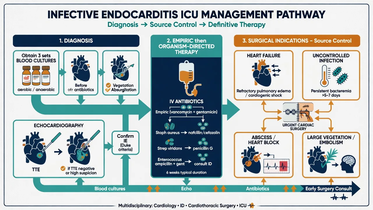
Endocarditis: culture-guided intravenous antibiotics and timing of surgery (early surgery for heart failure, an uncontrolled infection, a large vegetation, or a perivalvular abscess). Dissection: surgical repair of the dissection with resuspension or replacement of the aortic valve. Prosthetic dehiscence: redo surgery with a new valve.
Urgent surgical AVR
Acute severe AR from any mechanism is a surgical disease — the medical therapy is a holding manoeuvre, not a treatment. Mobilise the cardiothoracic and the cardiac-anaesthesia teams in parallel with the resuscitation; the patient who stabilises on the infusion can still decompensate abruptly as the LV fails.
Rheumatic mitral stenosis — the obstructed inflow
Rheumatic MS remains the dominant cause of MS worldwide; the pathological signature is commissural fusion with thickened, tethered chordae and a "fish-mouth" orifice. The obstruction raises the left atrial pressure, which is transmitted to the pulmonary veins and the capillaries — the pulmonary capillary wedge pressure (PCWP) rises, and the patient develops exertional dyspnoea, orthopnoea, and, at the extreme, haemoptysis and pulmonary hypertension. The stenotic valve also stretches the left atrium, which is why atrial fibrillation is so common (and so badly tolerated) in MS.[1]
[1]Balloon mitral valvulotomy — definitive when the valve is favourable
Percutaneous balloon mitral valvuloplasty (BMV, also called percutaneous mitral commissurotomy) splits the fused rheumatic commissures with an Inoue balloon across the atrial septum. It is the procedure of choice for the symptomatic patient with severe MS (a valve area under 1.5 cm²) and a favourable valve — and it can be definitive, unlike balloon aortic valvuloplasty. The decision hinges on the Wilkins echo score.[2]
The Wilkins score
Four features, graded 0–4
- Leaflet **mobility** (0 = fully mobile, 4 = fixed).
- Subvalvular **thickening** of the chordae (0 = thin, 4 = heavily thickened and shortened).
- Leaflet **thickening** (0 = normal, 4 = over 8 mm).
- **Calcification** (0 = none, 4 = extensive).
- A total score under 8 (favourable) predicts a good BMV result; over 8 (unfavourable) favours surgery. A pliable, non-calcified valve with thin chordae does well; a thick, calcified, tethered valve does not.
BMV — when it works
The favourable valve
- A Wilkins score under 8, no left atrial thrombus, no more than mild MR, and no heavy calcification.
- The valve area typically doubles (from around 1.0 to around 1.8 cm²) and the mean gradient halves, with the benefit durable for 10 years or more in the right patient.
- Performed via a trans-septal puncture from the right femoral vein, advancing the Inoue balloon across the atrial septum and inflating it across the stenotic valve to split the commissures.
Surgery — when BMV cannot
The unfavourable valve
- A Wilkins score over 8, heavy calcification, significant MR, a left atrial thrombus, or restenosis after a previous BMV.
- Open mitral commissurotomy (in the developing world) or, more commonly, **mitral valve replacement** (mechanical in the younger patient on warfarin, bioprosthetic in the elderly).
- A higher procedural risk than BMV but definitive — and the only option when the valve anatomy is hostile.
Severe MS with flash pulmonary oedema — the immediate pathway
Rate control is the first manoeuvre
IV metoprolol or esmolol to slow the ventricular response (target under 80 bpm, under 100 if in AF). In AF, the rate, not the rhythm, is the priority in the first hour — the PCWP falls as the diastolic filling time lengthens. Add digoxin if beta-blockade is contraindicated.
Sit up, oxygen, diurese gently
Sit the patient upright, high-flow oxygen (or NIV if the work of breathing is high — CPAP reduces the preload and the afterload on the pulmonary vasculature). IV frusemide 20–40 mg — the oedema is from the high LA pressure, so the diuresis is a pressure manoeuvre, not a volume one. Avoid over-diuresis: the underfilled LV depends on the available inflow.
Cardiovert if newly in AF and unstable
A new AF with a rapid response is the commonest trigger for an MS crisis. If the patient is shocked or the pulmonary oedema is refractory, synchronised DC cardioversion (with anticoagulation and TOE guidance if the onset is uncertain) can restore sinus rhythm and the atrial kick — which contributes disproportionately to filling across the stenotic valve.
Avoid vasodilators and the vasodilating anaesthetic
The LV is underfilled and the output is fixed by the obstruction — any drop in the SVR is uncompensated and hypotensive. NO nitrates, NO morphine, NO ACE-inhibitors acutely. Phenylephrine or a low-dose noradrenaline for hypotension, accepting a slight rise in the afterload that the failing forward flow can cope with.
Definitive BMV or MV replacement
Once stabilised, plan the definitive procedure: a trans-septal balloon mitral valvotomy for the favourable valve (Wilkins under 8), or a surgical MV replacement for the unfavourable one. In pregnancy, BMV is the preferred definitive therapy in the second trimester for symptomatic severe MS.
Native versus prosthetic valve dysfunction
The failing prosthetic valve presents differently from a failing native valve, and the two are managed along entirely different lines. The prosthesis introduces metal shadowing on echo, an anticoagulation status that colours every management decision, and a susceptibility to thrombosis and endocarditis that a native valve does not share.[1][1]
Native valve failure
A wear-and-tear or a destructive process
- Mechanisms: degenerative (calcific AS, a Barlow prolapse), rheumatic (MS, mixed MR), ischaemic (functional MR), bicuspid-related, or infective endocarditis.
- No anticoagulation (unless AF). No metal shadowing on echo — the TTE is fully diagnostic.
- Failure is usually a slow progression (degenerative, rheumatic) or an acute event (endocarditis, papillary muscle rupture).
- Definitive: repair or replacement with a mechanical or bioprosthetic prosthesis, or a percutaneous option (TAVR, MitraClip, BMV).
Prosthetic valve dysfunction
Thrombosis, pannus, dehiscence, endocarditis
- A mechanical valve requires lifelong warfarin (INR 2.5–3.5 depending on position); a bioprosthetic valve requires 3–6 months of anticoagulation then aspirin, with a structural deterioration over 10–15 years.
- Dysfunction presents as: **valve thrombosis** (an INR drift, a new embolic event, a muffled or absent click, an obstructive gradient on echo), **pannus ingrowth** (a slow fibrotic narrowing of the orifice), **dehiscence** (a paravalvular leak, often after endocarditis), or **prosthetic endocarditis** (the highest-risk subgroup).
- A **TOE is mandatory** — the TTE is blinded by the metal. Look for reduced leaflet mobility, a thrombus mass, a rocking sewing ring (dehiscence), a paravalvular leak, or a vegetation.
- Management: left-sided valve thrombosis — surgery preferred (the embolic risk of thrombolysis on a left-sided valve is 10–20 per cent); right-sided valve thrombosis — thrombolysis is first-line. Prosthetic endocarditis — prolonged culture-guided antibiotics, with surgery for heart failure, an uncontrolled infection, a paravalvular abscess, or a large mobile vegetation.
Vasoactive drug selection — the valve-by-valve summary
The single most testable bedside decision in the valvular ICU patient is the choice of vasoactive drug. The four valves have diametrically opposed requirements, and the wrong choice is catastrophic. The table below is the one-page summary.[1]
Aortic stenosis
Stiff LV, fixed output
- USE: phenylephrine (pure alpha-1) to restore the SVR and the coronary perfusion without a tachycardia.
- AVOID: vasodilators (GTN, morphine, anaesthetic induction), tachycardia, and hypovolaemia.
- Pacing if bradycardic — the fixed output means the rate is the only lever.
Aortic regurgitation
Dilated LV, volume overload
- USE: a vasodilator (nitroprusside) plus an inotrope (dobutamine) — afterload reduction lowers the regurgitant fraction; the inotrope keeps the rate and the output up.
- AVOID: bradycardia and pure afterload-raising vasopressors.
- Chronotropy is therapeutic — a rate around 80–90 shortens the diastolic regurgitation time.
Mitral regurgitation
Volume overload, low-resistance LA
- USE: a vasodilator (nitroprusside, GTN) and an **IABP** for the acute case — systolic unloading directly reduces the regurgitant fraction.
- AVOID: afterload increase (raises the regurgitant fraction) and bradycardia (lengthens systole).
- Inotrope if the LV is failing; do not let the forward output fall while waiting for theatre.
Mitral stenosis
Underfilled LV, obstructed inflow
- USE: a beta-blocker for rate control, phenylephrine or low-dose noradrenaline for hypotension, cautious preload.
- AVOID: vasodilators (the LV cannot compensate for a falling SVR), tachycardia (shortens the filling time and raises the LA pressure), and over-diuresis.
- Rate control is the dominant lever — the PCWP is exquisitely rate-sensitive.
Severe valvular disease — the numbers that matter
Red flags
Evidence and trials
Valvular disease — the guidelines and the landmark trials
2020 ACC/AHA Guideline for the Management of Patients With Valvular Heart Disease (Otto, JACC, PMID 33342586).[1] The contemporary North American standard. Codifies the severe criteria (AS area under 1.0 cm², mean gradient over 40 mmHg, peak velocity over 4.0 m/s), the low-flow low-gradient workup, the TAVR-across-the-risk-spectrum position, the Wilkins-score-guided BMV decision, and the surgical thresholds for asymptomatic severe AR and MR. The single reference for the valvular ICU topic.
2017 ESC/EACTS Guidelines for the management of valvular heart disease (Baumgartner, Eur Heart J, PMID 28886619).[2] The European parallel. Same severity criteria, a parallel TAVR position, and the canonical description of the Wilkins score and the BMV decision tree. Together with the ACC/AHA document, the two references that frame every valvular decision.
PARTNER cohort B (Leon, NEJM 2010, PMID 20961243) and cohort A (Smith, NEJM 2011, PMID 21639811).[3][4] The trials that established TAVR — first in the inoperable patient (TAVR halved the 1-year mortality versus medical therapy), then in the high-risk surgical candidate (TAVR non-inferior to surgery). The foundation of the entire TAVR field.
PARTNER 2A (Leon, NEJM 2016, PMID 27040324) and SURTAVI (Reardon, NEJM 2017, PMID 28304219).[5][6] The intermediate-risk trials — TAVR non-inferior to surgery with the balloon-expandable (SAPIEN) and the self-expanding (CoreValve) platforms respectively. Extended TAVR down a risk stratum and into the mainstream.
Evolut Low Risk (Popma, NEJM 2019, PMID 30883053).[7] The low-risk trial with the self-expanding Evolut — TAVR non-inferior to surgery on death or disabling stroke at 24 months, with less bleeding, less AKI, and far less new atrial fibrillation, but more pacemakers and more paravalvular regurgitation. Closed the loop: TAVR is now a first-line option across the risk spectrum.
COAPT (Stone, NEJM 2018, PMID 30280640) versus MITRA-FR (Obadia, NEJM 2018, PMID 30145927).[8][9] The two MitraClip trials in secondary MR that reached opposite conclusions, reconciled by the proportionate-versus-disproportionate-MR framework. MitraClip works when the MR is disproportionate to the LV size (COAPT) and is futile when it is proportionate (MITRA-FR).
IABP-SHOCK II (Thiele, NEJM 2012, PMID 22920912).[10] The trial that removed the routine IABP from MI-related cardiogenic shock broadly — but acute mechanical MR and acute VSD, the on-label haemodynamic-rescue indications, were under-represented. The IABP remains the bedside manoeuvre of choice for acute severe MR while theatre is mobilised.
Exam practice
SAQ — Severe aortic stenosis with new atrial fibrillation and shock
10 minutes · 10 marks
A 78-year-old man with known severe aortic stenosis (echocardiogram 3 months ago: aortic valve area 0.7 cm², mean gradient 48 mmHg, peak velocity 4.6 m/s, normal LV function) is admitted to the ICU with sudden dyspnoea. He is in atrial fibrillation at 140/min, BP 78/50 (on no vasoactive drug), SpO₂ 90 per cent on 15 L oxygen, and the bedside lung ultrasound shows bilateral B-lines. A bedside echo confirms the calcified aortic valve, a small hypertrophied hyperdynamic LV, no pericardial effusion.
SAQ — Acute severe mitral regurgitation after an inferior STEMI
10 minutes · 10 marks
A 64-year-old woman is on the ICU on day 4 of an inferior STEMI (successful primary PCI to the right coronary artery). She suddenly develops severe dyspnoea, a cough productive of pink frothy sputum, and shock. BP 84/60, HR 118 sinus, SpO₂ 88 per cent on high-flow oxygen. A bedside echo shows a flail posterior mitral leaflet with a large anteriorly-directed regurgitant jet and a hyperdynamic LV. The murmur at the apex is only grade 2/6.
References
- [1]Otto CM, Nishimura RA, Bonow RO, et al. 2020 ACC/AHA Guideline for the Management of Patients With Valvular Heart Disease: A Report of the American College of Cardiology/American Heart Association Joint Committee on Clinical Practice Guidelines J Am Coll Cardiol, 2021.PMID 33342586
- [2]Baumgartner H, Falk V, Bax JJ, et al. 2017 ESC/EACTS Guidelines for the management of valvular heart disease Eur Heart J, 2017.PMID 28886619
- [3]Leon MB, Smith CR, Mack M, et al. Transcatheter aortic-valve implantation for aortic stenosis in patients who cannot undergo surgery N Engl J Med, 2010.PMID 20961243
- [4]Smith CR, Leon MB, Mack MJ, et al. Transcatheter versus surgical aortic-valve replacement in high-risk patients N Engl J Med, 2011.PMID 21639811
- [5]Leon MB, Smith CR, Mack MJ, et al. Transcatheter or Surgical Aortic-Valve Replacement in Intermediate-Risk Patients N Engl J Med, 2016.PMID 27040324
- [6]Reardon MJ, Van Mieghem NM, Popma JJ, et al. Surgical or Transcatheter Aortic-Valve Replacement in Intermediate-Risk Patients N Engl J Med, 2017.PMID 28304219
- [7]Popma JJ, Deeb GM, Yakubov SJ, et al. Transcatheter Aortic-Valve Replacement with a Self-Expanding Valve in Low-Risk Patients N Engl J Med, 2019.PMID 30883053
- [8]Stone GW, Lindenfeld J, Abraham WT, et al. Transcatheter Mitral-Valve Repair in Patients with Heart Failure N Engl J Med, 2018.PMID 30280640
- [9]Obadia JF, Messika-Zeitoun D, Leurent G, et al. Percutaneous Repair or Medical Treatment for Secondary Mitral Regurgitation N Engl J Med, 2018.PMID 30145927
- [10]Thiele H, Zeymer U, Neumann FJ, et al. Intraaortic balloon support for myocardial infarction with cardiogenic shock N Engl J Med, 2012.PMID 22920912