ICU · Diagnostics
ICU biomarkers: procalcitonin, lactate, troponin, BNP, and CRP
Also known as ICU biomarkers · Procalcitonin · Lactate · Troponin · BNP · NT-proBNP · CRP · D-dimer · suPAR · Copeptin · MINS · hs-cTn
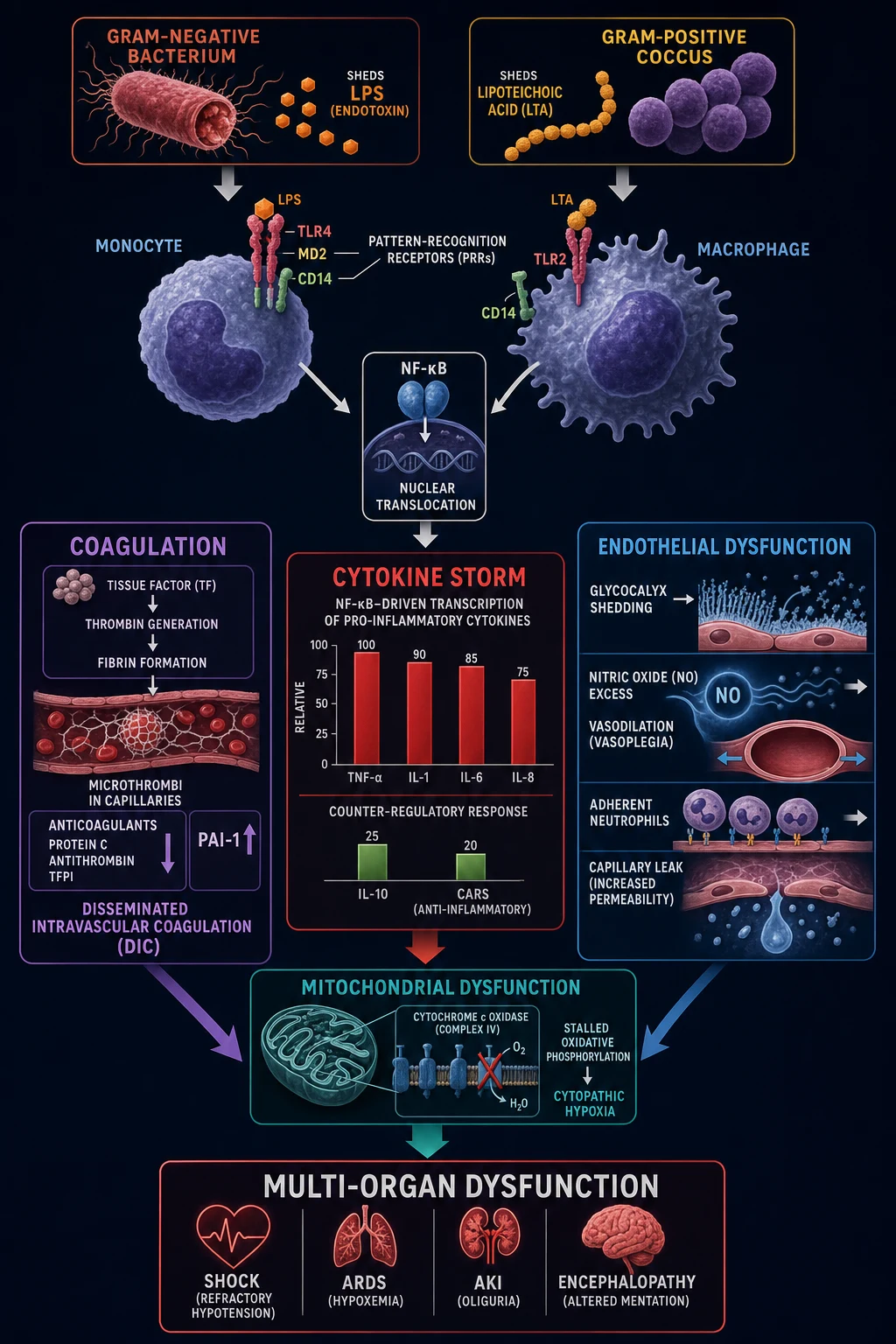
Biomarkers in ICU: objective measures that guide diagnosis, prognosis, and treatment. KEY biomarkers: (1) PROCALCITONIN (PCT): bacterial infection marker — rises in bacterial sepsis, stays low in viral/non-infective. Guides antibiotic duration (PRORATA, Bouadma 2010 — antibiotic stewardship). (2) LACTATE: tissue hypoperfusion marker — elevated in shock, sepsis, mesenteric ischaemia. Guide resuscitation (lactate clearance 10%/h; Jansen 2010, ANDROMEDA-SHOCK 2019). (3) TROPONIN (hs-cTn): myocardial injury — type 1 MI (atherosclerotic plaque rupture), type 2 MI (supply-demand mismatch — common in ICU), and MINS (myocardial injury after non-cardiac surgery — AHA 2021). (4) BNP/NT-proBNP: heart failure — elevated in volume overload, cardiac dysfunction. (5) CRP: inflammation — nonspecific, trends useful. (6) D-DIMER: fibrin turnover — elevated in VTE, DIC, arterial thrombosis, inflammation, malignancy, pregnancy (non-specific, high negative predictive value). (7) suPAR (soluble urokinase-type plasminogen activator receptor): inflammation/prognosis — predicts mortality in sepsis and critical illness. (8) COPEPTIN (CT-proAVP): vasopressin surrogate — quantifies stress response and arginine-vasopressin axis, prognostic in shock. Each has strengths, limitations, and specific clinical applications — always interpret in clinical context, not in isolation.
On this page & tools
Your progress
Saved locally on this device.
3 MCQs with explanations
Target exams
Red flags
Key ICU biomarkers comparison
| Biomarker | What it measures | When elevated | Clinical use | Limitations |
|---|---|---|---|---|
| Procalcitonin | Bacterial infection (calcitonin precursor) | Bacterial sepsis (>2 ng/mL), bacterial pneumonia; NOT viral/non-infective | Antibiotic stewardship (stop when <0.5 or falls ≥80%) | False positive (trauma, surgery, cardiogenic shock), false negative (first 6-12 h, localised) |
| Lactate | Tissue hypoperfusion / anaerobic metabolism | Shock, sepsis, mesenteric ischaemia, seizures, burns | Guide resuscitation (clearance >10%/h), prognosis | Also raised: β2-agonists, malignancy, mitochondrial toxins, liver failure |
| Troponin / hs-cTn | Myocardial injury (cardiomyocyte necrosis) | Type 1 MI, type 2 MI, MINS, sepsis, PE, myocarditis, renal failure | Diagnose MI, grade cardiac injury, prognostic | Type 2 in ICU (not atherosclerotic) — do NOT reflexively PCI; renal failure elevates |
| BNP / NT-proBNP | Cardiac stretch / wall tension (volume overload) | Heart failure, volume overload, PE, sepsis | Diagnose heart failure, guide diuresis, prognostic | Raised: renal failure, PE, sepsis, age, AF; LOWER in obesity (haemodilution/clearance) |
| CRP | Acute-phase inflammation | Infection, inflammation, tissue injury, malignancy, autoimmune | Trend (rising = worsening), monitor infection response | Nonspecific, slow (24-48 h lag), many causes |
| D-dimer | Cross-linked fibrin degradation (coagulation + fibrinolysis) | VTE (DVT/PE), DIC, arterial thrombosis, inflammation, malignancy, pregnancy, surgery | Rule OUT VTE/DIC (high NPV); ISTH DIC scoring | Non-specific (low specificity); rises with age — use age-adjusted cut-off (age × 10 µg/L in >50 yr) |
| suPAR | Soluble urokinase plasminogen activator receptor (immune activation) | Sepsis, septic shock, multi-organ failure, inflammation | Prognostic — predicts mortality independent of severity scores | Nonspecific; influenced by renal function, malignancy; not yet routine bedside |
| Copeptin (CT-proAVP) | C-terminal fragment of pro-arginine-vasopressin (AVP surrogate) | Shock, sepsis, stress, hypotension, hyponatraemia | Surrogate for hard-to-measure AVP; prognostic in shock; guides vasopressin consideration | Released with stress; non-specific; influenced by osmolality, fluid status |
Practical use of biomarkers in septic shock
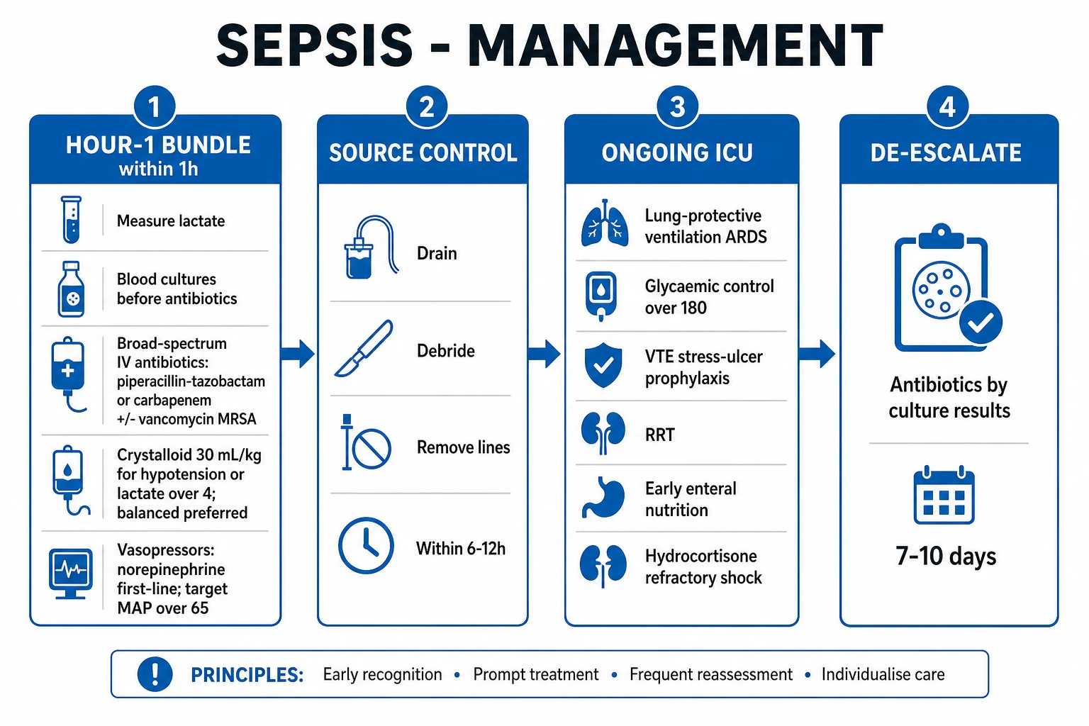
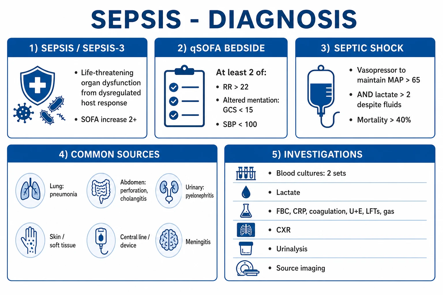
- On presentation — lactate (baseline — guide resuscitation), procalcitonin (baseline — guide antibiotics), CRP (baseline — trend later)
- Resuscitation guided by lactate — target lactate clearance >10%/h (or normalisation within 2-6h). If not clearing → ongoing hypoperfusion (assess volume, cardiac output, source control)
- Procalcitonin trend — if PCT falls >80% from baseline or <0.5 → consider stopping antibiotics (PROORATA algorithm). Repeat daily
- Troponin — check if cardiac symptoms, arrhythmia, or unexplained hypotension. Elevated → assess for type 1 (ECG, ACS) vs type 2 MI (treat underlying cause)
- BNP/NT-proBNP — check if heart failure suspected (dyspnoea, oedema). Low → heart failure unlikely. High → heart failure or other cause (renal, PE, sepsis)
- Serial CRP — trend every 24-48h. Falling → responding. Rising or persistently high → treatment failure, complication, ongoing infection
Clinical pearls
Red flags
Prognosis
PRORATA trial (Bouadma 2010, Lancet) — procalcitonin-guided antibiotic therapy
RCT: 621 ICU patients with suspected bacterial infection. Procalcitonin-guided (stop antibiotics when PCT <0.5 or fell ≥80%) vs standard (physician decision).
- Antibiotic days (days 1-28): PCT 9.3 vs standard 14.9 (p<0.0001) — REDUCED by 5.6 days
- Mortality (day 28): PCT 20.3% vs standard 20.4% (NO difference — safe)
- Relapse of infection: similar
- CONCLUSION: Procalcitonin-guided antibiotic therapy REDUCES antibiotic exposure (5.6 days) without increasing adverse outcomes. Now standard in many ICU antibiotic stewardship programmes. [1]
Jansen (lactate clearance, 2010): lactate-guided resuscitation reduced mortality in septic shock with lactate ≥3.0 (20% vs 44%, p=0.006). BNP prognosis: elevated BNP in ICU → 2-3x higher mortality.
Lactate — deep dive (perfusion, clearance, prognostics)
Lactate Type A (hypoperfusion / ischaemic) vs Type B (non-hypoperfusion / impaired clearance)
| Feature | Type A — hypoperfusion / tissue ischaemia | Type B — non-hypoperfusion (impaired metabolism or clearance) |
|---|---|---|
| Mechanism | Anaerobic glycolysis from oxygen debt; pyruvate shunted to lactate | Aerobic glycolysis (β2-agonists), impaired oxidation (mitochondrial toxins), reduced hepatic clearance |
| Lactate/pyruvate ratio | High (>10-15) | Normal |
| Examples | Septic / haemorrhagic / cardiogenic shock, mesenteric ischaemia, severe burns, limb ischaemia, CO poisoning, seizures (delayed) | Salbutamol / ritodrine / adrenaline infusions, metformin (MALA), cyanide, thiamine deficiency (beriberi), malignancy (lymphoma, leukaemia — Warburg), liver failure, ethanol, propofol infusion, linezolid |
| Clinical sign | Cool, mottled, prolonged cap refill, oliguria, hypotension | Warm, well-perfused, normal ScvO2, may be haemodynamically stable |
| Management | Resuscitate (fluids, blood, inotropes), source control, restore oxygen delivery | Remove trigger (stop β2-agonist / metformin), haemodialysis (metformin, linezolid), thiamine (alcoholic), treat malignancy |
| Prognosis | Mortality ∝ lactate level and clearance half-life | Driven by underlying cause |
Lactate clearance protocol in septic shock (Jansen 2010 algorithm)
- Draw baseline lactate at recognition of septic shock / suspected sepsis with lactate ≥3.0 mmol/L
- Resuscitate with crystalloid 30 mL/kg bolus (Surviving Sepsis Campaign) + early broad-spectrum antibiotics within 1 h + source control
- Recheck lactate at 2 h — calculate clearance: (baseline − current) / baseline × 100
- If clearance ≥10%/h → on target; continue therapy, recheck at 4-6 h
- If clearance <10%/h → reassess: echocardiography (cardiac output / fluid responsiveness), MAP target ≥65 mmHg, consider vasopressor (noradrenaline), inotrope (dobutamine) if low CO, source control (drain abscess, debride tissue), consider ongoing occult bleeding
- If lactate rising despite resuscitation → red flag: mesenteric ischaemia, mitochondrial dysfunction (severe sepsis), β2-agonist, metformin toxicity, limb/bowel ischaemia. Escalate: surgical review, CT angiography
- Target — lactate normalisation (≤2 mmol/L) within 2-6 h; persistent hyperlactataemia at 24 h = poor prognosis
Troponin in ICU — type 1 MI, type 2 MI, and MINS
Type 1 MI vs Type 2 MI vs MINS — distinguishing myocardial injury in ICU
| Feature | Type 1 MI | Type 2 MI | MINS (myocardial injury after non-cardiac surgery) |
|---|---|---|---|
| Mechanism | Atherosclerotic plaque rupture/erosion → in-situ thrombus | Supply-demand mismatch (no plaque rupture) | Perioperative supply-demand mismatch ± non-ischaemic injury |
| Troponin | Rise AND fall, with ≥1 value >99th centile | Rise AND fall, with ≥1 value >99th centile | Peak troponin ≥99th centile within 30 d post-op (no ischaemic symptom required) |
| Symptoms | Ischaemic chest pain/symptoms | Often absent or atypical in ICU | Usually SILENT (85% without ischaemic features) — detected by routine troponin screening |
| ECG | ST-elevation, ST-depression, T-wave inversion, new LBBB | Often non-specific, sinus tachycardia, or changes of underlying cause | Non-specific; new ST changes uncommon |
| Imaging | Regional wall motion abnormality, plaque on angiography | May be normal or fixed defect | Variable |
| Management | DAPT, anticoagulation, urgent PCI (STEMI) / early invasive strategy (NSTE-ACS) | Treat UNDERLYING cause (oxygen, BP, rate, sepsis, anaemia) — routine PCI not indicated | Risk stratify, address modifiable factors, start aspirin + statin, cardiology follow-up; troponin trend alone |
| Prognosis | Driven by infarct size and treatment | Mortality ~2-3× non-injured ICU patients | 30-day mortality ~8-10%; 1-year mortality ~15-20% |
Troponin elevation in ICU — how to distinguish type 1 MI from type 2 MI / MINS
- Confirm rise/fall pattern with serial hs-cTn (0, 1, 3 h). Isolated single rise ≠ acute MI — consider chronic injury (renal failure, structural heart disease)
- Assess ischaemic symptoms — chest pain, dyspnoea, diaphoresis, radiation (absent in many type 2 / MINS)
- ECG — look for ST-elevation (STE), ST-depression, T-wave inversion, new LBBB, posterior MI. Dynamic ST changes favour type 1. Diffuse / non-specific changes favour type 2
- Bedside echo — new regional wall motion abnormality (especially in a coronary territory) favours type 1; global hypokinesis / EF reduction favours type 2
- Identify supply-demand drivers of type 2 — tachyarrhythmia (AF with RVR), hypoxia, hypotension, severe anaemia, sepsis, severe hypertension
- Confirm non-cardiac triggers of troponin rise — PE, myocarditis, sepsis, renal failure, cardioversion, seizures (rule these out before calling it MI)
- Decision — STEMI / NSTE-ACS (type 1) → DAPT + anticoag + cardiology + early invasive strategy. Type 2 → treat underlying cause, monitor, antiplatelet/ statin if 10-yr ASCVD risk warrants. MINS → aspirin + statin + surveillance
BNP / NT-proBNP — heart failure diagnosis, volume status, prognosis
BNP vs NT-proBNP — what's the difference?
| Property | BNP | NT-proBNP |
|---|---|---|
| Origin | Cleaved from pro-BNP by corin → active BNP + inert NT-proBNP | Inert N-terminal fragment (released in 1:1 stoichiometry) |
| Half-life | ~20 min | ~120 min |
| Stability | Less stable (requires cold processing) | More stable (room temperature OK for hours) |
| Removed by | Neutral endopeptidases + clearance receptors | Renal clearance predominantly |
| Affected by neprilysin inhibitors (sacubitril) | ↑ measured BNP (false high) | No significant effect → preferred in patients on ARNI |
| Bedside assay | Available | Most labs batch; not always point-of-care |
| Heart-failure rule-out (dyspnoea) | BNP <100 pg/mL | NT-proBNP <300 pg/mL (rule-out across all ages) |
D-dimer — fibrin turnover in VTE, DIC, arterial thrombosis
D-dimer elevation — causes and significance
| Cause | Typical magnitude | Use |
|---|---|---|
| Acute VTE (DVT, PE) | Moderately-high | Rule OUT (negative D-dimer + low pre-test probability → VTE excluded, no imaging needed). High NPV (≥97%) |
| DIC (overt) | Very high + falling fibrinogen + rising PT + low platelets | ISTH overt DIC score — D-dimer contributes 0-3 points |
| Arterial thrombosis (MI, stroke, mesenteric) | Mild-moderate | Not diagnostic — supports prothrombotic state |
| Aortic dissection / aneurysm | Mild-moderate | Aortic Dissection Detection Risk Score uses D-dimer >500 µg/L |
| Pregnancy (3rd trimester) | Physiologically elevated | Use pregnancy-adapted YEARS algorithm / PE rule-out criteria |
| Malignancy, inflammation, sepsis, post-op, trauma | Variable, often high | Non-specific; cannot rule out VTE reliably |
| Ageing | Rises ~10 µg/L per year | Use age-adjusted cut-off: age × 10 µg/L FEU in >50 yr |
D-dimer interpretation in suspected PE in ICU
- Stratify pre-test probability — Wells score (low/moderate/high) or PERC if low-risk and young. Imaging is the rule in unstable patients
- Low/intermediate pre-test probability → D-dimer useful. Normal D-dimer (with sensitive assay, incl. age-adjusted cut-off in >50 yr) excludes PE — stop workup. The 2017 van Es IPD meta-analysis confirmed Wells + age-adjusted D-dimer gives a 3-month VTE failure rate <1.5% in PE-rule-out patients, including older patients.[18]
- High pre-test probability — do NOT rely on D-dimer (specificity too low). Go straight to CT pulmonary angiography
- Haemodynamically unstable — bedside echo (RV strain), in-lab pulmonary angiography if unstable for CT. D-dimer is academic
- Pregnancy — use YEARS algorithm: D-dimer + clinical signs of DVT + haemoptysis + most-symptomatic leg is PE → CT; if not, use D-dimer cut-off (rule-out at <1000 if no PE signs; <500 if signs present)
- Critically ill patient — D-dimer almost always elevated (sepsis, surgery, trauma, lines) → low rule-out utility; rely on imaging. The value in ICU is in trajectory (rising in DIC, falling in resolving thrombosis) and ISTH DIC scoring
suPAR — soluble urokinase-type plasminogen activator receptor
Copeptin — vasopressin surrogate
Procalcitonin — antibiotic stewardship algorithm
PRORATA-based procalcitonin algorithm for antibiotic de-escalation
- Draw baseline PCT at antibiotic initiation (do NOT delay empiric antibiotics for suspected sepsis)
- Day 1-3 (escalation phase) — continue antibiotics. Recheck PCT daily
- Stop antibiotics if PCT <0.5 ng/mL OR has fallen ≥80% from peak (and clinical improvement: defervescence, falling WBC, haemodynamic stability, source controlled)
- Continue if PCT >0.5 ng/mL AND <80% fall — reassess for: inadequate source control, resistant organism, wrong drug, new infection, non-bacterial cause (PCT can stay high in cardiogenic shock, severe trauma, medullary thyroid cancer)
- Special populations — PCT less reliable in neonates, localised infection (endocarditis, abscess), after major surgery/trauma (false positive), early infection <6 h (false negative), and immunosuppressed patients
- NEVER withhold or delay antibiotics in septic shock based on a single PCT value — clinical judgement overrides
Procalcitonin vs CRP — which is the better infection biomarker?
| Feature | Procalcitonin | CRP |
|---|---|---|
| Biologically specific for bacterial infection | Yes (calcitonin precursor from extra-thyroidal tissues) | No — generic acute-phase reactant |
| Onset after insult | 6-12 h | 24-48 h (slower) |
| Falls with effective therapy | ~50% per day | Slow (days) |
| Useful for antibiotic stewardship | Yes — PRORATA, SAPS, SABS validated algorithms | No — too slow and non-specific |
| Differentiates bacterial vs viral | Reasonably (low in pure viral) | No |
| False positives | Major surgery, severe trauma, cardiogenic shock, prolonged cardiac arrest, medulla of thyroid (calcitonin-producing tumour) | Any inflammation, malignancy, autoimmune, tissue injury |
| Cost / availability | More expensive | Cheap, universal |
Combining biomarkers — multimarker panels improve prediction
How to combine biomarkers in common ICU scenarios
| Scenario | Leading biomarkers | What each tells you | Integrated interpretation |
|---|---|---|---|
| Septic shock | Lactate + PCT + CRP + suPAR | Lactate → perfusion; PCT → bacterial likelihood; CRP → inflammation; suPAR → prognosis | High lactate + high PCT + rising suPAR = severe, escalate; clearance of lactate + falling PCT = improving |
| Acute dyspnoea (HF vs COPD) | BNP/NT-proBNP | Low → not HF; high → likely HF (mind renal/AF/obesity) | Combine with echo + clinical to confirm |
| Post-op troponin rise | hs-cTn trend | MINS vs type 1 MI | Aspirin + statin; invasive strategy only if ischaemic features |
| Suspected PE | D-dimer (low pre-test) | Normal D-dimer rules out PE in low-risk | If elevated → CTPA |
| Bleeding + abnormal coagulation | D-dimer + fibrinogen + PT + platelets | Overt DIC if ISTH ≥5 | Treat trigger + factor replacement |
| Septic shock on rising vasopressors | Copeptin + lactate + ScvO2 | Copeptin → AVP reserve; lactate → perfusion; ScvO2 → oxygen utilisation | Low copeptin (vasopressin depleted) + refractory shock → add vasopressin 0.03 U/min |
Clinical pearls — second set (deep dive)
Red flags — extended set
[1]Trial cards — the evidence base
ANDROMEDA-SHOCK (Hernández 2019, JAMA) — peripheral perfusion vs lactate-targeted resuscitation
Multicentre RCT (Latin America, n=424) in septic shock: capillary refill time (CRT)-guided vs lactate-guided resuscitation.
- 28-day mortality: CRT 34.9% vs lactate 43.4% (RR 0.75, 95% CI 0.55-1.02; OR 0.61 in adjusted analysis; p=0.06 unadjusted but adjusted was significant)
- Fluids given at 8 h: significantly LESS in CRT group (~1.8 L vs 2.3 L) — fewer harms of fluid overload
- SOFA, vasopressor days, RRT use: favoured CRT strategy
- CONCLUSION: A peripheral-perfusion (CRT, mottling, central-toe temperature) strategy may improve outcomes vs a lactate-chasing strategy in septic shock, in part by avoiding excessive fluid resuscitation. Practical: combine both — lactate for prognosis, perfusion for when to stop resuscitating.[17]
Jansen lactate clearance trial (2010, AJRCCM) — early lactate-guided therapy
Multicentre open-label RCT (n=348) of ventilated ICU patients with lactate ≥3.0 mmol/L: lactate-clearance-guided (target ≥20% per 2 h) vs no lactate guidance.
- In-hospital mortality: lactate-guided 23% vs no guidance 33% (statistically significant in the subgroup with lactate ≥3.0 entry criterion — 20% vs 44% in severe shock subgroup)
- Less organ failure in lactate-guided arm
- CONCLUSION: Early lactate-guided therapy reduced mortality and organ failure in patients with hyperlactataemia. Lactate clearance ≥10-20%/h became the standard resuscitation target, integrated into the Surviving Sepsis Campaign 2021.[2][7]
MINS — the VISION cohort data (Devereaux, Curr Opin Cardiol 2014; AHA 2021 Scientific Statement)
Pooled cohort data from the international VISION (Vascular Events In Noncardiac Surgery Patients Cohort Evaluation) study (>30,000 patients across multiple countries):
- Incidence of MINS: ~8% of all non-cardiac surgery; 20-30% in vascular surgery; higher with prior CAD, HF, stroke, emergency surgery
- 30-day mortality post-MINS: ~9% (3-4× higher than patients without MINS)
- Independent association with cardiac arrest, cardiogenic shock, acute HF, stroke — even when asymptomatic
- ~85% of MINS cases have no ischaemic symptoms — detected by routine postoperative troponin screening (AHA recommends hs-cTn at 24-48 h post-op in high-risk patients)
- AHA 2021 management: aspirin + statin, glycaemic control, intensive surveillance, cardiology follow-up; PCI reserved for high-risk trajectories (ischaemic features / dynamic ECG)
- PREVENT-MINS (Szczeklik 2025, Circulation): ivabradine did NOT reduce MINS — supports the supply-demand (rate-independent) pathophysiology in most cases.[9][10][11]
Backes 2012 systematic review (Intensive Care Med) — suPAR as biomarker in systemic inflammation
Systematic review of suPAR in sepsis, septic shock, and systemic inflammation across 14 studies (>3,500 patients):
- suPAR is an independent predictor of mortality in sepsis and septic shock, often superior to CRP, procalcitonin, IL-6 for prognosis
- Higher suPAR → higher 28-day and 90-day mortality; cut-off ~12 ng/mL carries mortality >50%
- Less useful for DIAGNOSIS of infection (PCT remains better for distinguishing bacterial from non-bacterial)
- Confirmed by Gussen 2019 (J Intensive Care): neutrophils are the dominant source of circulating suPAR; suPAR marks exhausted neutrophil function — high suPAR = dysfunctional, migratory-impaired neutrophils → worse outcome
- Status: not yet routine bedside in most ICUs, but a high-yield exam concept (immunoparalysis, neutrophil exhaustion).[13][14]
van Es 2017 IPD meta-analysis (J Thromb Haemost) — Wells rule + age-adjusted D-dimer for PE exclusion
Individual patient data meta-analysis (>7,000 patients with suspected PE) of the original and simplified Wells rules combined with age-adjusted D-dimer cut-off (age × 10 µg/L FEU in >50 yr):
- 3-month VTE failure rate after PE exclusion: <1.5% (well below the 3% safety threshold)
- Age-adjusted cut-off safely increased the proportion of older patients in whom PE could be ruled out by D-dimer alone (without imaging) — from ~6% to ~30% in >50 yr
- CONCLUSION: A negative D-dimer (using the age-adjusted threshold) combined with a low/intermediate Wells score safely excludes PE. In ICU, D-dimer is rarely rule-out useful (most critically ill have elevated D-dimer) — rely on imaging, except for low-risk young patients.[18]
Lee 2022 BMJ systematic review — NT-proBNP rule-in/rule-out pathway for acute heart failure
Meta-analysis (>40,000 patients with acute dyspnoea) validating NT-proBNP thresholds for acute heart failure:
- Rule-out: NT-proBNP <300 ng/L → NPV ~98% across all ages (heart failure unlikely)
- Rule-in (age-stratified): <50 yr >450 ng/L; 50-75 yr >900 ng/L; >75 yr >1800 ng/L → specificity ~90%
- Confounders: renal failure raises (use higher thresholds or alternative diagnosis), obesity lowers (use 50% lower thresholds), AF raises independently
- CONCLUSION: NT-proBNP is a powerful rule-out for acute heart failure in the dyspnoeic ICU patient; rule-in is age-stratified and confounder-aware. Trending NT-proBNP during decongestion predicts outcome.[3]
Special populations
Biomarker interpretation pitfalls by population
| Population | Pitfall | Workaround |
|---|---|---|
| Renal failure (eGFR <30, dialysis) | Troponin and NT-proBNP chronically elevated | Use delta troponin (>20% rise) for acute injury; rely on echo, JVP, fluid balance |
| Obesity (BMI ≥30) | BNP/NT-proBNP falsely low | Use obesity cut-offs (50% lower); use POCUS B-lines / JVP / echo |
| Pregnancy | D-dimer physiologically elevated from 1st trimester; alkaline phosphatase ↑ | Use YEARS algorithm; do NOT use plain D-dimer threshold |
| Cirrhosis / liver failure | Lactate clearance impaired; coagulopathy (PT, fibrinogen) altered; CRP often low (decreased hepatic synthesis) | Lactate may be mildly high without hypoperfusion; do not over-resuscitate |
| Cardiac surgery / post-op | Troponin, BNP, CRP all elevated at baseline | Use trends; post-CABG troponin rise is expected (surgical injury) — MINS definition (≥99th centile) still applies but threshold often higher |
| Malignancy | D-dimer chronically high; CRP high; suPAR high | D-dimer cannot rule out VTE — image; suPAR adds prognosis |
| Neutropenia / post-chemotherapy | PCT, CRP may be blunted; suPAR high | Do not rely on fever or PCT — start empiric antibiotics in any febrile neutropenic; serial CRP/PCT |
| On sacubitril/valsartan (ARNI) | BNP falsely high | Use NT-proBNP |
Exam synthesis — CICM / FFICM / EDIC one-minute answer
[1]One-glance biomarker-cause summary table
| Marker up | Bacterial infection | Hypoperfusion | Cardiac | Inflammation | Thrombosis | Stress / prognosis |
|---|---|---|---|---|---|---|
| Procalcitonin | ✓✓✓ | — | — | ✓ (surgery, trauma) | — | ✓ |
| Lactate | ✓ (sepsis) | ✓✓✓ | ✓ (shock) | — | — | ✓✓ |
| Troponin | — | ✓ (shock) | ✓✓✓ | ✓ (myocarditis) | ✓ (type 1) | ✓ |
| BNP/NT-proBNP | — | — | ✓✓✓ | ✓ (sepsis) | — | ✓ |
| CRP | ✓ | — | — | ✓✓✓ | — | — |
| D-dimer | — | — | — | ✓ (DIC) | ✓✓✓ (VTE/DIC) | — |
| suPAR | ✓✓ | — | — | ✓✓ | — | ✓✓✓ |
| Copeptin | — | ✓ (shock) | ✓ (stress) | — | — | ✓✓✓ |
Exam practice
SAQ — Procalcitonin-guided antibiotic stewardship in severe community-acquired pneumonia
10 minutes · 10 marks
A 68-year-old man is admitted to ICU with a 3-day history of fever, productive cough and pleuritic chest pain. He is confused, HR 112, BP 92/55 (MAP 67), RR 30, SpO2 91% on 6 L nasal prongs. Chest X-ray shows right middle and lower lobe consolidation. Lactate 2.6 mmol/L, WCC 18.5, CRP 185 mg/L, procalcitonin 8.4 ng/mL. Blood cultures have been taken and empirical piperacillin-tazobactam started.
SAQ — Postoperative troponin elevation: distinguishing MINS, type 1 MI and type 2 MI
10 minutes · 10 marks
A 72-year-old man with a background of hypertension and type 2 diabetes undergoes an emergency laparotomy for perforated diverticulitis. Twelve hours post-op in ICU he develops new atrial fibrillation with rapid ventricular response (HR 140), SpO2 92% on 2 L, BP 88/50 (MAP 63). He has no chest pain. High-sensitivity troponin T (hs-cTnT) is 14 ng/L pre-op, 280 ng/L at 12 hours post-op, and 360 ng/L at 15 hours (99th centile URL 40 ng/L). ECG shows AF with lateral T-wave inversion. Lactate 2.4 mmol/L. Haemoglobin 96 g/L.
References
- [1]Bouadma L, Luyt CE, Tubach F, et al. Use of procalcitonin to reduce patients' exposure to antibiotics in intensive care units (PRORATA trial): a multicentre randomised controlled trial Lancet, 2010.PMID 20097417
- [2]Jansen TC, van Bommel J, Schoonderbeek FJ, et al. Early lactate-guided therapy in intensive care unit patients: a multicenter, open-label, randomized controlled trial Am J Respir Crit Care Med, 2010.PMID 20463176
- [3]Lee KK, Doudesis D, Anwar M, et al. Development and validation of a decision support tool for the diagnosis of acute heart failure: systematic review, meta-analysis, and modelling study BMJ, 2022.PMID 35697365
- [4]Chapman AR, Adamson PD, Shah ASV, et al. High-Sensitivity Cardiac Troponin and the Universal Definition of Myocardial Infarction Circulation, 2020.PMID 31587565
- [5]Póvoa P. C-reactive protein: a valuable marker of sepsis Intensive Care Med, 2002.PMID 11904651
- [6]Vincent JL, Bakker J. Blood lactate levels in sepsis: in 8 questions Curr Opin Crit Care, 2021.PMID 33852499
- [7]Evans L, Rhodes A, Alhazzani W, et al. Executive Summary: Surviving Sepsis Campaign: International Guidelines for the Management of Sepsis and Septic Shock 2021 Crit Care Med, 2021.PMID 34643578
- [8]Bularga A, Hung J, Daghem M, et al. Coronary Artery and Cardiac Disease in Patients With Type 2 Myocardial Infarction: A Prospective Cohort Study Circulation, 2022.PMID 35341327
- [9]Ruetzler K, Smilowitz NR, Berger JS, et al. Diagnosis and Management of Patients With Myocardial Injury After Noncardiac Surgery: A Scientific Statement From the American Heart Association Circulation, 2021.PMID 34601955
- [10]Khan J, Alonso-Coello P, Devereaux PJ. Myocardial injury after noncardiac surgery Curr Opin Cardiol, 2014.PMID 25029449
- [11]Ekeloef S, Alamili M, Devereaux PJ, et al. Troponin elevations after non-cardiac, non-vascular surgery are predictive of major adverse cardiac events and mortality: a systematic review and meta-analysis Br J Anaesth, 2016.PMID 27799170
- [12]Levi M, Toh CH, Thachil J, et al. Guidelines for the diagnosis and management of disseminated intravascular coagulation. British Committee for Standards in Haematology Br J Haematol, 2009.PMID 19222477
- [13]Backes Y, van der Sluijs KF, Mackie DP, et al. Usefulness of suPAR as a biological marker in patients with systemic inflammation or infection: a systematic review Intensive Care Med, 2012.PMID 22706919
- [14]Gussen H, Hohlstein P, Bartneck M, et al. Neutrophils are a main source of circulating suPAR predicting outcome in critical illness J Intensive Care, 2019.PMID 31061709
- [15]Lee JH, Chan YH, Lai OF, et al. Vasopressin and copeptin levels in children with sepsis and septic shock Intensive Care Med, 2013.PMID 23344831
- [16]Dabla PK, Dabla V, Arora S. Co-peptin: Role as a novel biomarker in clinical practice Clin Chim Acta, 2011.PMID 20920496
- [17]Hernández G, Ospina-Tascón GA, Damiani LP, et al. Effect of a Resuscitation Strategy Targeting Peripheral Perfusion Status vs Serum Lactate Levels on 28-Day Mortality Among Patients With Septic Shock: The ANDROMEDA-SHOCK Randomized Clinical Trial JAMA, 2019.PMID 30772908
- [18]van Es N, Kraaijpoel N, Klok FA, et al. The original and simplified Wells rules and age-adjusted D-dimer testing to rule out pulmonary embolism: an individual patient data meta-analysis J Thromb Haemost, 2017.PMID 28106338