ICU · Diagnostics
Point-of-care ultrasound (POCUS): comprehensive ICU applications
Also known as POCUS · Point-of-care ultrasound · Bedside ultrasound · Focused ultrasound · RUSH protocol · BLUE protocol · FAST exam
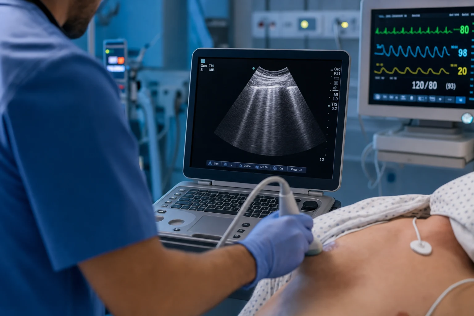
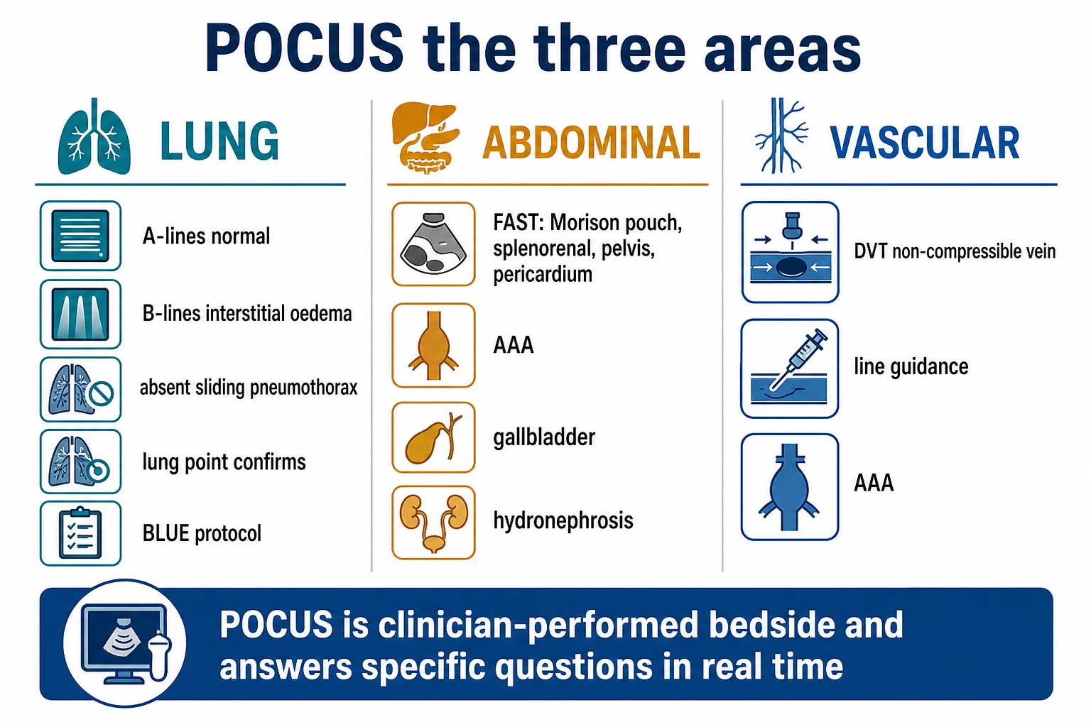
POCUS is real-time ultrasound performed and interpreted by the treating clinician at the bedside to answer specific clinical questions. In ICU: DIAGNOSTIC (cardiac, lung, abdominal, vascular) + PROCEDURE GUIDANCE (CVC insertion, thoracentesis, paracentesis, arthrocentesis). Protocols: RUSH (Rapid Ultrasound in Shock), BLUE (Bedside Lung Ultrasound in Emergency), FAST (Focused Assessment with Sonography in Trauma), FATE (Focus Assessed Transthoracic Echo). POCUS changes management in 30-50% of ICU examinations. Core principle: CLINICAL INTEGRATION — POCUS does NOT replace formal echo/imaging — it answers focused questions to guide immediate management.
On this page & tools
Your progress
Saved locally on this device.
Target exams
Red flags
Focused echocardiography (FOCUS / FATE)
FOCUS (Focused Cardiac Ultrasound) and FATE (Focus Assessed Transthoracic Echocardiography) are the two most widely adopted ICU focused-echo protocols. Both answer the same five questions within 2–3 minutes: (1) Is LV systolic function normal, hyperdynamic, or depressed? (2) Is the RV normal or dilated? (3) Is there a pericardial effusion, and is there tamponade physiology? (4) Is the patient volume-depleted (small/hyperdynamic LV, small IVC) or volume-overloaded (distended IVC, B-lines)? (5) Is there gross structural/valvular abnormality? FATE differs only in mandating a subcostal view first (obtainable in nearly any supine ventilated patient) and in explicitly integrating lung and pleural windows.[9][12]
Probe and machine setup. Use a phased-array (cardiac) probe at 1.5–5 MHz, depth 15–18 cm for adults, cardiac preset. The cardiac indicator (probe marker) is on the RIGHT side of the screen. Depth on the lung preset drops to 4–6 cm to see the pleural line.
[12]The four standard FOCUS/FATE views
[12]1. Parasternal long-axis (PLAX)
Place the probe at the 3rd–4th left intercostal space at the parasternal edge, indicator toward the RIGHT shoulder. The optimal plane slices from the right shoulder to the left hip and demonstrates: RV outflow tract (nearest field), LV cavity, left atrium, descending thoracic aorta (far field, longitudinal), the anterior mitral leaflet and aortic valve, and the pericardium as a bright echogenic line.
[8]What to assess:
- LV size and systolic function. Normal LV end-diastolic diameter (LVEDD) ≈ 3.5–5.3 cm (women) / 3.5–5.8 cm (men) on ASE 2015 reference. Dilated (>5.8 cm) = chronic dilated cardiomyopathy / chronic AR/MR. Eyeball EF: (a) hyperdynamic — LV walls nearly obliterate the cavity in systole ("kissing walls", EF >70%) → hypovolaemia or high-output/distributive state; (b) normal — roughly half the chamber shortens (EF 50–70%); (c) mild–moderately depressed (EF 30–50%); (d) severely depressed — barely any inward motion (EF <30%). Inter-rater agreement of eyeball EF by a competent operator is ±10% of biplane Simpson's.
- RV size. The RV should be only a small sliver at the top of PLAX; if it occupies >1/3 of the image width or is globular, suspect RV dilatation.
- Aortic root. Measure at end-systole, leading-edge to leading-edge at the level of the sinus of Valsalva: >3.8 cm is dilated. A dissection flap is an intimal linear echo in the descending aorta (far field).
- Pericardial effusion. An anechoic stripe anterior to the RVOT and/or posterior to the LV. Posterior-only effusion suggests chronic; circumferential suggests acute. Anechoic fluid tracking ONLY anterior to the aortic root is a pericardial fat pad, not an effusion.
- Valves (gross). Heavy calcification of the AV = aortic stenosis; prolapsing//flail MV leaflet = severe MR; a "rocking" AV or MV = endocarditis until disproven (but vegetations <2 mm are missed on POCUS — formal echo required).
2. Parasternal short-axis (PSAX)
From PLAX, rotate 90° clockwise so the indicator points to the LEFT shoulder. The LV appears as a circular doughnut. Fan from base to apex through three levels: mitral level (fish-mouth MV), papillary level (symmetric papillary muscles — the workhorse view), and apical level.
[5]What to assess:
- Global and regional LV function. All segments should thicken and move inward symmetrically. A hypokinetic/akinetic territory = regional wall motion abnormality (LAD = anterior/septal; RCA = inferior; LCx = lateral/posterior).
- Septal motion — the RV overload sign. A normal interventricular septum is concave toward the RV (the LV is round). Flattening of the septum producing a D-shaped LV indicates RV pressure/volume overload: diastolic flattening = volume overload; systolic flattening = pressure overload (PE, pulmonary HTN). This is one of the most reliable POCUS signs of RV strain.
- RV size. The RV is normally a thin crescent; if it wraps around the anterior LV and equals or exceeds LV size, RV is dilated.
- Pericardial effusion and tamponade. Circumferential effusion is best quantified here. Look for RV free-wall diastolic collapse — invagination of the RV wall in early diastole — a sensitive sign of tamponade.
3. Apical 4-chamber (A4C)
Place the probe at the point of maximal impulse with the patient in a slight LEFT lateral decubitus, indicator pointing to the patient's LEFT. This is the single most informative view for the intensivist because all four chambers are compared side-by-side.
[8]What to assess:
- RV:LV ratio. In A4C the RV should be <60% of the LV area in end-diastole (ASE 2015). RV:LV >0.6 = RV dilatation; >1.0 = severe. RV size larger than LV in this view is abnormal and the key POCUS trigger to think PE, pulmonary HTN, RV infarct, ARDS cor pulmonale, or chronic RV failure.
- McConnell's sign. RV apex hyperkinetic with free-wall hypokinesis — highly specific (94%) but insensitive (~48%) for PE. Also seen in RV infarct.
- Atrial size. RA/RV larger than LA/LV suggests chronic right-sided pathology (pulmonary HTN, tricuspid disease). Dilated LA = chronic LV failure / mitral disease / AF.
- LV function & apical thrombus. Look for apical ballooning (takotsubo, LAD infarct) and LV apical thrombus (post-MI).
- Tricuspid regurgitation jet (TR v-max). If colour Doppler available: v-max >2.8 m/s or 3.0 m/s suggests elevated pulmonary artery systolic pressure (PASP). Continuous-wave can estimate PASP = 4 × (TR v-max)² + RA pressure (estimated from IVC).
- Pericardial effusion / RA collapse. Right atrial collapse in late diastole/systole for >1/3 of the cardiac cycle is the most sensitive echocardiographic sign of tamponade.
4. Subcostal
Place the probe just below the xiphoid process, flat against the abdomen, aimed toward the left shoulder, indicator to the patient's LEFT. Fan through the liver to obtain a 4-chamber view. This is the CPR view — the only view obtainable without interrupting chest compressions.
[1]What to assess:
- Pericardial effusion. Fluid is most easily distinguished here from the bright liver (anechoic fluid between liver/diaphragm and RV free wall). Tamponade: RA systolic collapse, RV diastolic collapse, IVC plethora.
- IVC for volume. From the subcostal 4-chamber, rotate the probe 90° counterclockwise to image the IVC in long-axis entering the RA. Measure 1–2 cm from the RA junction. Cut-offs (with major caveats — see IVC pearl): <1.5 cm and >50% collapse with sniff → CVP <5 mmHg (likely fluid responsive); >2.5 cm and <20% collapse → CVP >15 mmHg (unlikely fluid responsive). Intermediate = indeterminate.
- Subcostal IVC in cardiac arrest. A small, completely collapsed IVC during CPR suggests reversible hypovolaemia; a distended, fixed IVC with no collapse suggests obstructive cause (tamponade, massive PE, tension PTX).
- Subcostal lung windows. Angle to either side of the heart to look for B-lines, consolidation, or effusion in dependent lung.
Tamponade — the POCUS checklist. Effusion + (RA systolic collapse OR RV diastolic collapse) + IVC plethora + tachycardia + hypotension / muffled heart sounds = tamponade until proven otherwise. POCUS can also identify loculated post-surgical effusions (regional tamponade — no IVC plethora, asymmetric chamber collapse) which formal echo is needed to characterise. If clinically tamponading and unstable → pericardiocentesis guided by POCUS.[9]
Lung ultrasound and the BLUE protocol
Technique. Use the phased-array or curvilinear probe in lung preset, depth 4–6 cm (just deep enough to see the pleural line). The pleural line lies between the shadow of the ribs (bat sign) — a bright horizontal line that shimmers with respiration (lung sliding). Assess a minimum of 8 zones (4 per hemithorax: upper/lower anterior, upper/lower lateral) — the BLUE protocol uses 3 points per side (upper anterior, lower anterior, lateral) and adds posterior zones if supine.
[4]The 5 fundamental lung signs (Lichtenstein)
- A-lines — horizontal reverberation artefacts below the pleural line = normal aeration (air).
- B-lines — vertical comet-tail artefacts arising from the pleural line, erasing A-lines, moving with sliding. ≥3 B-lines in a single intercostal space in a longitudinal plane = interstitial syndrome. Causes: pulmonary oedema, interstitial pneumonia, pulmonary fibrosis, ARDS.
- Consolidation (hepatization / tissue-like pattern) — lung looks like liver/spleen with dynamic air bronchograms (pneumonia) or static bronchograms (atelectasis).
- Pleural effusion — anechoic (black) collection above the diaphragm with atelectatic lung floating inside; can be quantified (depth × simple formulas approximate volume).
- Absent lung sliding ± lung point — pneumothorax. The lung point (junction of sliding and non-sliding lung) is pathognomonic and specific (near 100%); it also allows crude size estimation (more lateral/large field = larger PTX).
BLUE protocol — diagnosing acute respiratory failure
Lichtenstein's algorithm (90.5% correct first diagnosis in the original derivation cohort) integrates lung findings with venous (DVT) ultrasound. The order of decision-making:
[4]Caveats to the profiles. The BLUE profiles are a scaffold, not gospel: decompensated COPD can produce B-lines (cor pulmonale); ARDS produces bilateral B-lines (indistinguishable from pulmonary oedema by pattern alone — differentiate by the heart: depressed LV + B-lines = cardiogenic; normal heart + B-lines + reduced lung compliance = ARDS). Asymmetric pneumonia can be missed if scanning zones are too few. The original 90.5% accuracy is a derivation-set figure; real-world performance is lower and drops further in mixed ICU populations — POCUS narrows, it does not close, the differential.[4][2]
Abdominal ultrasound (FAST, bladder, AAA, organs)
Use the curvilinear probe (2–5 MHz), abdominal preset, depth 15–20 cm.
[11]FAST / eFAST
Four (five with eFAST) windows looking for free peritoneal/pericardial (or pleural) fluid:
- RUQ — Morison's pouch (hepatorenal recess): the most dependent supine peritoneal space and the most sensitive single FAST view. Anechoic stripe between liver and kidney = positive.
- LUQ — splenorenal recess + subphrenic space: left-sided bleeds (splenic injury) are easily missed; angle the probe coronally in the mid-to-posterior axillary line.
- Suprapubic (pelvic): bladder as an acoustic window; look behind/female pouch of Douglas/male retrovesical. A full bladder improves the view; ask to scan before Foley decompression.
- Subxiphoid (pericardial): pericardial fluid (tamponade) — same window as subcostal cardiac.
- eFAST — bilateral anterior thorax: lung sliding present = no PTX; absent sliding ± lung point = PTX. Replaces the supine CXR for occult PTX in trauma.
Sensitivity for clinically significant intra-abdominal bleed ≈ 60–90% (better for >500 mL); negative FAST does NOT exclude injury — unstable with blunt trauma goes to theatre regardless. FAST detects free fluid only — it misses retroperitoneal bleed (pelvic fracture), hollow viscus injury, and solid-organ injury without haemoperitoneum.[3]
Bladder volume
Place the probe suprapubically in transverse, measure width (W) and height (H); rotate 90° to sagittal and measure depth/length (L). Volume ≈ W × H × L × 0.75 (mL) (or ×0.7). Use: post-void residual (PVR >150–200 mL suggests urinary retention), confirm retention before Foley, estimate urine output in oliguria when no catheter, and guide suprapubic catheter placement.
[6]Abdominal aortic aneurysm (AAA) screening
From the subcostal/xiphoid window, scan in the midline in transverse, following the aorta from the diaphragmatic hiatus to the bifurcation. Measure the antero-posterior diameter, outer-to-outer wall, in transverse at the largest point (usually just below the renal arteries). ≥3 cm = aneurysmal. Risk of rupture rises sharply above 5.5 cm (the threshold for elective repair in men; 5.0 cm in women in many guidelines). In unexplained hypotension/back pain in an older man, an aortic dissection flap (intimal linear echo separating true/false lumen) or a ruptured AAA (loss of wall continuity, retroperitoneal fluid) must be sought — a POCUS AAA finding in shock mandates immediate vascular surgical input, not waiting for confirmatory CT if unstable.
Other abdominal applications
- Gallbladder (RUQ): stones (echogenic with shadow), wall thickening >3 mm (oedematous — cholecystitis, or hypoalbuminaemia/fluid overload in ICU), pericholecystic fluid, sonographic Murphy's.
- Kidneys: hydronephrosis (dilated anechoic collecting system) — obstructive AKI; small echogenic kidneys = chronic disease; enlarged = AKI.
- Bowel: pneumatosis / absent peristalsis / fluid-filled loops in mesenteric ischaemia (low sensitivity); dilated loops >3 cm + intra-luminal fluid = ileus/obstruction.
- Ascites / collections: map the largest pocket for paracentesis/drains; mark entry site and measure depth to avoid bowel/solid organs.
Vascular access ultrasound
Ultrasound guidance for central and peripheral vascular access is now standard of care — it reduces mechanical complications of IJV cannulation by ~70% (pneumothorax, arterial puncture, haematoma) and increases first-pass success.[7][11]
Central venous catheter (CVC) — internal jugular vein
Set-up. High-frequency linear probe (5–13 MHz), vascular preset, depth 3–4 cm. Patient supine, head turned slightly away, in slight Trendelenburg. The IJV lies lateral/superficial, the carotid medial and deep; light transducer pressure (the IVC/IJV collapse easily with pressure).
Short-axis (out-of-plane) vs long-axis (in-plane):
- Short-axis / out-of-plane (SAX-OOP): probe transverse to the vessel; vessel is round, needle seen as a bright dot crossing into it. Tracks needle tip accurately across the screen, easier to keep both vessel and carotid in view, lower risk of posterior wall puncture if tip is followed. Most commonly used.
- Long-axis / in-plane (LAX-IP): probe aligned with the vessel; entire needle shaft seen advancing into a tubular vessel. Higher first-pass success but operator must keep the needle in the ultrasound plane — lateral/medial deviation is invisible, so risk of inadvertent carotid/posterior wall puncture is real if alignment is lost.
- Best practice. Pre-procedure "scout" scan (confirm patency — ensure the vein is fully compressible, rule out thrombus), then real-time guidance during puncture, with the needle tip visualised at all times. Confirm venous (compressible, non-pulsatile, resizable with Valsalva) vs arterial (pulsatile, thick wall, non-compressible).
Arterial line guidance
Use a high-frequency linear probe, depth 1.5–2.5 cm. The radial artery is identified at the wrist lateral to the flexor carpi radialis tendon; short-axis/out-of-plane with dynamic needle tip tracking is the workhorse for difficult radial cannulation, and modified Seldinger/catheter-over-wire kits improve success. For the femoral artery, in-plane guidance lowers the risk of posterior wall injury and pseudoaneurysm. Ultrasound is especially valuable in: obesity, oedema, scars, hypotension/shock (non-palpable pulses), paediatric/pulseless patients, and after failed landmark attempts.
Peripheral IV under ultrasound (deep / difficult access)
Useful when superficial veins are exhausted. Identify the deep brachial, basilic, or cephalic veins in the upper arm with the linear probe; these are compressible, anechoic, non-pulsatile. In-plane technique with a longer cannula (≥4.5 cm to span the vessel wall fully) is recommended. Pitfalls: inadvertently cannulating the brachial artery (always confirm compressibility and lack of pulsatility), transfixing the vein, and catheter kinking if the vein is too deep.
Compression ultrasound for DVT
Two-point or whole-leg compression: apply probe pressure to collapse the femoral/popliteal vein against the underlying bone. Fully compressible = no DVT; non-compressible = DVT (the most important criterion; Doppler adds little in symptomatic patients). This is the "pipes" step of RUSH and the venous step of BLUE — a non-compressible femoral vein in a shocked, dyspnoeic patient is strong support for PE.
Clinical pearls
Red flags
Prognosis
POCUS evidence and outcomes
ACEP Emergency Ultrasound Guidelines (2016) — scope, training, and QA (PMID 27251108)
International evidence-based recommendations for point-of-care lung ultrasound (Volpicelli 2012/2020) — the lung-POCUS standard (PMID 35403953)
Karakitsos 2006 — real-time ultrasound-guided IJV catheterisation vs landmark (PMID 16957148)
Exam SAQ — densified leaf
10 minutes · 10 marks
In structured CICM/FFICM style: (1) define the core entity in one sentence; (2) list three immediate ICU priorities; (3) state two investigations that change management; (4) name one evidence landmark or guideline anchor; (5) give one fatal exam trap.
Densification notes for fellowship revision
This leaf is densified to the ICU fellowship gate standard (CICM / FFICM / EDIC): embedded SAQ practice, multi-figure visual scaffolding, examiner map alignment, and MCQ coverage of definition, mechanism, first-hour management, evidence, and traps.
[5]- Revision checkpoint 1: restate definition, one number examiners expect, and one absolute do-not-miss action.
- Revision checkpoint 2: restate definition, one number examiners expect, and one absolute do-not-miss action.
- Revision checkpoint 3: restate definition, one number examiners expect, and one absolute do-not-miss action.
- Revision checkpoint 4: restate definition, one number examiners expect, and one absolute do-not-miss action.
- Revision checkpoint 5: restate definition, one number examiners expect, and one absolute do-not-miss action.
- Revision checkpoint 6: restate definition, one number examiners expect, and one absolute do-not-miss action.
- Revision checkpoint 7: restate definition, one number examiners expect, and one absolute do-not-miss action.
- Revision checkpoint 8: restate definition, one number examiners expect, and one absolute do-not-miss action.
- Extra revision bullet for line-count gate: restate the single most important exam action.
- Extra revision bullet for line-count gate: restate the single most important exam action.
- Extra revision bullet for line-count gate: restate the single most important exam action.
- Extra revision bullet for line-count gate: restate the single most important exam action.
- Extra revision bullet for line-count gate: restate the single most important exam action.
- Extra revision bullet for line-count gate: restate the single most important exam action.
- Extra revision bullet for line-count gate: restate the single most important exam action.
- Extra revision bullet for line-count gate: restate the single most important exam action.
References
- [1]Mayo PH, et al. American College of Chest Physicians/La Société de Réanimation de Langue Française statement on competence in critical care ultrasonography. Chest, 2009.PMID 19188546
- [2]Volpicelli G, et al. International evidence-based recommendations for point-of-care lung ultrasound. Intensive Care Med, 2012.PMID 22392031
- [3]Perera P, et al. The RUSH exam: Rapid Ultrasound in SHock in the evaluation of the critically lll. Emerg Med Clin North Am, 2010.PMID 19945597
- [4]Lichtenstein DA, Mezière GA. Relevance of lung ultrasound in the diagnosis of acute respiratory failure: the BLUE protocol. Chest, 2008.PMID 18403664
- [5]Frankel HL, et al. Guidelines for the Appropriate Use of Bedside General and Cardiac Ultrasonography in the Evaluation of Critically Ill Patients-Part I: General Ultrasonography. Crit Care Med, 2015.PMID 26468699
- [6]Pourmand A, et al. The utility of point-of-care ultrasound in the assessment of volume status in acute and critically ill patients. World J Emerg Med, 2019.PMID 31534598
- [7]Karakitsos D, et al. Real-time ultrasound-guided catheterisation of the internal jugular vein: a prospective comparison with the landmark technique in critical care patients. Crit Care, 2006.PMID 17112371
- [8]Lichtenstein D. Fluid administration limited by lung sonography: the place of lung ultrasound in assessment of acute circulatory failure (the FALLS-protocol). Expert Rev Respir Med, 2012.PMID 22455488
- [9]Via G, et al. International evidence-based recommendations for focused cardiac ultrasound. J Am Soc Echocardiogr, 2014.PMID 24951446
- [10]Moore CL, Copel JA. Point-of-care ultrasonography. N Engl J Med, 2011.PMID 21345104
- [11]American College of Emergency Physicians. Emergency ultrasound guidelines. Ann Emerg Med, 2009.PMID 19303521
- [12]Holm JH, et al. Perioperative use of focus assessed transthoracic echocardiography (FATE). Anesth Analg, 2012.PMID 23051882