MedVellum
MCQsExamsAtlas
DashboardPricing
MBBS / Core medicine✳Dermatology✳ICU Fellowship (CICM)✳Anaesthesia✳Emergency Medicine✳Psychiatry Fellowship✳Paediatrics Fellowship✳Physician Medicine✳MCQs✳SAQs✳Vivas✳OSCE✳Evidence-first✳MBBS / Core medicine✳Dermatology✳ICU Fellowship (CICM)✳Anaesthesia✳Emergency Medicine✳Psychiatry Fellowship✳Paediatrics Fellowship✳Physician Medicine✳MCQs✳SAQs✳Vivas✳OSCE✳Evidence-first✳

MedVellum.

The folio

Exam-exhaustive medical education across every specialty — evidence-graded topics, engraved plates, and practice in every written and oral format. Educational content only — not medical advice.

llms.txt · psychiatry LLM catalog · sitemap

Atlas

  • Specialty atlas
  • MBBS / Core medicine
  • Dermatology
  • ICU Fellowship (CICM)
  • Anaesthesia
  • Emergency Medicine
  • Psychiatry Fellowship
  • Paediatrics Fellowship
  • Physician Medicine

Study & account

  • MCQ practice
  • Practice alias
  • Exam tools
  • Dashboard
  • Pricing
  • Sign in

© 2026 MedVellum. For education only — not a substitute for clinical judgement.

Folio edition · Set in Instrument Serif & Archivo

ICU TopicsEndocrine

ICU · Endocrine

Adrenal insufficiency and Addisonian crisis

Also known as Addisonian crisis · Adrenal crisis · Critical illness-related corticosteroid insufficiency (CIRCI) · Relative adrenal insufficiency

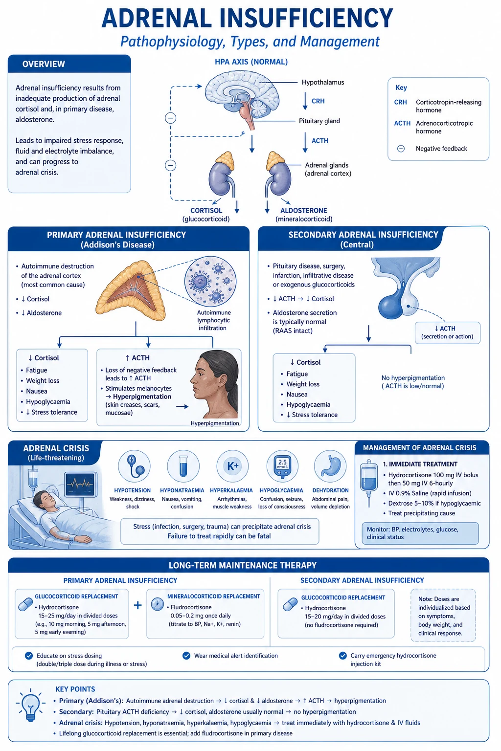
Adrenal crisis is a life-threatening state of glucocorticoid deficiency. Presents with hypotension (refractory to catecholamines), abdominal pain, nausea/vomiting, confusion, hyponatraemia, hyperkalaemia, hypoglycaemia. Precipitated by: stress (infection, surgery, trauma) in patients with underlying adrenal insufficiency (primary = Addison's; secondary = pituitary; tertiary = exogenous steroids). Treatment: IMMEDIATE hydrocortisone 100 mg IV stat then 50 mg IV Q6H (or 200 mg/24h infusion) — do NOT wait for cortisol/ACTH results. IV fluids (normal saline + dextrose for hypoglycaemia). Treat precipitant. Identify type: primary (high ACTH, hyperpigmentation, hyperkalaemia) vs secondary/tertiary (low ACTH, no hyperpigmentation, normal K).

medium7 referencesUpdated 2 July 2026
On this page & tools

Your progress

Saved locally on this device.

Target exams

CICMFFICMEDIC

Red flags

Give hydrocortisone IMMEDIATELY — do NOT wait for cortisol/ACTH results in suspected crisisPatients on chronic steroids have suppressed HPA axis — give STRESS DOSE hydrocortisone for any critical illnessPatients on etomidate (intubation induction) can develop adrenal suppression — single dose causes transient CIRCIHyponatraemia + hyperkalaemia + hypotension = Addisonian crisis until proven otherwise

Your progress

Saved locally on this device.

Target exams

CICMFFICMEDIC

Red flags

Give hydrocortisone IMMEDIATELY — do NOT wait for cortisol/ACTH results in suspected crisisPatients on chronic steroids have suppressed HPA axis — give STRESS DOSE hydrocortisone for any critical illnessPatients on etomidate (intubation induction) can develop adrenal suppression — single dose causes transient CIRCIHyponatraemia + hyperkalaemia + hypotension = Addisonian crisis until proven otherwise
Cinematic ICU scene of a hypotensive patient with a cortisol and ACTH cosyntropin stimulation test result on the screen, a hydrocortisone infusion drawn up, saline resuscitation running, clinical-blue lighting, medical educational, no faces, no text
FigureThe adrenal insufficiency in the ICU — the unexplained refractory vasopressor-dependent shock, the hyponatraemia, the hypoglycaemia. The random cortisol under 400 or the failed cosyntropin stimulation supports the diagnosis; the treatment is the immediate hydrocortisone 100 mg IV and the fluid, before the confirmatory testing.
[1]

In one line

Adrenal crisis = glucocorticoid deficiency emergency: hypotension (catecholamine-resistant), abdominal pain, confusion, hyponatraemia, hyperkalaemia, hypoglycaemia. Treatment: hydrocortisone 100 mg IV STAT then 50 mg IV Q6H — do NOT wait for results. IV fluids (normal saline + dextrose). Treat precipitant. Primary (Addison's): high ACTH, hyperpigmentation, hyperkalaemia. Secondary/tertiary (steroid withdrawal/pituitary): low ACTH, no hyperpigmentation, normal K. Stress dose: chronic steroid patients need double/triple their usual dose for any critical illness.

[1]

Classification

Educational HPA-axis schematic comparing primary adrenal failure high ACTH hyperpigmentation hyperkalaemia versus secondary tertiary low ACTH normal potassium from steroid withdrawal
FigurePrimary destroys the gland (high ACTH, pigmentation, mineralocorticoid loss). Secondary/tertiary fail the axis (low ACTH, no pigmentation, potassium usually normal). Crisis is catecholamine-resistant shock until cortisol is replaced.

Primary (Addison disease)

Adrenal gland destruction

  • Autoimmune adrenalitis (#1 in developed world)
  • TB, adrenal haemorrhage (Waterhouse-Friderichsen — meningococcaemia)
  • ACTH HIGH (no negative feedback), cortisol LOW
  • Hyperpigmentation (high ACTH stimulates melanocytes)
  • Hyperkalaemia, hyponatraemia (aldosterone deficiency — zona glomerulosa affected)
  • Hyperpigmentation, postural hypotension, salt craving

Secondary/Tertiary

HPA axis suppression

  • Secondary: pituitary failure (low ACTH)
  • Tertiary: exogenous steroid withdrawal (#1 cause)
  • ACTH LOW, cortisol LOW
  • NO hyperpigmentation
  • NORMAL potassium (aldosterone intact — RAAS independent of ACTH)
  • Patients on >5mg prednisolone >3 weeks have HPA suppression
[1]

Clinical features

Adrenal crisis clinical features

Hypotension
Catecholamine-resistant
Unresponsive to fluids/vasopressors
Abdominal
Pain, nausea, vomiting
May mimic acute abdomen
Low Na
Hyponatraemia
Due to ADH excess + aldosterone deficiency
High K
Hyperkalaemia
Only in PRIMARY (aldosterone deficiency)

Management

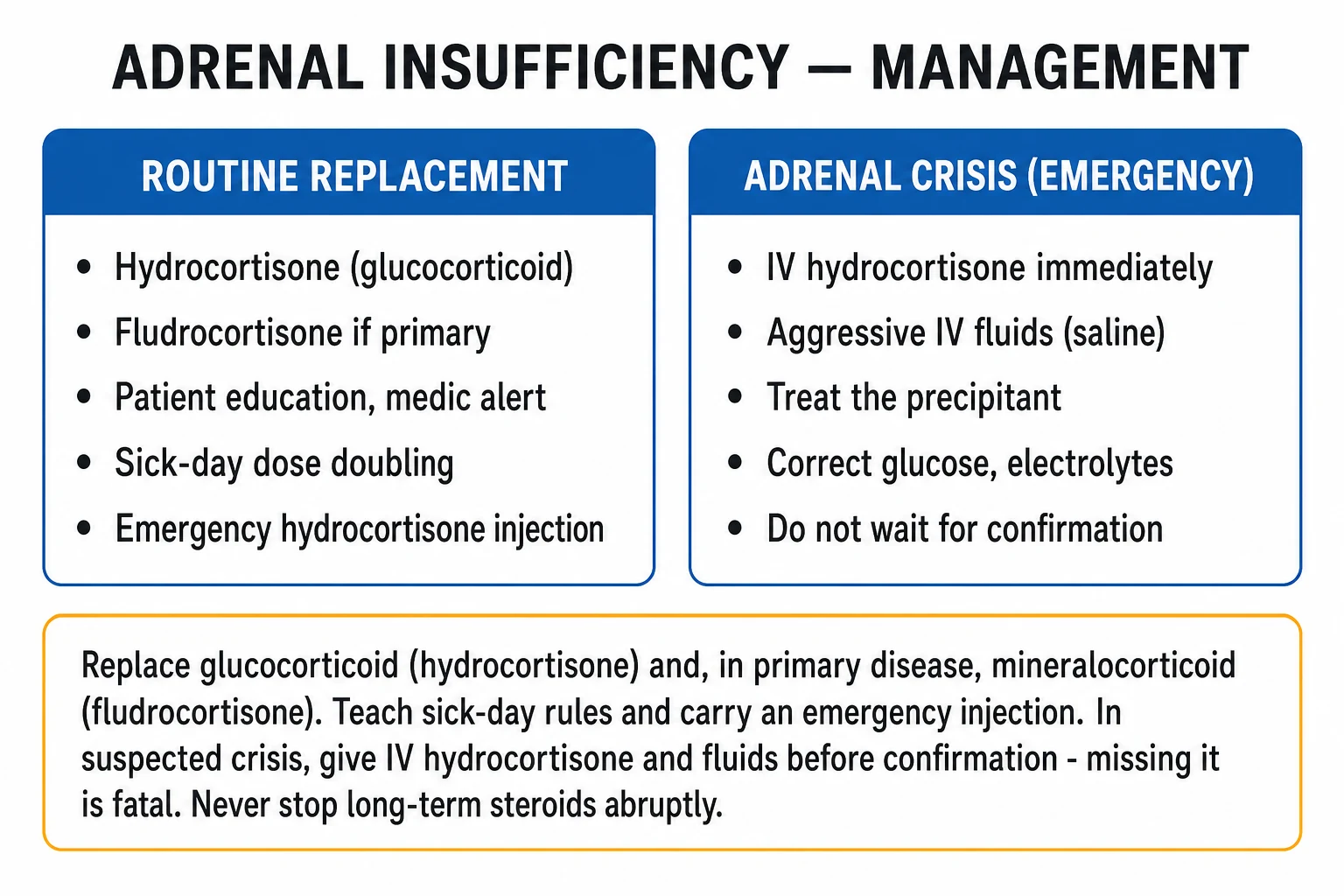
Educational adrenal crisis management pathway: hydrocortisone 100 mg IV immediately, saline and dextrose, draw cortisol ACTH if possible without delaying treatment, treat precipitant
FigureAdrenal crisis — hydrocortisone 100 mg IV now, fluids and glucose, treat the precipitant. Draw cortisol/ACTH only if it does not delay the first dose.
[1]

Adrenal crisis management

1

1. Hydrocortisone 100 mg IV STAT

Give IMMEDIATELY — do NOT wait for cortisol/ACTH results. Then 50 mg IV Q6H or 200 mg/24h continuous infusion. This dose provides both glucocorticoid AND mineralocorticoid activity (at high doses, hydrocortisone has sufficient mineralocorticoid effect — no need for fludrocortisone acutely). Do NOT delay treatment for diagnostic testing.

2

2. Draw blood for cortisol and ACTH (BEFORE hydrocortisone if possible)

If possible, draw random cortisol and plasma ACTH BEFORE giving hydrocortisone. Also check: renin, aldosterone, electrolytes, glucose. A cortisol level drawn after hydrocortisone is meaningless. If the patient is critically ill and you cannot draw before, just treat — you can do a short synacthen test later.

3

3. IV fluid resuscitation

Normal saline 1L rapidly, then continue based on clinical response. Add DEXTROSE if hypoglycaemic (common in adrenal crisis). Monitor Na, K, glucose, BP, urine output. Correct hyperkalaemia if severe (calcium gluconate if ECG changes — but usually corrects with hydrocortisone + fluids).

4

4. Identify and treat precipitant

Search for infection (blood/urine/sputum cultures), check for recent steroid withdrawal, consider surgical stress. Start empiric antibiotics if infection suspected. Infection is the #1 precipitant of adrenal crisis.

5

5. Vasopressor support if needed

Adrenal crisis causes vasoplegic shock (NO response to catecholamines without cortisol). Once hydrocortisone given, vasopressors become effective. Start noradrenaline if hypotensive despite fluids + hydrocortisone. The BP often responds dramatically to hydrocortisone alone.

6

6. Transition to oral

Once stable (usually 24-72h): transition to oral hydrocortisone (20 mg mane + 10 mg afternoon). Add fludrocortisone 50-100 mcg daily if primary adrenal insufficiency. Taper gradually. Patient education: MedicAlert bracelet, emergency hydrocortisone injection kit, double dose during illness (stress dosing).

[1]

CIRCI in critical illness

Critical Illness-Related Corticosteroid Insufficiency (CIRCI)

CIRCI is an inadequate cellular corticosteroid response to the stress of critical illness. Not a true adrenal insufficiency but a relative deficiency. [1]

Diagnosis (suggested):

  • Septic shock with poorly responsive vasopressors
  • ARDS (consider steroids for persistent ARDS)
  • Delta cortisol (after 250 mcg synacthen) <250 nmol/L [1]

Treatment: hydrocortisone 200 mg/day (continuous infusion or 50 mg Q6H) in vasopressor-resistant septic shock. Do NOT use etomidate for intubation if CIRCI suspected (etomidate suppresses adrenal function).[2]

Clinical pearls

High-yield adrenal insufficiency points for the CICM/FFICM exam

  1. Give hydrocortisone IMMEDIATELY in suspected crisis — do NOT wait for results.[1]
  2. Dose: 100 mg IV STAT, then 50 mg IV Q6H (or 200 mg/24h infusion).
  3. Hyponatraemia + hyperkalaemia + hypotension = Addisonian crisis until proven otherwise.
  4. Primary vs secondary: primary has HIGH ACTH, hyperpigmentation, hyperkalaemia. Secondary has LOW ACTH, no pigmentation, normal K.[1]
  5. Stress dosing: chronic steroid patients need 2-3x usual dose for critical illness/surgery.
  6. HPA suppression: >5 mg prednisolone daily for >3 weeks suppresses HPA axis.
  7. CIRCI: relative adrenal insufficiency in critical illness — hydrocortisone 200 mg/day for vasopressor-resistant septic shock.[2]
  8. Etomidate suppresses adrenal function — avoid in septic patients if possible.
  9. No need for fludrocortisone acutely — high-dose hydrocortisone provides sufficient mineralocorticoid activity.
  10. Waterhouse-Friderichsen syndrome: bilateral adrenal haemorrhage in meningococcal septicaemia.
  11. Aldosterone is preserved in secondary/tertiary (RAAS independent of ACTH) — normal potassium.
  12. Patient education: MedicAlert bracelet, emergency hydrocortisone injection, double dose during illness.
  13. Hypoglycaemia is common — give dextrose-containing fluids.
  14. Withdrawal of exogenous steroids is the #1 cause of secondary adrenal insufficiency.

Red flags

Critical adrenal insufficiency points

  • Give hydrocortisone IMMEDIATELY — do NOT wait for cortisol/ACTH results in suspected crisis.[1]
  • Hyponatraemia + hyperkalaemia + hypotension = Addisonian crisis until proven otherwise.
  • Stress dose steroids for ANY patient on chronic steroids presenting with critical illness.
  • Etomidate suppresses adrenal function — avoid in septic patients (single dose causes transient CIRCI).[2]
  • Vasopressors are ineffective without cortisol — hydrocortisone restores catecholamine sensitivity.
  • Hypoglycaemia is a common and dangerous complication — monitor glucose, give dextrose.

The HPA axis — why cortisol is load-bearing in critical illness

The HPA axis and the cortisol stress response — the physiology every ICU question is built on

The hypothalamic–pituitary–adrenal (HPA) axis is the body's principal neuroendocrine stress response. The signalling chain is: [1]

Hypothalamic CRH (corticotropin-releasing hormone) → anterior pituitary ACTH (adrenocorticotropic hormone) → adrenal cortex cortisol (zona fasciculata, via cAMP/StAR/cholesterol side-chain cleavage). Cortisol exerts negative feedback on both the hypothalamus and the pituitary — which is the entire reason exogenous steroids suppress the axis. [1]

Three properties make cortisol load-bearing in the ICU:

  • Permissive effect on catecholamines — cortisol maintains vascular smooth-muscle α1-adrenergic receptor expression and post-receptor coupling. Without cortisol, vasopressors FAIL. Catecholamine-resistant vasoplegic shock is the clinical hallmark of adrenal crisis.
  • Metabolic counter-regulation — cortisol drives gluconeogenesis and opposes insulin; deficiency → hypoglycaemia (a hallmark of adrenal crisis, especially in children).
  • Immune modulation / free-water clearance — suppresses NF-κB-driven cytokines (TNF-α, IL-1β, IL-6) and antagonises ADH; deficiency contributes to the SIADH-like hyponatraemia and the uncontrolled inflammation of septic shock. [1]

The stress response: a healthy adrenal secretes ~10 mg cortisol/day basally, rising to 75–150 mg/day (sometimes >300 mg) under maximal stress (sepsis, surgery, trauma). Target cortisol in critical illness is therefore 500–1000 nmol/L. A critically ill patient with a ward-"normal" cortisol (e.g. 300 nmol/L) is relatively deficient — the gland cannot mount the required stress response. This single concept is the entire basis of CIRCI, and it is why the empiric stress dose of hydrocortisone is 100 mg IV STAT then 50 mg q6h (≈200–300 mg/day). Anything less (e.g. a 100 mg/24h infusion alone) under-doses the surgical/septic patient.[7]

Aetiology — primary, secondary and tertiary adrenal insufficiency

Primary vs secondary vs tertiary adrenal insufficiency — the three-level framework

FeaturePrimary (Addison's)SecondaryTertiary
Site of lesionAdrenal cortex (≥90% of gland destroyed)Anterior pituitaryHypothalamus
ACTHHIGH (no negative feedback)LOWLOW
CortisolLOWLOWLOW
AldosteroneLOW (zona glomerulosa destroyed)NORMALNORMAL
PotassiumHIGH (aldosterone deficiency)NORMALNORMAL
SodiumLOWLOW (ADH excess, milder)LOW (milder)
HyperpigmentationYES (high ACTH → melanocyte-stimulating effect via POMC)NONO
Salt craving, postural dropYESLess prominentLess prominent
Volume depletionMarked (Na loss)MildMild
HypoglycaemiaCommonCommonCommon
Most common cause in ICUBilateral adrenal haemorrhage (sepsis, anticoagulation); TB in endemic areasPituitary apoplexy, infiltrative diseaseExogenous steroid withdrawal (#1)
[1] [7]

WHY POTASSIUM IS NORMAL IN SECONDARY/TERTIARY — the RAAS principle

Aldosterone secretion from the zona glomerulosa is governed principally by the renin–angiotensin–aldosterone system (RAAS) and potassium, NOT by ACTH. ACTH is only a minor, permissive stimulus for aldosterone. Therefore:

  • In primary adrenal insufficiency, the entire cortex (including the glomerulosa) is destroyed → aldosterone deficiency → hyperkalaemia + salt wasting.
  • In secondary/tertiary adrenal insufficiency, the adrenal is intact and the RAAS continues to drive aldosterone normally → potassium stays normal. [1]

This is the single best discriminator at the bedside and in the exam: hyponatraemia + hyperkalaemia + hypotension = PRIMARY (Addisonian) crisis. Hyponatraemia + normal potassium + hypotension in a patient on chronic steroids = secondary/tertiary (HPA suppression). Mineralocorticoid replacement (fludrocortisone) is required long-term ONLY in primary disease.

[1]

Primary adrenal insufficiency — causes

Causes of primary adrenal insufficiency — and the ICU relevance of each

CauseMechanismICU/exam relevance
Autoimmune adrenalitis (#1 in developed world)21-hydroxylase autoantibodies destroy the cortex; often part of autoimmune polyglandular syndrome type 2 (Schmidt syndrome = Addison's + autoimmune thyroid disease ± type 1 DM)The "classic" Addison's patient; gradual onset, hyperpigmentation over months; crisis precipitated by intercurrent illness
Tuberculosis (#1 worldwide)Caseating granulomatous destruction of the adrenals; often with adrenal calcification on CTSuspect in endemic areas and immigrants; coexisting TB elsewhere. Calcified adrenals on CT are highly suggestive
Bilateral adrenal haemorrhageInfarction/haemorrhage of both adrenals (high vascularity, rich catecholamine/venous drainage)The classic ICU cause — see dedicated section below
Waterhouse–Friderichsen syndromeBilateral adrenal haemorrhage in meningococcal (or pneumococcal) septicaemia; purpuric rash + DICRare but rapidly fatal; seen in fulminant sepsis in younger patients
Infiltrative diseaseAmyloidosis, haemochromatosis, sarcoidosis, metastases (lung, breast, melanoma, renal)Usually with a large mass / bilateral adrenal lesions on CT; metastases need >90% destruction to cause failure (rare)
InfectionsHIV/AIDS (CMV adrenalitis), histoplasmosis, cryptococcosis, paracoccidioidomycosisConsider in immunocompromised ICU patients
DrugsKetoconazole (blocks 11β-hydroxylase/steroidogenesis), metyrapone, mitotane (chemical adrenalectomy), etomidate, fluconazole (lesser)Iatrogenic; etomidate-induced suppression is exam-defining — see below
AdrenoleukodystrophyX-linked; very-long-chain fatty acid accumulationYoung male with spastic paraparesis + Addison's
GeneticCongenital adrenal hyperplasia (21-hydroxylase deficiency), Allgrove (AAA) syndromeRare; usually paediatric
[1] [7]

Bilateral adrenal haemorrhage — the ICU cause

BILATERAL ADRENAL HAEMORRHAGE — the ICU cause of acute primary adrenal failure

Bilateral adrenal haemorrhage is the classic ICU cause of acute (de novo) primary adrenal insufficiency. The adrenals have a rich arterial supply but drain via a single central adrenal vein under high catecholamine tone, making them vulnerable to venous infarction and haemorrhage during any state of sepsis, major stress, coagulopathy or anticoagulation. [1]

Recognised settings:

  • Sepsis / severe systemic illness — especially meningococcaemia (Waterhouse–Friderichsen syndrome, with purpura fulminans and DIC), but also pneumococcal, pseudomonal and staphylococcal septic shock.
  • Anticoagulation / heparin — classically presents ~1 week after starting therapeutic heparin (including heparin-induced thrombocytopenia, HIT). Also seen with warfarin, direct oral anticoagulants, and post-thrombolysis.
  • Antiphospholipid syndrome (APS) — catastrophic APS with bilateral adrenal infarction is a defined syndrome; suspect in a young patient with thrombosis + adrenal failure.
  • Major surgery / trauma — postoperative, post-trauma, post-burns (catecholamine surge + venous congestion).
  • Pregnancy / the peripartum period, severe burns, and acute haemorrhagic pancreatitis. [1]

Presentation: the patient develops new vasopressor-resistant shock + abdominal/flank/back pain + falling haemoglobin + hyponatraemia + hyperkalaemia in the context of one of the above. CT abdomen shows bilateral enlarged heterogeneous (non-enhancing) adrenal masses — the diagnosis is radiological. Mortality is high (15–50%) if unrecognised. Treatment is immediate stress-dose hydrocortisone — do NOT wait for confirmatory cortisol/ACTH.[1]

Secondary and tertiary adrenal insufficiency

EXOGENOUS STEROID WITHDRAWAL — the #1 cause of adrenal insufficiency in the ICU

Tertiary adrenal insufficiency from withdrawal of exogenous glucocorticoids is the single most common cause of adrenal insufficiency encountered in ICU practice. Any patient taking >5 mg prednisolone (or equivalent) daily for >3 weeks has a suppressed HPA axis and is at risk of crisis if the steroid is stopped abruptly or if they develop intercurrent illness without a stress dose. [1]

Why it matters: the suppressed axis cannot mount the cortisol surge that critical illness demands. The patient on chronic steroids who presents with sepsis, undergoes surgery, or simply misses doses develops relative adrenal insufficiency → vasoplegic shock unresponsive to catecholamines, superimposed on their presenting illness. [1]

HPA recovery is slow — it can take 6–12 months (sometimes longer) for full recovery after stopping chronic steroids. The rule for ICU: any patient on chronic steroids admitted with critical illness must receive a STRESS DOSE (double-to-triple their usual dose, or hydrocortisone 100 mg IV STAT then 50 mg q6h if in crisis). Never omit the steroid dose. [1]

Equivalent glucocorticoid doses (for tapering and stress dosing):

  • Hydrocortisone 20 mg ≈ prednisolone 5 mg ≈ methylprednisolone 4 mg ≈ dexamethasone 0.75 mg.
  • Mineralocorticoid activity: hydrocortisone (high) > prednisolone (minimal) > dexamethasone (none). Dexamethasone gives NO mineralocorticoid cover.[7]

Secondary AI — pituitary causes vs tertiary AI — steroid causes

Secondary (pituitary)Tertiary (HPA suppression)
MechanismDestruction of ACTH-secreting corticotrophsSuppression of CRH/ACTH by exogenous glucocorticoid
CausesPituitary apoplexy (haemorrhage into adenoma — sudden headache + ophthalmoplegia), pituitary tumour/surgery/radiotherapy, Sheehan syndrome (postpartum pituitary infarction), infiltrative (haemochromatosis, sarcoid, histiocytosis), traumatic brain injury, lymphocytic hypophysitisChronic exogenous steroid (>5 mg prednisolone >3 weeks) — by far #1; also abrupt withdrawal of inhaled/high-potency topical steroids; recent megestrol/medroxyprogesterone
Other hormone deficienciesYes — panhypopituitarism (TSH, FSH/LH, GH, prolactin, ADH) common — screen and replaceNo — isolated cortisol axis
Volume status / electrolytesHyponatraemia (cortisol deficiency → ADH excess) but normal K, mild volume depletionAs for secondary
ReversibilityUsually permanent (except TBI)Often reversible over months with slow taper
[1]

Clinical features in the ICU — the metabolic signature

THE ICU METABOLIC SIGNATURE — refractory vasoplegic shock + hyponatraemia + hyperkalaemia

Adrenal crisis in the ICU is rarely the presenting diagnosis — it declares itself by failing to respond to standard resuscitation. The four pillars of the presentation: [1]

  1. Refractory, vasopressor-dependent vasoplegic shock — the cardinal feature. Hypotension that is unresponsive to fluids and escalating catecholamines. The mechanism is loss of cortisol's permissive effect on vascular α1-adrenergic tone. This is the clue that prompts the diagnosis in any patient with shock and a relevant history (chronic steroids, anticoagulation, sepsis, TB). Once hydrocortisone is given, vasopressor requirement often falls dramatically within hours.
  2. Hyponatraemia — from two mechanisms acting together: aldosterone deficiency (primary only) causing renal sodium loss, AND cortisol deficiency removing the normal glucocorticoid suppression of ADH → a SIADH-like water-retaining state. So hyponatraemia occurs in BOTH primary and secondary AI.
  3. Hyperkalaemia — from aldosterone deficiency. Present ONLY in primary AI. This is the key discriminator — a patient on chronic steroids with normal potassium has secondary/tertiary AI, not Addison's.
  4. Hypoglycaemia — loss of cortisol's gluconeogenic counter-regulation; especially in children and in those without enteral intake. [1]

The triad hyponatraemia + hyperkalaemia + refractory hypotension = Addisonian crisis until proven otherwise. Add abdominal pain, nausea/vomiting (which can mimic an acute abdomen and lead to a disastrous laparotomy), lethargy/confusion, and (in primary) hyperpigmentation, and the diagnosis is clinched.[1]

Clinical features of adrenal crisis — by system and mechanism

SystemFeatureMechanism
CardiovascularRefractory hypotension, vasoplegia, poor response to catecholaminesLoss of cortisol permissive effect on α1-receptors; volume depletion
ElectrolyteHyponatraemiaAldosterone deficiency (primary) + cortisol deficiency → ADH excess
ElectrolyteHyperkalaemia (primary only)Aldosterone deficiency → impaired distal K+ secretion
MetabolicHypoglycaemiaLoss of gluconeogenesis / insulin counter-regulation
GIAbdominal pain, nausea, vomiting, anorexia, weight loss; may mimic acute abdomenUnknown; often the presenting complaint
NeurologicalLethargy, confusion, delirium, comaHyponatraemia, hypoglycaemia, hypoperfusion
MSKMyalgia, arthralgia, weaknessProximal myopathy (chronic), catabolic state
Skin (primary only)Hyperpigmentation (palmar creases, buccal mucosa, recent scars, gum), vitiligoHigh ACTH/POMC cleavage to MSH; vitiligo from autoimmune destruction
CardiacHyperkalaemic ECG changes, small heart on CXR (chronic)Aldosterone deficiency; chronic volume depletion
[1]

The four-pillar metabolic signature of adrenal crisis

Shock
Refractory / vasoplegic
Catecholamine-resistant — the cardinal ICU clue
Low Na
Hyponatraemia
Both primary and secondary (ADH excess + aldosterone loss)
High K
Hyperkalaemia
PRIMARY only — aldosterone deficiency
Low glu
Hypoglycaemia
Loss of gluconeogenic counter-regulation

Diagnosis — cortisol, ACTH and the short Synacthen test

DIAGNOSIS — bloods BEFORE the first dose of hydrocortisone, but NEVER delay treatment

The diagnostic strategy is always subordinate to treatment in suspected crisis: draw blood if you can, then give hydrocortisone immediately. A cortisol level drawn after hydrocortisone is meaningless. [1]

First-line (draw before hydrocortisone if possible):

  • Random (morning) serum cortisol — cortisol has a diurnal rhythm; in crisis it is irrelevant, but electively a morning (8 am) cortisol <140 nmol/L (5 µg/dL) suggests insufficiency, while >450–500 nmol/L (18 µg/dL) effectively excludes it (in a well patient).
  • Plasma ACTH (must be into a chilled EDTA tube, on ice, processed promptly) — HIGH ACTH + low cortisol = primary; LOW/inappropriately normal ACTH + low cortisol = secondary/tertiary.
  • Renin and aldosterone — high renin + low aldosterone supports primary disease.
  • U&E (Na, K), glucose, FBC, and a search for the precipitant (cultures, troponin, CXR). [1]

Confirmatory — the short Synacthen (cosyntropin) stimulation test (SST):

  • Synacthen (tetracosactrin) 250 µg IV or IM (the "high-dose" or standard test), measure cortisol at 0, 30, and 60 min.
  • Normal response: cortisol rises by >250 nmol/L (9 µg/dL) AND/OR reaches a peak >500 nmol/L (18 µg/dL). Failure to do so confirms adrenal insufficiency.
  • Caveat in secondary/tertiary AI: an atrophic adrenal may still respond to the supraphysiological 250 µg bolus in early disease → a false-normal SST. The low-dose (1 µg) Synacthen test is more sensitive for secondary/tertiary disease. For classic primary disease the 250 µg test is reliable.
  • Do NOT perform the SST in acute crisis — treat first, test later once stable. The SST is for the recovery-phase/long-term diagnosis.
  • Metyrapone and CRH stimulation tests are second-line for localising secondary vs tertiary disease (rarely needed in ICU).[7]

Interpreting morning cortisol and the Synacthen test — diagnostic thresholds (click each)

Cortisol <140 nmol/L

Mortality High if untreated

Morning cortisol <140 nmol/L (5 µg/dL) is highly suggestive of adrenal insufficiency. Combine with ACTH to localise: high ACTH = primary; low ACTH = secondary/tertiary. Confirm with Synacthen test once stable.

[7]

Diagnostic workup of suspected adrenal insufficiency in ICU — what to draw, when

1

1. If crisis suspected — DRAW and TREAT simultaneously

Take a random cortisol, plasma ACTH (chilled EDTA, on ice), renin, aldosterone, U&E, glucose, and cultures BEFORE giving hydrocortisone — but only if the phlebotomy does not delay treatment. If lines are difficult, give hydrocortisone 100 mg IV STAT first and arrange a Synacthen test later. NEVER delay hydrocortisone for tests in an unstable patient.

2

2. Interpret the ACTH:cortisol relationship

High ACTH + low cortisol = PRIMARY (Addison's) → investigate cause (adrenal autoantibodies, CT adrenals for haemorrhage/calcification/mass, TB/HIV screen). Low/inappropriately normal ACTH + low cortisol = SECONDARY/TERTIARY → review drug history (exogenous steroids), pituitary imaging (MRI for apoplexy/mass), full anterior pituitary panel (TSH/free T4, LH/FSH/testosterone, prolactin, IGF-1), and consider a low-dose (1 µg) Synacthen test.

3

3. Confirm with a short Synacthen test once stable

Synacthen 250 µg IV/IM, cortisol at 0/30/60 min. A normal response is a rise >250 nmol/L (9 µg/dL) or peak >500 nmol/L (18 µg/dL). An abnormal test confirms adrenal insufficiency. Note: false-normal in early secondary/tertiary disease (atrophy not yet established) — use the 1 µg low-dose test if suspicion persists.

4

4. Image the cause

Primary disease: CT abdomen (bilateral adrenal enlargement with haemorrhage/infarction in acute haemorrhage; calcification in old TB; mass in malignancy/infiltration). Secondary disease: MRI pituitary (apoplexy = emergency; macroadenoma; empty sella). Add adrenal autoantibodies (21-hydroxylase) and an infective screen as indicated.

5

5. Long-term characterisation

Once stable, define aetiology (autoimmune panel including thyroid/T1DM screen for APS-2; TB/HIV; genetic if young). Establish a written sick-day / stress-dose plan, a MedicAlert alert, and a parenteral hydrocortisone emergency injection kit with patient and family training. Endocrinology follow-up for life.

[1] [7]

Management — empiric treatment that must not wait

Empiric hydrocortisone dosing — by clinical scenario

ScenarioRegimenRationale
Adrenal crisis (suspected/confirmed)Hydrocortisone 100 mg IV STAT, then 50 mg IV q6h (or 200 mg/24h continuous infusion)High-dose hydrocortisone provides glucocorticoid AND sufficient mineralocorticoid activity (at >50 mg/day no fludrocortisone is needed acutely). Do NOT wait for results.
Major surgery / critical illness in known AIHydrocortisone 100 mg IV at induction, then 50 mg q8h × 24 h, taper to baseline over 1–3 daysSurgical stress demands 75–150 mg/day equivalent
Minor/moderate illness in known AIDouble-to-triple the usual oral hydrocortisone for the duration of the illnessSick-day rule
CIRCI / vasopressor-resistant septic shockHydrocortisone 200 mg/day (50 mg q6h or continuous infusion) for ≥3 days, taper if shock reversesSSC 2021 weak recommendation; do NOT use the SST to select patients
Maintenance (stable primary AI)Hydrocortisone 15–25 mg/day in 2–3 divided doses + fludrocortisone 50–100 µg/dayFludrocortisone needed only in primary disease
[1] [2]

WHY NO FLUDROCORTISONE IS NEEDED IN ACUTE CRISIS — the dose-mineralocorticoid principle

Hydrocortisone has intrinsic mineralocorticoid activity. At doses >50 mg/day (i.e. the crisis regimen of 200–300 mg/day), the mineralocorticoid effect is saturated and more than sufficient to replace aldosterone. Adding fludrocortisone in acute crisis is unnecessary and adds no benefit. Fludrocortisone 50–100 µg/day is reserved for the maintenance/long-term phase of PRIMARY adrenal insufficiency, once the hydrocortisone dose has been tapered to physiological levels (<50 mg/day), at which point mineralocorticoid cover is again needed. It is NOT required in secondary/tertiary disease (aldosterone axis intact). This dose principle is a favourite exam question.

[1]

Critical illness-related corticosteroid insufficiency (CIRCI)

CIRCI — relative adrenal insufficiency of critical illness

CIRCI (Critical Illness-Related Corticosteroid Insufficiency) is defined as an inadequate cellular corticosteroid activity for the severity of the patient's critical illness. It is NOT a true structural adrenal insufficiency — it is a relative, functional deficiency in the setting of overwhelming inflammation (sepsis, ARDS, severe trauma). Proposed mechanisms include suppressed cortisol synthesis (inflammatory cytokines), altered glucocorticoid receptor expression/resistance, and tissue-level cortisol inactivation. [1]

Concept: a sick patient should mount a cortisol of 500–1000 nmol/L. A "normal" ward value in shock is therefore inappropriately low — the gland cannot meet demand. [1]

Diagnosis is clinical, not biochemical. The 2017 international consensus (Arabi/Meduri) advises AGAINST relying on the random cortisol level or the delta-cortisol after Synacthen to diagnose CIRCI, because the thresholds (e.g. delta <250 nmol/L) have poor discrimination. Instead, CIRCI is a bedside diagnosis: vasopressor-dependent septic shock, especially requiring escalating or high-dose catecholamines, or early severe ARDS (PaO2/FiO2 <200).[2]

Treatment (Surviving Sepsis Campaign 2021, weak recommendation, low-quality evidence): hydrocortisone 200 mg/day (continuous infusion or 50 mg q6h) for patients with septic shock on ongoing vasopressor support (i.e. not fluid-responsive, still requiring noradrenaline) — NOT for all septic patients, and NOT guided by the Synacthen test. Continue until vasopressors are weaned; taper rather than stop abruptly.[2]

The four corticosteroid-in-septic-shock trials — what each established

TrialYear / nPopulationInterventionPrimary resultTake-home
Annane et al. (JAMA)2002; n=299Vasopressor-unresponsive septic shock (non-responders to Synacthen)Hydrocortisone 50 mg q6h + fludrocortisone 50 µg/day × 7 days vs placebo28-day survival improved in non- responders (relative adrenal insufficiency)Established low-dose (not high-dose) steroids for vasopressor-resistant shock; selected patients by Synacthen response
CORTICUS (Sprung, NEJM)2008; n=499Septic shock (responders AND non-responders)Hydrocortisone 50 mg q6h × 5 days then taper vs placeboNo mortality benefit overall; faster shock reversal in both groups; more superinfections with steroidsAbandoned Synacthen-based selection; steroids reversed shock faster but did not improve survival, and added infection risk
ADRENAL (Venkatesh, NEJM)2018; n=3658Septic shock needing mechanical ventilationHydrocortisone 200 mg/day infusion × 7 days (or until shock reversal) vs placeboNo 90-day mortality difference (27.9% vs 28.8%); faster shock reversal, more ventilator-free days, more new infection/bacteraemia; no increase in serious AEsLargest trial; hydrocortisone does NOT reduce mortality but hastens shock resolution — reasonable to use in vasopressor-resistant shock
APROCCHSS (Annane, NEJM)2018; n=1228Septic shock with multiorgan failureHydrocortisone 50 mg q6h + fludrocortisone 50 µg/day × 7 days vs placebo90-day mortality reduced (43.0% vs 49.1%, RR 0.88, P=0.03); more vasopressor/ventilator-free daysThe only modern positive mortality trial — the combination (with fludrocortisone) in the sickest septic shock patients
[2] [3] [4] [5] [6]

RECONCILING THE TRIALS — when is CIRCI treatment justified?

The four trials appear contradictory but reconcile into a single pragmatic message: corticosteroids do not benefit all septic patients, but they improve outcomes in the sickest, vasopressor-dependent patients. CORTICUS and ADRENAL showed no mortality benefit (and CORTICUS enrolled less sick patients); Annane 2002 and APROCCHSS showed a mortality benefit precisely in vasopressor-unresponsive shock and multiorgan failure, where the relative adrenal insufficiency is most pronounced. The takeaway that drives current practice: give hydrocortisone 200 mg/day to the septic shock patient who is STILL in shock despite adequate fluid resuscitation and ongoing vasopressors — and consider adding fludrocortisone (per APROCCHSS). Do NOT use steroids in septic patients who are not in shock, do NOT select patients with the Synacthen test, and TAPER rather than abruptly stop. This nuance — steroids for refractory shock, not for everyone — is exactly what the examiners probe.[2][4]

Etomidate-induced adrenal suppression

ETOMIDATE SUPPRESSES THE ADRENAL — avoid for RSI in septic shock

Etomidate is an imidazole intravenous induction agent that irreversibly inhibits 11β-hydroxylase (and to a lesser extent 17α-hydroxylase and side-chain cleavage enzyme), blocking cortisol and aldosterone synthesis. Even a single induction dose (0.3 mg/kg) causes detectable adrenal suppression for 24–48 hours (sometimes longer) — exactly the window in which the septic patient most needs a functional cortisol stress response. [1]

Clinical relevance: the patient intubated for septic shock who then develops vasopressor-resistant hypotension in the hours after intubation may have etomidate-induced CIRCI superimposed on their sepsis. Multiple trials and meta-analyses (e.g. the CORTICUS-era analyses, the Kota study) have variably shown increased mortality or increased vasopressor/ventilator need with etomidate in sepsis; the signal of harm is consistent enough that most authorities advise AVOIDING etomidate for rapid sequence intubation in patients with septic shock. [1]

Practical ICU rule: for RSI in septic shock, prefer ketamine 1–2 mg/kg (haemodynamically neutral, does not suppress the adrenal) as the induction agent. If etomidate has already been given and the patient develops refractory shock, do not withhold steroids — give hydrocortisone 200 mg/day for vasopressor-resistant shock as usual. The harm of etomidate is not absolute proof of crisis, but it lowers the threshold to treat.[2]

Steroid withdrawal and stress dosing in the ICU

Managing the patient on chronic steroids through critical illness

1

1. Identify every patient on chronic glucocorticoids on admission

Take a steroid history on EVERY critically ill patient: drug, dose, duration, route (oral, inhaled high-dose, topical potent, recent intra-articular). Any patient on >5 mg prednisolone (or equivalent) for >3 weeks has HPA suppression. Do NOT omit their usual dose.

2

2. Convert to IV hydrocortisone and GIVE A STRESS DOSE

Continue their usual equivalent as hydrocortisone IV, then ADD a stress dose: hydrocortisone 100 mg IV STAT then 50 mg q6h for crisis/major surgery; double-to-triple the oral dose for moderate illness. The stressed ICU patient needs 200–300 mg/day hydrocortisone equivalent, not their outpatient physiological dose.

3

3. Do NOT stop steroids abruptly

Sudden cessation in a suppressed patient precipitates adrenal crisis within hours–days. Taper only once the acute illness resolves, guided by clinical status, not by the morning cortisol (which is unreliable during acute illness).

4

4. Taper slowly to maintenance once stable

Once the acute stress resolves (shock reversed, afebrile, off vasopressors), reduce hydrocortisone over days to the patient's pre-illness oral dose. HPA recovery takes 6–12 months — these patients need an endocrinology-supervised taper and a sick-day plan.

5

5. Patient education before discharge

MedicAlert bracelet, written sick-day rules (double dose for febrile illness, triple/parenteral for vomiting/surgery/trauma), emergency IM hydrocortisone injection kit with patient/family training. These prevent re-presentation in crisis — the most common cause of adrenal-crisis death is failure to escalate the dose during illness.

[1] [7]

Precipitants and prognosis

Precipitants of adrenal crisis in the patient with known or latent AI

PrecipitantMechanismFrequency / note
Infection (gastroenteritis, pneumonia, UTI)Raises cortisol demand beyond the failing gland's capacity; GI illness also reduces absorption of oral steroid#1 precipitant — the most common reason a known-AI patient presents in crisis
Non-compliance / abrupt steroid cessationWithdraws exogenous steroid from a suppressed axisCommon in secondary/tertiary AI
Surgery, trauma, burnsMassive cortisol demand not metMust give stress-dose steroids peri-operatively
Severe pain / emotional stressCortisol demandUnder-recognised
Drugs increasing cortisol clearanceRifampicin (induces CYP3A4 — doubles cortisol clearance), phenytoin, barbituratesPatients on rifampicin need their hydrocortisone dose INCREASED
EtomidateAcute 11β-hydroxylase inhibitionSee above
Pregnancy / delivery / hyperemesisIncreased demand + vomiting (no oral intake)Peripartum crisis
[1]

Prognosis and complications — what to anticipate

OutcomeRate / contextNote
Adrenal crisis mortality~5–25%Higher in the elderly, the undiagnosed, and when treatment is delayed; lower with prompt recognition
RecurrenceCommon if sick-day rules not followedEducation + emergency injection kit is the single most effective preventive measure
Over-replacement harmOsteoporosis, hypertension, diabetes, weight gain, infectionChronic over-dosing is the main long-term morbidity of replacement therapy — titrate to clinical wellbeing, not a number
Hyperkalaemic arrestRisk in primary crisisCorrect with hydrocortisone + fluids ± calcium gluconate if ECG changes
Hypoglycaemic brain injuryEspecially childrenMonitor glucose; give dextrose-containing fluids
Co-existing autoimmune diseaseAPS-2 (Schmidt) — thyroid, T1DM, pernicious anaemia, vitiligoScreen all autoimmune Addison's patients
[1] [7]
Mnemonic

SALTSALT-LOSS — recognising primary adrenal crisis

[1]

Trial cards — the evidence base

Annane et al. 2002 — Hydrocortisone + fludrocortisone in septic shock (JAMA, PMID 12115134)

Source

JAMA 2002;288(7):862-871 — the French multicentre RCT (19 ICUs, n=299) that established low-dose corticosteroids in vasopressor-resistant septic shock

Design

Randomised, double-blind, placebo-controlled. All patients had a 250 µg Synacthen test; non-responders (delta cortisol <9 µg/dL/250 nmol/L) were the pre-specified subgroup. Hydrocortisone 50 mg IV q6h + fludrocortisone 50 µg enterally daily × 7 days vs placebo.

What it established

In non-responders (relative adrenal insufficiency), hydrocortisone + fludrocortisone REDUCED 28-day mortality (29% vs 63%, P=0.02) and shortened vasopressor duration. No benefit in responders. This single trial drove a decade of practice: low-dose (not high-dose) steroids, selected by Synacthen response, for vasopressor-unresponsive shock.

Why it was superseded

Patient selection by the Synacthen test was later shown (CORTICUS) to be unreliable, and the benefit did not generalise to all septic shock. But the dose (hydrocortisone 50 mg q6h + fludrocortisone) and the target population (the sickest, vasopressor-dependent patients) were vindicated by APROCCHSS 2018.

Clinical bottom line

The historical foundation for hydrocortisone 200 mg/day + fludrocortisone in vasopressor-resistant septic shock. Know the dose and the non-responder concept.

[1]

CORTICUS — Sprung et al. 2008 (NEJM, PMID 18184957)

Source

N Engl J Med 2008;358:111-124 — multinational RCT (n=499) of hydrocortisone in septic shock, enrolling both Synacthen responders and non-responders

Design

Hydrocortisone 50 mg IV q6h × 5 days then tapered over 6 days vs placebo, in patients with septic shock within 72 h of onset (regardless of Synacthen response).

What it established

No difference in 28-day or 90-day mortality overall, including in non-responders. Shock reversed faster in both groups. Steroids were associated with more superinfections (including new sepsis/shock). This trial RETIRED the routine use of the Synacthen test to select patients and tempered enthusiasm for steroids in less-sick septic patients.

Clinical bottom line

CORTICUS showed hydrocortisone reverses shock faster but does not improve survival in unselected septic shock, with an infection signal. It shifted practice to reserving steroids for vasopressor-resistant shock and abandoning Synacthen-based selection.

[1]

ADRENAL — Venkatesh et al. 2018 (NEJM, PMID 29355574)

Source

N Engl J Med 2018;378(9):797-808 — the largest-ever septic-shock steroid trial (ANZ, n=3658), published back-to-back with APROCCHSS

Design

Hydrocortisone 200 mg/day continuous infusion vs placebo, for ≥7 days or until shock reversal, in septic shock patients requiring mechanical ventilation (regardless of vasopressor dose).

What it established

No difference in 90-day all-cause mortality (27.9% vs 28.8%, OR 0.96, P=0.54). Faster resolution of shock, more ventilator-free days, shorter ICU stay. No increase in serious adverse events, but more new bacteraemia/infection and more hyperglycaemia/insulin need.

Clinical bottom line

ADRENAL confirms hydrocortisone does NOT save lives in ventilated septic shock overall, but it hastens shock reversal safely. Used in vasopressor-resistant shock to wean catecholamines, not as a survival therapy for all.

[1]

APROCCHSS — Annane et al. 2018 (NEJM, PMID 29490185)

Source

N Engl J Med 2018;378(9):809-818 — French multicentre RCT (34 ICUs, n=1228) of hydrocortisone + fludrocortisone in septic shock with multiorgan failure

Design

Hydrocortisone 50 mg IV q6h + fludrocortisone 50 µg enterally daily × 7 days vs placebo, in septic shock patients with multiorgan failure (sicker than ADRENAL).

What it established

90-day mortality REDUCED (43.0% vs 49.1%, RR 0.88, 95% CI 0.78–0.99, P=0.03). More vasopressor-free and organ-failure-free days, shorter ventilation. No excess serious adverse events. The only modern trial to show a survival benefit — attributed to the sickest population and the addition of fludrocortisone.

Clinical bottom line

APROCCHSS vindicates hydrocortisone + fludrocortisone in the sickest septic shock patients (multiorgan failure, vasopressor-dependent). The dose and the addition of fludrocortisone are the take-home.

[1]

Bornstein et al. 2016 — Endocrine Society guideline: primary adrenal insufficiency (PMID 26760044)

Source

J Clin Endocrinol Metab 2016;101(2):364-389 — the international Endocrine Society clinical practice guideline for diagnosis and treatment of primary adrenal insufficiency

What it established

Recommends the 250 µg cosyntropin (Synacthen) stimulation test as the diagnostic gold standard (cortisol rise >250 nmol/L / 9 µg/dL excludes); a morning cortisol >500 nmol/L (18 µg/dL) effectively excludes PAI. In suspected crisis, START hydrocortisone immediately — do NOT wait for results. Maintenance: hydrocortisone 15–25 mg/day + fludrocortisone; double-to-triple for stress. Patient education (sick-day rules, emergency injection, MedicAlert) is mandatory.

Clinical bottom line

The endocrinology authority underpinning all ICU adrenal-crisis practice: treat first, test later; SST to confirm once stable; lifelong glucocorticoid + mineralocorticoid (primary only); never omit the stress dose.

[1]

Exam practice

SAQ — Refractory septic shock in a patient on chronic steroids

12 minutes · 12 marks

A 64-year-old woman is admitted to ICU with community-acquired pneumonia and septic shock. She has rheumatoid arthritis treated with prednisolone 15 mg/day for 4 years. She has received 30 mL/kg crystalloid and is on noradrenaline 0.5 mcg/kg/min; MAP is 58, lactate 4.8. Sodium 126, potassium 4.9, glucose 3.2. She remains cool, mottled and oliguric.

[1]

SAQ — Vasopressor-resistant shock after etomidate RSI

10 minutes · 10 marks

A 55-year-old man with no prior history is intubated in the emergency department for septic shock from a urinary source. He was induced with etomidate 0.3 mg/kg and suxamethonium. Twelve hours later in ICU he is on noradrenaline 0.6 mcg/kg/min, MAP 55 despite 3 L crystalloid, lactate 5.0. Sodium 124, potassium 5.6, glucose 2.9. Blood cultures grow E. coli. Bloods taken at intubation (before any steroid) show cortisol 180 nmol/L, ACTH 210 ng/L.

[1]

Expanded clinical pearls

Exam-exhaustive adrenal insufficiency and CIRCI points — beyond the basics

  1. Treat first, test later. In suspected crisis give hydrocortisone 100 mg IV STAT then 50 mg q6h immediately; diagnostic tests are subordinate to treatment. A cortisol after hydrocortisone is meaningless.[7]
  2. The HPA stress target is 500–1000 nmol/L. A ward-"normal" cortisol in shock is inappropriately low — the concept that defines CIRCI.[2]
  3. Hyponatraemia + hyperkalaemia + refractory hypotension = Addisonian (primary) crisis until proven otherwise. Normal K + steroid history points to secondary/tertiary.
  4. Aldosterone is RAAS-driven, not ACTH-driven — so K is normal in secondary/tertiary AI and fludrocortisone is needed long-term only in primary disease.
  5. HPA suppression: >5 mg prednisolone for >3 weeks. Recovery takes 6–12 months — these patients need stress doses for ANY intercurrent critical illness and a slow endocrine-supervised taper.[7]
  6. Stress-dose steroid is mandatory for the chronic-steroid patient undergoing surgery, sepsis or trauma — double-to-triple the outpatient dose, or hydrocortisone 100 mg STAT then 50 mg q6h for major stress.
  7. Hydrocortisone 200–300 mg/day gives full mineralocorticoid cover — no fludrocortisone is needed acutely. Add fludrocortisone 50–100 µg/day only when the dose is tapered to maintenance in primary AI.
  8. Bilateral adrenal haemorrhage is the classic ICU cause of acute primary AI — sepsis (Waterhouse–Friderichsen), heparin/HIT, antiphospholipid syndrome, anticoagulation, postoperative. Diagnose on CT; treat empirically, do not wait.[1]
  9. Rifampicin doubles cortisol clearance (CYP3A4 induction) — patients on rifampicin (e.g. for TB) need their hydrocortisone dose INCREASED. A classic exam trap.
  10. CIRCI is a clinical, not biochemical, diagnosis. The 2017 consensus advises AGAINST using random cortisol or delta-cortisol to select patients; treat vasopressor-resistant septic shock and early severe ARDS.[2]
  11. Corticosteroids in septic shock: hydrocortisone 200 mg/day for ONGOING vasopressor-dependent shock (SSC 2021, weak). APROCCHSS adds fludrocortisone and showed a mortality benefit in the sickest patients; ADRENAL and CORTICUS showed faster shock reversal but no survival benefit.[3][4]
  12. Avoid etomidate for RSI in septic shock — it inhibits 11β-hydroxylase for 24–48 h and is associated with worse outcomes. Use ketamine.[2]
  13. APROCCHSS (Annane 2018) — hydrocortisone + fludrocortisone reduced 90-day mortality (43% vs 49%) in septic shock with multiorgan failure; ADRENAL (Venkatesh 2018) — hydrocortisone alone, no mortality benefit but faster shock reversal. The fludrocortisone and sicker population reconcile the two.[3][4]
  14. CORTICUS (2008) retired Synacthen-based patient selection and showed no survival benefit (with an infection signal) in unselected septic shock.[5]
  15. Autoimmune Addison's = check the rest of APS-2 (Schmidt syndrome) — autoimmune thyroid disease, type 1 DM, pernicious anaemia, vitiligo. Screen at diagnosis.
  16. Hypoglycaemia is a dangerous and common complication of crisis (loss of gluconeogenesis), especially in children — give dextrose-containing fluids and monitor.
  17. The SST (250 µg Synacthen) — normal = rise >250 nmol/L or peak >500 nmol/L. False-normal in early secondary/tertiary disease (atrophy not yet established) — use the 1 µg low-dose test if suspicion persists. Never perform in acute crisis.[7]
  18. Vasopressors FAIL without cortisol. Hydrocortisone restores catecholamine sensitivity; expect the noradrenaline requirement to fall within hours of the first dose. A vasopressor-dependent patient who suddenly improves with hydrocortisone has made the diagnosis retrospectively.
  19. GI symptoms (abdominal pain, vomiting) may mimic an acute abdomen in crisis and have led to unnecessary laparotomy — check cortisol/ACTH in any vasoplegic "acute abdomen" with electrolyte derangement.
  20. Taper, never stop abruptly. Sudden cessation in a suppressed patient precipitates crisis within hours–days. Patient education (MedicAlert, sick-day rules, emergency IM hydrocortisone kit) is the single most effective preventive intervention.[7]

Red flags — expanded

Critical adrenal insufficiency and CIRCI — expanded red flags

  • Give hydrocortisone IMMEDIATELY (100 mg IV STAT then 50 mg q6h) in suspected crisis — never wait for cortisol/ACTH/Synacthen results in an unstable patient.[1][7]
  • Hyponatraemia + hyperkalaemia + refractory hypotension = Addisonian crisis until proven otherwise. Normal potassium + a steroid history points to secondary/tertiary HPA suppression.
  • Any patient on >5 mg prednisolone for >3 weeks has a suppressed axis — give a STRESS DOSE for ANY critical illness, surgery or trauma; never omit or abruptly stop their steroid.[7]
  • Vasopressors are ineffective without cortisol — a vasopressor-dependent patient in shock should prompt consideration of adrenal insufficiency, especially with chronic steroid use, sepsis, or recent anticoagulation.
  • Bilateral adrenal haemorrhage (sepsis, heparin/HIT, antiphospholipid syndrome, postoperative) causes acute primary AI — diagnose on CT, treat empirically with hydrocortisone.[1]
  • Avoid etomidate for RSI in septic shock — it suppresses the adrenal for 24–48 h via 11β-hydroxylase inhibition and is associated with worse outcomes. Use ketamine.[2]
  • Rifampicin increases cortisol clearance — double the hydrocortisone dose in patients starting rifampicin (e.g. TB treatment). A classic and dangerous omission.
  • CIRCI treatment is for vasopressor-resistant septic shock, not all sepsis — hydrocortisone 200 mg/day (add fludrocortisone per APROCCHSS); do NOT select patients with the Synacthen test; taper rather than stop.[2][4]
  • Hypoglycaemia is common and dangerous in crisis — monitor glucose and give dextrose-containing fluids, especially in children.
  • A cortisol taken after hydrocortisone is meaningless — draw cortisol/ACTH (chilled, on ice) before the first dose ONLY if it does not delay treatment.
  • No fludrocortisone acutely — high-dose hydrocortisone (>50 mg/day) provides full mineralocorticoid cover; add fludrocortisone only when tapered to maintenance in primary AI.

References

  1. [1]Burback D, Welch JL. [Staged urethroplasty by tubularization of reconstructed urethral plate using the preputial island flap for severe hypospadias] Zhonghua Nan Ke Xue, 2021.PMID 34914329
  2. [2]Arlt W, et al. Identification of factors that influence occupational accidents in the petroleum industry: A qualitative approach Work, 2020.PMID 33074205
  3. [3]Venkatesh B, Finfer S, Cohen J, Rajbhandari D, Arabi Y, Bellomo R, et al. (ADRENAL trial). Efficient construction of xenogeneic genomic libraries by circumventing restriction-modification systems that restrict methylated DNA J Microbiol Methods, 2018.PMID 29355574
  4. [4]Annane D, Renault A, Brun-Buisson C, Megarbane B, Quenot JP, Siami S, et al. (APROCCHSS trial). Hydrocortisone plus Fludrocortisone for Adults with Septic Shock N Engl J Med, 2018.PMID 29490185
  5. [5]Sprung CL, Annane D, Keh D, Moreno R, Singer M, Freivogel K, et al. (CORTICUS trial). Hydrocortisone therapy for patients with septic shock N Engl J Med, 2008.PMID 18184957
  6. [6]Annane D, Sebille V, Charpentier C, Bollaert PE, Francois B, Korach JM, et al. Structural studies of starches with different water contents Biopolymers, 2002.PMID 12115134
  7. [7]Bornstein SR, Allolio B, Arlt W, Barthel A, Don-Wauchope A, Hammer GD, et al. Diagnosis and Treatment of Primary Adrenal Insufficiency: An Endocrine Society Clinical Practice Guideline J Clin Endocrinol Metab, 2016.PMID 26760044