ICU · endocrine
Acute Severe DKA and HHS — Comprehensive Integrated Diabetic Emergencies
Also known as Diabetic ketoacidosis (DKA) · Hyperosmolar hyperglycaemic state (HHS) · Hyperglycaemic emergencies · Diabetic coma · Diabetic ketoacidosis · Fixed-rate intravenous insulin infusion (FRIII) · Beta-hydroxybutyrate · Cerebral oedema in DKA · Hyperosmolar non-ketotic coma (HONK) · Euglycaemic DKA (SGLT2 inhibitor)
Diabetic ketoacidosis (DKA) and hyperosmolar hyperglycaemic state (HHS) are the two life-threatening hyperglycaemic emergencies of diabetes, lying on a spectrum of insulin deficiency. DKA = absolute insulin deficiency → hyperglycaemia (11 mmol/L), metabolic acidosis (venous pH <7.3, bicarbonate <15 mmol/L), and ketosis (beta-hydroxybutyrate 3 mmol/L, anion gap 12); occurs mainly in type 1 diabetes. HHS = relative insulin deficiency with enough residual insulin to suppress lipolysis/ketogenesis but not enough to control glucose → extreme hyperglycaemia (33 mmol/L), hyperosmolality (320 mOsm/kg), profound dehydration (6-9 L), and NO significant ketoacidosis (pH 7.3, bicarbonate 15); occurs mainly in elderly type 2 diabetics. Precipitants are identical for both: infection (1, ~30-50%), missed insulin / non-adherence, new-onset diabetes, myocardial infarction, acute pancreatitis, stroke, and drugs (steroids, SGLT2 inhibitors, thiazides, atypical antipsychotics). The four pillars of management are the SAME for both: (1) aggressive fluid resuscitation (0.9% saline 15-20 mL/kg or 1 L in the first hour, then 0.45% saline if corrected sodium normal/high, add 5% dextrose when glucose falls below 14 mmol/L), (2) fixed-rate intravenous insulin infusion 0.1 U/kg/h for DKA (0.05 U/kg/h for HHS — half the dose, NEVER bolus, continue insulin even after adding dextrose to suppress ketogenesis), (3) potassium replacement — ALWAYS check K+ BEFORE giving insulin (if K+ <3.3 hold insulin and give K+ 20-40 mmol/h FIRST, target 3.3-5.5, add 20-40 mmol KCl per litre of fluid), (4) identify and treat the precipitant. Bicarbonate is given ONLY if arterial pH <6.9 (100 mmol NaHCO3 in 400 mL sterile water with 20 mmol KCl over 2 h) — never for pH 7.0, and it increases cerebral oedema risk in children. Monitoring: glucose hourly, potassium and venous/arterial blood gas every 2-4 h. Cerebral oedema is the feared complication of DKA treatment (mainly children, 0.5-1%, mortality 20-40%) — avoid rapid osmolar correction; in children use 0.9% (not 0.45%) saline initially. HHS differs: more dehydrated, less acidotic, no ketones, lower insulin doses, slower glucose correction (do not lower 3-4 mmol/L/h), higher mortality (10-20% vs 5% for DKA). Resolution criteria for DKA: ketones <0.6 mmol/L, venous pH 7.3, bicarbonate 15. Transition to subcutaneous insulin only when DKA resolved AND patient eating — give first SC dose 1-2 h before stopping the IV infusion to avoid rebound hyperglycaemia/ketoacidosis.
On this page & tools
Your progress
Saved locally on this device.
3 MCQs with explanations
Target exams
Red flags
Overview
These are the two emergencies every CICM/FFICM candidate must manage to resolution, not just diagnose. The intensivist encounters them in three forms: the classical DKA in a young type 1 diabetic (often new diagnosis or intercurrent infection), the HHS in an elderly type 2 diabetic presenting obtunded with extreme hyperosmolality, and the increasingly common euglycaemic DKA (glucose <14 mmol/L with ketosis) in patients on SGLT2 inhibitors, post-surgery, or in pregnancy. The unifying insight is that DKA and HHS share the same four-pillar management (fluids, insulin, potassium, treat the precipitant) and differ mainly in the degree of insulin deficiency, dehydration, and ketosis — which in turn dictates the dose of insulin, the rate of correction, and the mortality. The single most common and most dangerous error is giving insulin before checking potassium — insulin drives K+ intracellularly and converts a safe serum K+ into a peri-arrest one.[1][2]
Pathophysiology — the spectrum of insulin deficiency
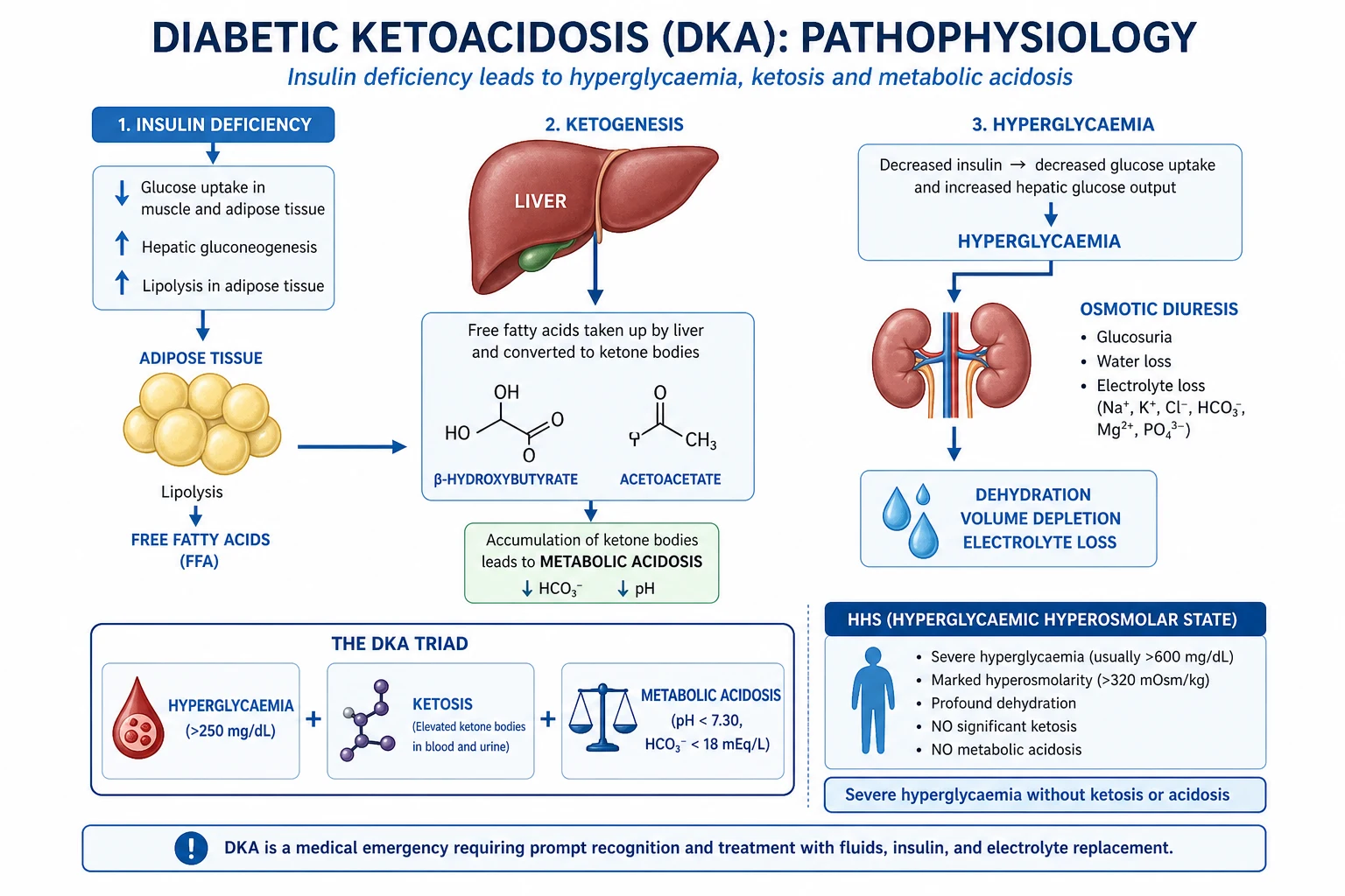
DKA and HHS are not distinct diseases but two points on a spectrum determined by how much insulin remains. In DKA, insulin deficiency is absolute (type 1 autoimmune destruction, or complete beta-cell failure in advanced type 2). Without insulin, three things happen simultaneously: (1) glucose cannot enter cells → hyperglycaemia; the hyperglycaemia overwhelms the renal glucose threshold (~10 mmol/L) → osmotic diuresis → polyuria, electrolyte loss (K+, Na+, phosphate), and dehydration. (2) The counter-regulatory hormones surge (glucagon, catecholamines, cortisol, growth hormone) in response to the intracellular glucose starvation and volume depletion → further hepatic gluconeogenesis, glycogenolysis, and lipolysis. (3) Unopposed lipolysis releases free fatty acids, which the liver converts (under glucagon drive) into ketone bodies — beta-hydroxybutyrate, acetoacetate, and acetone — via beta-oxidation. The ketones are strong organic acids that consume bicarbonate → high anion gap metabolic acidosis. The Kussmaul breathing (deep, rapid) is the compensatory respiratory alkalosis blowing off CO2 to buffer the acidosis.[1][6]
In HHS, a small amount of residual insulin (endogenous or from ongoing oral therapy) is sufficient to suppress hormone-sensitive lipase and therefore lipolysis and ketogenesis — but insufficient to suppress hepatic glucose output or promote peripheral glucose uptake. The result is extreme hyperglycaemia without ketosis: glucose climbs far higher than in DKA (>33 mmol/L vs >11), driving a more prolonged osmotic diuresis → more profound dehydration (6-9 L vs 3-6 L) and higher osmolality. The acidosis is absent or mild (lactic acidosis from hypoperfusion only). Because HHS develops more insidiously over days to weeks, the brain adapts to the chronic hyperosmolality, which is why mental status changes dominate and why rapid correction risks cerebral oedema.[5]
DKA vs HHS — pathophysiology and biochemistry
| Parameter | DKA | HHS |
|---|---|---|
| Insulin state | Absolute deficiency (no residual) | Relative deficiency (residual insulin present) |
| Predominant diabetes type | Type 1 (some type 2 — "ketosis-prone") | Type 2, elderly |
| Glucose | >11 mmol/L (typically 15-30) | >33 mmol/L (often >40) |
| Venous pH | <7.30 (typically 6.8-7.2) | >7.30 (normal or mildly low) |
| Bicarbonate | <15 mmol/L | >15 mmol/L |
| Beta-hydroxybutyrate | >3 mmol/L (often >6) | Minimal/absent (<1) |
| Anion gap | >12 (elevated — ketones) | Normal or mildly elevated (lactate) |
| Urine ketones | Strongly positive (+++) | Negative/trace |
| Osmolality | Variable (usually <320) | >320 mOsm/kg (often >340) |
| Dehydration | 3-6 L | 6-9 L (profound) |
| Mental status | Alert or mildly confused (Kussmaul, fruity breath) | Often obtunded / stupor / coma |
| Kussmaul breathing | Yes (metabolic acidosis compensation) | No |
| Onset | Hours to 1-2 days | Days to weeks (insidious) |
| Insulin dose | 0.1 U/kg/h (standard FRIII) | 0.05 U/kg/h (half — more sensitive, fluid-led) |
| Bicarbonate therapy | Only if pH <6.9 | Not indicated (not acidotic) |
| Mortality | ~5% (higher in elderly/comorbid) | 10-20% (10x DKA) |
The diagnostic criteria — DKA severity (Kitabchi/ADA)
| Parameter | Mild DKA | Moderate DKA | Severe DKA |
|---|---|---|---|
| Venous pH | 7.25-7.30 | 7.00-7.24 | <7.00 |
| Bicarbonate | 15-18 mmol/L | 10 to <15 | <10 |
| Mental status | Alert | Alert / drowsy | Stupor / coma |
| Anion gap | >10 | >12 | >12 |
| Glucose | >11 mmol/L | >11 mmol/L | >11 mmol/L |
| Beta-hydroxybutyrate | >3 mmol/L | >3 mmol/L | >3 mmol/L |
All three severity grades are managed identically (fluids + FRIII + K+); severity guides monitoring intensity (severe → ICU, hourly gas) and bicarbonate consideration (only when pH <6.9).[1]
Diagnostic approach — confirm and exclude mimics
Diagnosis requires simultaneous demonstration of the triad (DKA) or dyad (HHS) on a single blood set, plus immediate bedside tests to exclude the dangerous mimics. Send: venous blood gas (pH, bicarbonate, lactate, glucose), beta-hydroxybutyrate (the preferred ketone — far more sensitive than urine ketones, which miss the dominant DKA ketone), U&E (sodium, potassium, creatinine), calculated osmolality (2 × Na + glucose + urea), corrected sodium (Na + 1.6 × (glucose − 5.5)/5.5 — or 2.4 with the Na/glucose product method), full blood count, and ECG (silent MI is a common precipitant and hyper-/hypo-kalaemia show on ECG). Crucially, also send a venous/arterial blood gas potassium BEFORE giving any insulin.[1][2]
Initial blood panel and what each result dictates
| Test | Why | What it changes |
|---|---|---|
| Venous blood gas (pH, HCO3, glucose, lactate, K+) | Confirms DKA (pH <7.3, HCO3 <15) vs HHS (pH >7.3); gives the K+ BEFORE insulin | Determines bicarbonate need, insulin timing, K+ strategy |
| Beta-hydroxybutyrate | The pathognomonic ketone (>3 mmol/L); superior to urine ketones | Confirms ketosis; used to monitor DKA resolution (<0.6) |
| Sodium + corrected Na | Corrected Na guides fluid tonicity | Corrected Na low → 0.9% saline; normal/high → 0.45% saline |
| Potassium | Total body K+ depleted despite normal/high serum; insulin will drop it | K+ <3.3 → HOLD insulin, give K+ first. K+ 3.3-5.5 → KCl in fluids + insulin. K+ >5.5 → insulin first, no K+ yet |
| Creatinine/urea | AKI is common (dehydration); urea high = severe dehydration (cerebral oedema risk in children) | Guides fluid rate; urea elevated is a Glaser risk factor |
| Osmolality (calculated: 2Na + glucose + urea) | Confirms HHS (>320); monitors correction rate | Do not lower >3-4 mOsm/kg/h in HHS |
| ECG | Silent MI precipitant; K+ effects (peaked T, wide QRS) | Treat hyperkalaemia if ECG changes before insulin |
| Cultures, troponin, CXR, urinalysis | Find the precipitant (infection #1, MI) | Targeted antibiotics / cardiology |
The two calculations you must perform at the bedside
Corrected sodium (hyperglycaemia dilutes measured sodium — 1 mmol/L glucose rise above 5.5 pulls ~1.6 mmol/L Na into cells): Na_corrected = Na_measured + 1.6 × (glucose − 5.5) / 5.5. If corrected Na is low or normal → use 0.9% saline; if high → switch to 0.45% saline after the initial resuscitation. A rising corrected sodium during treatment is expected and reassuring; a falling one suggests over-rapid dilution. [1]
Calculated osmolality = 2 × Na + glucose + urea (all in mmol/L). Normal ~285-295; HHS >320 (often >340). The rate of osmolality fall should be monitored in HHS — too fast risks cerebral oedema.[1]
Precipitants — the same five causes, in the same order, for both
The precipitant determines recurrence risk and often mortality. Infection is the single commonest precipitant (~30-50% of cases). A hyperglycaemic emergency with no identifiable precipitant in a previously well-controlled patient should prompt a search for occult sepsis, silent MI, or a new diagnosis of diabetes.[1][6]
Precipitants of DKA/HHS — frequency and evaluation
| Precipitant | Approximate frequency | Evaluation |
|---|---|---|
| Infection (#1) | 30-50% | Blood/urine/sputum cultures, CXR, urinalysis, CRP/procalcitonin; empiric antibiotics if septic |
| Missed insulin / non-adherence | 20-30% | History; review pump/pen use; psychosocial assessment |
| New-onset diabetes | 10-25% (DKA); often young | No prior diagnosis; autoimmune markers (anti-GAD, anti-IA2); C-peptide low |
| Myocardial infarction | 5-10% (higher in elderly, often silent) | ECG, troponin — MI may be painless in diabetics |
| Acute pancreatitis | variable | Lipase/amylase, CT abdomen; hypertriglyceridaemia causes both pancreatitis AND DKA |
| Stroke / CNS event | <5% | CT brain if focal neurology or obtunded |
| Drugs | variable | Steroids, SGLT2 inhibitors (euglycaemic DKA), thiazides, sympathomimetics, atypical antipsychotics, cocaine |
| Endocrine | rare | Thyrotoxicosis, phaeochromocytoma, Cushing's, acromegaly |
| Pregnancy | DKA risk | Insulin resistance + vomiting; lower glucose threshold for DKA |
Clinical presentation — recognise the pattern
The clinical features map directly to the biochemistry: dehydration (polyuria, polydipsia, tachycardia, hypotension, dry mucous membranes, reduced skin turgor), acidosis (Kussmaul breathing — deep and rapid; nausea, vomiting, abdominal pain that can mimic an acute abdomen; fruity/pear-drop breath from acetone), and encephalopathy (lethargy, confusion, obtundation — more pronounced in HHS due to hyperosmolality). Abdominal pain is common in DKA (gastric stasis and ileus from acidosis) and must be distinguished from a surgical cause; it usually resolves as the acidosis corrects.[1]
Management — the four pillars (applied identically to DKA and HHS, dose-adjusted)
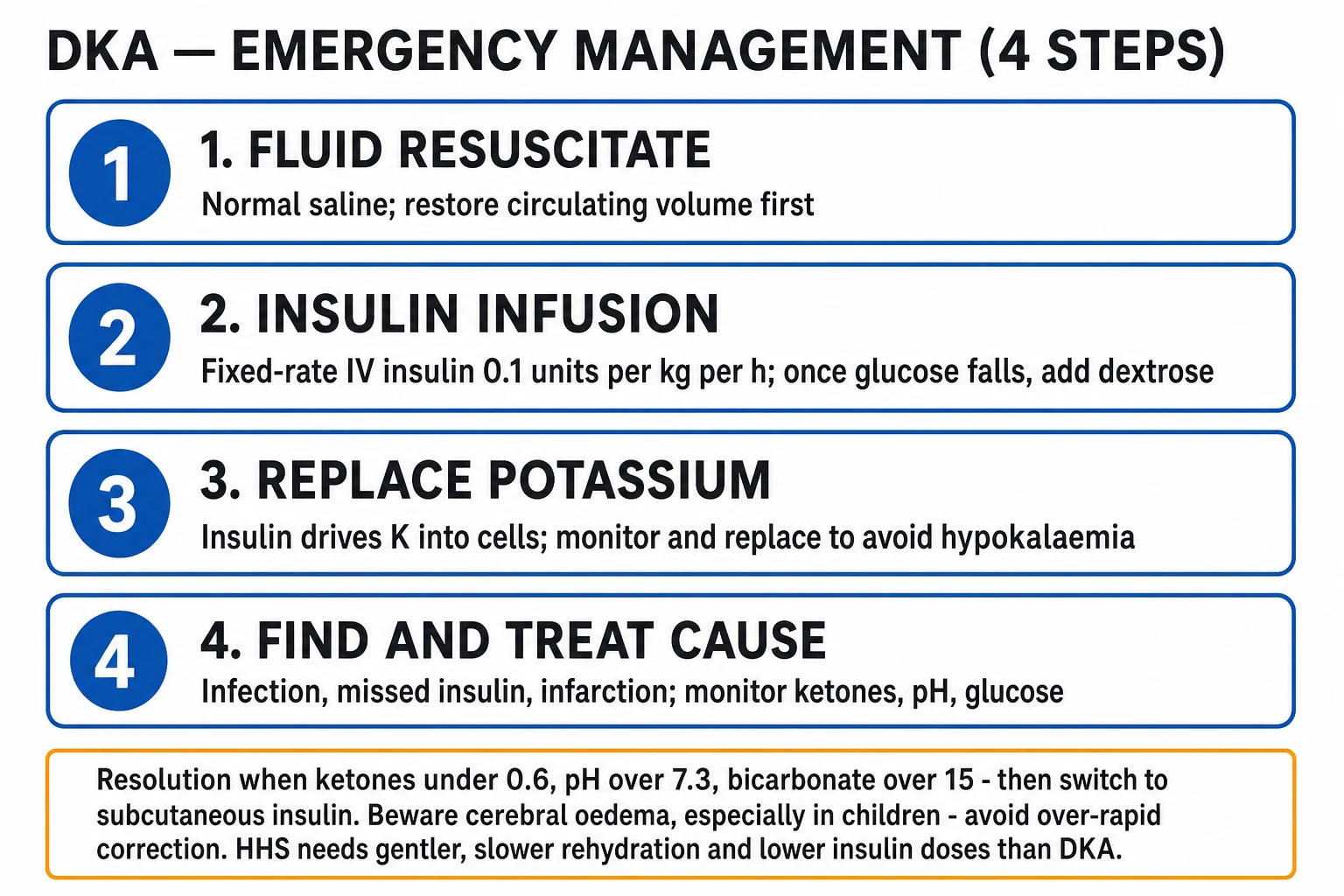
Management is a race to restore intravascular volume, suppress ketogenesis, and correct electrolytes — without precipitating cerebral oedema, hypokalaemia, or hypoglycaemia. The order of operations is critical and is the sequence in which every CICM/FFICM answer must flow: fluids FIRST → check potassium → insulin → bicarbonate only if pH <6.9 → monitor → treat precipitant → transition.[1][2]
DKA / HHS management protocol — the order of operations
-
FLUID RESUSCITATION FIRST — restore intravascular volume before insulin. The patient is volume-depleted from osmotic diuresis. 0.9% saline 15-20 mL/kg/h (~1 L) in the first hour (slower in the elderly / cardiac/renal — 250-500 mL/h). Then titrate: if corrected sodium is LOW or NORMAL → continue 0.9% saline 250-500 mL/h; if corrected sodium is HIGH or NORMAL-HIGH → switch to 0.45% saline 250-500 mL/h. Goal: restore perfusion, improve GFR, promote glucose excretion (glucose falls 2-5 mmol/L with fluids ALONE, before any insulin). When glucose falls to 14 mmol/L → ADD 5% dextrose (dextrose-containing fluid) alongside the saline; insulin continues. In HHS, fluids are the PRIMARY treatment and the glucose-lowering driver; the corrected sodium and osmolality, not glucose alone, guide fluid choice. Estimate total deficit: DKA 3-6 L, HHS 6-9 L; replace over 24 h (DKA) to 24-48 h (HHS).[1][2]
-
CHECK POTASSIUM BEFORE INSULIN — the cardinal rule. Total body potassium is DEPLETED (osmotic diuresis + vomiting) despite a normal or even high serum K+ (acidosis shifts K+ extracellular). Insulin will drive K+ back into cells, unmasking the deficit → life-threatening hypokalaemia. Stratify by serum K+: K+ <3.3 → HOLD insulin, give KCl 20-40 mmol/h FIRST, recheck in 1-2 h, start insulin once K+ ≥3.3. K+ 3.3-5.5 → add KCl 20-40 mmol per litre of IV fluid, start insulin. K+ >5.5 → start insulin WITHOUT potassium; check K+ every 2 h, add K+ once it falls below 5.5. Target 3.3-5.5 mmol/L throughout. Cardiac monitoring mandatory (K+ shifts → arrhythmias).[1][2]
-
FIXED-RATE IV INSULIN INFUSION (FRIII) — never bolus. Soluble (regular) insulin 0.1 U/kg/h (DKA) or 0.05 U/kg/h (HHS — half the dose, because HHS patients are more insulin-sensitive and fluids do most of the work). Do NOT give an IV insulin bolus — it causes unpredictable hypoglycaemia and osmolar shifts. Goal: glucose falls 3-4 mmol/L/h. If glucose not falling by ≥3 mmol/L in the first hour → check the line/pump and increase FRIII (DKA: recheck calculations; may increase to 0.15 U/kg/h). When glucose <14 mmol/L → add 5% dextrose (do NOT stop insulin) and continue FRIII at 0.05-0.1 U/kg/h; the dextrose prevents hypoglycaemia while insulin continues to clear ketones. Insulin is the ONLY therapy that stops ketogenesis — it must continue until DKA is resolved.[1][3]
-
BICARBONATE — ONLY if arterial pH <6.9. Give 100 mmol NaHCO3 in 400 mL sterile water with 20 mmol KCl over 2 hours, then recheck pH. Do NOT give bicarbonate if pH ≥7.0. Rationale against routine bicarbonate: it (a) does not improve outcome above pH 7.0; (b) worsens central nervous system acidosis (CO2 crosses the blood-brain barrier faster than bicarbonate, paradoxically lowering CSF pH); (c) delays ketone clearance; (d) shifts the oxyhaemoglobin curve left (worsens tissue O2 delivery); and (e) increases cerebral oedema risk in children (Glaser 2001). It is reserved for the severely acidotic patient (pH <6.9) with haemodynamic compromise where the acidosis itself is impairing catecholamine responsiveness. In HHS, bicarbonate is essentially never indicated (pH is normal).[1][4]
-
MONITOR intensively — glucose hourly, K+ and blood gas every 2-4 h. Glucose hourly (capillary, confirmed by lab if falling fast). Potassium and venous blood gas every 2-4 h (every 2 h initially in severe DKA). Sodium and corrected sodium each blood gas (rising corrected Na is expected). Ketones (beta-hydroxybutyrate) every 2-4 h — the best marker of DKA resolution. Hourly urine output (catheterise). Continuous cardiac monitoring (K+ shifts). Neurological observations hourly — cerebral oedema signs (headache, vomiting, drowsiness, bradycardia, hypertension, abnormal pupils) warrant immediate mannitol/hypertonic saline. Calculate corrected sodium and osmolality at each measurement.[2]
-
IDENTIFY AND TREAT THE PRECIPITANT — without this, DKA/HHS recurs. Send cultures (blood, urine, sputum), CXR, urinalysis, ECG and troponin, lipase. Start empiric broad-spectrum antibiotics early if any septic features (infection is #1). Treat MI per ACS protocol. Stop offending drugs (SGLT2 inhibitors, steroids if possible). Address non-adherence / insulin pump failure with diabetes educator and psychosocial input. In new-onset type 1, start diabetes education and autoimmune workup.[1][6]
-
PHOSPHATE AND MAGNESIUM — correct only if symptomatic/severe. Hypophosphataemia and hypomagnesaemia are universal (osmotic diuresis). Replace phosphate only if <0.3 mmol/L AND with respiratory failure/haemolysis/heart failure — routine replacement does not improve outcome and can cause hypocalcaemia. Replace magnesium if <0.5 mmol/L or refractory hypokalaemia.[1]
-
THROMBOPROPHYLAXIS — HHS and severe DKA are prothrombotic (dehydration, high viscosity, inflammation). Give prophylactic LMWH unless contraindicated.[5]
-
TRANSITION TO SUBCUTANEOUS INSULIN — only when DKA RESOLVED and eating. DKA resolution criteria: ketones <0.6 mmol/L, venous pH >7.3, bicarbonate >15 mmol/L, AND patient is haemodynamically stable and able to eat. Give the FIRST subcutaneous insulin dose (long-acting basal such as glargine/detemir 0.25 U/kg, or the patient's usual regimen) 1-2 hours BEFORE stopping the IV insulin infusion — the overlap prevents rebound hyperglycaemia and recurrent ketogenesis (subcutaneous insulin takes 1-2 h to act; IV insulin has a half-life of only minutes). Continue a rapid-acting insulin with meals. Never stop the IV infusion and then give SC insulin — the gap causes recurrence. In new-onset type 1, calculate the total daily dose (~0.5-0.8 U/kg/day) and split into basal/bolus. In type 2, may transition to oral agents if suitable once stable.[1][2]
Potassium strategy — what to do based on the initial serum K+
| Initial serum K+ | Total body K+ | Action | Insulin timing |
|---|---|---|---|
| <3.3 mmol/L (severe depletion) | Severely depleted | HOLD insulin. Give IV KCl 20-40 mmol/h (max 20 mmol/h peripheral, 40 mmol/h central). Recheck in 1-2 h. Cardiac monitor. | Start insulin only once K+ ≥3.3 |
| 3.3-5.5 mmol/L | Depleted (masked by acidosis) | Add KCl 20-40 mmol per litre of IV fluid. Start insulin. | Start insulin NOW (with K+ in fluids) |
| >5.5 mmol/L | Variable (often still depleted; extracellular shift from acidosis/AKI) | Start insulin WITHOUT added potassium. Check K+ every 2 h. | Start insulin NOW; add K+ once K+ <5.5 |
The key teaching point: a "normal" serum K+ in DKA is a depleted patient with an extracellular shift — insulin WILL unmask the deficit. Pre-empt by adding K+ to fluids even when K+ looks normal, and by holding insulin entirely when K+ <3.3.[1][2]
Cerebral oedema — the feared complication of DKA treatment
Cerebral oedema is the leading cause of DKA-related death in children and adolescents (incidence 0.5-1%, mortality 20-40%, ~15% of survivors have permanent neurological injury). It is rare in adults. It typically occurs 4-12 hours after starting treatment (rarely before), as the brain — adapted to chronic hyperosmolality — swells when extracellular osmolality falls too quickly. The Glaser 2001 case-control study (NEJM) defined the risk factors: (a) lower initial PaCO2 (a marker of acidosis severity — hypocapnia causes cerebral vasoconstriction; on correction, vasodilation → hyperaemia), (b) higher initial serum urea nitrogen (severity of dehydration), and (c) treatment with bicarbonate (independent risk factor, RR 4.2). Importantly, the rate of fluid or glucose correction was NOT independently associated in this study, but paediatric guidelines still recommend conservative correction.[4]
Prevention: in children, use 0.9% saline (not 0.45%) for initial resuscitation; do not lower glucose faster than ~3 mmol/L/h; avoid bicarbonate; do not give insulin until after the first hour of fluids. Recognition: headache, vomiting, altered mental status/lethargy, incontinence, bradycardia, hypertension (Cushing response), abnormal pupillary reflexes, seizures. Treatment is an emergency: mannitol 0.5-1 g/kg IV OR hypertonic 3% saline 5 mL/kg; reduce fluid rate; intubate and ventilate if GCS <8 (target normocapnia, avoid hyperventilation which worsens cerebral ischaemia); urgent CT to exclude other causes.[2][4]
HHS — what is different (and what is the same)
HHS shares the four-pillar management of DKA but with five critical adjustments: (1) FLUIDS dominate — HHS patients are more dehydrated (6-9 L) and glucose falls with fluids alone; resuscitate even more aggressively (cautiously in the elderly/cardiac). (2) HALF the insulin dose — 0.05 U/kg/h (HHS patients are more insulin-sensitive and fluid-led; standard 0.1 risks rapid osmolar shifts). (3) Slower glucose correction — do not lower glucose >3-4 mmol/L/h or osmolality >3 mOsm/kg/h; cerebral oedema and osmotic demyelination are real risks. (4) No bicarbonate (pH is normal). (5) Higher mortality (10-20%) — elderly, comorbidities; the precipitant (MI, sepsis, stroke) often drives outcome. HHS may take 24-72 h to resolve (vs 12-24 h for DKA); do not rush.[5]
Management: where DKA and HHS differ
| Management element | DKA | HHS |
|---|---|---|
| Insulin dose | FRIII 0.1 U/kg/h | FRIII 0.05 U/kg/h (half) |
| Primary glucose-lowering therapy | Insulin (also clears ketones) | Fluids (insulin is adjunctive) |
| Rate of glucose correction | 3-4 mmol/L/h | 3-4 mmol/L/h (do NOT exceed) |
| Dextrose added when glucose < | 14 mmol/L | 14-16 mmol/L |
| Bicarbonate | Only if pH <6.9 | Essentially never (not acidotic) |
| Total fluid deficit | 3-6 L (replace over 24 h) | 6-9 L (replace over 24-48 h) |
| Resolution marker | Ketones <0.6, pH >7.3, HCO3 >15 | Osmolality normalising, glucose stable, eating |
| Time to resolution | 12-24 h | 24-72 h |
| Thromboprophylaxis | Consider | Strongly recommended (very prothrombotic) |
| Mortality | ~5% | 10-20% |
Complications of treatment — anticipate and prevent
Complications of DKA/HHS treatment and how to prevent them
| Complication | Cause | Prevention / management |
|---|---|---|
| Hypokalaemia (#1 danger) | Insulin shifts K+ intracellularly; total body depletion unmasked | Check K+ before insulin; add K+ to fluids; hold insulin if K+ <3.3; monitor q2h |
| Hypoglycaemia | Insulin excess | Add 5% dextrose when glucose <14; halve FRIII if glucose falling fast; hourly glucose |
| Cerebral oedema (children) | Rapid osmolar shift | 0.9% saline initially (children); avoid bicarbonate; do not over-correct; mannitol/3% saline if occurs |
| Acute respiratory distress syndrome (ARDS) | Rare; from rapid fluid / capillary leak | Careful fluid balance; monitor oxygenation |
| Hypophosphataemia | Osmotic loss + refeeding-like | Replace only if severe (<0.3) + symptomatic |
| Thromboembolism | Dehydration, hyper viscosity | LMWH prophylaxis (especially HHS) |
| Recurrent ketoacidosis | Stopping IV insulin without SC overlap | Give SC insulin 1-2 h before stopping IV |
Exam practice — SAQs
SAQ — Severe DKA in an adolescent with cerebral oedema risk
10 minutes · 10 marks
A 16-year-old girl (55 kg) with a 2-week history of weight loss, polyuria and polydipsia is brought to the emergency department drowsy and breathing deeply. She is clinically severely dehydrated. GCS 13 (E3V4M6), HR 128, BP 92/54, RR 34 with Kussmaul breathing, fruity breath. Capillary glucose 33 mmol/L. Venous blood gas: pH 6.92, bicarbonate 7 mmol/L, pCO2 14 mmHg, potassium 3.1 mmol/L, sodium 128 mmol/L, urea 14 mmol/L, beta-hydroxybutyrate 9.2 mmol/L. New-onset type 1 diabetes is diagnosed.
SAQ — Hyperosmolar hyperglycaemic state in an elderly patient with acute kidney injury
10 minutes · 10 marks
A 78-year-old man with type 2 diabetes, hypertension and ischaemic heart disease is found unresponsive at home by his family. GCS 8 (E2V2M4), temp 38.2°C, HR 108 (atrial fibrillation), BP 96/52, RR 22. Capillary glucose 54 mmol/L. Venous blood gas: pH 7.32, bicarbonate 19 mmol/L, sodium 152 mmol/L, potassium 5.8 mmol/L, urea 28 mmol/L, creatinine 320 µmol/L (baseline 95), lactate 2.4. Calculated osmolality 360 mOsm/kg. Urinalysis: glucose ++, ketones trace. CXR shows right lower lobe consolidation.
Clinical pearls
Red flags
Prognosis
DKA and HHS outcomes and prognostic factors
| Factor | Outcome | Notes |
|---|---|---|
| DKA overall mortality | ~5% (adults); <1% in young otherwise-well | Higher in elderly, comorbid, severe acidosis (pH <7.0), renal failure; cause of death is usually the precipitant (sepsis/MI) or cerebral oedema (children) |
| HHS mortality | 10-20% | ~10× DKA — elderly, comorbidities, profound dehydration, obtunded presentation; the precipitant (MI, sepsis, stroke) often determines outcome |
| Cerebral oedema | 0.5-1% incidence (children); 20-40% mortality; ~15% permanent neuro injury | Leading cause of DKA death in children; risk factors (Glaser): low PaCO2, high urea, bicarbonate treatment |
| Severe acidosis (pH <6.9) | Higher mortality | May warrant bicarbonate; indicates severe DKA and need for ICU |
| Recurrent DKA | Up to 25% recurrence in type 1 | Usually non-adherence / psychosocial; insulin pump failure; diabetes education is key |
| Hypokalaemia from treatment | Major iatrogenic cause of death | Preventable — check K+ before insulin |
| Age and comorbidity | Strongest prognostic modifiers | Elderly, CKD, cardiac failure → worse outcome in both |
| Time to resolution | DKA 12-24 h; HHS 24-72 h | HHS resolves more slowly — do not rush correction |
Key trials and evidence
Kitabchi 2009 — ADA consensus: Hyperglycemic crises in adult patients with diabetes (PMID 19564476)
Source
Diabetes Care 2009;32(7):1335-1343 — the seminal ADA consensus statement, still the global management backbone
What it established
Diagnostic criteria (DKA: glucose >13.9 mmol/L, pH <7.3, HCO3 <18, ketones positive, anion gap >10; HHS: glucose >33.3, osmolality >320, pH >7.3, HCO3 >15, stupor), severity grading (mild/moderate/severe), and the four-pillar management protocol: fluids → K+ → insulin 0.1 U/kg/h → treat precipitant
Key contribution
Standardised the potassium-first rule (hold insulin if K+ <3.3), bicarbonate only if pH <6.9, and the glucose <14 → add dextrose (continue insulin) algorithm. Established HHS at half the insulin dose.
Clinical bottom line
The exam-defining reference — every DKA/HHS guideline since (JBDS, UK, ANZ) is built on this framework
Glaser 2001 — Cerebral oedema risk factors in paediatric DKA (NEJM) (PMID 11172153)
Source
New England Journal of Medicine 2001;344(4):264-269 — multicentre case-control study, 61 children with cerebral oedema vs 181 random + 174 matched controls
What it found
Independent risk factors for cerebral oedema: (1) lower initial PaCO2 (hypocapnia, RR 3.4 per 7.8 mmHg decrease), (2) higher initial serum urea nitrogen (dehydration severity, RR 1.7 per 9 mg/dL), and (3) treatment with bicarbonate (RR 4.2, p=0.008)
Key contribution
Provided the evidence base for AVOIDING bicarbonate in DKA and for conservative fluid/osmolar correction in children; rate of fluid or glucose correction was NOT independently associated but paediatric guidelines remain conservative
Clinical bottom line
The reason bicarbonate is restricted to pH <6.9, and why children get 0.9% saline (not 0.45%) with slow glucose correction
Dhatariya 2022 — JBDS-IP updated DKA management guideline (PMID 35224769)
Source
Diabetic Medicine 2022;39(6):e14788 — the Joint British Diabetes Societies for Inpatient Care updated adult DKA guideline (most recent indexed update; March 2023 web version is the current standard)
What it established
Refined the FRIII (fixed-rate intravenous insulin infusion) approach: when glucose <14 mmol/L add 10% glucose (not stop insulin); consider REDUCING FRIII from 0.1 to 0.05 U/kg/h if glucose or ketones fall too fast or hypoglycaemia/hypokalaemia develop; resolution defined by ketones <0.6 mmol/L
Key contribution
Emphasised beta-hydroxybutyrate (not pH/HCO3) as the primary resolution marker; added explicit guidance on SGLT2-inhibitor euglycaemic DKA, young adults 16-18, AKI, and pregnancy
Clinical bottom line
The most widely used adult DKA protocol in the UK/ANZ — the practical bedside algorithm that operationalises Kitabchi
Pasquel & Umpierrez 2014 — HHS historic review (Diabetes Care) (PMID 25342831)
Source
Diabetes Care 2014;37(11):3124-3131 — the definitive review of HHS presentation, diagnosis, and treatment
What it established
HHS diagnostic criteria (glucose >33.3 mmol/L, effective osmolality >320 mOsm/kg, no significant ketoacidosis), the 10-20% mortality (10× DKA), and that NO prospective randomised trials exist for HHS treatment — management is extrapolated from DKA
Key contribution
Codified the fluid-led approach (fluids are primary, insulin adjunctive at half-dose), the slower correction targets, and thromboprophylaxis; highlighted that cerebral oedema risk from over-rapid correction is real in HHS
Clinical bottom line
The HHS counterpart to Kitabchi — read it for every HHS question
References
- [1]Kitabchi AE, Umpierrez GE, Miles JM, Fisher JN. Hyperglycemic crises in adult patients with diabetes Diabetes Care, 2009.PMID 19564476
- [2]Dhatariya KK, Joint British Diabetes Societies (JBDS) for Inpatient Care. The management of diabetic ketoacidosis in adults-An updated guideline from the Joint British Diabetes Society for Inpatient Care Diabet Med, 2022.PMID 35224769
- [3]Umpierrez GE, Cuervo R, Karabell A, Latif K, Freire AX, Kitabchi AE. Treatment of diabetic ketoacidosis with subcutaneous insulin aspart Diabetes Care, 2004.PMID 15277410
- [4]Glaser N, Barnett P, McCaslin I, et al.; Pediatric Emergency Medicine Collaborative Research Committee of the American Academy of Pediatrics. Risk factors for cerebral edema in children with diabetic ketoacidosis. The Pediatric Emergency Medicine Collaborative Research Committee of the American Academy of Pediatrics N Engl J Med, 2001.PMID 11172153
- [5]Pasquel FJ, Umpierrez GE. Hyperosmolar hyperglycemic state: a historic review of the clinical presentation, diagnosis, and treatment Diabetes Care, 2014.PMID 25342831
- [6]Dhatariya K, Mustafa O, Stathi D. In: Feingold KR, et al., editors. Endotext. Hyperglycemic Crises 2000.PMID 25905280