ICU · Endocrine & metabolic emergencies
Diabetic Ketoacidosis (DKA) & Hyperosmolar Hyperglycaemic State (HHS)
Also known as Diabetic ketoacidosis · DKA · Hyperosmolar hyperglycaemic state · HHS · Hyperosmolar non-ketotic coma · HONK · Fixed-rate intravenous insulin infusion · FRIII · Euglycaemic DKA · Hyperglycaemic crisis · Ketonaemia · Beta-hydroxybutyrate
DKA and HHS are the two hyperglycaemic emergencies. DKA (typically type 1 diabetes) is the triad of absolute insulin deficiency, ketogenesis and a high-anion-gap metabolic acidosis — glucose above 11 mmol/L, venous pH below 7.3, bicarbonate below 15, ketones. HHS (typically type 2, elderly) is severe hyperglycaemia (above 30-33 mmol/L), osmolality above 320, profound dehydration with minimal ketosis and a near-normal pH. Management is fluid first (1 L isotonic saline in the first hour), a fixed-rate insulin infusion (0.1 unit/kg/h for DKA; lower for HHS), potassium replacement before insulin if serum K is below 3.3, and identification plus treatment of the precipitant. The DKA resolution target is ketone clearance, NOT glucose normalisation.
On this page & tools
Your progress
Saved locally on this device.
Target exams
Red flags

Definition and classification
DKA and HHS are not separate diseases but points on a spectrum of decompensated diabetes determined by the degree of insulin deficiency and counter-regulatory hormone excess.[3][6]
- Diabetic ketoacidosis (DKA) — near-absolute insulin deficiency. Predominantly type 1 diabetes but increasingly seen in ketosis-prone type 2 and in type 2 under stress. Defined by the triad of hyperglycaemia, ketonaemia, and a high-anion-gap metabolic acidosis.
- Hyperosmolar hyperglycaemic state (HHS) — a relative, not absolute, insulin deficiency sufficient to suppress lipolysis and ketogenesis but inadequate to prevent hyperglycaemia. Predominantly type 2 diabetes in older patients; evolves over days to weeks, producing extreme dehydration and hyperosmolality with little or no acidosis.
- Overlap states exist, and euglycaemic DKA (DKA with a near-normal glucose) is increasingly recognised with SGLT2 inhibitors, pregnancy and starvation.[11]
DKA
Absolute insulin deficiency
- Typically type 1 diabetes; younger patient; onset hours to days
- Glucose above 11 mmol/L (often much higher)
- Ketonaemia (beta-hydroxybutyrate above 3 mmol/L) and ketonuria
- Venous pH below 7.30, bicarbonate below 15 mmol/L — high anion gap
- Total-body potassium depleted despite normal/high serum
- Mortality 1-5% (cerebral oedema in the young)
HHS
Relative insulin deficiency
- Typically type 2 diabetes; older, comorbid patient; onset over days to weeks
- Marked hyperglycaemia (above 30-33 mmol/L)
- Effective osmolality above 320 mOsm/kg
- Venous pH above 7.30, bicarbonate above 15 — minimal ketosis
- Profound dehydration (average 6-9 L deficit); altered conscious state
- Mortality 10-20% (thromboembolism, comorbidity, sepsis)
Diagnostic criteria
Biochemical diagnostic criteria — DKA severity by pH and bicarbonate (click each)
pH below 7.00 / HCO3 below 10
Severe DKA. pH below 7.00, bicarbonate below 10. ICU admission. Consider IV bicarbonate if pH below 7.0 (controversial). Highest risk of cerebral oedema in the young.
The biochemical diagnosis of DKA requires ALL of:[1][4]
- Hyperglycaemia — blood glucose above 11 mmol/L (or documented diabetes). Euglycaemic DKA can present with glucose below 11.[11]
- Acidosis — venous pH below 7.30 and/or bicarbonate below 15 mmol/L with a high anion gap.
- Ketonaemia — beta-hydroxybutyrate above 3 mmol/L (preferred), or moderate ketonuria (++ or more).
- Marked hyperglycaemia — above 30-33 mmol/L (JBDS uses above 30).
- Hyperosmolality — effective osmolality above 320 mOsm/kg (calculated as 2 x [Na + K] + glucose).
- Absence of significant ketosis/acidosis — pH above 7.30, bicarbonate above 15.
- Significant dehydration — with altered conscious level, often obtunded. [1]
Pathophysiology
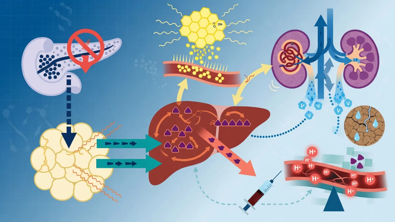
Both states arise from insulin deficiency combined with an excess of counter-regulatory hormones (glucagon, catecholamines, cortisol, growth hormone) — most often triggered by infection, missed insulin, or a new metabolic insult.[3][4]
In DKA the insulin deficit is profound enough to permit unopposed lipolysis: free fatty acids released from adipose tissue are taken up by the liver and oxidised to ketone bodies (beta-hydroxybutyrate, acetoacetate, acetone). In HHS a residual, low level of insulin suppresses lipolysis but is inadequate to control gluconeogenesis and peripheral glucose uptake, so ketogenesis is minimal.[6]
Four downstream consequences follow: [1]
- Hyperglycaemia from impaired glucose uptake plus gluconeogenesis and glycogenolysis. Glycosuria exceeds the renal threshold, producing an osmotic diuresis with free water, sodium, potassium, magnesium, phosphate and chloride losses.[5]
- Ketosis (DKA). Beta-hydroxybutyrate and acetoacetate dissociate and consume bicarbonate, generating a high-anion-gap metabolic acidosis. The falling pH drives Kussmaul respiration, which lowers PaCO2 as compensation.[4]
- Electrolyte deficit. Despite a normal or even high serum potassium on presentation, total-body potassium is depleted (typically 3-5 mmol/kg deficit) through the osmotic diuresis and vomiting. Insulin therapy and correction of the acidosis drive potassium intracellularly and unmask this deficit — the basis for the potassium protocol.[1]
- Hyperosmolality and dehydration (especially HHS). Free-water loss exceeds solute loss; patients may be 6-9 L deficit. Altered conscious state correlates with osmolality, not glucose.[8]
Clinical presentation
- DKA: polyuria, polydipsia, weight loss, nausea, vomiting and abdominal pain (which can mimic an acute abdomen). Kussmaul respiration (deep, sighing, rapid) is the compensatory response to metabolic acidosis. Breath may smell of acetone ("pear-drop"/nail-polish remover). Dehydration signs (dry mucous membranes, reduced skin turgor, tachycardia, hypotension) and a varying degree of altered conscious state. Features of the precipitant dominate the history.[5][7]
- HHS: an insidious onset over days to weeks of profound dehydration, profound thirst, weakness, and progressive neurological depression ranging from confusion to obtundation and coma (correlating with osmolality, not glucose). Seizures may occur. The patient is typically older with type 2 diabetes and significant comorbidity.[8][12]
Precipitants
Finding and treating the precipitant determines survival as much as the metabolic correction.[1]
DKA triggers
Commonest first
- Infection (most common) — pneumonia, UTI, sepsis
- Missed or inadequate insulin (the commonest preventable cause)
- New-onset type 1 diabetes
- Acute myocardial infarction / ischaemia
- Acute pancreatitis
- Drugs — steroids, SGLT2 inhibitors, atypical antipsychotics
- Pregnancy, substance misuse, surgery
HHS triggers
Often multifactorial
- Infection (the commonest — pneumonia, UTI, sepsis)
- Undertreated or newly diagnosed type 2 diabetes
- Acute illness — MI, stroke, pancreatitis, GI bleed
- Poor fluid intake in the elderly / care-home patient
- Drugs — steroids, diuretics, beta-blockers, SGLT2 inhibitors
- Dialysis/renal failure, burns
Investigations
A focused, parallel panel is required at presentation:[1][5]
- Capillary and laboratory glucose (capillary meters may under-read at very high glucose or in shock — send a laboratory sample).
- Venous blood gas — pH, bicarbonate, pCO2 (a venous gas is sufficient; arterial is unnecessary unless hypoxia or respiratory compromise is suspected). Beta-hydroxybutyrate.
- Urea and electrolytes — sodium, potassium, creatinine. Correct the sodium for hyperglycaemia: corrected Na = measured Na + 1.6 x ([glucose mmol/L - 5.5] / 5.5). Anion gap = Na - (Cl + HCO3).
- Ketones — capillary/laboratory beta-hydroxybutyrate plus urine ketones.
- Full blood count, liver function, magnesium, phosphate, calcium, CRP.
- Cultures (blood, urine, sputum) and a chest radiograph; ECG (silent MI is a common precipitant, and the ECG also reflects potassium).
- Troponin, amylase/lipase if pancreatitis is suspected. Urinalysis and a pregnancy test in women of child-bearing age. [1]
Management
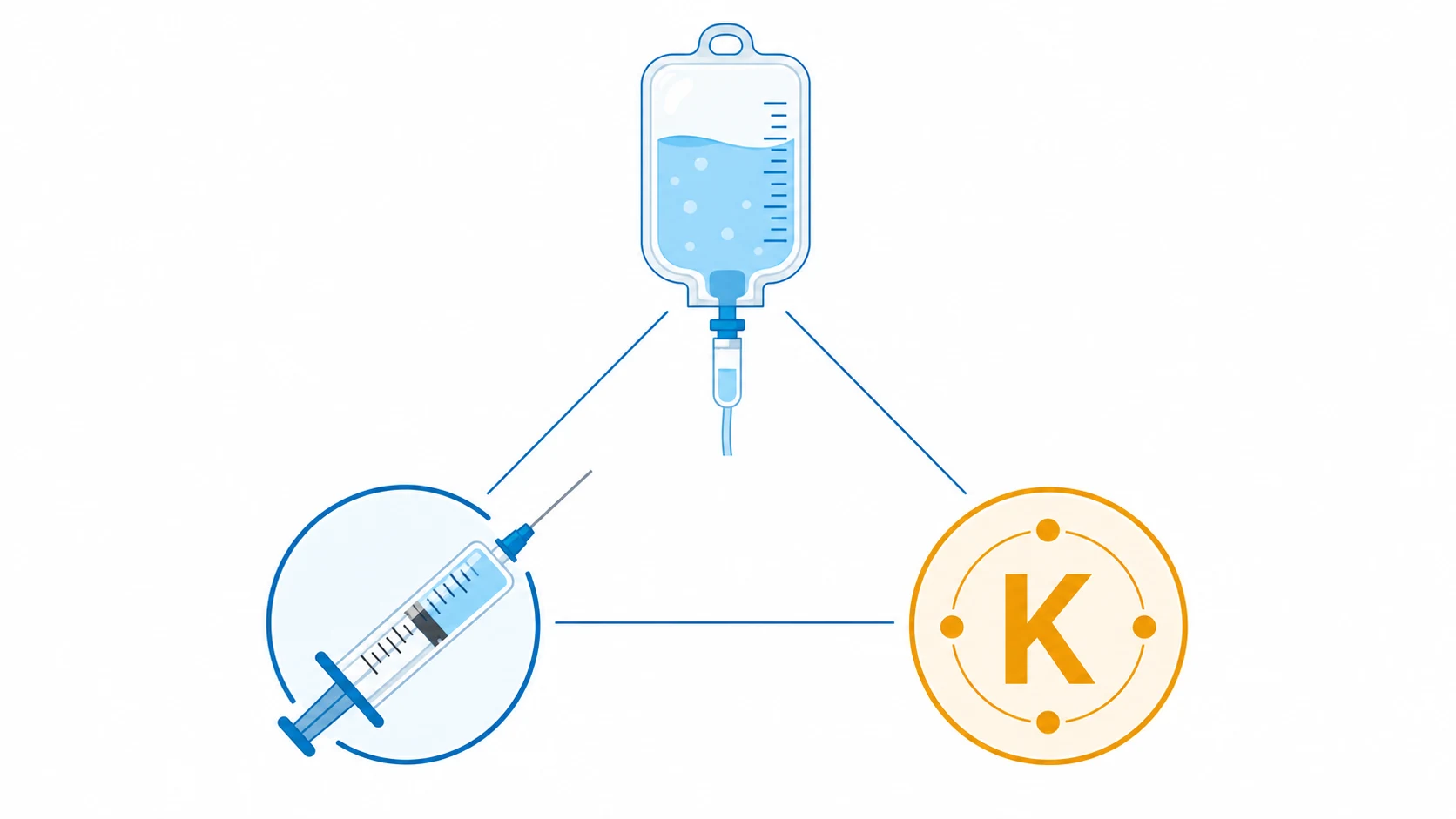
DKA and HHS share the same therapeutic spine, with important differences in emphasis. The four pillars are fluid, insulin, potassium, and treat the trigger.[1][3]
1. Fluid resuscitation
Restoration of circulating volume is the first intervention and improves perfusion, lowers the counter-regulatory surge, and partly lowers glucose (through improved insulin sensitivity and renal glucose clearance) even before insulin is given.[1][10]
Fluid protocol (JBDS 2022 — DKA)
1 L of 0.9% sodium chloride over the first hour
Give stat to the hypovolaemic, shocked patient. Use balanced crystalloid (Hartmann/Plasma-Lyte) as an acceptable alternative; 0.9% saline is the JBDS default. Reassess cardiovascular status and consider a second litre over the next hour if still hypovolaemic.
Then 1 L 0.9% saline over 2 hours, then over 4 hours, then over 8 hours
The stepped schedule replaces the estimated 3-6 L deficit over 12-24 hours. Titrate to clinical markers (BP, perfusion, urine output, corrected sodium). Reduce the rate in heart failure or the elderly; invasive monitoring if shocked or cardiac/renal impairment.
Switch to 10% glucose at 125 mL/h once glucose is below 14 mmol/L
In DKA, continue insulin AND add glucose — the insulin is still needed to clear ketones. Use 10% (not 5%) glucose as it allows continued insulin at a meaningful rate. Do NOT stop insulin to allow the glucose to rise.
Avoid hypotonic solutions in the resuscitation phase
Hypotonic fluids (0.45% saline, dextrose-only) risk cerebral oedema, especially in children and young adults. Use them only later if the corrected sodium is rising significantly, and cautiously.
The choice of crystalloid remains debated. A 2024 review highlights that 0.9% saline carries a risk of hyperchloraemic metabolic acidosis (which confounds bicarbonate monitoring) and that balanced crystalloids (Hartmann/Plasma-Lyte) may resolve ketosis slightly faster, but the JBDS still endorses 0.9% saline and no single trial has shown a mortality difference.[10]
2. Fixed-rate intravenous insulin infusion (FRIII)
A weight-based fixed-rate infusion, NOT a sliding scale, is the standard for DKA. The aim is to suppress ketogenesis, not merely to lower glucose.[1][4]
Insulin protocol (JBDS 2022 — DKA)
FRIII at 0.1 unit/kg/h (soluble insulin, e.g. Actrapid)
Weight-based fixed rate (typically 5-8 unit/h for an average adult). NO routine bolus. Make up 50 unit in 50 mL 0.9% saline. Aim for a fall in ketones of at least 0.5 mmol/L/h and/or a rise in bicarbonate of at least 3 mmol/L/h and/or a fall in glucose of at least 3 mmol/L/h.
Check response at 1 hour
If ketones are NOT falling by at least 0.5 mmol/L/h (or glucose not falling by at least 3 mmol/L/h), give a bolus of 0.1 unit/kg and continue FRIII at the same rate. Re-check at the next hour. Review adherence of the infusion and the IV access.
Continue FRIII until ketone clearance criteria met
Resolution = beta-hydroxybutyrate below 0.6 mmol/L AND venous pH above 7.3 AND bicarbonate above 15 mmol/L. The patient must also be medically well and able to eat. Glucose alone is NOT a resolution target.
Start glucose when glucose below 14 mmol/L
Add 10% glucose at 125 mL/h alongside the FRIII so insulin can continue to suppress ketogenesis without hypoglycaemia. The rate of FRIII may need to reduce if glucose keeps falling despite glucose infusion.
3. Potassium
Total-body potassium is depleted but the serum may be normal or high. Insulin and correction of the acidosis shift potassium intracellularly; hypokalaemia is the most dangerous preventable complication and can be fatal.[1][5]
K below 3.3
Hold insulin — replete first
- DEFER the FRIII until potassium is above 3.3 mmol/L
- Give IV potassium (e.g. 40 mmol in 1 L of fluid) and re-check
- Insulin will worsen the hypokalaemia — the commonest cause of arrhythmic death in DKA
K 3.3-5.5
Add potassium to the bag
- Add 40 mmol/L potassium to the maintenance fluid (or per local protocol)
- Start the FRIII concurrently
- Re-measure potassium every 2 hours for the first 6 hours
K above 5.5
No potassium yet
- Give insulin + fluid WITHOUT added potassium
- Recheck potassium within 2 hours; add potassium once below 5.5
- Ensure urine output is adequate before any potassium
4. Treat the trigger + general measures
A systematic search for and treatment of the precipitant is decisive.[1][3]
- Infection: cultures before early broad antibiotics if sepsis suspected.
- Myocardial infarction: ECG and troponin; treat per ACS protocol — silent MI is common in diabetic patients.
- Missed insulin: identify and plan the subcutaneous regimen.
- Venous thromboembolism prophylaxis: DKA and especially HHS are pro-thrombotic — prophylactic LMWH is recommended in all HHS and in DKA unless contraindicated.[2]
- Monitoring: glucose hourly, ketones and VBG 1-2 hourly, potassium and U&E 2 hourly, urine output (catheterise the obtunded), GCS, continuous cardiac monitoring. A dedicated DKA/HHS paper or electronic protocol chart reduces error.
- ICU admission: severe DKA (pH below 7.0, HCO3 below 10, obtunded, haemodynamically unstable, on insulin for HHS), all HHS needing insulin, or significant comorbidity.
HHS-specific modifications
HHS is managed with more fluid, less insulin, and slower osmolality correction.[2][12]
- Fluid is the primary therapy. Replace the 6-9 L deficit over 24 hours using 0.9% saline initially, switching to 0.45% saline if the corrected sodium is normal or rising. Lower the osmolality gradually (no more than 3 mOsm/kg per hour) to reduce cerebral oedema risk.
- Insulin at a lower rate (0.05 unit/kg/h, or 0.1 unit/kg/h only if ketones are present). Insulin in HHS can precipitate a fall in osmolality and shift water — start it only after fluid resuscitation has begun and potassium is known. Glucose often falls with fluid alone.
- Thromboprophylaxis is mandatory. HHS carries a high arterial and venous thromboembolism rate; give prophylactic LMWH unless contraindicated.[2]
- Do not aim for normoglycaemia — keep glucose 10-15 mmol/L in the acute phase to avoid osmotic shifts.
Phosphate, magnesium and bicarbonate
- Phosphate: routine replacement is NOT recommended — it does not improve outcome and may cause hypocalcaemia. Replace only if phosphate is below 0.3 mmol/L with respiratory depression, cardiac dysfunction or haemolysis.[5]
- Magnesium: replete if low (contributes to refractory hypokalaemia).
- Bicarbonate: routine use is NOT recommended — it may delay ketone clearance, lower CSF pH paradoxically, and increase cerebral oedema risk. Reserved for pH below 7.0-7.1, and even then given cautiously alongside standard therapy until the pH rises above 7.0.[1][13]
DKA / HHS — key numbers
Transition to subcutaneous insulin
Once resolution criteria are met AND the patient is well and eating, transition to the long-term subcutaneous regimen.[1][5]
Transition from IV to subcutaneous insulin
Calculate the long-term regimen BEFORE stopping the infusion
For known type 1 diabetes, restart the usual basal-bolus regimen. For new-onset diabetes, start basal insulin 0.25 unit/kg and a rapid-acting analogue with meals (total ~0.5-0.7 unit/kg/day). Never stop the IV insulin before the subcutaneous dose is given.
Give the subcutaneous insulin, then OVERLAP the IV infusion for 30-60 min
Give rapid-acting SC insulin with the first meal and/or the basal dose, and CONTINUE the FRIII for 30-60 minutes afterwards. The IV insulin has a half-life of only a few minutes — stopping it without overlap causes rapid rebound ketosis.
Stop the IV infusion; monitor glucose before meals and at bedtime
Once stopped, monitor capillary glucose pre-meals and overnight. Discontinue the glucose infusion at the meal. Aim for glucose 6-10 mmol/L.
Diabetes team review and education
Involve the diabetes inpatient team for sick-day rules, ketone monitoring, pump management, and prevention of recurrence. The commonest cause of recurrence is premature cessation of insulin.
Evidence and guidelines
JBDS DKA 2022
Diabet Med 2022
UK consensus guideline — adults with DKA
Key finding
FRIII 0.1 unit/kg/h; resolution defined by beta-hydroxybutyrate below 0.6, pH above 7.3, bicarbonate above 15. 0.9% saline as default; no routine bicarbonate.
Practice change
Ketone-based resolution replaces glucose-based resolution
JBDS HHS 2023
Diabet Med 2023
UK consensus guideline — adults with HHS
Key finding
Fluid-first; low-dose insulin (0.05 U/kg/h); lower osmolality gradually; mandatory VTE prophylaxis.
Practice change
Standardised HHS protocol; emphasis on thromboprophylaxis
2024 Consensus Report
Cleve Clin J Med 2025
US multi-society consensus on hyperglycaemic crises
Key finding
Refined diagnostic thresholds and integrated DKA/HHS on a spectrum; reaffirmed FRIII and ketone-based monitoring.
Practice change
Harmonised transatlantic diagnostic criteria
Fluid choice
Curr Opin Clin Nutr 2024
Review of crystalloid choice in DKA
Key finding
Balanced crystalloids may resolve ketosis slightly faster than 0.9% saline; no mortality difference; hyperchloraemic acidosis with saline confounds bicarbonate monitoring.
Practice change
Balanced crystalloid a reasonable alternative; saline remains acceptable
Complications
- Hypokalaemia — the most dangerous preventable complication; managed by the potassium protocol above. Hyperkalaemia (over-treatment, AKI) also occurs.[1]
- Cerebral oedema — predominantly in children and young adults (and occasionally adults); presents with headache, altered behaviour, bradycardia, hypertension and falling GCS, often 4-12 hours into therapy. Risk factors include severe acidosis, high urea, and rapid osmolality/tonicity correction with hypotonic fluids. Avoid hypotonic fluids; use 0.9% saline; correct gradually. Treat with mannitol or hypertonic saline.[4][13]
- Acute respiratory distress syndrome (ARDS) — rare but described, related to fluid resuscitation and capillary leak; monitor oxygenation.[13]
- Hypoglycaemia — from over-aggressive insulin or failure to start glucose at glucose below 14 mmol/L; the reason glucose is added rather than insulin stopped.
- Hyperchloraemic metabolic acidosis — from large-volume 0.9% saline; a normal anion gap with rising chloride; confounds bicarbonate-based monitoring (use ketones).[10]
- Hypokalaemia-induced arrhythmia — the lethal early event; preventable.
- Thromboembolism (DVT/PE/stroke) — especially HHS; prevented with LMWH.[2]
- Acute kidney injury — from dehydration; usually pre-renal and resolves with fluid.
- Recurrent DKA — from premature cessation of insulin at transition.
Prognosis
Outcomes
Prognosis depends on the precipitant and the patient's comorbidity more than on the biochemistry. In adults with prompt treatment DKA mortality is low (1-5%); HHS mortality is substantially higher (10-20%) driven by age, comorbidity, severe dehydration and thromboembolism. The single most important prognostic step is identifying and treating the trigger.[2][3][8]
Exam practice
SAQ — Severe DKA management
10 minutes · 10 marks
A 24-year-old man with type 1 diabetes is brought to the emergency department drowsy and breathing deeply and rapidly. He has had a flu-like illness for three days and has omitted his insulin for the last 36 hours. Examination: HR 124, BP 96/58, RR 30, deep sighing respirations, dry mucous membranes, GCS 14. Glucose 34 mmol/L. Venous gas: pH 7.02, pCO2 2.1 kPa, HCO3 6 mmol/L, base excess -24. Sodium 132, potassium 3.1, chloride 100, urea 11, creatinine 140. Beta-hydroxybutyrate 8.2 mmol/L. Urinalysis: ketones +++.
Clinical pearls
Red flags
References
- [1]Dhatariya KK, Joint British Diabetes Societies for Inpatient Care Group. The management of diabetic ketoacidosis in adults-An updated guideline from the Joint British Diabetes Society for Inpatient Care Diabet Med, 2022.PMID 35224769
- [2]Mustafa OG, Haq M, Dashora U, et al. Management of Hyperosmolar Hyperglycaemic State (HHS) in Adults: An updated guideline from the Joint British Diabetes Societies (JBDS) for Inpatient Care Group Diabet Med, 2023.PMID 36370077
- [3]Fayfman M, Pasquel FJ, Umpierrez GE. Management of Hyperglycemic Crises: Diabetic Ketoacidosis and Hyperglycemic Hyperosmolar State Med Clin North Am, 2017.PMID 28372715
- [4]Dhatariya KK, Glaser NS, Codner E, Umpierrez GE. Diabetic ketoacidosis Nat Rev Dis Primers, 2020.PMID 32409703
- [5]Karslioglu French E, Donihi AC, Korytkowski MT. Diabetic ketoacidosis and hyperosmolar hyperglycemic syndrome: review of acute decompensated diabetes in adult patients BMJ, 2019.PMID 31142480
- [6]Umpierrez G, Korytkowski M. Diabetic emergencies - ketoacidosis, hyperglycaemic hyperosmolar state and hypoglycaemia Nat Rev Endocrinol, 2016.PMID 26893262
- [7]Eledrisi MS, Elzouki AN. Management of Diabetic Ketoacidosis in Adults: A Narrative Review Saudi J Med Med Sci, 2020.PMID 32952507
- [8]Pasquel FJ, Umpierrez GE. Hyperosmolar hyperglycemic state: a historic review of the clinical presentation, diagnosis, and treatment Diabetes Care, 2014.PMID 25342831
- [9]Rodriguez Alvarez P, San Martin VT, Morey-Vargas OL. Hyperglycemic crises in adults: A look at the 2024 consensus report Cleve Clin J Med, 2025.PMID 40032308
- [10]Ramanan M, Delaney A, Venkatesh B. Fluid therapy in diabetic ketoacidosis Curr Opin Clin Nutr Metab Care, 2024.PMID 38126191
- [11]Long B, Lentz S, Koyfman A, Gottlieb M. Euglycemic diabetic ketoacidosis: Etiologies, evaluation, and management Am J Emerg Med, 2021.PMID 33626481
- [12]Long B, Willis GC, Lentz S, Koyfman A, Lin A. Diagnosis and Management of the Critically Ill Adult Patient with Hyperglycemic Hyperosmolar State J Emerg Med, 2021.PMID 34256953
- [13]Dunn BK, Coore H, Bongu N, et al. Treatment Challenges and Controversies in the Management of Critically Ill Diabetic Ketoacidosis (DKA) Patients in Intensive Care Units Cureus, 2024.PMID 39360087