ICU · Endocrine
Hyperosmolar hyperglycaemic state (HHS)
Also known as Hyperosmolar hyperglycaemic state (HHS) · Hyperosmolar non-ketotic coma (HONK) · Hyperosmolar hyperglycaemic non-ketotic syndrome · Hyperosmolar non-ketotic state (HNS)
HHS is a severe hyperglycaemic emergency of type 2 diabetes characterised by: severe hyperglycaemia (33 mmol/L), high serum osmolality (320 mOsm/kg), profound dehydration (6-9 L), and ABSENCE of significant ketosis (minimal/no ketoacidosis — pH 7.3, bicarbonate 15). The defining pathophysiology is RELATIVE insulin deficiency — enough residual insulin to suppress lipolysis and prevent ketoacidosis, but not enough to prevent hyperglycaemia. Presents in elderly type 2 diabetics, often precipitated by infection (1 single precipitant), MI, stroke, or new medication (steroids, thiazides, atypical antipsychotics). Mortality 10-20% (higher than DKA — elderly, comorbidities). Management: aggressive fluid resuscitation FIRST (0.9% saline 15-20 mL/kg in the first hour), potassium replacement BEFORE insulin, low-dose insulin (0.05 U/kg/h — half the DKA dose, started ONLY after fluids), and anticoagulation for high thrombotic risk. Identify and treat the precipitant.
On this page & tools
Your progress
Saved locally on this device.
Target exams
Red flags

Diagnostic criteria and the biochemical signature
HHS diagnostic thresholds vs DKA vs mixed picture
| Parameter | HHS | DKA | Mixed / overlap |
|---|---|---|---|
| Glucose | >33 mmol/L (often 40-60) | >13.9 mmol/L (typically 15-30) | High; can overlap |
| Arterial/venous pH | >7.30 | <7.30 (mild 7.25-7.30, mod 7.00-7.24, severe <7.00) | May be <7.30 |
| Bicarbonate | >15 mmol/L | <15 mmol/L | Variable |
| Ketones (β-OHB) | Minimal/absent (<3 mmol/L) | Positive (>3 mmol/L) | Moderate |
| Anion gap | <12 (normal; may be mildly ↑ from lactate) | >12 (high-gap ketoacidosis) | Elevated |
| Osmolality | >320 mOsm/kg (often 340-360) | Variable, usually <320 | High |
| Mental status | Altered/stupor/coma (≈30% comatose) | Usually alert / mildly confused | Clouded |
| Free-water deficit | 6-9 L (~9-12% body weight) | 3-6 L (~6% body weight) | Large |
| Typical patient | Elderly type 2 DM | Type 1 DM (or type 2 ketosis-prone) | Type 2 |
| Insulin requirement | LOW (0.05 U/kg/h) | STANDARD (0.1 U/kg/h) | Standard |
| Onset | Days to weeks (insidious) | Hours to <24 h (rapid) | Variable |
| Cerebral oedema risk | Lower than paediatric DKA but real in adults | Classic in children, rare in adults | Present |
The HHS numbers to memorise
HHS vs DKA comparison
HHS
Type 2 diabetes, elderly
- Glucose: >33 mmol/L (typically higher than DKA)
- pH: >7.30 (minimal acidosis)
- Bicarbonate: >15 mmol/L (no significant ketoacidosis)
- Ketones: minimal/absent (urine/serum)
- Osmolality: >320 mOsm/kg (typically much higher — profound dehydration)
- Anion gap: normal or mildly elevated (lactic acidosis from hypoperfusion)
- Dehydration: 6-9 L (more severe than DKA)
- Mental status: often altered/confused/comatose (from hyperosmolality)
- Insulin dose: LOW (0.05 U/kg/h — half DKA dose)
DKA
Type 1 (and some type 2) diabetes
- Glucose: >13.9 mmol/L (typically lower than HHS)
- pH: <7.30 (metabolic acidosis)
- Bicarbonate: <15 mmol/L (ketoacidosis)
- Ketones: positive (urine/serum — beta-hydroxybutyrate)
- Osmolality: variable (less dehydration than HHS)
- Anion gap: elevated (ketones — beta-hydroxybutyrate, acetoacetate)
- Dehydration: 3-6 L
- Mental status: usually alert or mildly confused
- Insulin dose: STANDARD (0.1 U/kg/h)
Pathophysiology — the cascade
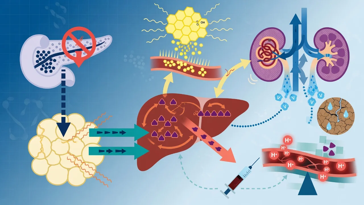
HHS pathophysiology — stepwise from insulin resistance to coma
Relative insulin deficiency + counter-regulatory excess
Insulin secretion falls (or resistance rises) but enough insulin remains to suppress hormone-sensitive lipase → NO ketogenesis. Counter-regulatory hormones (glucagon, catecholamines, cortisol, GH) surge in response to a precipitant (infection, MI, stroke, steroids). Net effect: hepatic gluconeogenesis/glycogenolysis unchecked, peripheral glucose uptake impaired.
Uncontrolled hyperglycaemia
Glucose climbs over hours-to-days (HHS is insidious — symptoms develop over days-weeks, unlike the hours of DKA). Glucose typically exceeds 33 mmol/L and may reach 60-80 mmol/L. The slower onset allows the brain time to accumulate "idiogenic osmoles", which paradoxically INCREASES the cerebral-oedema risk if glucose is later dropped too fast.
Osmotic diuresis
Glucose filtered load exceeds the renal Tm → glycosuria. Glucose acts as an obligate osmole in the tubule, dragging water AND electrolytes (Na, K, Mg, PO4) into the urine. The result is the HALLMARK free-water deficit of 6-9 L plus total-body potassium depletion of ~5-10 mmol/kg (despite often normal/high serum K from acidosis/hypovolaemia/insulin deficiency).
Hyperosmolality and cellular dehydration
Rising extracellular glucose and sodium raise serum osmolality above 320 mOsm/kg. Water shifts out of cells (including neurons). Consciousness deteriorates in proportion to osmolality: mild confusion → obtundation → coma. Osmolality >340 is usually associated with significant neurological depression.
Hypovolaemia and end-organ hypoperfusion
Intravascular volume depletion → tachycardia, hypotension, prerenal AKI, lactate rise (a "normal anion gap" lactic acidosis is common and distinguishes HHS from the high-gap DKA). Severe volume depletion → shock. The hyperosmolar, dehydrated state is profoundly PROTHROMBOTIC (hyperviscosity, high factor VIII/vWF, low-flow stasis). Prerenal AKI can mask the true glucose (renal clearance falls) — glucose may be even higher than expected.
Compounded by the precipitant
The precipitating illness (infection, MI, stroke) is often itself the major driver of mortality. HHS is as much a marker of serious systemic illness in an elderly type-2 patient as it is a metabolic emergency. Treating HHS without finding and treating the trigger guarantees recurrence and worsens outcome.
Precipitating causes — find the trigger
Precipitants of HHS — search for these in every patient
| Precipitant | Mechanism / note | Frequency |
|---|---|---|
| Infection (pneumonia, UTI, sepsis) | Raises counter-regulatory hormones; #1 single precipitant | Most commonly identified single trigger (~40-50%)[5] |
| New/occult type 2 diabetes | First presentation — HHS may be the debut illness | ~30%[5] |
| Acute cardiovascular event | Silent MI (autonomic neuropathy in elderly diabetics), stroke, ACS | Major mortality contributor |
| Medication-induced | Glucocorticoids (raise gluconeogenesis + insulin resistance), thiazides, beta-blockers (mask counter-regulation), atypical antipsychotics (olanzapine, clozapine), phenytoin, sympathomimetics | Common; review ALL drugs |
| Non-adherence / inadequate insulin | Missed oral hypoglycaemics or insulin | Common in known diabetics |
| Substance misuse | Cocaine, alcohol | Often overlooked |
| Acute illness (non-infective) | Pancreatitis, GI bleed, trauma, surgery, dialysis, TPN | ~50% in pooled series[5] |
| Total parenteral nutrition / high-dextrose | Iatrogenic glucose load | ICU/ward iatrogenic |
Management
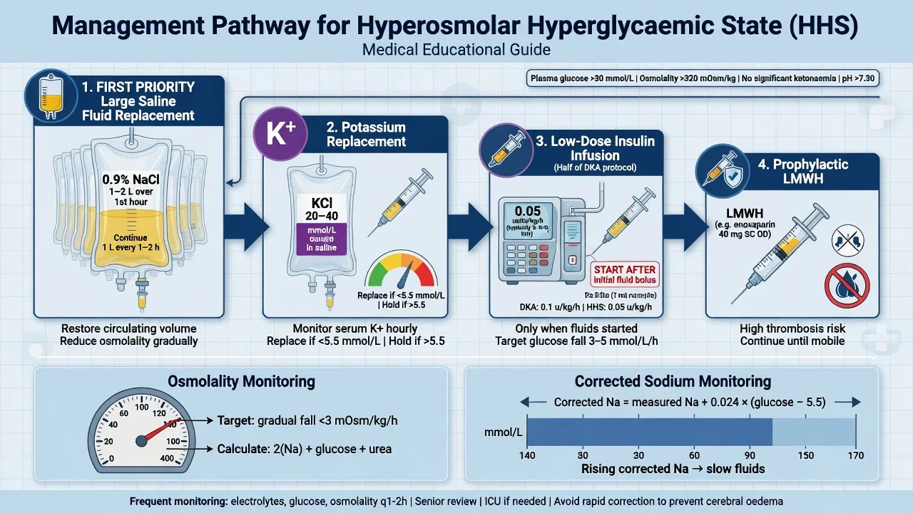
HHS management protocol
Aggressive fluid resuscitation (PRIMARY treatment)
0.9% saline: 15-20 mL/kg in first hour (1-1.5 L). Then titrate based on: corrected serum sodium, hydration status, urine output, cardiac status. Goal: restore intravascular volume → improve tissue perfusion → improve renal function → promote glucose excretion. Glucose falls 2-5 mmol/L with fluids ALONE before any insulin given. Switch to 0.45% saline if corrected Na is normal/high after initial resuscitation. Add 5% dextrose when glucose reaches 14-16 mmol/L.
Potassium replacement BEFORE insulin
Check K+ immediately. If K+ <3.3: hold insulin, give K+ 40 mmol/h. If K+ 3.3-5.2: give K+ 20-30 mmol/L of IV fluid, start insulin. If K+ >5.2: start insulin, check K+ every 2h, hold K+ replacement. Insulin drives K+ into cells → hypokalaemia risk. HHS patients have TOTAL BODY POTASSIUM DEPLETION (despite normal/initially high serum K+ from acidosis/dehydration).
Low-dose insulin (HALF DKA dose) — ONLY after fluids
Insulin infusion: 0.05 U/kg/h (NOT 0.1 U/kg/h as in DKA — HHS patients are more sensitive to insulin). Goal: lower glucose 3-4 mmol/L/h. If glucose not falling: increase insulin to 0.1 U/kg/h. When glucose reaches 14-16 mmol/L: add 5% dextrose infusion + reduce insulin rate to maintain glucose 10-14 mmol/L. Do NOT lower glucose too fast — risk of cerebral oedema (osmotic shift).
Monitor closely
Glucose: hourly. Electrolytes (Na, K, Mg, PO4): every 2-4h. Creatinine, venous pH: every 4-6h. Urine output: hourly (catheterise). Cardiac monitoring (K+ shifts → arrhythmias). Neurological status (cerebral oedema risk — headache, vomiting, altered mental status). Calculate corrected sodium and osmolality at each measurement.
Identify and treat precipitant
Search for: (1) Infection (#1 — chest, urine, blood cultures, antibiotics). (2) MI (ECG, troponin — silent MI common in elderly diabetics). (3) Stroke (CT brain if focal neurology). (4) New medication (steroids, thiazides, beta-blockers, atypical antipsychotics). (5) Non-adherence to diabetes medication. (6) Substance abuse. Without treating the precipitant: HHS will recur.
Anticoagulate (thromboprophylaxis)
HHS is a profoundly prothrombotic state — hyperviscosity, dehydration, high FVIII/vWF, immobility, and (often) central lines/catheters. Give prophylactic LMWH unless contraindicated (e.g. active bleeding). Arterial events (MI, stroke) and VTE are leading complications.
Transition to subcutaneous insulin
When stable (glucose 10-14 mmol/L, normal osmolality, eating): transition to subcutaneous insulin. Give first subcutaneous dose 1-2 hours before stopping IV infusion (overlap to prevent rebound hyperglycaemia). Long-acting insulin (glargine/detemir) + rapid-acting insulin with meals. Type 2 diabetes: may transition to oral hypoglycaemics if suitable. Adjust based on HbA1c, body weight, renal function.
Fluid resuscitation — the cornerstone, done correctly
Potassium management — replace before, monitor during
Insulin therapy — half the DKA dose, started only after fluids
Anticoagulation — the prothrombotic emergency
Monitoring protocol
Hourly and serial monitoring in HHS — what to measure and how often
Hourly
Capillary glucose (titrate insulin/fluids); neurological observation (GCS — rising GCS = osmolality improving; falling GCS or new headache/vomiting = cerebral oedema until proven otherwise); urine output via urinary catheter (target ≥0.5 mL/kg/h); continuous cardiac monitoring (K+ shifts → arrhythmias); vital signs and fluid balance.
Every 2 hours
Venous blood gas (pH, HCO3, lactate, calculated anion gap); serum potassium (drive insulin and K+ replacement off this); capillary β-hydroxybutyrate if any ketotic overlap to confirm resolution. Recalculate corrected sodium and osmolality.
Every 4-6 hours
Full U&E (Na, K, Cl, bicarbonate, urea, creatinine); magnesium and phosphate (replace if low — phosphate <0.5 may impair respiratory muscle function and tissue oxygen delivery); venous pH. Calculate effective osmolality trend — target fall ≤3-5 mOsm/kg/h.
Daily / ongoing
FBC, CRP, coagulation; review cultures and imaging; troponin if any cardiac suspicion; CXR if oxygen requirement or suspected aspiration. Nutritional assessment and a plan for subcutaneous insulin conversion once eating. Pressure-area care and DVT prophylaxis (LMWH) — immobile dehydrated elderly patients are at very high VTE risk.
Targets to achieve before stopping the IV insulin
Glucose stable at 10-14 mmol/L on a dextrose-containing regimen; osmolality normalising (≤315, falling); acidosis (if any) resolved; potassium in range on a reducing replacement rate; the precipitant identified and being treated; the patient eating and ready for subcutaneous conversion.
Complications
Complications of HHS — anticipate and prevent
| Complication | Mechanism | Prevention / management |
|---|---|---|
| Cerebral oedema | Rapid osmolar shift; idiogenic osmole accumulation. Rare in adults vs paediatric DKA, but fatal when it occurs | Limit glucose fall to 3-4 mmol/L/h; do NOT give insulin before fluids; watch for headache, vomiting, falling GCS. Treat: mannitol / hypertonic saline |
| Hypokalaemia | Intracellular K+ shift once insulin started; ongoing kaliuresis | Replace K+ before insulin (hold if <3.3); recheck q2h; replace Mg²⁺ concurrently |
| Acute kidney injury | Profound hypovolaemia + osmotic diuresis; nephrotoxic precipitant | Aggressive early fluids; avoid NSAIDs; monitor creatinine; usually prerenal and reversible |
| Acute coronary syndrome / stroke | Hyperviscosity, hypercoagulability, dehydration; silent MI in elderly | ECG + troponin on admission; LMWH; maintain perfusion; low threshold to image brain |
| Venous thromboembolism | Prothrombotic state (high FVIII/vWF), immobility, central lines | Prophylactic LMWH for all; mechanical prophylaxis; investigate new swelling/dyspnoea |
| Pulmonary oedema / ARDS | Over-aggressive fluid resuscitation in elderly with cardiac disease | Titrate fluids to corrected Na, urine output, perfusion; consider CVP/POCUS; use balanced approach |
| Hypoglycaemia | Insulin overdose / failure to add dextrose at the switch point | Hourly glucose; add 5% dextrose at glucose 14-16; halve insulin rate |
| Hypophosphataemia | Cellular uptake with insulin; respiratory muscle weakness, tissue hypoxia | Replace if PO4 <0.5 mmol/L (esp. if ventilated) |
| Aspiration pneumonia | Reduced GCS + vomiting | Airway protection; early NG tube if obtunded; head-up positioning |
Outcomes and mortality
HHS mortality and outcome — what the pooled data show
Prompt recognition, treated in ICU
When HHS is recognised early and managed with fluid-first, low-dose insulin, electrolyte replacement and anticoagulation, outcome approaches that of uncomplicated DKA. The median hospital stay in pooled series is 7.5 days; ICU admission rate ~40%.
Transition and ongoing care
From IV insulin to subcutaneous — the safe conversion
Confirm stability before switching
Glucose stable at 10-14 mmol/L; osmolality ≤315 and falling; acidosis resolved; K+ in range; the precipitant identified and treated; the patient alert and eating. Premature conversion risks rebound hyperglycaemia.
Calculate the total daily subcutaneous dose
A pragmatic starting point is **0.5-0.8 U/kg/day** total insulin, split into ~50% basal (glargine/detemir/degludec) and ~50% rapid-acting (lispro/aspart) with meals. Alternatively, base the new regimen on the average IV insulin rate over the preceding 6-12 h. Err conservative — over-dosing causes hypoglycaemia, the commonest inpatient adverse event.
Overlap — NEVER stop IV before SC is active
Give the first subcutaneous (basal) dose **1-2 hours before** stopping the IV infusion. Rapid-acting meal-time insulin is given with the first meal after stopping IV. Stopping IV insulin without overlap causes rapid rebound hyperglycaemia within 1-2 h (IV insulin has a half-life of minutes).
Long-term plan — insulin vs oral agents
HHS often first presents in previously undiagnosed type 2 DM (~30%). After stabilisation, characterise the diabetes (C-peptide, antibodies if phenotype ambiguous), check HbA1c (reflects the preceding weeks), and arrange endocrinology follow-up. Many type-2 patients can later transition to oral agents (metformin first-line if renal function permits), but most HHS survivors need insulin for at least the short-to-medium term.
Education and prevention
Sick-day rules (do NOT stop insulin during illness; check glucose/CBG more often; maintain hydration); glucose-monitoring education; medication review (stop offending drugs — steroids if possible, thiazides); podiatry and diabetes-nurse input; structured diabetes education. Recurrence is common if the precipitant and the underlying poor control are not addressed.
Evidence and landmark data
French et al. 2026 — HHS systematic review and meta-analysis (BMJ Open Diabetes Res Care, PMID 42373194)
Source
BMJ Open Diabetes Res Care 2026;14(3):e005765 — the largest pooled dataset of HHS cases to date (PRISMA-compliant, 27 studies, 63,935 cases), aggregating HHS case series and cohorts across all regions
Design
Systematic review and meta-analysis of studies reporting HHS cases from database inception to Feb 2025. Outcomes: demographics, biochemistry, precipitants, complications, mortality, stratified by region.
What it established
Median age 63.8 (IQR 56.1-72.0). Precipitants: non-infective illness 49.5%, infection 44.0%, new diabetes diagnosis 31.7%. ICU admission 40.8% (highest in North America 70%). Median hospital stay 7.5 days (Asia 17.5 days, Americas 4 days). Overall mortality 21.1% (Africa 40%, Asia 18.2%, North America 4.8%). Complications: AKI 7.6%, pulmonary oedema 4.8%, acute coronary syndrome 3.9%. Biochemistry is similar globally; outcomes vary markedly by region and resources.
Clinical bottom line
The definitive contemporary epidemiology. Confirms HHS mortality is high (≈21%) and regionally unequal; complications are dominated by dehydration/vascular events (AKI, pulmonary oedema, ACS) rather than the glucose itself. Underlines that precipitant identification, fluid-first care, and thromboprophylaxis — not faster glucose correction — are the mortality-reduction levers.
Umpierrez et al. 2024 — ADA Consensus Report: Hyperglycemic Crises in Adults (Diabetes Care, PMID 39052901)
Source
Diabetes Care 2024;47(8):1257-1275 — the current American Diabetes Association / international consensus report on DKA and HHS, authored by Umpierrez, Dhatariya, Fadini and colleagues
What it established
Modern unified definitions and management of hyperglycaemic emergencies. Endorses the conceptual framework of a SPECTRUM (DKA ↔ HHS) rather than two discrete diseases — mixed/overlap presentations are common. Recommends fluid-first resuscitation, potassium correction before insulin, low-dose fixed-rate insulin (0.05 U/kg/h for HHS, 0.1 for DKA), and glucose/osmolality-fall ceilings to prevent cerebral oedema. Emphasises precipitant identification, thromboprophylaxis, and structured transition to subcutaneous insulin.
Clinical bottom line
The current authoritative guidance for HHS. Know the numbers: glucose >33 mmol/L, osmolality >320, pH >7.30, HCO3 >15, insulin 0.05 U/kg/h, glucose fall cap 3-4 mmol/L/h.
Dhatariya & Vellanki 2017 — DKA/HHS UK vs USA (Curr Diab Rep, PMID 28364357)
Source
Curr Diab Rep 2017;17(5):37 — a widely cited review reconciling the UK (JBDS-IP) and US (ADA) protocols for DKA and HHS
What it established
Highlights the practical differences between the UK JBDS and US ADA approaches (e.g. fluid tonicity selection, insulin infusion rates, the role of bicarbonate — none recommended — and the speed of correction). Reinforces that HHS is managed with lower insulin doses and slower correction than DKA, and that the two syndromes can overlap.
Clinical bottom line
A concise comparative reference for the CICM/FFICM exam — cite it when asked to contrast UK vs US fluid and insulin strategies in HHS.
Wei et al. 2022 — HHS and VTE: a nationwide cohort (J Pers Med, PMID 35207789)
Source
J Pers Med 2022;12(2):317 — nationwide population-based cohort study quantifying the association between HHS and venous thromboembolism in diabetic patients
What it established
Diabetic patients hospitalised with HHS have a significantly higher incidence of VTE than matched diabetic controls without HHS, validating the clinical impression that HHS is a prothrombotic emergency. Supports routine thromboprophylaxis in HHS.
Clinical bottom line
The evidence behind 'give prophylactic LMWH to every HHS patient' — a frequent exam point and a practical bedside rule.
Clinical pearls
[1]Mnemonic
HHSH-H-S-S-S — the five pillars of HHS management
Exam practice
SAQ — Confusion and hyperglycaemia in an elderly patient
12 minutes · 12 marks
A 76-year-old man with type 2 diabetes, hypertension and chronic kidney disease is brought to the ED drowsy and confused. His family report progressive polyuria, thirst and malaise over the past week and reduced oral intake for two days. He takes metformin, empagliflozin, perindopril and furosemide. On examination he is cachectic, dry mucous membranes, HR 112, BP 96/58, RR 22, T 37.9°C. GCS 13 (E3 V4 M6). CBG reads 'HI'. Venous gas: pH 7.34, glucose 48 mmol/L, Na 128, K 4.8, Cl 94, HCO3 22, urea 18, creatinine 210, lactate 2.1. Urine ketones trace.
SAQ — Differentiating HHS from DKA and managing the overlap
10 minutes · 10 marks
A 58-year-old woman with known type 2 diabetes presents unwell. Venous gas: pH 7.28, glucose 39 mmol/L, HCO3 14, anion gap 16, β-hydroxybutyrate 2.8 mmol/L, calculated osmolality 338, Na corrected 144, K 3.1. She is drowsy (GCS 14) but conversing.
Expanded clinical pearls
Red flags
[1]References
- [1]Kitabchi AE, Umpierrez GE, Miles JM, Fisher JN. Hyperglycemic crises in adult patients with diabetes Diabetes Care, 2009.PMID 19564476
- [2]Dhatariya KK, Vellanki P. Treatment of Diabetic Ketoacidosis (DKA)/Hyperglycemic Hyperosmolar State (HHS): Novel Advances in the Management of Hyperglycemic Crises (UK Versus USA) Curr Diab Rep, 2017.PMID 28364357
- [3]Stoner GD. Hyperosmolar Hyperglycemic State Am Fam Physician, 2017.PMID 29431405
- [4]Umpierrez GE, Davis GM, ElSayed NA, Fadini GP, Galindo RJ, Hirsch IB, Klonoff DC, McCoy RG, Misra S, Gabbay RA, Bannuru RR, Dhatariya KK. Hyperglycemic Crises in Adults With Diabetes: A Consensus Report Diabetes Care, 2024.PMID 39052901
- [5]French J, Bomphrey L, Manta A, Scandrett K, Malhotra K, Kempegowda P. Hyperosmolar hyperglycaemic state: a systematic review and meta-analysis BMJ Open Diabetes Res Care, 2026.PMID 42373194
- [6]Kitabchi AE, Umpierrez GE, Murphy MB, Kreisberg RA. Hyperglycemic crises in adult patients with diabetes: a consensus statement from the American Diabetes Association Diabetes Care, 2006.PMID 17130218
- [7]Wei WT, Lin SM, Hsu JY. Association between Hyperosmolar Hyperglycemic State and Venous Thromboembolism in Diabetes Patients: A Nationwide Analysis in Taiwan J Pers Med, 2022.PMID 35207789