ICU · environmental
Acute Decompression Illness — Comprehensive ICU Management (Diving Emergencies)
Also known as Decompression illness (DCI) · Decompression sickness (DCS) · The bends · Arterial gas embolism (AGE) · Pulmonary barotrauma · The chokes (pulmonary DCS) · The staggers (vestibular DCS) · Skin bends · Recompression therapy · Hyperbaric oxygen (HBO) · US Navy Treatment Table 6 · Dysbarism
Acute decompression illness (DCI) — the umbrella term for the two dysbaric injuries of ascent: decompression sickness (DCS, 'the bends') and arterial gas embolism (AGE). Both arise from bubbles formed when ambient pressure falls. DCS is driven by inert-gas (nitrogen) supersaturation during/after ascent — in-situ bubbles in tissues and blood — classically Type 1 (musculoskeletal joint pain 'the bends', skin itching/mottling 'skin bends', lymphatic) and Type 2 (neurological — spinal cord is the MOST SERIOUS form with paralysis, paraesthesia, bladder/bowel dysfunction; pulmonary 'the chokes' — cough, dyspnoea, substernal chest pain; vestibular 'the staggers' — vertigo, nystagmus, nausea). AGE is pulmonary barotrauma of ascent — breath-holding or too-rapid ascent over-expands alveoli → alveolar rupture → gas enters pulmonary veins → left heart → systemic arteries (brain most often) → stroke-like presentation during or within minutes of surfacing. Risk is increased by depth, bottom time, repetitive dives, rapid ascent, cold, exertion, dehydration, and a patent foramen ovale (paradoxical embolisation). Onset: AGE within minutes; DCS usually within minutes-to-hours, 98% within 24 h. Management is identical for both: 100% oxygen immediately (denitrogenates tissues and accelerates bubble resolution), IV isotonic glucose-free fluids (correct immersion diuresis and dehydration, avoid hypovolaemia), and DEFINITIVE recompression therapy — hyperbaric oxygen, most commonly US Navy Treatment Table 6 (100% oxygen at 2.8 ATA), which shrinks bubbles by Boyle's law (to ~one-third of surface volume), oxygenates ischaemic tissue, reduces oedema, and denitrogenates. Retrieve to the nearest hyperbaric chamber urgently — call the Divers Alert Network (DAN) 24-hour hotline. Delay to recompression worsens outcome — recompress as soon as feasible.
On this page & tools
Your progress
Saved locally on this device.
Target exams
Red flags
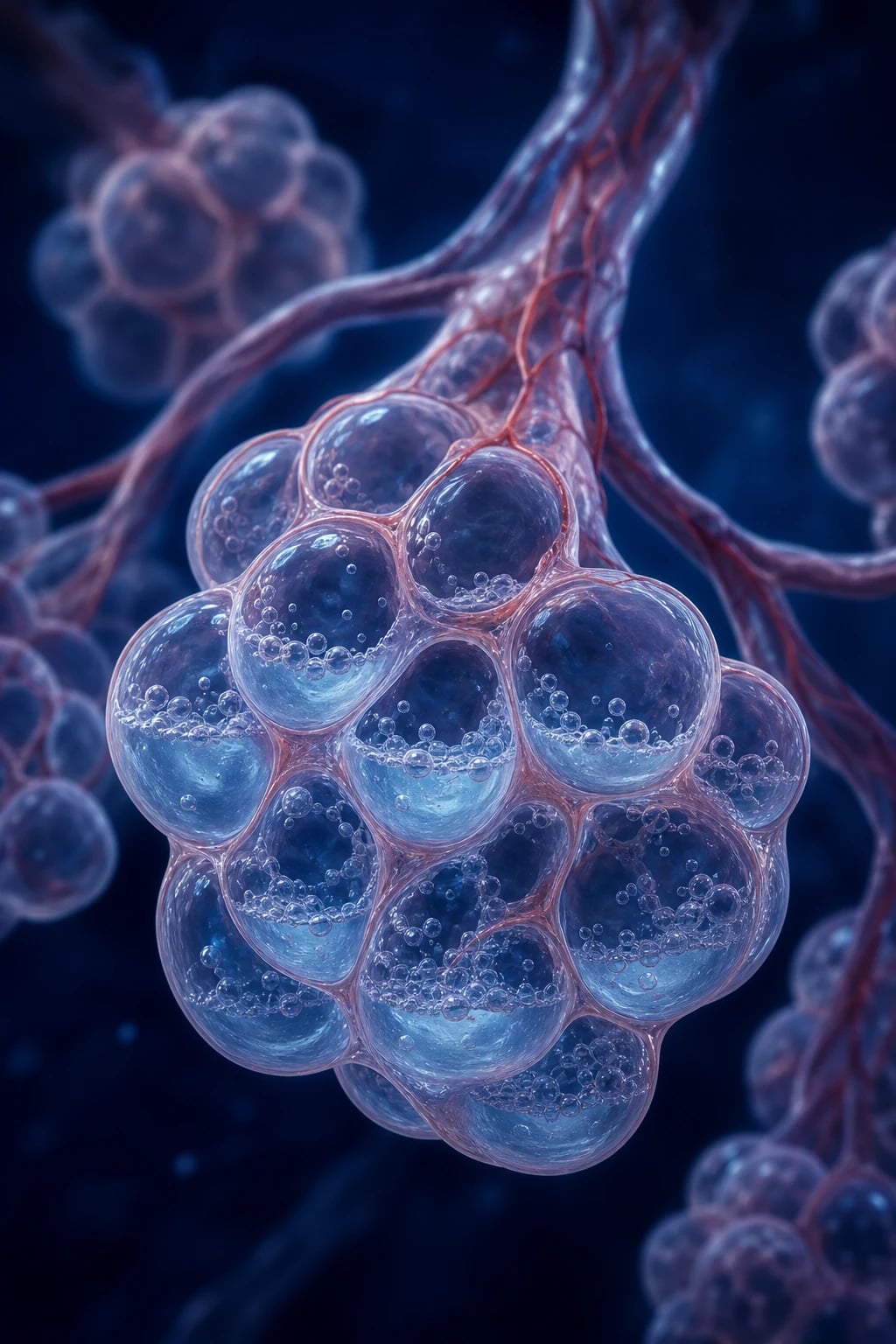
Overview
Pathophysiology — the unifying mechanism is bubbles on ascent
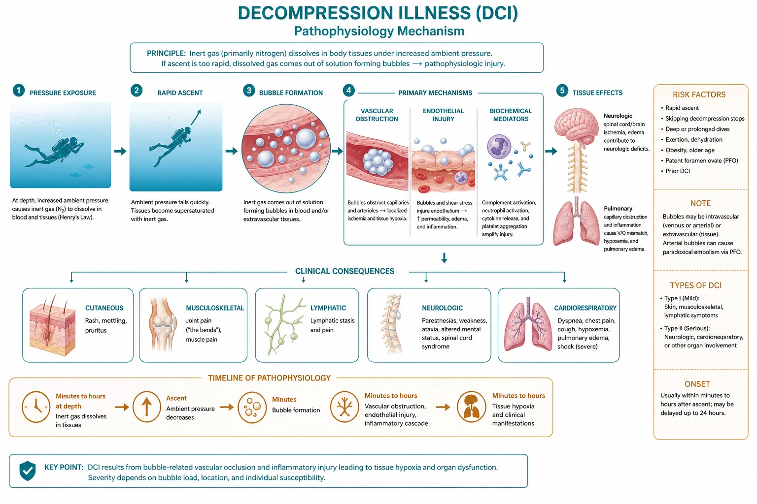
Both syndromes under the DCI umbrella share a single root cause: gas bubbles form when ambient pressure falls faster than dissolved inert gas can be cleared. Understanding the physics (Boyle's law, Henry's law) and the two distinct routes to bubble injury is the key to the whole topic.[1][5]
-
DCS — in-situ bubble formation from dissolved inert gas. Breathing compressed air at depth dissolves extra nitrogen in tissues (Henry's law: more pressure → more dissolved gas; fat dissolves ~5x more nitrogen than water, so adipose and myelin-rich spinal cord white matter are nitrogen reservoirs). On ascent, if ambient pressure falls faster than nitrogen can diffuse out into blood and be exhaled, the tissue becomes supersaturated and nitrogen comes out of solution as bubbles in situ — within tissues, the venous circulation, and lymphatics. These bubbles cause injury by four mechanisms: (a) mechanical disruption of tissue (especially spinal cord white-matter tracts), (b) vascular occlusion (venous and capillary blockage → ischaemia), (c) platelet activation and coagulation at the bubble-blood interface, and (d) endothelial dysfunction and capillary leak (complement activation, leukocyte adhesion, lipid peroxidation) → oedema and a secondary inflammatory cascade that continues after the bubbles themselves have shrunk.[3][5]
-
AGE — alveolar gas forced into the circulation by pulmonary barotrauma. During ascent the gas in the lungs expands (Boyle's law: at 30 m / 4 ATA, lung volume quadruples on ascent to the surface if not exhaled). If a diver holds their breath, ascends too fast, or has trapped gas (obstructed airway, mucus plug, bleb), the expanding gas over-distends and ruptures alveoli. Gas then tracks into the pulmonary venous circulation → left heart → systemic arterial tree. The brain is the most common final destination, producing a focal cerebral deficit; coronary embolisation can cause arrhythmia or ischaemia. AGE occurs during or within minutes of surfacing — the timing is the cardinal clue that distinguishes it from DCS. Note that AGE can also arise iatrogenically (central line placement, cardiac surgery, haemodialysis) and is not unique to diving.[1]
-
The crossover: paradoxical embolism via a right-to-left shunt. Venous bubbles from DCS are normally filtered by the pulmonary circulation (where they are gradually reabsorbed). But in a diver with a patent foramen ovale (PFO) — present in ~25-30% of the population — or a pulmonary arteriovenous malformation, venous bubbles can cross directly into the arterial system and embolise the brain or inner ear. This explains why some divers develop cerebral or inner-ear DCS disproportionate to the dive profile, and why PFO is over-represented in recurrent neurological DCS.[1][6]
DCI — the two syndromes and how to tell them apart
DCS
Inert-gas bubbles
- Mechanism: nitrogen supersaturation → in-situ bubbles in tissues/venous blood
- Onset: minutes to hours after surfacing; 98% within 24 h
- Tied to depth, bottom time, repetitive dives, rapid ascent, cold, exertion
- Type 1 (joint/skin) or Type 2 (spinal cord, pulmonary, vestibular, cerebral)
AGE
Pulmonary barotrauma
- Mechanism: alveolar rupture on ascent → gas into pulmonary veins → arteries
- Onset: DURING or within minutes of surfacing
- Often a breath-hold ascent or rapid ascent from as little as 2 m
- Stroke-like: hemiparesis, aphasia, visual loss, seizure, coma; can be cardiac
Shared
Treatment is identical
- Both: 100% oxygen immediately + IV fluids + recompression (HBO)
- Both can coexist in the same diver — do not wait to distinguish them
- Differentiation is academic at the bedside: treat both as DCI
DCS — the Type 1 / Type 2 classification
The traditional classification divides DCS by severity and organ system. Modern diving medicine increasingly describes DCS by symptom/organ system (because the type boundary is blurred and prognosis tracks the organ involved), but the Type 1/Type 2 framework remains the exam staple and is useful for triage.[1][5]
DCS Type 1 versus Type 2
Type 1
Mild (pain/skin)
- MUSCULOSKELETAL — "the bends": periarticular joint pain (shoulders, elbows, knees, hips), classically a dull deep ache that is poorly localised, worsens with movement, and is relieved only by recompression; local tenderness is minimal and out of proportion to the pain
- SKIN — "skin bends": itching (formication), mottling/marbling (cutis marmorata), rash, patchy cyanosis
- LYMPHATIC — localised lymphoedema, regional node tenderness
- Generally NOT life-threatening but can herald evolving Type 2 — do not dismiss
Type 2
Serious (neuro/pulm/vestibular)
- SPINAL CORD — the MOST SERIOUS form: girdle/torso pain, progressive limb weakness or paralysis, paraesthesia, sensory level, bladder retention, bowel incontinence, sexual dysfunction; venous infarction of the cord
- PULMONARY — "the chokes": substernal chest pain, cough, dyspnoea, haemoptysis; pulmonary vascular obstruction → can progress to shock and collapse
- VESTIBULAR/COCHLEAR — "the staggers": vertigo, nystagmus, nausea, vomiting, tinnitus, hearing loss; can be hard to distinguish from inner-ear barotrauma
- CEREBRAL — headache, visual disturbance, confusion, hemiparesis, seizure (often via PFO paradoxical embolism)
Why the spinal cord is so vulnerable
The spinal cord is the classic and most feared target of serious DCS for three reasons: it is rich in lipid (myelin), which dissolves large amounts of nitrogen; its venous anatomy (long, valveless epidural venous plexus) allows bubbles to accumulate and obstruct venous outflow, producing venous infarction of white-matter tracts; and the resulting oedema in a confined spinal canal compounds the ischaemia. The result is a anterior cord / mixed picture: motor weakness (often ascending), sensory disturbance, and autonomic failure (bladder retention is an early and important sign). Spinal cord DCS is the scenario most likely to leave permanent deficit even after appropriate treatment, which is why any diver with limb weakness, gait disturbance, or urinary symptoms after a dive is treated as a hyperbaric emergency.[3][6]
AGE — the stroke of ascent
Arterial gas embolism must be suspected in any diver who loses consciousness, seizes, or develops a focal neurological deficit during ascent or within minutes of reaching the surface. The mechanism is mechanical: the expanding alveolar gas ruptures into the pulmonary vasculature. It can occur after ascent from surprisingly shallow depths — even a few metres — in a breath-hold diver, and is the leading cause of death in recreational diving fatalities that involve pulmonary barotrauma.[1][5]
Distinguishing AGE from DCS at the bedside
Timing
The strongest clue
- AGE: during ascent or within minutes (typically under 10 min) of surfacing
- DCS: usually minutes-to-hours; up to 24 h; onset is delayed as bubbles grow
Neuro pattern
Focal vs spinal
- AGE: focal CORTICAL signs — hemiparesis, aphasia, visual field loss, seizure, coma
- DCS: often SPINAL — ascending weakness, sensory level, bladder/bowel; can be cerebral
Provocation
Ascent mechanics
- AGE: breath-hold / rapid / panic ascent; may have chest pain or pneumothorax
- DCS: depth/time profile, repetitive dives, missed decompression stops
Bottom line
Do not delay
- Both get 100% oxygen, fluids, and recompression — distinguish only after treatment started
- A pneumothorax from the same barotrauma event needs a chest drain BEFORE recompression (it will expand in the chamber)
Diagnosis — clinical, with adjuncts
DCI is a clinical diagnosis made on the basis of a diving/altitude exposure and compatible symptoms within the time window. There is no blood test that confirms DCS, and waiting for investigations must never delay 100% oxygen and contact with a hyperbaric service.[1][3]
[1] [5]The mimics — what else to consider
Several conditions overlap with DCI and must not be missed: inner-ear barotrauma (alternative cause of vertigo/hearing loss during descent — NO recompression, distinguished by onset during descent and no systemic features); pneumothorax (dyspnoea and chest pain from the same barotrauma — drain before chamber); acute stroke in a cerebral presentation (the diving history and time course usually point to AGE/DCS, but thrombolysis may be appropriate in genuine stroke — discuss with neurology and hyperbarics); carbon monoxide poisoning from contaminated breathing gas (check COHb); and immersion pulmonary oedema. The history almost always resolves the diagnosis.[1]
Management — oxygen, fluids, recompression, retrieval
The management ladder is the same for DCS and AGE, and every step below should be initiated in parallel, not sequentially. The single most important phone call is to the nearest hyperbaric service or the Divers Alert Network (DAN) — retrieval logistics dominate outcome.[1][3]
Decompression illness management protocol
1. 100% oxygen immediately via non-rebreather mask
Apply high-flow 100% oxygen via a NON-REBREATHER mask at 15 L/min with a tight face-seal (reservoir bag kept more than two-thirds full) for EVERY suspected case of DCI, even mild "skin bends". Breathing 100% oxygen creates a maximal alveolar-to-tissue gradient for nitrogen, accelerating its washout from bubbles and supersaturated tissues (denitrogenation), and oxygenates ischaemic tissue. It is first aid AND definitive therapy for most altitude DCS. Intubate and ventilate with 100% oxygen if the airway is threatened (coma, GCS under 8) or breathing is inadequate. Continue 100% oxygen throughout retrieval and recompression.
2. Resuscitate and position the patient
Assess ABCDE. Keep the diver HORIZONTAL (supine) — the traditional 30-degree head-down Trendelenburg position is NO LONGER routinely recommended (it increases cerebral venous pressure and oedema, and risks aspiration); a flat supine position is preferred. If AGE is suspected and airway protected, left lateral decubitus may limit cerebral embolic load but flat supine is the safe default. Treat pneumothorax with a chest drain BEFORE recompression (a trapped pneumothorax will expand dangerously under pressure). Avoid unnecessary movement.
3. IV isotonic, glucose-free fluids
Give isotonic crystalloid (normal saline or balanced crystalloid) intravenously to correct the dehydration of immersion (cold-water immersion diuresis plus reduced oral intake) and to maintain perfusion of bubble-injured tissue. Target euvolaemia and a good urine output (around 1-1.5 mL/kg/h) — over-hydration risks pulmonary and cerebral oedema. AVOID glucose-containing fluids: hyperglycaemia worsens neurological outcome in DCI (as it does in acute stroke and spinal cord injury). In unconscious or spinal-cord cases, catheterise early to monitor output and relieve retention.
4. Contact the hyperbaric service / DAN immediately and arrange retrieval
This is the single most important call. Contact the nearest hyperbaric chamber directly, or the Divers Alert Network (DAN) 24-hour emergency hotline, which coordinates retrieval and chamber location worldwide. Retrieval should be by the fastest appropriate means — air retrieval is often required. CRITICAL retrieval rules: the patient must remain on 100% oxygen throughout transport; cabin pressure in a pressurised aircraft should be maintained at sea-level equivalent (avoid unpressurised aircraft above 300 m / 1000 ft, as further decompression worsens bubbles); if a commercial aircraft is unavoidable, request sea-level cabin pressure. Give the hyperbaric service a full dive and symptom history.
5. Recompression therapy — the definitive treatment
Recompression in a hyperbaric chamber breathing 100% oxygen is DEFINITIVE. The standard schedule is US Navy Treatment Table 6 (USN TT6): recompression to 60 feet of seawater (18 m, 2.8 ATA) breathing 100% oxygen with intermittent air breaks, then a staged ascent to 30 feet (1.9 ATA), total run time approximately 4-5 hours. It works by THREE mechanisms — (a) Boyle’s law: increased pressure shrinks bubbles to about one-third of their surface volume (1/2.8), restoring perfusion; (b) oxygenation of ischaemic tissue from the very high inspired PO2 (around 6 vol% dissolved oxygen in plasma at 2.8 ATA meets resting metabolic demand independent of haemoglobin); and (c) denitrogenation, accelerating inert-gas washout and reducing oedema. Additional treatments (generally one to two, occasionally more) are given for residual manifestations until clinical stability.
6. Adjunctive and supportive care
Lidocaine infusion is sometimes used in severe neurological DCI (proposed neuroprotection, mixed evidence) but is not standard. NSAIDs (tenoxicam) added to recompression may reduce the number of repeat recompressions required (though they do not improve the odds of recovery, per Bennett 2010). Heliox tables (helium-oxygen) are an alternative for refractory cases and may reduce the need for multiple recompressions. Maintain normoglycaemia and normothermia. Give prophylaxis against venous thromboembolism in paralysed/immobilised patients (proven spinal cord DCS is a VTE-risk state). Treat seizures with benzodiazepines; arrhythmia and ischaemia conventionally. Avoid nitrous oxide (expands nitrogen bubbles) and avoid over-sedation before chamber treatment.
7. Repeat recompression for residual deficit
After the initial USN TT6, reassess. Residual neurological symptoms warrant further recompression treatments (usually daily TT6 or Table 5/6 until no further improvement). Severe cases may require several treatments over days. Roughly three-quarters of divers achieve complete recovery; a minority are left with residual deficit even after multiple recompressions — completeness of recovery at discharge is the strongest long-term predictor.
8. Follow-up, fitness-to-dive, and PFO assessment
After recovery, address recurrence risk: counsel on safe diving practice (ascent rates, decompression stops, hydration, avoiding exertion after diving). For DIVERS WITH RECURRENT or DISPROPORTIONATE NEUROLOGICAL/INNER-EAR DCS, investigate for a PATENT FORAMEN OVALE with bubble-contrast echocardiography; PFO closure may be considered (controversial). Document residual deficit, arrange rehabilitation (especially for spinal cord DCS with paraparesis), and give a formal fitness-to-dive assessment before return to diving. Report the event to the relevant diving medicine body for surveillance.
The three mechanisms of recompression — know them by name
How US Navy Table 6 fixes a bubble injury
| Mechanism | Physics / physiology | Effect |
|---|---|---|
| Bubble shrinkage | Boyle's law (P1V1 = P2V2): at 2.8 ATA a bubble is 1/2.8 (~36%) of its surface volume | Restores flow through occluded vessels; relieves mechanical pressure on tissue |
| Tissue oxygenation | Henry's law: ~6 vol% oxygen dissolves in plasma at 2.8 ATA | Meets resting tissue O2 demand independent of haemoglobin; rescues ischaemic cord/brain |
| Denitrogenation | Breathing 100% O2 creates maximal N2 gradient out of tissue/bubbles | Accelerates bubble resolution; reduces oedema and the inflammatory cascade |
Evidence on recompression and adjuncts — what the trials show
Recompression is the universally accepted standard of care for DCI, yet the evidence base for which table and which adjuncts is surprisingly thin. A fellowship candidate should know what is established and what is genuinely uncertain.[2][3]
Recompression itself is standard but unproven by RCT. No randomised trial has ever compared recompression against no recompression — and none ever will, because withholding it would be unethical. The US Navy Treatment Table 6 (2.8 ATA, 100% oxygen, ~4-5 h) is the global default on the basis of decades of observational experience; Moon 2014 confirms it as the recommended schedule for most cases of DCS, with additional treatments for residual manifestations.[3]
Adjunctive therapy — the Cochrane / Bennett view. Bennett 2010 (a systematic review of all RCTs) found only two trials that could be pooled in no useful way. One showed that adding the NSAID tenoxicam to standard recompression did not improve the odds of recovery but did reduce the number of repeat recompressions required (a likely economic and symptom-duration benefit). The other showed a heliox table reduced the odds of multiple recompressions versus an oxygen table. Neither improved the odds of recovery. The bottom line: recompression is standard; adjuncts (NSAID, heliox) may reduce the treatment burden but are not proven to improve outcome, and the overall RCT evidence is sparse.[2]
Does delay matter? — the timing evidence. General teaching and clinical practice insist on recompressing as soon as feasible, and most series show that earlier treatment trends toward better outcome. Hadanny 2015 showed that even delayed recompression (started 48 h or more after surfacing) still achieved complete recovery in 76% of divers, with a (non-significant) trend favouring the US Navy Table 6 protocol over shorter schedules — so a delayed presentation is never a reason to withhold recompression. Blatteau 2011, in a large spinal-cord DCS cohort, found that after statistical adjustment the time-to-recompression did not independently predict recovery, whereas clinical severity (motor deficit, bladder dysfunction, a high severity score) and symptom progression before treatment did. The synthesis: recompress as soon as you can, but never refuse a delayed case — late is still better than never.[3][4][6]
Clinical pearls
Red flags
Prognosis
DCI outcomes and predictors
| Scenario / factor | Outcome | Notes |
|---|---|---|
| Mild Type 1 DCS, prompt recompression | Excellent | Joint/skin bends usually resolve fully with early recompression |
| Spinal cord DCS with motor deficit | Guarded-poor | Most serious form; ~25% incomplete recovery at 1 month (Blatteau 2011) |
| Bladder dysfunction at presentation | Poor | Independent predictor of bad recovery (OR ~3.8); a red-flag sign |
| Symptom progression before recompression | Poor | Worsening en route to chamber predicts worse outcome (OR ~2.07) |
| Depth at or beyond 39 m | Worse | Independent risk factor for severe spinal cord DCS |
| Age at or above 42 years | Worse | Independent predictor of incomplete recovery in spinal cord DCS |
| AGE with loss of consciousness | Variable | Often good if recompressed promptly; can leave residual focal deficit |
| Delay to recompression | Generally worse | Recompress ASAP, but late (>48 h) treatment still works (~76% complete recovery) |
| Residual deficit after initial table | Needs more treatments | Repeat recompression (daily TT5/6) until no further improvement |
| PFO with recurrent neurological DCS | Recurrence risk | Investigate with bubble-contrast echo; consider closure (controversial) |
Most divers treated promptly recover completely. The chief predictor of incomplete recovery is clinical severity at presentation — particularly spinal cord involvement with motor deficit and bladder dysfunction, and progression of symptoms before treatment reaches the chamber. Time-to-recompression matters at the population level (recompress as soon as feasible) but, after adjustment, severity dominates: Blatteau 2011 found that age, depth, bladder dysfunction, symptom progression, and a high severity score independently predicted bad recovery in spinal cord DCS, while the absolute time to recompression and the choice of initial table did not. The practical message is to treat aggressively and early, but never abandon a delayed or severe case — late recompression and repeat treatments still achieve meaningful recovery, and residual neurological deficit warrants ongoing rehabilitation and formal fitness-to-dive assessment.[1][3][4][6]
Key trials and evidence
Vann 2011 — Decompression illness (Lancet) (PMID 21215883)
Type
Definitive narrative review of the two-syndrome DCI concept
Key points
DCI = DCS (in-situ inert-gas bubbles) + AGE (alveolar gas forced into arteries via barotrauma); immersion, exercise, and temperature modify risk
Management
First aid 100% oxygen; definitive recompression breathing 100% oxygen; adjunctive fluids and VTE prophylaxis in paralysed patients
Prognosis
Treatment effective in most cases; residual deficit can persist in serious cases even after several recompressions
Clinical bottom line
The authoritative modern reference for the unifying bubble pathophysiology and the standard management ladder
Moon 2014 — Hyperbaric oxygen treatment for DCS (Undersea Hyperb Med) (PMID 24851553)
Type
Evidence-based review of HBOT for decompression sickness
Pathophysiology
In-situ bubble formation → mechanical disruption, vascular occlusion, platelet activation, endothelial dysfunction, capillary leak
Recommended schedule
100% oxygen at 2.82 ATA — US Navy Treatment Table 6 or equivalent — for most cases; additional treatments for residual manifestations
Adjuncts
Isotonic, glucose-free fluids for prevention/treatment of hypovolaemia; evidence-based review of adjunctive therapies
Clinical bottom line
The single best source for the standard recompression protocol and the why behind it (Boyle, Henry, denitrogenation)
Bennett 2010 — Recompression & adjunctive therapy for DCI: systematic review (Anesth Analg) (PMID 20332190)
Design
Systematic review of all RCTs comparing recompression schedules or adjunctive therapies
Findings
Only 2 RCTs met criteria; pooling not possible. Tenoxicam (NSAID): no improvement in recovery odds but fewer repeat recompressions needed (P=0.01). Heliox table: lower odds of multiple recompressions vs oxygen table (RR 0.56)
Limitation
Recompression is the universal standard yet has no RCT evidence — adjuncts may reduce treatment burden, not recovery odds
Clinical bottom line
Recompression remains standard; NSAID or heliox may reduce the number of treatments required but are not proven to improve outcome
Hadanny 2015 — Delayed recompression for DCS (PLoS One) (PMID 25906396)
Design
Retrospective cohort: 76 divers recompressed at 48 h or more vs 128 treated earlier than 48 h
Result
Complete recovery in 76% of the delayed group vs 78% of the early group — comparable outcomes
Protocol finding
US Navy Table 6 trended toward better outcome than a shorter 90-min 2-ATA schedule (OR 2.79, not significant)
Clinical bottom line
Late recompression (days late) still has clinical value — never withhold the chamber from a delayed presentation
Blatteau 2011 — Prognostic factors of spinal cord DCS (Neurocrit Care) (PMID 20734244)
Design
Multicentre retrospective analysis of 279 recreational divers with spinal cord DCS (France and Belgium)
Result
26% had incomplete resolution at 1 month
Independent predictors of poor recovery
Age at or above 42, depth at or beyond 39 m, bladder dysfunction (OR ~3.8), symptom progression before recompression (OR ~2.07), high severity score
Nuance
Time-to-recompression and choice of initial table did not significantly affect recovery after adjustment
Clinical bottom line
In spinal cord DCS, clinical severity (not delay alone) drives prognosis — but treat early and aggressively regardless
Exam practice — SAQ
SAQ — Decompression illness after recreational dive
10 minutes · 10 marks
A 38-year-old recreational diver surfaces from a 32 m, 28-minute air dive with a rapid ascent. Within 5 minutes he has severe dyspnoea then left hemiparesis. SpO2 90% on air.
Examiner densification notes
[1] [1]References
- [1]Vann RD, Butler FK, Mitchell SJ, Moon RE. Decompression illness Lancet, 2011.PMID 21215883
- [2]Bennett MH, Lehm JP, Mitchell SJ, Wasiak J. Recompression and adjunctive therapy for decompression illness: a systematic review of randomized controlled trials Anesth Analg, 2010.PMID 20332190
- [3]Moon RE. Hyperbaric oxygen treatment for decompression sickness Undersea Hyperb Med, 2014.PMID 24851553
- [4]Hadanny A, Fishlev G, Bechor Y, et al. Delayed recompression for decompression sickness: retrospective analysis PLoS One, 2015.PMID 25906396
- [5]Bove AA. Diving medicine Am J Respir Crit Care Med, 2014.PMID 24869752
- [6]Blatteau JE, Gempp E, Simon O, et al. Prognostic factors of spinal cord decompression sickness in recreational diving: retrospective and multicentric analysis of 279 cases Neurocrit Care, 2011.PMID 20734244