ICU · Environmental
Electrical and radiation injury in ICU
Also known as Electrocution · Lightning injury · Radiation injury · Acute radiation syndrome · Electrical burns
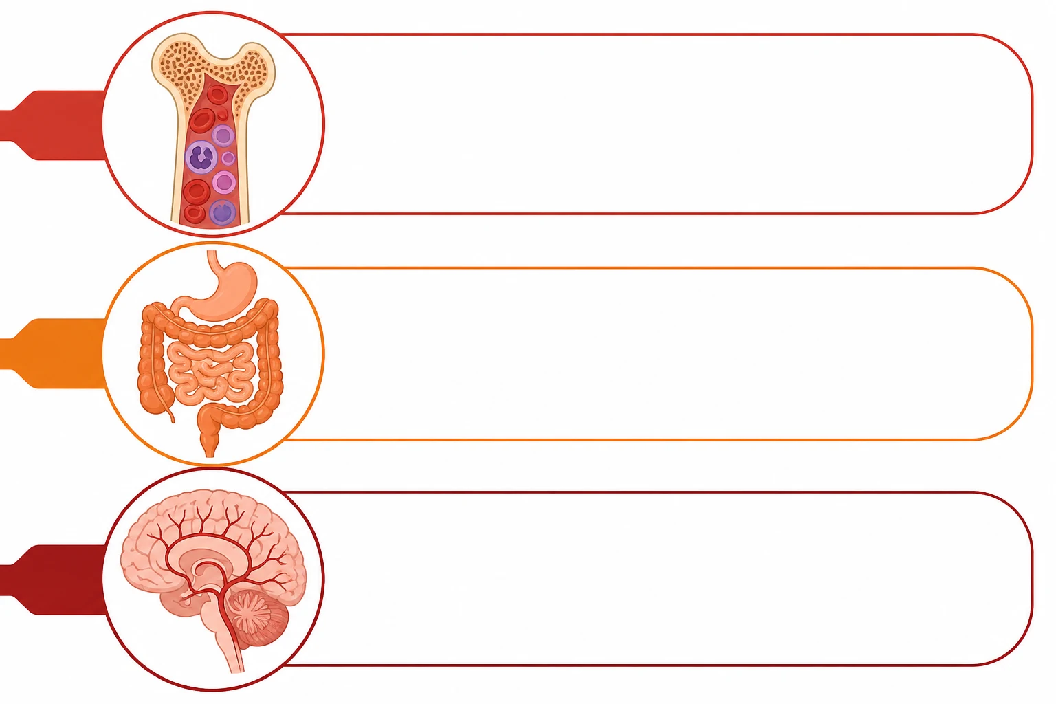
Electrical injury: damage from electricity passing through body. Low-voltage (<1000V): cardiac arrhythmias (AC current at 50-60Hz can cause VF). High-voltage (1000V): deep tissue injury (muscle, nerve, vessel), compartment syndrome, rhabdomyolysis, AKI, cardiac arrest, secondary trauma (fall). Lightning: massive DC discharge, causes immediate cardiac arrest (asystole), respiratory arrest (neurological), Lichtenberg figures (fern-like skin markings). Radiation injury: acute radiation syndrome (ARS) from whole-body radiation 1 Gy. Syndromes: haematopoietic (2-6 Gy), gastrointestinal (6-8 Gy), neurovascular (8 Gy, fatal). Management: electrical — ACLS, fluid resuscitation (rhabdomyolysis protocol), fasciotomy for compartment syndrome, cardiac monitoring. Radiation — supportive, transfusion, growth factors, bone marrow transplant (selected).
On this page & tools
Your progress
Saved locally on this device.
Target exams
Red flags
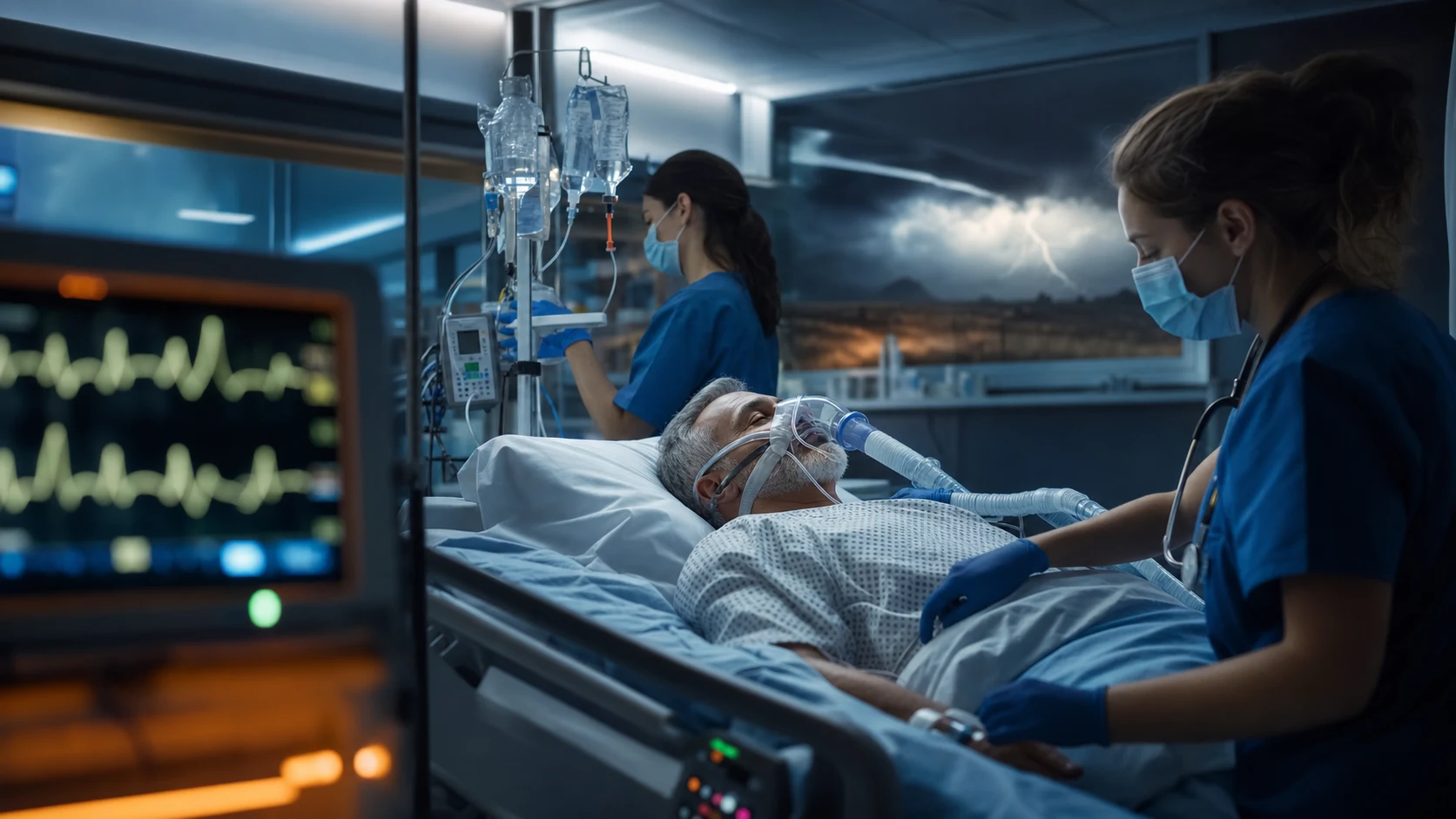

Low-voltage vs high-voltage vs lightning injury
| Feature | Low-voltage (<1000V) | High-voltage (>1000V) | Lightning |
|---|---|---|---|
| Current type | AC (alternating) | AC or DC | Massive DC (direct current) |
| Cardiac | VF (tetanic contraction) | Asystole, VF, cardiac arrest | Asystole (DC current) |
| Tissue injury | Superficial (entry/exit wounds) | DEEP (muscle, nerve, vessel) | Variable (flash over — often less than expected) |
| Muscle/Rhabdo | Mild | SEVERE (massive) | Variable |
| Compartment syndrome | Rare | COMMON | Variable |
| Skin signs | Entry/exit burns | Entry/exit + deep burns | Lichtenberg figures (fern-like), linear burns |
| Secondary trauma | Rare (fall) | COMMON (thrown — fractures, head injury) | Blast injury (tympanic, fall) |
| Mortality | Low (if no VF) | High (20-30%) | Variable (10-30% of victims, often at scene) |
| Management | ACLS (VF → defib), cardiac monitor | Fluid (rhabdomyolysis), fasciotomy, trauma survey | Prolonged CPR (reverse triage), ACLS, trauma survey |
Management of high-voltage electrical injury
- Ensure scene safety — power source disconnected before approaching. Rescuer safety first
- ACLS — cardiac arrest (VF → defibrillation; asystole → CPR/ACLS). Lightning: prolonged CPR (victims may recover after long down-time — reverse triage)
- Primary survey (ABCDE) — airway (may have facial/airway burns), breathing, circulation (arrhythmias, hypovolaemia), disability (neurological injury), exposure (all skin for entry/exit wounds)
- ECG monitoring for 4-6h — arrhythmia risk. Troponin. If normal ECG + no symptoms at 6h, very low risk of late arrhythmia
- Fluid resuscitation — rhabdomyolysis protocol — crystalloid to maintain urine output 1-1.5 mL/kg/h (HIGHER than burn — muscle damage more extensive). Monitor CK, myoglobin, urine output
- Trauma survey — secondary injuries (fall, blast): fractures, head injury, spinal injury, tympanic membrane rupture
- Compartment syndrome assessment — check all limbs for tense compartments, pain on passive stretch. Fasciotomy if signs present
- Laboratory — CK, troponin, U&E (AKI), LFTs, coagulation (DIC), ABG (acidosis). Urine: myoglobin (dark, dipstick blood+ but no RBC)
- Surgical consultation — burns, plastic, orthopaedic (fasciotomy, escharotomy), trauma
- Tetanus prophylaxis — if not immune
Short answer questions
SAQ — High-voltage electrical injury with rhabdomyolysis and compartment syndrome
10 minutes · 10 marks
A 41-year-old linesman is brought to the emergency department after the boom of his crane contacted an 11 000 V overhead AC power line. He was thrown from the crane and landed on his right side; bystanders report a brief loss of consciousness. On arrival he is confused (GCS 13), HR 122, BP 104/64, SpO2 96 per cent on room air. There is a charred 3 cm contact burn on the right palm and a ragged exit wound over the medial right calf. The right forearm and leg are tense and exquisitely painful on passive movement, and the urine is dark red-brown. ECG shows sinus tachycardia with peaked T waves. CK 24 000 U/L, K+ 6.8 mmol/L, creatinine 142 micromol/L, venous pH 7.22.
SAQ — Mass-casualty radiation exposure: triage and disposition
10 minutes · 10 marks
An industrial radiography source containing caesium-137 is breached at a metropolitan worksite, contaminating twelve workers. The emergency department receives advance warning that casualties are inbound within thirty minutes. Several patients have external skin contamination with caesium dust, and three are thought to have significant whole-body gamma exposure of uncertain dose. The first symptomatic patient vomited ninety minutes after exposure, has erythema over the trunk, and an absolute lymphocyte count of 0.6 × 10⁹/L at twelve hours. You are the ICU consultant leading the hospital radiation response.
SAQ — Acute radiation syndrome: haematopoietic management and the role of stem-cell transplant
10 minutes · 10 marks
A 34-year-old male nuclear-power-plant technician was exposed to an estimated 4.5 Gy whole-body gamma radiation during a containment breach six hours ago. He has had two episodes of vomiting, the last at three hours post-exposure, and is currently nauseated but haemodynamically stable. Absolute lymphocyte count is 0.9 × 10⁹/L at six hours. He has no burns and no residual external contamination after decontamination. The haematology and transplant teams have been notified. You are the intensivist planning his ICU admission.
Clinical pearls
Red flags
Prognosis
Outcomes of high-voltage electrical injury (Waldmann 2018)
Cohort study of high-voltage electrical injuries:
- Mortality: 20-30% (higher with cardiac arrest, falls, secondary trauma)
- Amputation rate: 10-20% (deep muscle necrosis, compartment syndrome)
- Long-term sequelae: neurological (neuropathy, chronic pain), psychiatric (PTSD), cataracts, cardiac (arrhythmias)
- AKI requiring RRT: 5-10% (rhabdomyolysis) [1]
Lightning mortality: 10-30% of victims (many die at scene). Of survivors: 70% have long-term sequelae (neurological, cardiac, psychological). ARS mortality: <1 Gy → survival. 2-6 Gy → survival with treatment (growth factors, isolation). 6-8 Gy → high mortality even with treatment. >8 Gy → uniformly fatal.
Physics and determinants of electrical injury
The severity of an electrical injury is set by current, not voltage — by Ohm's law $I = V/R$ the same 240 V applied to wet skin (≈1000 Ω) drives ~240 mA, while dry skin (≈100 000 Ω) limits it to ~2 mA, the difference between lethal VF and a tingle. Six variables therefore determine outcome: voltage, current, resistance, current type (AC vs DC), pathway through the body, and contact duration. Heat is governed by Joule's law $H = I^2 R t$: deep, high-resistance tissues (bone, tendon) become the hottest and conduct that heat to adjacent low-resistance muscle, nerve and vessel — the structural basis of the 'iceberg' injury in which trivial surface burns conceal massive deep-tissue necrosis.[2] }
Current magnitude produces a stereotyped clinical ladder. Roughly 1 mA is the threshold of perception; 5-9 mA is the maximum harmless current; 10-20 mA is the 'let-go' threshold above which tetanic forearm-flexor contraction fixes the victim's grip onto a live AC source, prolonging contact and converting a survivable shock into a lethal one; ~100 mA of 50-60 Hz AC traversing the chest precipitates ventricular fibrillation; currents exceeding 1-2 A produce direct myocardial standstill, deep burns and, if the victim survives, extensive rhabdomyolysis.[2] }
Determinants of electrical tissue injury
| Factor | Effect on severity |
|---|---|
| Current (I) | True determinant of injury. 1 mA = perception; 10-20 mA = let-go threshold (tetany); 100 mA AC = VF; >1-2 A = standstill + deep burns |
| Voltage | Higher voltage overcomes skin resistance to drive current. >1000 V = high-voltage, deep injury |
| Resistance (R) | Dry skin ≈100 000 Ω (protective); wet skin ≈1000 Ω (100× current surge). Order high→low: bone > fat > tendon > dry skin > wet skin > muscle > blood > nerve |
| Current type | AC (50-60 Hz) most dangerous — tetany + VF; DC = single spasm, victim thrown, secondary trauma |
| Pathway | Hand-to-hand/hand-to-foot across thorax → arrhythmia; along a limb → compartment syndrome; through head → CNS/retina/cataract |
| Duration | Longer contact = more Joule heat + longer current flow across myocardium |
| Contact area | Small area = high current density = severe local burn; large area dissipates energy |
Alternating vs direct current — mechanism
AC vs DC electrical injury
| Feature | Alternating current (AC) | Direct current (DC) |
|---|---|---|
| Frequency | 50-60 Hz (mains) | Zero (constant) |
| Myocardial risk | VF at ~100 mA — 50-60 Hz sits near the cardiac vulnerable period, readily precipitating VF | Higher threshold for VF; large DC more often causes asystole (standstill) |
| Muscle effect | Tetanic contraction at >10-20 mA → 'stuck to source', prolonged contact, drowning of electricians in water | Single violent spasm — victim thrown clear (short contact, more secondary trauma) |
| Let-go threshold | ~10-20 mA (forearm flexors outstrength extensors) | Much higher — victim rarely 'stuck' |
| Typical sources | Household mains, industrial supply | Batteries, railways, defibrillators, lightning |
| Skin/contact burns | Entry/exit, often small | Entry/exit, often larger/exploding exit |
The vulnerability of AC at 50-60 Hz is not coincidence: this band straddles the effective refractory window of cardiac myocytes, so successive cycles deliver current into the repolarising myocardium and trigger re-entry → VF. The let-go threshold exists because forearm flexor muscles are stronger than extensors, so tetanic flexion locks the hand closed on the conductor. This is why the first action at a low-voltage electrocution scene is to cut the power before touching the victim — pulling a gripped victim from a live source risks further injury to both patient and rescuer.[2] }
Lightning injury in detail
A lightning bolt carries 100 million to 1 billion volts, 10 000-200 000 A, lasting only ~30-50 milliseconds. Despite these staggering figures, internal injury is often surprisingly modest thanks to flashover — the current takes the lower-resistance route over the body surface (sweat-dampened skin) rather than through the torso. The same physics means entry/exit wounds and deep-muscle necrosis are far less prominent than in equivalent-voltage industrial electrocution.[1] }
Three lightning-specific signs the examiner wants to hear: [1]
- Lichtenberg figures — fern-like, feathery, branching skin markings that are pathognomonic for lightning. They are not thermal burns (no tissue necrosis) — thought to be an electron shower in superficial capillaries (a keraunographic mark). They fade within 24 h, so their absence later does not exclude lightning injury.
- Fixed, dilated pupils — in lightning, dilated unreactive pupils reflect transient autonomic (ciliary) dysfunction, NOT brain death. A lightning victim with fixed pupils and apnoea must never be declared dead on that basis; outcomes after prolonged CPR are excellent. Combined with the 'reverse triage' principle, this is a high-yield exam pearl.
- Keraunoparalysis (Charcot's keraunoparalysis) — transient flaccid paralysis with sensory loss, pale/cold/blue extremities and absent pulses (vasomotor spasm). Limbs are preferentially affected and resolve over hours. Mimics spinal cord injury or acute limb ischaemia; always re-examine before imaging decisions. [1]
The 'reverse triage' / 'resuscitate the apparently dead first' principle is unique to mass-casualty lightning incidents. Lightning causes simultaneous asystole (massive DC standstill) and respiratory arrest (medullary paralysis). The cardiac rhythm often auto-restarts within minutes, but if the victim remains apnoeic they suffer a secondary hypoxic arrest ('dead-in-bed' sequence). Therefore: in a group of lightning casualties, the conscious walking wounded can wait while the apnoeic, pulseless victims are intubated and CPR'd first — they may recover fully after very long down-times.[1] }
Lightning injury management — reverse triage
- Scene safety — lightning can strike twice; move casualties to shelter before assessment. Apply reverse triage: resuscitate the apparently dead first, conscious walking wounded can wait
- Airway and breathing — intubate apnoeic patients immediately. The cause of death is usually secondary hypoxic arrest from ongoing apnoea, so ventilatory support is the highest-yield intervention
- Circulation / ACLS — asystole (DC standstill) → CPR ± adrenaline. Prolonged CPR is justified — down-times of 30+ min with good neurological recovery are described; the myocardium is healthy and only needs to be reoxygenated
- Do NOT use fixed/dilated pupils as a sign of brain death — autonomic dysfunction is reversible. Continue resuscitation
- Secondary survey / trauma — blast injury (tympanic membrane rupture in up to 50%), falls and being thrown (fractures, head injury, spinal), thermal burns (skin/clothing ignition), eye trauma (cataracts, retinal)
- 12-lead ECG and cardiac monitoring — arrhythmias, ST changes, QT prolongation, troponin rise
- Document and photograph Lichtenberg figures early — they fade within 24 h and provide forensic confirmation of lightning injury
- Neurological assessment — keraunoparalysis resolves in hours; persistent deficits warrant imaging for spinal/head injury
- Obstetric assessment in pregnant patients — fetal demise can occur with apparently minor maternal injury; monitor
- Admit for observation even if asymptomatic at first — delayed arrhythmias, cataracts and neuropsychological sequelae are described
Radiation — principles and radiation types
Ionising radiation transfers energy to tissue by ionising atoms, generating free radicals that damage DNA. Four types differ in penetration and hazard: [1]
Radiation types — penetration and clinical hazard
| Type | Stopped by | External hazard | Internal hazard | Typical sources |
|---|---|---|---|---|
| Alpha | A sheet of paper / dead skin | None (cannot penetrate) | High if inhaled/ingested | Plutonium-239, polonium-210, radon, uranium |
| Beta | Clothing / a few mm aluminium | Skin burns ('beta burns') | Moderate if internal | Iodine-131, strontium-90, tritium |
| Gamma / X-ray | Lead / concrete | High — whole-body | High | Cobalt-60, caesium-137, iridium-192 |
| Neutron | Hydrogen-rich material (water, concrete, wax) | High — whole-body, induces radioactivity | High | Nuclear weapons, reactor cores |
A crucial triage distinction: an irradiated patient (exposed to a beam, e.g. gamma) emits no radiation and poses no risk to staff — manage them like any other critically ill patient. A contaminated patient (radioactive dust/liquid on skin, wounds, or inhaled/ingested) is radioactive and requires decontamination and personal protective equipment. Incorporation describes uptake of a radionuclide into tissues (e.g. iodine into thyroid, strontium into bone), which then requires decorporation therapy.[3] }
Acute radiation syndrome (ARS) — phases
ARS follows whole-body exposure above ~1 Gy (100 rad) delivered acutely. It unfolds in four phases whose timing and severity are dose-dependent, allowing the clinician to estimate dose prospectively: [1]
- Prodromal phase (minutes-hours, occasionally up to 2 days): nausea, vomiting, anorexia, fatigue, fever, diarrhoea. Time to vomiting is a bedside dose estimator — onset <1 h implies ≥6 Gy (usually fatal); 1-2 h ~4-6 Gy (serious); >2 h ~2-4 Gy (moderate).
- Latent phase (days to weeks): the patient looks and feels well — a dangerous interval that can mislead clinicians into under-resourcing. Higher doses shorten the latent phase.
- Manifest illness phase: the dose-dependent syndrome emerges (haematopoietic, GI, neurovascular — see table).
- Recovery or death over weeks to months.[6] }
ARS dose-dependent syndromes
ARS dose-dependent syndromes (whole-body acute dose)
| Syndrome | Dose (Gy) | Latent | Manifest illness features | Prognosis |
|---|---|---|---|---|
| Cerebrovascular / neurovascular | >8-10 | None-immediate | Confusion, ataxia, seizures, cerebral oedema, vasomotor collapse, death within 24-72 h | Uniformly fatal — supportive/palliative only |
| Gastrointestinal | 6-8 | 3-5 days | Severe bloody diarrhoea, mucosal sloughing, fluid loss, sepsis, GI bleeding | High mortality even with maximal ICU support |
| Haematopoietic | 2-6 | 1-3 weeks | Pancytopenia → infection, bleeding (petechiae, mucosal haemorrhage), anaemia | Survivable with growth factors, isolation, transfusion |
| Subclinical / mild | 1-2 | — | Transient nausea, mild lymphopenia | Survival |
Cutaneous radiation injury (CRI) can co-exist: erythema appears at ~2 Gy, dry desquamation at 6 Gy, moist desquamation/blistering at 10-15 Gy, and ulceration/necrosis above 20 Gy. Unlike a thermal burn, radiation skin injury recurs and progresses over weeks as a 'wave' of erythema — biopsy avoidance and specialist wound care are key. [1]
Radiation biodosimetry
Three complementary tools estimate dose when history is unreliable: [1]
- Serial lymphocyte count — lymphocytes are the most radiosensitive circulating cell; a 50% drop within 24-48 h approximates a 2-4 Gy exposure, and an absolute lymphocyte count <0.5 × 10⁹/L at 48 h implies a potentially lethal dose. Repeat every 6-12 h.
- Time-to-vomiting (see above) — the fastest bedside estimator.
- Dicentric chromosome assay — the gold standard biodosimetry; dicentric chromosomes in cultured peripheral lymphocytes are scored, but the assay takes days and is available only in specialist reference laboratories. Useful for confirmation and dose reconstruction.[8] }
Radiation decontamination and medical management
Radiation casualty — decontamination and management priorities
- Triage and resuscitate first — life-saving medical treatment never waits for decontamination. Airway, breathing, circulation take priority over radiation concerns; an irradiated (non-contaminated) patient is managed normally
- Determine exposure type — irradiation only (no staff hazard) vs external/internal contamination (PPE + decontamination). Notify the hospital radiation safety officer and regional radiation emergency centre immediately
- External decontamination — remove all clothing (removes ~90% of external contamination); bag and label as radioactive waste. Wash skin and hair with lukewarm soap and water (avoid abrading skin), covering wounds first. Flush eyes, ears, nose
- Survey with a Geiger counter — document residual counts; repeat washing until counts approach background or plateau. Never shave body hair
- Internal contamination / decorporation — selected by radionuclide (see table). Treat within hours where possible
- Supportive care — antiemetics (5-HT3 antagonist ± dexamethasone), fluids, electrolyte correction, analgesia, nutritional support (early enteral). Reverse isolation once pancytopenic
- Haematopoietic syndrome — G-CSF / GM-CSF started within 24-72 h; prophylactic antiviral/antibacterial/antifungal cover; irradiated leukoreduced blood products; consider stem-cell transplant in young patients with 4-8 Gy exposure and no major burns (consult transplant centre)
- GI syndrome — gut rest, parenteral nutrition, somatostatin analogue for diarrhoea, broad-spectrum antibiotics, platelet support for bleeding
- Psychosocial support and follow-up — long-term cancer screening, cataract surveillance, fertility counselling
Radionuclide-specific decorporation therapy
| Radionuclide | Antidote / therapy | Mechanism |
|---|---|---|
| Iodine-131 | Potassium iodide (KI) | Saturates thyroid with stable iodine → blocks radioiodine uptake. Most effective <4 h post-exposure; some benefit up to 24 h; little benefit after 24 h. No effect on other radionuclides |
| Caesium-137, thallium | Prussian blue (ferric hexacyanoferrate) | Insoluble ion-exchange resin traps Cs/Tl in gut lumen → faecal excretion. Used after Goiânia (1987) and Litvinenko (Tl) cases |
| Plutonium, americium, curium (transuranics) | Ca-DTPA then Zn-DTPA (IV/inhaled) | Chelation — Ca-DTPA more effective initially but depletes trace metals; switch to Zn-DTPA for maintenance |
| Uranium | Sodium bicarbonate alkalinisation | Promotes urinary uranium excretion; chelation with DTPA less effective |
| Tritium | Forced oral/intravenous fluids | Dilutes and accelerates urinary excretion |
| Strontium-90 | Calcium, alginate, ammonium chloride | Competitively blocks bone deposition |
Potassium iodide warrants a specific exam point: KI does not protect against external radiation, caesium, strontium or any radionuclide other than radioiodine, and it works only if given before or shortly after intake while the thyroid is still taking up iodine. The American Thyroid Association endorses KI distribution to those <40 years within the plume of a radioiodine release.[7] }
Chernobyl and Fukushima — lessons for ICU
Two nuclear accidents — what the ICU must know
| Feature | Chernobyl (1986) | Fukushima (2011) |
|---|---|---|
| Cause | Reactor fire / steam explosion during a test | Tsunami disabled emergency cooling after earthquake |
| Acute deaths | 2 immediate; 28 first-responders died of ARS within 4 months | No ARS deaths in workers or public |
| Highest ARS cases | 134 plant workers/firefighters with 0.8-16 Gy | Max occupational dose ~0.68 Gy — below ARS threshold |
| Main lesson | Mass KI distribution + early evacuation reduces thyroid cancer in children (the demonstrable long-term harm) | Iodine prophylaxis + rapid 20-km evacuation kept public doses low; need resilient on-site power |
| ICU relevance | Demonstrated stem-cell transplant poor outcomes when combined with burns; bone-marrow transplant rarely indicated; supportive care + G-CSF is the backbone | Evacuation itself can harm frail/elderly patients — weigh against radiation risk |
Chernobyl taught intensivists three durable lessons: (1) iodine prophylaxis works — prompt KI to children around Chernobyl demonstrably reduced radiation-induced thyroid cancer, the single clearest preventable long-term outcome; (2) haematopoietic growth factors (G-CSF), reverse isolation and transfusion support are the mainstay of the haematopoietic syndrome, while allogeneic stem-cell transplant was largely disappointing in the 1986 cohort (especially when ARS was complicated by burns); and (3) contaminated first-responders can overwhelm the system — decontamination logistics matter as much as biology.[4] }[6] }
Additional clinical pearls
Red flags (additional)
Prognosis and trial evidence
Waldmann 2018 — electrical cardiac injuries: current concepts (Eur Heart J)
Review of arrhythmia risk and monitoring after electrical injury:
- Ventricular arrhythmias (VF, VT) are the immediate cause of death; conduction abnormalities (bundle-branch block, heart block, QT prolongation) and troponin elevation occur in a minority
- Late arrhythmia is rare if the admission ECG and troponin are normal; asymptomatic low-voltage injury with a normal ECG can usually be observed briefly and discharged
- High-voltage injury, loss of consciousness, cardiac symptoms, or abnormal ECG warrant prolonged (≥24 h) monitoring
- Long-term: structural cardiomyopathy is uncommon but described; high-voltage survivors need cardiac follow-up
Radulovic 2019 — long-term sequelae after electrical injury (BMJ Open)
Retrospective cohort of electrical-injury survivors:
- Persistent symptoms in a substantial proportion: chronic pain, sensory neuropathy, weakness, neuropsychological complaints (memory, concentration, depression) and post-traumatic stress
- Return-to-work was impaired in a clinically important subgroup, independent of visible injury severity — supporting that 'minor' electrical injury can have major functional consequences
- Implication for ICU: counsel patients and families that full recovery can take months and that neuropsychological follow-up is appropriate even after an apparently minor shock
Leung 2017 — ATA Scientific Statement on potassium iodide in a nuclear emergency (Thyroid)
Consensus on KI thyroid blockade:
- KI saturates the thyroid with stable iodine, reducing radioiodine uptake when given before or shortly after exposure
- Greatest benefit in those <40 years; in adults >40 the risk of KI-induced thyroid dysfunction generally outweighs benefit at low-dose exposures
- Single dose effective for ~24 h; repeat daily if exposure persists (and benefit outweighs risk)
- No protection against caesium, strontium or external radiation; KI complements but does not replace evacuation, sheltering and food/milk controls
Waselenko 2004 / Dainiak 2011 — Strategic National Stockpile guidance for ARS (Ann Intern Med; Disaster Med Public Health Prep)
Landmark consensus on ARS medical management:
- Biodosimetry with serial lymphocyte counts and dicentric chromosome assay underpins dose estimation
- Haematopoietic syndrome (2-6 Gy): G-CSF/GM-CSF within 24-72 h, reverse isolation, antimicrobial prophylaxis, irradiated leukoreduced blood products; stem-cell transplant in carefully selected younger patients with 4-8 Gy exposure
- GI syndrome (6-8 Gy): aggressive fluid/electrolyte support, nutritional support, antiemetics, antibiotics, platelet transfusion
- Neurovascular syndrome (>8 Gy): uniformly fatal — focus on symptom control and palliation
- Cutaneous and combined injuries carry the worst prognosis and reduce the benefit of haematological support
Chernobyl 1986 — outcomes of ARS in 134 first-responders
- 134 plant workers and firemen developed clinically evident ARS (estimated 0.8-16 Gy)
- 28 deaths from ARS within the first 4 months; several more over subsequent years from burns, infections and bone-marrow failure
- Key lesson: prompt KI and evacuation of children reduced thyroid cancer; combined ARS + thermal burns carried the worst prognosis; allogeneic bone-marrow transplant was used in a handful and did not clearly improve survival
- Implication: in any future high-dose radiation event, expect burns, infections and GI complications to drive mortality alongside marrow failure
Fukushima 2011 — what did NOT happen
- No ARS deaths among workers or the public; maximum occupational dose (~0.68 Gy) stayed below the ARS threshold
- Effective controls: rapid 20-km evacuation, KI distribution, food/milk monitoring, sheltering
- Counter-harm: evacuation itself (especially of frail elderly from hospitals and care homes) caused more attributable deaths than radiation — a reminder that the radiological risk must be weighed against the harms of displacement in critical-care populations
Quick-reference summary
One-page exam summary — electrical, lightning, radiation
| Domain | Electrical (low V) | Electrical (high V) | Lightning | Radiation (ARS) |
|---|---|---|---|---|
| Cardinal injury | VF (AC tetany) | Deep tissue + rhabdo | Asystole + apnoea | Bone-marrow/GI/CNS |
| Defining dose/feature | <1000 V, AC | >1000 V | Massive DC, flashover, Lichtenberg | >1 Gy whole-body |
| First action | Cut power, ACLS (defib VF) | Scene safety, trauma survey | Reverse triage, ventilate apnoeic | Resuscitate, then decontaminate |
| Kidney risk | Low | High (myoglobinuria) | Variable | Low (unless sepsis/shock) |
| Key antidote / therapy | Defibrillation | Fluids (rhabdo), fasciotomy | Prolonged CPR + ventilation | G-CSF, KI, Prussian blue, DTPA |
| Prognostic marker | ECG, troponin | CK, compartment pressure | Lichtenberg, tympanic membrane | Lymphocytes, time-to-vomiting, dicentrics |
| Classic late sequela | Cataract, neuropathy | Amputation, chronic pain | Cataract, neuropsychiatric | Cancer, cataract, infertility |
References
- [1]Waldmann V, Narayanan K, Combes N, et al. Electrical cardiac injuries: current concepts and management Eur Heart J, 2018.PMID 28444167
- [2]Fish RM, Geddes LA. Conduction of electrical current to and through the human body: a review Eplasty, 2009.PMID 19907637
- [3]Kazzi ZN, Buzzell J. Emergency department management of patients internally contaminated with radioactive material Emerg Med Clin North Am, 2015.PMID 25455668
- [4]Dainiak N, Gent RN, Carr Z, et al. First global consensus for evidence-based management of the hematopoietic syndrome resulting from exposure to ionizing radiation Disaster Med Public Health Prep, 2011.PMID 21987000
- [5]Radulovic N, Mason SA, et al. Acute and long-term clinical, neuropsychological and return-to-work sequelae following electrical injury: a retrospective cohort study BMJ Open, 2019.PMID 31092649
- [6]Waselenko JK, MacVittie TJ, Blakely WF, et al. Medical management of the acute radiation syndrome: recommendations of the Strategic National Stockpile Radiation Working Group Ann Intern Med, 2004.PMID 15197022
- [7]Leung AM, Bauer AJ, et al. American Thyroid Association Scientific Statement on the Use of Potassium Iodide Ingestion in a Nuclear Emergency Thyroid, 2017.PMID 28537500
- [8]Testa A, Palma V. Dicentric Chromosome Assay (DCA) and Cytokinesis-Block Micronucleus (CBMN) Assay in the Field of Biological Dosimetry Methods Mol Biol, 2019.PMID 31473956