ICU · Environmental emergencies
Radiation Injury
Also known as Acute radiation syndrome · ARS · Radiation sickness · Hematopoietic subsyndrome · GI subsyndrome · Neurovascular subsyndrome · Cutaneous radiation syndrome · Radiation emergency · Deterministic tissue reaction · Stochastic radiation effect · Internal contamination / radionuclide incorporation
The acute radiation syndrome (ARS) is the illness caused by a high dose of whole-body ionising radiation delivered over a short time, producing three dose-dependent subsyndromes: the HEMATOPOIETIC (1-2 Gy, bone-marrow suppression, pancytopenia, infection and bleeding), the GASTROINTESTINAL (6-8 Gy, mucosal sloughing, fluid loss and sepsis) and the NEUROVASCULAR (above 30 Gy, cerebral oedema, seizures, coma, cardiovascular collapse). It evolves through four classical phases — prodromal, latent, manifest illness, and recovery or death. Radiation injury is best understood through two effect categories: DETERMINISTIC (tissue reactions with a dose THRESHOLD and severity that rises with dose — e.g. skin erythema 2 Gy, cataracts 0.5 Gy, bone-marrow suppression 1 Gy, GI syndrome 6-8 Gy, neurovascular syndrome above 30 Gy) and STOCHASTIC (cancer and heritable effects with NO threshold where PROBABILITY, not severity, rises with cumulative dose). The four radiation types behave very differently as hazards: ALPHA (stopped by paper/dead skin — an internal hazard only if inhaled/ingested), BETA (penetrates a few mm — causes skin burns), GAMMA and X-RAY (highly penetrating — a whole-body external hazard) and NEUTRON (penetrating and induces secondary radioactivity). Management is built on five pillars: (1) scene safety and external DECONTAMINATION (removing clothing removes ~90% of external contamination), (2) supportive ICU care with irradiated blood products, (3) cytokines (G-CSF / GM-CSF / pegfilgrastim) to accelerate neutrophil recovery, (4) treatment of internal contamination with specific countermeasures (Ca/Zn-DTPA for plutonium and transuranics, Prussian blue for caesium-137 and thallium, potassium iodide for radioactive iodine) and (5) haematopoietic stem-cell transplant for the most severe, irreversible marrow injury.
On this page & tools
Your progress
Saved locally on this device.
Target exams
Red flags
Overview & definition
The acute radiation syndrome (ARS) — the illness from the whole-body the ionising the radiation. The three subsyndromes (the hematopoietic, the GI, the neurovascular) determined by the dose (the 1 to 2 Gy → the hematopoietic; the 6 to 8 Gy → the GI; the above 30 Gy → the neurovascular). The classic phases: the prodromal, the latent, the manifest. The management — the supportive, the cytokines, the transfusion, the stem cell transplant.[1][1]
Ionising radiation carries enough energy to eject electrons from atoms, producing ions and free radicals (especially the hydroxyl radical from water radiolysis) that damage DNA, lipid membranes and proteins. Two fundamentally different categories of biological effect flow from this damage — deterministic (tissue) reactions and stochastic effects — and the entire exam topic hinges on keeping these straight. The deterministic effects are what cause the acute radiation syndrome (a threshold dose must be exceeded before the syndrome appears, and the higher the dose the more severe the effect). The stochastic effects are the long-term cancer risk with no threshold, where cumulative dose determines probability but not severity.[1][1]
Radiation types — alpha, beta, gamma/X-ray, neutron
The biological hazard of a given radiation depends on two things: how much ENERGY it deposits per unit path length (the linear energy transfer, LET) and how far it PENETRATES tissue. High-LET radiation (alpha, neutron) deposits a lot of energy over a very short distance — biologically devastating if it reaches living cells, but only if it can get to them. Low-LET radiation (beta, gamma, X-ray) penetrates further but spreads its energy out. The clinical implication: alpha is an internal hazard only; beta is a skin hazard; gamma/X-ray and neutron are whole-body penetrating hazards.[4][1]
The four radiation types — penetration, hazard and clinical relevance
| Type | Description | Penetration / range in tissue | LET | Primary hazard | Stopped by | Clinical relevance |
|---|---|---|---|---|---|---|
| Alpha | Helium-4 nucleus (2 protons + 2 neutrons); heavy charged particle | A few cm in air; <0.1 mm in tissue (cannot cross dead skin) | HIGH | INTERNAL only (inhaled/ingested) | A sheet of paper / dead epidermis | Polonium-210 (Litvinenko), plutonium, radium, radon daughters — devastating if internalised; essentially harmless external |
| Beta | High-speed electron | A few metres in air; up to ~1-2 cm in tissue | Moderate-low | EXTERNAL: skin/burns; INTERNAL if ingested | A few mm of aluminium, plastic, glass | Strontium-90, tritium, carbon-14, phosphorus-32 — beta burns to skin; "beta burns" seen on Fukushima/Chernobyl responders |
| Gamma / X-ray | Electromagnetic photons | Highly penetrating — passes through the whole body | LOW | WHOLE-BODY external | Only dense material (lead, concrete, depleted uranium) reduces intensity (cannot be fully "stopped") | Cobalt-60, caesium-137, iodine-131 (gamma component) — the cause of whole-body ARS |
| Neutron | Uncharged particle from nuclear fission | Very penetrating; interacts with nuclei, can induce secondary radioactivity (make tissue radioactive) | HIGH (indirect) | WHOLE-BODY + activation products | Hydrogen-rich material (water, concrete, polyethylene); lead is poor | Criticality accidents, nuclear weapon fallout — most damaging per unit dose (RBE higher) |
Alpha — internal hazard only. Alpha particles are large, heavy and carry a +2 charge, so they interact intensely with matter and lose all their energy over a microscopic distance — they cannot even penetrate the dead, outermost layer of skin. Externally, an alpha source held in the hand is essentially harmless. The danger arises only when the alpha emitter gets INSIDE the body (inhaled, ingested, or through a wound): once among living cells it deposits an enormous, densely ionising track of energy directly into tissue. For this reason alpha emitters are among the most carcinogenic substances known when internalised. The classic example is polonium-210 (the Alexander Litvinenko assassination, 2006), an alpha emitter that is lethal in microgram quantities when ingested but harmless held externally. Radium (dial painters) and radon gas (inhaled — lung cancer in uranium miners) are other alpha hazards.[4]
Beta — skin burns and the "beta burn". Beta particles are fast electrons that penetrate a few millimetres to ~1-2 cm of tissue. Their external hazard is principally to the SKIN: a high surface contamination with a beta emitter causes a "beta burn" (erythema, blistering, ulceration). Beta emitters kept close to the skin for hours (e.g. in a pocket) produce localised deep skin injury. Internally, ingested beta emitters (e.g. strontium-90, which mimics calcium and concentrates in bone) are a long-term bone-cancer/leukaemia risk. Beta radiation is stopped by a few millimetres of aluminium, plastic or glass — PPE and a simple barrier offer good protection.[4][1]
Gamma and X-ray — penetrating, whole-body hazard. These are electromagnetic photons (X-rays produced by electron transitions, gamma rays by nuclear decay) that travel at the speed of light and pass through the whole body. They are the radiations that cause classical whole-body ARS when a person is exposed to a high dose (e.g. from a stolen industrial cobalt-60 or caesium-137 source such as Goiânia, Brazil 1987). Because they are uncharged they deposit energy sparsely along their path (low LET), but their penetration means the entire body — bone marrow, gut, brain — is irradiated. They cannot be fully "stopped"; only attenuated by dense shielding (lead, concrete, depleted uranium) and the inverse-square law (distance) and limiting time of exposure.[3][1]
Neutron — penetrating and induces radioactivity. Neutrons are released by nuclear fission (reactors, weapons, criticality accidents) and are exceptionally penetrating. Because they are uncharged they pass through electron clouds and interact directly with nuclei, where they can be CAPTURED — turning stable nuclei into radioactive ones ("neutron activation"). So a person exposed to a high neutron flux can themselves become transiently radioactive (sodium-24 from sodium-23, etc.). Neutrons have a high relative biological effectiveness (RBE) and are shielded best by hydrogen-rich materials (water, concrete, polyethylene, wax), not lead.[4]
The three cardinal principles of radiation protection at the scene follow directly from these properties — TIME (minimise duration of exposure), DISTANCE (intensity falls with the square of distance — the inverse-square law), and SHIELDING (dense material for gamma, hydrogenous material for neutron, barriers/clothing for alpha/beta).[1]
Deterministic vs stochastic effects — the exam-defining distinction
Radiation effects fall into two categories, and almost every exam question on radiation biology tests whether a candidate knows which is which.[1][1]
Deterministic (tissue reactions) vs stochastic effects
| Feature | Deterministic (tissue reactions) | Stochastic (probabilistic) effects |
|---|---|---|
| Examples | Skin erythema/burns, epilation, cataracts, bone-marrow suppression, GI mucosal denudation, sterility, acute radiation syndrome | Radiation-induced cancer, leukaemia, heritable (genetic) effects |
| Threshold | YES — a minimum dose must be exceeded before the effect occurs | NO threshold — any dose, however small, carries a finite (non-zero) probability |
| Severity vs dose | Severity INCREASES with dose above the threshold (more dose → worse injury) | Severity is INDEPENDENT of dose; only the PROBABILITY of occurrence increases with dose |
| Mechanism | Mass killing / dysfunction of cells in a tissue — clinical effect appears when enough cells are lost that the tissue can no longer function | Sublethal DNA damage (mutation, chromosomal translocation) in a single surviving cell that eventually drives malignant transformation |
| Clinical course | Acute (days-weeks) or late (months-years), appears after a defined latent period once threshold exceeded | Latency of years to decades; cancer may not appear for 5-40 years |
| Dose-response shape | Sigmoid (threshold then steep rise) | Linear (or linear-quadratic) with no threshold — the linear-non-threshold (LNT) model |
| Regulatory basis | ICRP Publication 118 (2012) — re-evaluated tissue-reaction thresholds | ICRP Publication 103 (2007) — LNT model, dose limits for workers/public |
| Exam one-liner | "Has a threshold; severity rises with dose" | "No threshold; probability rises with dose, severity does not" |
Deterministic effects — the dose thresholds
Deterministic (tissue-reaction) thresholds were comprehensively re-evaluated by ICRP Publication 118 (2012), which LOWERED several thresholds (notably cataracts and circulatory disease) compared with older figures. The clinically important thresholds to know for the exam are:[1]
Deterministic tissue reactions — threshold doses (single acute exposure)
| Tissue / effect | Approximate threshold dose | Onset | Clinical note |
|---|---|---|---|
| Bone-marrow suppression (hematopoietic subsyndrome) | ~1 Gy | Days-weeks | Lymphocytes most sensitive; pancytopenia; the threshold for clinical ARS |
| Temporary epilation | ~2 Gy | 2-3 weeks | Hair regrows after months if follicle stem cells survive |
| Skin erythema | ~2 Gy (transient); 5-6 Gy (main) | Hours (transient) then 1-4 weeks (main) | Early transient erythema then a main wave; the cutaneous radiation syndrome |
| Skin moist desquamation / blistering | ~12-20 Gy | 2-4 weeks | Wet desquamation in irradiated field; necrosis above ~18-25 Gy |
| Lens — cataracts | ~0.5 Gy (ICRP 118, lowered from ~2 Gy / 5 Gy) | Months-years | Threshold revised sharply DOWNWARD; the eye is more radiosensitive than historically thought |
| Permanent sterility — male | ~6 Gy (testes) | Weeks-months | Lower threshold for temporary sterility (~0.15 Gy) |
| Permanent sterility — female | ~2.5-6 Gy (ovaries) | Months | Age-dependent ovarian reserve |
| Gastrointestinal syndrome (GI subsyndrome) | ~6-8 Gy | 3-5 days | Mucosal denudation, diarrhoea, fluid/electrolyte loss, sepsis |
| Cardiovascular / cerebrovascular disease | ~0.5 Gy to heart/brain (ICRP 118) | Years | Late circulatory-system tissue reaction |
| Neurovascular syndrome | >30 Gy | Minutes-hours | Cerebral oedema, seizures, coma, vasomotor collapse — uniformly fatal |
| Lung — radiation pneumonitis | ~7-10 Gy (whole lung) | 1-3 months | Acute pneumonitis → late fibrosis |
| Thyroid — hypothyroidism | ~10-15 Gy (from I-131 etc.) | Months-years | Hypothyroidism, later cancer risk |
The lower the threshold, the more radiosensitive the tissue. The ranking of radiosensitivity broadly follows the law of Bergonié and Tribondeau: cells are most radiosensitive when they are highly mitotic, undifferentiated and have a long mitotic future — hence lymphocytes, haematopoietic stem cells, intestinal crypt cells, gonadal germ cells and the lens epithelium are the most radiosensitive tissues, while nerve, muscle and bone are the most radioresistant. This is exactly why the bone marrow, gut and (at very high dose) the neurovascular system are the organs that fail in ARS.[1]
Stochastic effects — cancer, no threshold
Stochastic effects are so named because their occurrence is a matter of chance (probability). The defining features: (1) no threshold — any radiation dose, however small, carries a non-zero probability of inducing the effect; (2) probability (not severity) increases with dose — a larger dose means a higher chance of cancer, but the cancer that results is not biologically worse; (3) the effect is all-or-nothing — a person either develops the cancer or does not. The principal stochastic effects are cancer (solid tumours and leukaemia) and heritable (genetic) effects in descendants (never definitively demonstrated in humans but assumed to exist).[1]
The quantitative basis is the linear-non-threshold (LNT) model adopted by the ICRP (Publication 103, 2007) for radiation protection: cancer risk is assumed to rise linearly with cumulative dose, with no safe threshold. The nominal risk coefficient is about 5% per sievert (Sv) for fatal cancer (i.e. a 5% excess fatal-cancer risk for a 1 Sv whole-body dose). For comparison, average annual background radiation is ~2-3 mSv. The LNT model is a deliberate conservative assumption for regulatory protection — it may slightly overestimate risk at very low doses, but it is the basis for the principle that all radiation exposure should be ALARA (as low as reasonably achievable).[1]
Clinically this matters for radiation workers and for survivors of accidents. The Life Span Study of atomic-bomb survivors showed a measurable increase in solid-cancer and leukaemia incidence that rises with dose, consistent with the LNT model, with excess cancers appearing after a latency of as little as 2-5 years for leukaemia (peak ~5-10 years) and 10-40 years for solid tumours. The takeaway for the ICU: a patient who survives ARS from a moderate dose has a lifelong elevated cancer risk that is proportional to their cumulative dose, and they need long-term surveillance — but this is a long-term concern, NOT something managed in the acute admission.[1]
The three subsyndromes
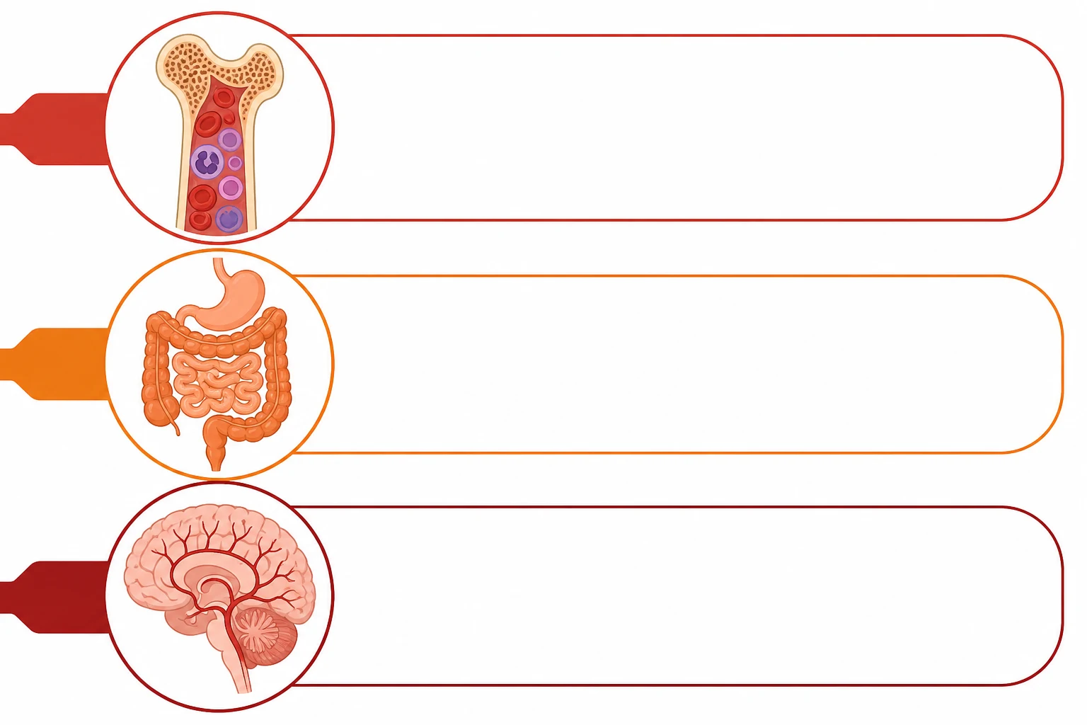
| Subsyndrome | Dose | Onset | Key features | Prognosis |
|---|---|---|---|---|
| Hematopoietic | 1-2 Gy | Days-weeks | Bone marrow suppression (pancytopenia), infection, bleeding | Treatable |
| GI | 6-8 Gy | Hours-days | Mucosal sloughing, diarrhoea, sepsis, fluid loss | Poor |
| Neurovascular | Above 30 Gy | Minutes-hours | Cerebral oedema, seizures, coma, cardiovascular collapse | Fatal |
The subsyndromes are dose-dependent and cumulative: as the dose rises, a more radioresistant system fails in addition to (not instead of) the more sensitive one. At 1-2 Gy the marrow is affected but the gut recovers. At 6-8 Gy BOTH the marrow and the gut fail (GI mortality from fluid loss and sepsis usually supersedes the marrow problem). Above 30 Gy the neurovascular system collapses so rapidly that the patient dies before the marrow and gut problems fully declare — the death is from cerebral oedema and vasomotor shock within hours to days.[1][3]
In practice a fourth subsyndrome — cutaneous radiation syndrome (CRS) — is recognised when localised skin doses are very high (a beta/gamma source held against the skin, or partial-body exposure). CRS runs its own timeline of erythema, dry/moist desquamation, blistering, necrosis and late fibrosis, and can dominate the clinical picture in partial-body accidents (e.g. industrial source exposure to a hand).[4][1]
The phases
The acute radiation syndrome evolves through four classical phases. The duration and severity of each phase is dose-dependent; at very high doses the phases compress and may merge.[1][1]
- The prodromal (the hours to the days) — the nausea, the vomiting, the fatigue, the headache, the fever. The time the to the onset → the dose the estimate (the earlier → the higher the dose).[1][1]
- The latent (the days to the weeks) — the apparent the recovery (the symptoms the subside). The deceptive.[1]
- The manifest illness (the days to the weeks) — the subsyndrome-specific (the pancytopenia, the GI, the neuro).[1][1]
- Recovery or death — determined by the whole-body dose and the adequacy of supportive care. With good support, survival is possible up to ~6-8 Gy; above ~8-10 Gy marrow recovery is unlikely without transplant; above ~10-12 Gy the GI syndrome is usually fatal regardless; above ~30 Gy death is rapid and inevitable.[1][3]
The prodromal phase is mediated by direct radiation injury to the gut mucosa and the vomiting centre, and by cytokine release. The onset, severity and duration of prodromal symptoms are dose-dependent: at 1-2 Gy vomiting starts 2-6 hours after exposure and lasts hours; at 6-8 Gy within 1-2 hours; above 10 Gy within minutes to an hour. This is the basis of the time-to-emesis rule for dose estimation.[3][4]
The latent phase is the deceptive interval (a few days to ~2-3 weeks) during which the patient looks and feels well — prodromal symptoms resolve — while the irradiated tissues are silently failing. The marrow stem cells that were lethally damaged die as they attempt division, and circulating cells are consumed without replacement; the gut mucosa thins. The latent phase is shorter the higher the dose (it may be absent altogether above ~10-15 Gy).[1]
The manifest illness phase is when the subsyndrome-specific failure declares: pancytopenia with infection and bleeding (hematopoietic), intractable diarrhoea with fluid loss and Gram-negative sepsis (GI), and cerebral oedema with seizures and coma (neurovascular).[1]
Dose estimation — time-to-emesis and the lymphocyte-depletion nomogram
Because patients usually do not know their exact dose, two clinical tools estimate whole-body dose at the bedside, and both drive triage and treatment intensity.[3][4]
1. Time to emesis (vomiting). The earlier the vomiting begins after exposure, the higher the dose. The Andrews / REAC/TS rules of thumb: vomiting within 1 hour → very high dose (above ~6 Gy, often fatal); within 1-2 hours → moderate-high dose (~4-6 Gy); after 2 hours → lower dose. Absence of vomiting within 4 hours makes a clinically significant (above ~2 Gy) exposure unlikely. Time-to-emesis is a rapid triage tool but is overridden by biological dosimetry.[4]
2. Serial absolute lymphocyte count (the Andrews lymphocyte-depletion nomogram). Lymphocytes are the most radiosensitive circulating cell and their rate of fall is a quantitative measure of whole-body dose. Draw a baseline lymphocyte count immediately and repeat at 24, 48 and 72 hours, then plot the fall against the Andrews nomogram. Rough landmarks: lymphocytes 1.0-1.5 x10^9/L at 24 hours implies ~1-2 Gy; 0.5-1.0 x10^9/L at 24 hours implies ~2-4 Gy; below 0.5 x10^9/L at 24 hours implies a severe exposure of above ~4 Gy; below 0.1 x10^9/L implies a lethal (above ~6-8 Gy) dose. A 50% drop in lymphocytes within 24 hours is itself a marker of significant exposure. The lymphocyte depletion kinetics are the single most reliable early biological dosimeter available in any hospital.[3][4]
3. Chromosomal dicentric assay (gold-standard biological dosimetry). Analysis of dicentric chromosomes in cultured peripheral lymphocytes is the most accurate method of dose estimation (the gold standard), but it takes several days and is available only in specialist reference laboratories. It is used to confirm/refine the bedside estimate, not to make acute decisions.[3]
The management
1. The decontamination + the isolation.[1]
- The remove the contaminated the clothing (the 90 per cent of the external the contamination). The wash the skin. The PPE for the staff.[1]
- Clothing removal eliminates approximately 90% of external contamination and must be done BEFORE any wash, transfer or further care. Bag clothing and all run-off as radioactive waste. Wash gently with soap and lukewarm water (do NOT scrub — skin damage increases absorption); pay attention to folds, wounds and body orifices. Staff use PPE (gowns, double gloves, booties, respiratory protection if aerosol risk) and follow time-distance-shielding principles. Critically ill patients are stabilised first, then decontaminated — life-saving care never waits for decontamination, but a contaminated patient is kept in a controlled area to avoid spreading contamination through the hospital.[3][1]
- The fluids, the electrolytes, the antiemetics. The transfusion (the RBC, the platelets, the irradiated the products).[1]
- The infection (the broad-spectrum the antibiotics; the antifungal; the antiviral — the neutropenic the sepsis).[1]
- Aggressive supportive care is the cornerstone of survival through the manifest-illness phase. The GI subsyndrome causes massive fluid and electrolyte losses from diarrhoea and vomiting — replace with crystalloid and electrolytes guided by haemodynamics. Antiemetics (5-HT3 antagonist such as ondansetron). All blood products must be irradiated and leukodepleted to prevent transfusion-associated graft-versus-host disease (TA-GvHD) — the immunocompromised ARS patient cannot reject donor T-lymphocytes, which then attack host tissues. Treat neutropenic sepsis empirically with broad-spectrum antipseudomonal beta-lactam, add antifungal (mould-active) and antiviral cover as indicated; reverse isolation and gut decontamination may be used.[1][3]
3. The cytokines.[2]
- The G-CSF / the GM-CSF (the granulocyte the colony the stimulating the factor — the accelerates the neutrophil the recovery).[2]
- Colony-stimulating factors (filgrastim, pegfilgrastim, sargramostim/GM-CSF) should be STARTED EARLY (within 24-72 hours of exposure) in any patient with a significant whole-body dose (above ~2 Gy) — do not wait for manifest neutropenia. They shorten the depth and duration of neutropenia and reduce infectious mortality. The Strategic National Stockpile Working Group (Waselenko 2004) specifically recommends early CSF use in adults exposed to above ~3 Gy. Pegfilgrastim offers convenient once-per-cycle dosing.[2][3]
4. The stem cell transplant (for the severe — the dose the above the 6-8 Gy; the bone the marrow the irreversible).[1]
- Haematopoietic stem-cell transplantation (HSCT) is considered when the marrow injury is judged irreversible — typically a whole-body dose above ~8-10 Gy without combined injury, with an HLA-matched donor available, and no lethal GI or neurovascular injury. Below ~8 Gy the marrow usually recovers with support alone; above ~10-12 Gy the refractory GI syndrome makes transplant futile. The European consensus (Cordelli 2025) emphasises early HLA typing and transfer to a transplant centre for the small, selected group who may benefit.[1]
Internal contamination — chelation and blocking/decorporation therapy
When a radionuclide has been inhaled, ingested or absorbed through a wound, internal contamination persists and continues to irradiate tissues until it is removed. Treatment is radionuclide-SPECIFIC and must be matched to the isotope. Three agents dominate and are among the highest-yield exam facts in radiation medicine: Ca/Zn-DTPA, Prussian blue and potassium iodide (KI).[5][1]
Medical countermeasures for internal contamination — match the agent to the radionuclide
| Agent | Radionuclide(s) targeted | Mechanism | Dose / route | Key clinical points |
|---|---|---|---|---|
| Ca-DTPA then Zn-DTPA (pentetic acid) | Plutonium (Pu-238/239), americium (Am-241), curium — the transuranics | Chelation — DTPA forms stable complexes with the metal that are renally excreted, accelerating removal | Ca-DTPA 1 g IV/NEB first (more effective initially), then Zn-DTPA 1 g IV/IM daily (better tolerated for maintenance; Ca-DTPA depletes trace metals — Mn, Zn, Mg) | Give as early as possible (most effective in first 24 h). Nebulised for inhaled contamination. Replete zinc/magnesium. FDA-approved |
| Prussian blue (ferric hexacyanoferrate, Radiogardase) | Caesium-137 (Chernobyl, Goiânia), thallium-201/208 | Ionic trapping in the gut — exchanges potassium for caesium/thallium, preventing intestinal (re)absorption and increasing faecal excretion | 1-3 g orally three times daily for at least 30 days | Traps the isotope in the lumen and interrupts enterohepatic recycling; stool (and the patient) remains radioactive — handle as waste. Constipation common. FDA-approved. NOT absorbed systemically |
| Potassium iodide (KI) | Radioactive iodine — I-131 (and I-132, I-133), e.g. reactor/fallout release | Thyroid blockade — saturates the thyroid with stable iodide so it cannot take up radioiodine (competitive inhibition of NIS) | Adults 130 mg PO once daily; most effective if given BEFORE or within ~4 h of exposure, little benefit after 24 h | Protects ONLY the thyroid and ONLY against radioiodine. Useless (and falsely reassuring) against caesium, strontium, plutonium. Risk of thyroid dysfunction in extremes of age — use selectively in adults >40 only when dose high; in children/pregnant at lower thresholds |
Ca-DTPA / Zn-DTPA for plutonium and transuranics. DTPA (diethylenetriaminepentaacetic acid) is a chelator chemically related to EDTA but with higher affinity for the heavy transuranic metals. The calcium salt (Ca-DTPA) is ~10 times more effective than the zinc salt (Zn-DTPA) for the FIRST dose after contamination, but repeated Ca-DTPA strips the body of essential trace metals (manganese, zinc, magnesium) and can be toxic; therefore the regimen is Ca-DTPA first, then Zn-DTPA for maintenance. DTPA is given IV for systemic contamination and by nebuliser for inhaled plutonium/americium. It is the specific antidote for the transuranics — but has NO role for caesium or iodine.[5][1]
Prussian blue for caesium and thallium. Prussian blue (insoluble ferric hexacyanoferrate) is not absorbed from the gut; it acts as an ion-exchange resin that traps caesium and thallium in the intestinal lumen, breaking their enterohepatic recirculation and shunting them into the stool. It was the principal agent used after the Goiânia accident (1987) — a scavenged caesium-137 radiotherapy source contaminated hundreds of people — and after Chernobyl to reduce body caesium burden. Treatment continues for weeks; the stool is radioactive and must be handled as waste. Crucially, Prussian blue does nothing for iodine or the transuranics.[5][1]
Potassium iodide (KI) for radioactive iodine. The thyroid concentrates iodide via the sodium-iodide symporter (NIS). Flooding the body with stable iodide saturates the transporter, so that radioiodine (I-131) cannot be taken up and is instead excreted renally. KI thus protects the thyroid — and ONLY the thyroid — against ONLY radioiodine. Timing is critical: it is most effective if given before or within ~4 hours of exposure, still useful up to ~12 hours, and offers little benefit after 24 hours. The classic population-level use is thyroid blockade after a reactor release (Chernobyl distributed KI to millions; this is credited with reducing paediatric thyroid cancer where given promptly). The major exam trap: KI does NOT protect against caesium (the principal Chernobyl/Goiania contaminant) or any non-iodine radionuclide — administering it after a caesium or unknown-source release is ineffective and gives a false sense of protection.[5][1]
Other, less common countermeasures: propylthiouracil/methimazole (block thyroid hormone synthesis from radioiodine), phosphates/pamidronate (for strontium-90, a calcium analogue that deposits in bone), alginates/Prussian blue analogues and barium sulphate (to reduce gut absorption of ingested radionuclides), and forced diuresis / haemodialysis for tritium (radioactive water) and other soluble isotopes.[5][1]
Prognosis
The hematopoietic (1-2 Gy) — the treatable (the supportive + the cytokines → the recovery). The GI (6-8 Gy) — the poor. The neurovascular (above 30 Gy) — the fatal.[1][1]
In practical terms: with modern supportive care and cytokines, survival is expected below ~4-5 Gy; possible but difficult at 5-8 Gy; unlikely above ~8-10 Gy without haematopoietic stem-cell transplant; and essentially impossible above ~10-12 Gy because of the refractory GI syndrome, even with transplant. Above ~30 Gy death occurs within hours to days from neurovascular collapse regardless of treatment. The prognostic boundary between "treatable" and "futile" is generally drawn at the 8-12 Gy whole-body dose, and triage decisions in a mass-casualty radiological event are driven by this — patients in the moderate (2-8 Gy) range get full ICU support and cytokines; those with lethal (above ~10-12 Gy) doses are designated expectant in a resource-constrained setting.[1][3]
Management of acute radiation syndrome (ARS)
- SCENE SAFETY + EXTERNAL DECONTAMINATION (do this first, but never delay life-saving care) — (a) PROTECT STAFF: PPE (gowns, double gloves, booties, eye/resp protection if aerosol risk); apply TIME, DISTANCE, SHIELDING (inverse-square law; dense material for gamma, hydrogenous for neutron, barriers for alpha/beta). (b) REMOVE CLOTHING — eliminates ~90% of external contamination; bag as radioactive waste. (c) WASH gently with soap and lukewarm water (NOT hot — vasodilates/increases absorption; NOT scrubbed — damages skin/increases absorption); attend to wounds, skin folds, hair, orifices. (d) SURVEY with a contamination monitor; repeat wash until count is at 2x background or no longer falling. (e) CONTAIN run-off. Critically ill patients: stabilise (ABC) THEN decontaminate, in a controlled area to prevent hospital spread.[3][1]
- DOSE ESTIMATION + TRIAGE (drives everything that follows) — (a) HISTORY: time, duration, distance, shielding, source type if known; time of exposure vs onset of vomiting. (b) TIME-TO-EMESIS: vomiting <1 h → above ~6 Gy (often fatal); 1-2 h → 4-6 Gy; after 2 h → lower dose; no vomiting by 4 h → below ~2 Gy likely. (c) SERIAL ABSOLUTE LYMPHOCYTE COUNT at 0/24/48/72 h plotted on the Andrews nomogram: lymphocytes below 1.0 at 24 h → above ~4 Gy; below 0.5 → above ~6 Gy; below 0.1 → lethal. (d) Send blood for DICENTRIC CHROMOSOME assay (gold standard, days to result). (e) TRIAGE: 0-1 Gy observe/discharge; 1-2 Gy admit, supportive; 2-8 Gy full ICU + cytokines ± transplant referral; above ~10-12 Gy expectant (refractory GI).[3][4]
- SUPPORTIVE ICU CARE through the manifest-illness phase — (a) AIRWAY/BREATHING: oxygen, lung-protective ventilation if ARDS (especially aspiration/pulmonary injury). (b) CIRCULATION/FLUIDS: GI subsyndrome causes massive fluid/electrolyte loss — aggressive crystalloid + electrolyte replacement guided by haemodynamics and UO; vasopressors for refractory shock. (c) ANTIEMETICS: ondansetron for prodromal vomiting. (d) NUTRITION: early enteral where possible; TPN if severe GI mucosal failure. (e) GI: treat diarrhoea, fluid/electrolyte loss; manage mucosal sloughing. (f) SKIN: cutaneous radiation syndrome — wound care, debridement of necrotic tissue, surgical excision/grafting for deep local injury.[1][1]
- BLOOD PRODUCTS — IRRADIATED AND LEUKODEPLETED ONLY — All transfusions in ARS must be irradiated (25 Gy) and leukodepleted to PREVENT transfusion-associated graft-versus-host disease (TA-GvHD): the patient's immunocompromised marrow cannot reject donor T-lymphocytes, which then mount a fatal graft-vs-host reaction. Transfuse RBCs for anaemia, platelets for thrombocytopenia/bleeding (keep platelets above 10-20, or above 50 if bleeding/invasive procedure). Avoid non-irradiated products absolutely.[1][3]
- INFECTION PROPHYLAXIS + TREATMENT (neutropenic sepsis is the leading cause of death) — (a) PROPHYLAXIS: reverse/laminar-flow isolation where possible; gut decontamination; antiviral (acyclovir if HSV-seropositive), antifungal (fluconazole/mould-active), antibacterial (fluoroquinolone) prophylaxis during profound neutropenia. (b) EMPIRIC TREATMENT of any fever in neutropenia: broad-spectrum antipseudomonal beta-lactam (pip-tazobactam/meropenem) immediately; escalate per local neutropenic-sepsis protocol; add antifungal for persistent fever. (c) Consider immunoglobulin for severe hypogammaglobulinaemia.[1][3]
- CYTOKINES — START EARLY (within 24-72 h) — Begin G-CSF/GM-CSF (filgrastim 5 microg/kg/day SC, or pegfilgrastim 6 mg once per cycle; sargramostim/GM-CSF 250 microg/m^2/day) in any patient with a significant whole-body dose (above ~2-3 Gy), BEFORE manifest neutropenia develops. CSFs shorten neutropenia depth/duration and reduce infectious mortality (Waselenko/SNS Working Group 2004; Herodin 2026). Continue until ANC recovery.[2][3]
- INTERNAL CONTAMINATION — MATCHED COUNTERMEASURE (give the right agent for the right isotope) — Identify the radionuclide. (a) PLUTONIUM/americium/curium (transuranics) → Ca-DTPA 1 g IV (first dose) then Zn-DTPA 1 g IV/IM daily; nebulised for inhalation; replete zinc/magnesium. (b) CAESIUM-137/thallium → Prussian blue 1-3 g PO TDS for 30+ days (interrupts enterohepatic recirculation; stool is radioactive). (c) RADIOACTIVE IODINE (I-131) → potassium iodide 130 mg PO daily — give BEFORE or within 4 h of exposure (little benefit after 24 h); protects the THYROID only and ONLY against iodine. (d) STRONTIUM → phosphate/pamidronate; TRITIUM → forced fluids/dialysis. Do NOT give KI for caesium or unknown-source contamination.[5][1]
- HAEMATOPOIETIC STEM-CELL TRANSPLANT — for the selected few with irreversible marrow — Consider HSCT when whole-body dose is above ~8-10 Gy (marrow recovery unlikely), there is a suitable HLA-matched donor, AND no lethal GI (dose above ~10-12 Gy) or neurovascular injury. Early HLA typing; transfer to transplant centre. Below ~8 Gy the marrow usually recovers with support alone and transplant is not needed; above ~10-12 Gy the refractory GI syndrome makes transplant futile. (Cordelli 2025 European consensus.)[1]
Exam practice — SAQs
SAQ — Industrial gamma-source exposure: acute radiation syndrome
10 minutes · 10 marks
A 38-year-old technician is brought to the emergency department 3 hours after accidental whole-body exposure to a damaged industrial cobalt-60 (Co-60) gamma source at a sterilisation facility. He vomited once, 90 minutes after the exposure, and has had two episodes of watery diarrhoea. He is anxious and nauseated but haemodynamically stable: HR 96, BP 112/68, RR 20, SpO2 98 percent on room air, GCS 15, temperature 37.4 degrees C. Baseline (time 0) absolute lymphocyte count was 1.8 x10^9/L; at 24 hours it is 0.6 x10^9/L. He has diffuse erythema over the anterior chest and abdomen. There is no associated trauma or burn.
SAQ — Acute radiation syndrome: neutropenic sepsis in the manifest-illness phase
10 minutes · 10 marks
A 45-year-old nuclear-facility technician is admitted to the ICU three weeks after an initially unrecognised whole-body radiation exposure (a sealed caesium-137 source was breached during a maintenance task; biological dosimetry later estimates ~3-4 Gy). He is febrile, tachycardic and hypotensive, with severe oral mucositis and bleeding gums. Investigations show profound pancytopenia: neutrophils 0.2 x10^9/L, platelets 12 x10^9/L, haemoglobin 65 g/L; blood cultures grow Pseudomonas aeruginosa. He did not receive any cytokine therapy at the time of exposure.
SAQ — Radiation emergency management: mass-casualty radiological dispersal device
10 minutes · 10 marks
A radiological dispersal device (commonly called a dirty bomb) detonates in a city centre, dispersing caesium-137 (Cs-137) over a populated area. Your hospital incident team begins receiving contaminated casualties, several of whom also have blast trauma and burns. You are the intensivist coordinating radiation emergency management.
Clinical pearls
Red flags
Prognosis, evidence and landmark data
Radiation injury evidence, consensus statements and outcomes
Waselenko 2004 — Strategic National Stockpile Radiation Working Group (Annals of Internal Medicine): the seminal consensus on medical management of ARS. Recommendations: (1) risk-stratify by whole-body dose; (2) early use of colony-stimulating factors (G-CSF/GM-CSF) in adults exposed to above ~3 Gy, started within days and continued to ANC recovery; (3) irradiated/leukodepleted blood products; (4) infection prophylaxis and empiric neutropenic-sepsis therapy; (5) consider HSCT above ~8-10 Gy with a matched donor and no lethal GI/neurovascular injury. Foundation of modern ARS management.[3] Cordelli 2025 / European consensus (Disaster Med Public Health Prep): updated European approach to ARS in a radiation emergency — early HLA typing, transfer to specialist/transplant centres for the selected few (above ~8-10 Gy, suitable donor, no lethal GI injury), cytokine use (G-CSF/pegfilgrastim), and the dose-stratified triage (2-8 Gy full support; above ~10-12 Gy expectant).[1] Herodin 2026 (J Radiol Prot): cytokine use in the hematopoietic subsyndrome — evidence for early (within 24-72 h) G-CSF/GM-CSF/pegfilgrastim to shorten neutropenia depth/duration and reduce infectious mortality; reinforces the 'start early, don't wait for manifest neutropenia' principle.[2] Flynn & Goans 2002 (Surg Clin North Am): triage and medical management of radiation and combined-injury casualties — the Andrews lymphocyte-depletion nomogram, the time-to-emesis rule for dose estimation, and the dose-stratified mass-casualty triage (the 8-12 Gy boundary between treatable and expectant). Defines the bedside biological dosimetry used worldwide.[4] Moulder 2005 (Health Physics): review of post-irradiation treatments for radiological-terrorism/accident casualties — the countermeasure pharmacopoeia: Ca/Zn-DTPA for the transuranics, Prussian blue for caesium/thallium, potassium iodide for radioiodine, and adjuncts (phosphates for strontium, forced diuresis for tritium). Emphasises matching the agent to the isotope.[5] ICRP Publication 118 (2012): re-evaluation of tissue-reaction thresholds — LOWERED the cataract threshold to ~0.5 Gy (from ~2-5 Gy) and established ~0.5 Gy for circulatory disease; the authoritative source for deterministic-effect thresholds. ICRP Publication 103 (2007): the linear-non-threshold model for stochastic (cancer) risk, ~5% per Sv, no threshold — the basis of all radiation-protection dose limits and ALARA.[1][1] Historical dose-response anchors: Chernobyl (1986) — mass caesium/iodine release; KI distribution credited with reducing paediatric thyroid cancer where prompt; ARS in ~134 liquidators, 28 early deaths. Goiania (1987) — a scavenged Cs-137 radiotherapy source contaminated ~250 people; Prussian blue reduced body burden; 4 deaths. Tokaimura (1999) — criticality accident, two workers received above 10 Gy, both died. These anchor the clinical dose-response and countermeasure data.[5][1] Mortality by dose (modern support + cytokines): below ~4-5 Gy — expected survival; 5-8 Gy — survival possible but difficult (full ICU, cytokines, ± HSCT); above ~8-10 Gy — marrow recovery unlikely (HSCT if donor + no lethal GI); above ~10-12 Gy — refractory GI syndrome, near-100% mortality despite transplant (expectant); above ~30 Gy — rapid neurovascular death within hours-days. Leading cause of death in the treatable range is neutropenic sepsis, then bleeding/haemorrhage.[1][3]
References
- [1]Cordelli E, et al. European Approach for Acute Radiation Syndrome's Management Facing Radiological and Nuclear Threats: A Call for Arms Disaster Med Public Health Prep, 2025.PMID 41234135
- [2]Herodin F, et al. Cytokine use in the hematopoietic subsyndrome of acute radiation syndrome (H-ARS): implications for the role of cytokines in a mass casualty radiologic/nuclear (R/N) emergency J Radiol Prot, 2026.PMID 41678840
- [3]Waselenko JK, MacVittie TJ, Blakely WF, Pesik N, Wiley AL, Dickerson WE, et al. Medical management of the acute radiation syndrome: recommendations of the Strategic National Stockpile Radiation Working Group Ann Intern Med, 2004.PMID 15197022
- [4]Flynn DF, Goans RE. [New hope for heart failure patients. Fatigued hearts need medication and electrical aids] MMW Fortschr Med, 2002.PMID 12116546
- [5]Moulder JE. Cardiac rehabilitation and getting to lipid goals J Cardiopulm Rehabil, 2005.PMID 16217228