ICU · Equipment, physics & clinical measurement
Radiation Safety
Also known as Radiation safety · ALARA · Sievert · Gray · Inverse square law · Stochastic vs deterministic effects · Film badge · Time distance shielding · Dose limits · Lead apron · Half value layer · CT dose index · Dosimetry · Effective dose
Radiation safety for the ICU First Part: the dose units (Gray for absorbed dose, Sievert for equivalent and effective dose), typical doses (chest X-ray ~0.02 mSv, CT chest ~7 mSv which is ~350 chest X-rays (commonly quoted as ~200-400), CT abdomen-pelvis 10-20 mSv), the ALARA principle (As Low As Reasonably Achievable) and the three protections (time - minimise exposure time; distance - inverse-square law, doubling distance quarters dose; shielding - lead aprons at least 0.5 mm Pb equivalent, thyroid shields, lead glasses, gonadal shielding), the distinction between stochastic effects (cancer, hereditary - NO threshold - probability proportional to cumulative dose) and deterministic effects (tissue reactions - skin erythema ~2 Gy, cataracts ~0.5 Gy, sterility - WITH a threshold), the dose limits (public 1 mSv/yr, occupational whole body 20 mSv/yr averaged over 5 years and no more than 50 mSv in any single year, lens 150 mSv/yr, skin and extremities 500 mSv/yr, pregnant worker fetal 1 mSv for the remainder of pregnancy), personal dosimetry (film badge, TLD, OSL dosimeter worn under the lead apron), and radiation in pregnancy (minimise CT - prefer ultrasound or MRI - if CT is necessary shield the abdomen and use a low-dose protocol).
On this page & tools
Your progress
Saved locally on this device.
8 MCQs with explanations
Target exams
Red flags
Overview
Ionising radiation is used in the ICU for portable chest imaging, computed tomography (CT), fluoroscopy during line placement and other procedures, and occasionally interventional radiology. The risks for an individual study are small but accumulate over a patient's admission and over a clinician's career, so radiation protection follows a defined set of units, principles, and limits. The governing goal is to obtain the diagnostic information needed at the lowest dose that is reasonably achievable - the ALARA principle (As Low As Reasonably Achievable).[1][1]
ALARA rests on two complementary justifications: any imaging study must first be justified (it must do more good than harm) and then optimised (the dose must be kept as low as possible while still answering the clinical question). For staff, protection is achieved through the three cardinal measures - time, distance, and shielding.[1]
Dose units
Three quantities are examinable, and candidates must keep them distinct:[1]
- Absorbed dose (the energy deposited per unit mass of tissue) is measured in Gray (Gy), where 1 Gy = 1 joule per kilogram. Absorbed dose describes the physical energy deposition and is independent of radiation type or tissue.
- Equivalent dose, measured in Sievert (Sv), multiplies the absorbed dose by a radiation weighting factor (wR) that accounts for the differing biological damage of radiation types. X-rays, gamma rays, and beta particles have a weighting factor of 1; alpha particles 20; neutrons 5-20 depending on energy. For the diagnostic X-rays used in the ICU, wR = 1, so absorbed dose in Gy and equivalent dose in Sv are numerically equal.
- Effective dose, also in Sievert, further multiplies the equivalent dose by a tissue weighting factor (wT) that reflects the variable radiosensitivity of organs (bone marrow, colon, lung, stomach, breast, and gonads are among the most sensitive). Effective dose gives a single whole-body risk estimate that allows doses from different examinations and to different body regions to be compared - it is the quantity used to express the dose of a CT chest or a chest X-ray.[1]
Absorbed, equivalent, and effective dose - the three quantities
| Quantity | Symbol | Unit | Definition | Radiation weighting factor (wR) | Tissue weighting factor (wT) |
|---|---|---|---|---|---|
| Absorbed dose | D | Gray (Gy) | Energy deposited per unit mass (1 Gy = 1 J/kg) | Not applied | Not applied |
| Equivalent dose | H_T | Sievert (Sv) | Absorbed dose x radiation weighting factor (per tissue) | Applied (X-ray = 1, alpha = 20) | Not applied |
| Effective dose | E | Sievert (Sv) | Sum over tissues of (equivalent dose x tissue weighting factor) | Applied | Applied (bone marrow, breast, gonads high) |
Because diagnostic X-rays have wR = 1, the Gray and the Sievert are interchangeable for X-rays (1 Gy = 1 Sv). The distinction matters only for particulate radiation. Practical submultiples: 1 mSv = 1000 microSv; background radiation is about 2-3 mSv per year (so 1 mSv is about 4-5 months of natural background). [1]
Typical doses
Knowing the approximate effective dose of common examinations is examinable and gives a sense of scale:[1][3]
- A chest X-ray is about 0.02 mSv (roughly 3 days of natural background radiation).[3]
- A CT chest is about 7 mSv - approximately 350 chest X-rays (commonly quoted as ~200-400, about 2 years of background).[2][3]
- A CT abdomen-pelvis is about 10-20 mSv (about 500-1000 chest X-rays, or 3-7 years of background).[3][4]
- A CT head is about 2 mSv; a CT pulmonary angiogram (CTPA) about 5-15 mSv.[3]
- Fluoroscopy dose depends on screening time and is measured as a dose-area product (DAP); bedside procedures add a few mSv of staff scatter per minute of screening.[1]
Typical effective doses of common imaging examinations
| Examination | Approximate effective dose | Equivalent chest X-rays | Equivalent background |
|---|---|---|---|
| Chest X-ray (PA) | ~0.02 mSv | 1 | ~3 days |
| Limb X-ray | < 0.01 mSv | < 1 | < 1 day |
| CT head | ~2 mSv | ~100 | ~8 months |
| CTPA (CT pulmonary angiogram) | ~5-15 mSv | ~250-750 | ~2-5 years |
| CT chest | ~7 mSv | ~350 | ~2 years |
| CT abdomen-pelvis | ~10-20 mSv | ~500-1000 | ~3-7 years |
| Annual natural background | ~2-3 mSv | ~100-150 | 1 year |
| ICRP annual occupational limit | 20 mSv | ~1000 | ~7 years |
The ALARA principle
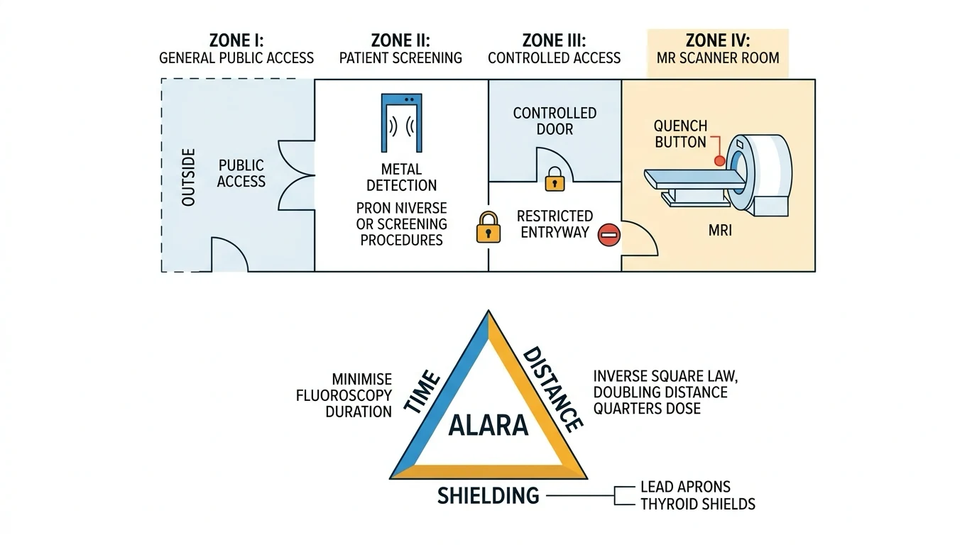
ALARA - As Low As Reasonably Achievable - is the foundational doctrine of radiation protection.[1] It is not a number but a philosophy: the dose from any imaging study should be no higher than is needed to obtain the diagnostic information. ALARA derives from the fact that stochastic effects (cancer, hereditary harm) have no threshold - every exposure, however small, adds some risk - so dose must be justified and then minimised.
ALARA has two pillars:[1]
- Justification - no examination should be performed unless it does more good than harm. The clinician ordering the study bears this responsibility: is the question answerable by ultrasound, MRI, or a study already done? The ICU patient accumulates scans quickly, and unnecessary CTs add real risk.
- Optimisation - once a study is justified, the dose should be kept as low as reasonably achievable through protocol selection (low-dose CT, reduced milliampere-seconds, limiting the scan range, shielding, collimation), and for staff through the three protections (time, distance, shielding). [1]
For staff, ALARA translates into the three cardinal protections described below. For the patient, ALARA is achieved by the radiographer/CT protocol (collimation, tube current modulation, pitch, limiting scan length) and by the clinician asking whether the scan is needed at all. [1]
The three protections - time, distance, and shielding
Radiation protection for staff (and for the patient where possible) rests on three cardinal measures, all of which reduce exposure. They are easiest to remember as a triad.[1][1]
1. Time - minimise exposure time
Minimise the time spent near the source. Dose is proportional to exposure time: doubling the screening time doubles the dose. Practical steps:[1]
- Use pulsed fluoroscopy at the lowest acceptable frame rate (e.g., 3-7.5 pulses per second rather than continuous 25-30 fps) - this alone can cut dose by half or more.
- Pre-position the patient and equipment before screening; use the last-image-hold function to review anatomy without re-screening.
- Plan tube angulation and tabletop position with the beam off.
- Take the fewest exposures that answer the question. [1]
2. Distance - maximise distance from the source
Dose falls with the inverse square of distance from the source. This is the inverse-square law and is the most powerful single protective measure.[1]
- Doubling the distance from the source quarters the dose (1/2^2 = 1/4).
- Trebling the distance reduces it to one-ninth.
- Halving the distance quadruples the dose. [1]
During any exposure - a portable chest X-ray on the unit, or fluoroscopy in theatre - staff should step back at least 2 metres from the patient and the X-ray tube. Scatter falls off rapidly, so a few steps dramatically reduce staff dose. Where possible, leave the room during portable imaging, standing behind the lead-lined control panel or a mobile lead screen.[1]
Inverse-square law worked example - dose vs distance from source
| Distance from source | Relative dose | Comment |
|---|---|---|
| 0.5 m | 1 (reference) | Close to patient - high scatter dose |
| 1 m | 1/4 | Doubling distance quarters dose |
| 2 m | 1/16 | Stepping back to the doorway |
| 3 m | 1/36 | Across the bed space |
| 4 m | 1/64 | Far side of the room |
3. Shielding - interpose a barrier
Place a radiation-attenuating barrier (most commonly lead) between the source and the person.[1]
- Lead aprons - worn for any fluoroscopy or proximity to the beam; a minimum of 0.5 mm lead (Pb) equivalent over the front (often 0.25 mm front and 0.25 mm back for a wraparound apron). The apron covers the radiosensitive bone marrow of the spine, pelvis, and sternum.[1]
- Thyroid shields - a separate collar of 0.5 mm Pb equivalent protecting the thyroid, highly radiosensitive.
- Lead glasses - protect the lens of the eye (the deterministic threshold for cataracts is low, ~0.5 Gy).
- Lead gloves - when hands are near the beam (rare; usually hands should be OUT of the beam entirely).
- Mobile lead screens and lead-lined walls/doors - structural shielding for fixed X-ray rooms.
- For the patient: gonadal shielding (where it does not obscure the region of interest), collimation of the beam to the area of interest (the single most important patient-dose reducer for plain film), and the lowest-dose protocol that answers the question.
Shielding materials and the half-value layer (HVL)
The effectiveness of a shield is described by the half-value layer (HVL): the thickness of a material that halves the intensity of the radiation beam. For diagnostic X-rays (around 100 kVp):[1]
- The HVL of lead is about 0.1-0.3 mm - hence a 0.5 mm lead apron attenuates the beam by far more than half (about 90-95 per cent at typical diagnostic energies).
- The HVL of concrete is about 40-50 mm, of barium plaster a few millimetres, and of water/tissue about 30-40 mm (which is why the patient is their own shield, and why scatter rather than the primary beam is what reaches staff). [1]
Lead equivalence is the practical descriptor of aprons: a 0.5 mm Pb-equivalent apron attenuates around 90-99 per cent of scatter at diagnostic energies, and is the minimum standard. Heavier 1.0 mm Pb-equivalent aprons offer more attenuation but are heavier and less well tolerated - the gain is small for the comfort cost. [1]
CT radiation dose
CT is the largest single source of medical radiation exposure for many ICU patients, and its use has risen steeply. A single CT chest delivers about 7 mSv - roughly 350 chest X-rays (commonly quoted as ~200-400, depending on technique), or about two years of natural background.[2][3] A CTPA for suspected pulmonary embolism delivers 5-15 mSv, and an abdomen-pelvis CT 10-20 mSv. Cumulative CT exposure over an admission is not trivial: in the largest dose-response studies, CT in childhood and adolescence was associated with a measurable increase in subsequent leukaemia and brain-tumour risk, with a dose-response relationship.[5][6]
CT dose is described by three quantities the candidate should know:[1]
- CT dose index (CTDIvol) - the absorbed dose within a single rotation, measured in mGy; a measure of scanner output for a given protocol.
- Dose-length product (DLP) - CTDIvol multiplied by the scan length (mGy.cm); a measure of total dose delivered for the examination.
- Effective dose (mSv) - estimated from the DLP using a region-specific conversion factor (k, in mSv/mGy.cm; roughly 0.014 for chest, 0.015 for abdomen, 0.002 for head). This is the figure quoted as "the dose" of a CT. [1]
Dose reduction in CT (all part of ALARA for the patient) is achieved by:[1]
- Automatic tube current modulation (ATCM) - the scanner reduces mA as it rotates through thin body regions.
- Reducing scan length to the region of interest only.
- Increasing pitch (table travel per rotation) where image quality allows.
- Lower kVp protocols (e.g., 80 or 100 kVp) for CTPA in smaller patients - the iodine contrast is higher relative to soft tissue at lower kVp, allowing lower dose with better vessel opacification.
- Iterative reconstruction algorithms that allow lower mA while preserving image quality.
- Avoiding non-indicated multiphase scans (non-contrast plus arterial plus venous plus delayed) - each phase roughly doubles dose. [1]
Reducing CT dose in the ICU patient (ALARA for the patient)
- Justify the scan first. Is the question already answered by a recent study, by ultrasound, or by clinical assessment? Cumulative ICU CT dose is a real risk - the requesting clinician carries this responsibility.[2][7]
- Choose the lowest-dose modality that answers the question. Ultrasound and MRI do not use ionising radiation. Where CT is needed, request the minimum: single phase, limited scan range, no non-contrast phase if contrast is diagnostic.
- Use a low-dose or weight-adjusted protocol. For CTPA in a smaller patient, request 80-100 kVp. For follow-up (e.g., serial abdominal collections), a low-dose protocol is usually sufficient.
- Limit multiphase scanning. Each contrast phase roughly doubles dose. Multi-phase protocols should be reserved for specific indications (e.g., characterising a liver lesion, renal mass, or vascular injury).
- Track cumulative dose in the chronically ventilated patient who accrues many scans - the lifetime attributable cancer risk rises with each study, particularly in younger patients.[8]
- Document the indication clearly so that the radiologist can tailor the protocol - ALARA is a shared clinician-radiographer responsibility.[1]
Stochastic versus deterministic effects
The biological effects of ionising radiation fall into two categories whose distinction is central to the exam and to dose limits.[1][1]
Stochastic effects - no threshold
Stochastic (probabilistic) effects are those in which the probability of harm rises with dose, but with no threshold - any exposure, however small, carries some risk. The two stochastic effects are cancer (somatic) and hereditary (genetic, affecting future offspring).[1][1]
Because there is no threshold, every exposure adds some risk, which is why ALARA applies even to small doses. The risk is roughly linear with cumulative dose. The lifetime excess cancer risk from a single 10 mSv CT is estimated at about 1 in 1000 to 1 in 2000 in adults, higher in children.[7][8]
Deterministic effects - with a threshold
Deterministic effects (now called tissue reactions by the ICRP) are those in which the severity of harm rises with dose above a threshold - below the threshold there is no clinically meaningful harm. Deterministic effects are the concern after high-dose fluoroscopy, prolonged interventional procedures, or accidental exposure.[1]
Key thresholds (ICRP 118):[1]
- Skin erythema - threshold around 2 Gy (early transient erythema may occur at ~2 Gy; main erythema at ~6 Gy; moist desquamation at ~15 Gy).
- Temporary epilation - around 3 Gy; permanent epilation above ~7 Gy.
- Cataracts (lens of eye) - threshold around 0.5 Gy (ICRP 118 lowered this substantially from the earlier ~0.5-2 Gy, with a single acute dose of ~0.5 Gy sufficient).
- Temporary sterility - testes ~0.15 Gy, ovaries ~0.6-1.5 Gy; permanent sterility at ~3.5-6 Gy (testes) and ~2.5-6 Gy (ovaries).
- Bone marrow depression - ~0.5 Gy; fatal marrow syndrome at whole-body doses above ~3-5 Gy.
- Depression of intellectual development in the fetus above ~100 mGy (8-25 weeks gestation is the window of maximal sensitivity). [1]
These thresholds underpin the dose limits: occupational limits keep the whole-body, lens, skin, and extremity doses well below the deterministic thresholds even over a working lifetime.[1]
Stochastic vs deterministic effects - the core distinction
| Feature | Stochastic effects | Deterministic (tissue reactions) |
|---|---|---|
| Threshold | None - any dose carries some risk | Yes - a threshold dose must be exceeded |
| What changes with dose | The probability of the effect | The severity of the effect |
| Examples | Cancer, hereditary (genetic) harm | Skin erythema, cataracts, sterility, marrow suppression, epilation |
| Relevance | Justify and minimise every exposure (ALARA) | Concern after high-dose fluoroscopy / accidental exposure |
| Underpins | Dose limits for cancer risk reduction | Dose limits that stay below tissue thresholds |
| Examinable thresholds | None (no threshold) | Skin erythema ~2 Gy; cataracts ~0.5 Gy; temporary sterility ~0.15-1.5 Gy |
Deterministic (tissue reaction) thresholds - examinable
| Tissue / effect | Approximate threshold dose | Notes |
|---|---|---|
| Lens of eye - cataracts | ~0.5 Gy (acute) | ICRP 118 (2012) lowered from earlier ~1.5 Gy; basis for the tightened lens limit |
| Skin - early transient erythema | ~2 Gy | Main erythema ~6 Gy; moist desquamation ~15 Gy |
| Skin - temporary epilation | ~3 Gy | Permanent epilation above ~7 Gy |
| Bone marrow - depression | ~0.5 Gy | Fatal syndrome above ~3-5 Gy whole body |
| Testes - temporary sterility | ~0.15 Gy | Permanent sterility ~3.5-6 Gy |
| Ovaries - sterility | ~2.5-6 Gy | Temporary effects at lower doses |
| Fetus - intellectual disability | ~100 mGy (8-25 weeks) | Window of maximal CNS sensitivity |
Dose limits
Dose limits translate the principles into numbers that staff and regulators can enforce. The ICRP 103 (2007) limits are widely adopted and are the exam answer:[1]
ICRP dose limits (Publication 103, 2007)
| Group / tissue | Annual limit | Averaged / notes |
|---|---|---|
| Occupational - effective dose (whole body) | 20 mSv/year | Averaged over 5 years (100 mSv in 5 years); no more than 50 mSv in any single year |
| Occupational - lens of eye | 150 mSv/year | Tightened to 20 mSv/year by ICRP 118 (2012) for new recommendations; many jurisdictions now adopt 20 mSv/year |
| Occupational - skin, hands, feet (extremities) | 500 mSv/year | To the most exposed 1 cm^2 of skin |
| Public - effective dose | 1 mSv/year | May be higher in a single year provided the 5-year average stays at or below 1 mSv/year |
| Pregnant worker - fetal dose | 1 mSv | For the remainder of the declared pregnancy (surface of the abdomen); dosimeter worn under the apron at waist level |
| Comforters and carers | ~5 mSv per episode | (e.g., a parent holding a child during imaging) |
Key points to remember:[1]
- The 20 mSv/year occupational whole-body limit is averaged over 5 years - a worker may receive up to 50 mSv in one year provided the 5-year average does not exceed 20 mSv/year.
- The 1 mSv/year public limit is far below occupational limits, reflecting that the public does not benefit from the work and includes children and pregnant women.
- The 500 mSv/year skin/extremity limit is much higher than the whole-body limit because skin and extremities are less radiosensitive than the bone marrow and gonads (which dominate the whole-body effective dose).
- The lens limit (150 mSv/year, tightened to 20 mSv/year) reflects the lower deterministic threshold for cataracts established in ICRP 118.
- The pregnant worker fetal limit (1 mSv for the remainder of pregnancy) is comparable to the public limit and is intended to keep fetal risk negligible. [1]
Personnel monitoring and dosimetry
Staff who work with ionising radiation wear a personal dosimeter to record cumulative dose. The dosimeter is worn under the lead apron at the level of greatest whole-body exposure (chest or waist), to estimate the dose to the bone marrow. A second dosimeter may be worn outside the apron at the collar or on the temple to estimate dose to the thyroid and lens.[1][1]
Three technologies are in common use:[1]
- Film badge - photographic film in a holder with filters; the darkening indicates dose and energy. Cheap but relatively insensitive at low doses and must be developed monthly.
- Thermoluminescent dosimeter (TLD) - lithium fluoride crystals store energy from radiation and release it as light when heated; more sensitive and accurate than film.
- Optically stimulated luminescence (OSL) - aluminium oxide crystals stimulated by a laser; highly sensitive, accurate, and the modern standard for personal dosimetry (e.g., the Landauer InLight badge).
- Electronic personal dosimeter (EPD) - real-time digital readout, useful in fluoroscopy where immediate feedback on dose rate encourages dose-reducing behaviour. [1]
Dosimeters are read at set intervals (monthly or quarterly) and the cumulative dose is logged against the worker's record; exceeding investigation levels triggers review of working practice. [1]
Personal dosimetry technologies
| Type | Principle | Sensitivity | Use |
|---|---|---|---|
| Film badge | Photographic film darkening | Moderate; needs monthly development | Traditional; cheap |
| TLD (thermoluminescent) | Crystal releases light when heated | High | Long-term monitoring |
| OSL (optically stimulated) | Crystal stimulated by laser | Very high | Modern standard (e.g., Landauer) |
| Electronic (EPD) | Solid-state detector, real-time | Very high, instantaneous | Fluoroscopy, immediate feedback |
Radiation and pregnancy
Pregnancy is the single setting in which radiation decisions most often cause anxiety and confusion; the principles are clear.[1][1]
For the pregnant patient
- Minimise CT. Where possible, answer the clinical question with ultrasound or MRI (neither uses ionising radiation).[1]
- If CT is necessary, shield the abdomen with lead where it does not obscure the region of interest, use a low-dose protocol (e.g., reduced mA, single phase, limited scan range), and image only the region needed. CTPA for suspected PE in pregnancy is a common justified indication; the fetal dose from a CTPA (around 0.1-0.5 mGy) is far below the deterministic threshold and the small stochastic risk is justified by the diagnostic need.[1]
- Fetal deterministic thresholds: above ~100 mGy (10 rad) there is a measurable risk of intellectual disability and growth restriction, with the 8-25 week window of maximal CNS sensitivity. Most diagnostic CT delivers fetal doses well below 50 mGy, so deterministic effects are not a concern for diagnostic studies - the relevant risk is the small stochastic risk of childhood cancer.[1]
- The traditional "10-day rule" (defer non-urgent irradiation until after the next period) and "28-day rule" have been superseded by the principle that any study that is clinically justified should proceed, and any that is not should be deferred regardless of cycle timing.[1]
- After an in-utero exposure of less than 100 mGy (the vast majority of diagnostic studies), the risk is not considered grounds for recommending termination of pregnancy.[1]
For the pregnant worker
- Once pregnancy is declared, the fetal dose is limited to 1 mSv for the remainder of the pregnancy.[1][1]
- The worker continues to use lead aprons, thyroid shields, and distance from scatter; a dosimeter is worn under the apron at waist level (over the fetus) to monitor fetal dose directly.
- In practice, most fluoroscopy and ICU imaging work can continue safely within these limits; the pregnant worker is not barred from the ICU or theatre, only from work that would exceed the fetal limit.
Imaging a pregnant ICU patient - the decision sequence
- Ask whether imaging is truly needed now. Defer non-urgent imaging. If the question is urgent, justify the study explicitly.[1]
- Choose the lowest-radiation modality that answers the question. Prefer ultrasound (bedside echo, abdominal, vascular) and MRI (no ionising radiation).[1]
- If ionising radiation (X-ray or CT) is necessary, shield the abdomen where it does not obscure the region of interest, and request a low-dose, single-phase, limited-scan-range protocol.[1]
- Consider the indication. A CTPA for suspected PE in pregnancy is justified - the fetal dose (~0.1-0.5 mGy) is far below deterministic thresholds and the small stochastic risk is outweighed by the diagnostic need. A V/Q scan is an alternative with comparable or lower fetal dose.[1]
- Reassure - for fetal doses < 100 mGy (essentially all diagnostic studies), the risk of deterministic harm is negligible and termination of pregnancy is not indicated. The relevant residual risk is the small stochastic risk of childhood cancer.[1]
- Document the indication, the modality chosen, and the dose-reduction measures - this is part of ALARA and medico-legally important.[1]
Paediatric considerations
Children are more sensitive to radiation than adults: their cells are dividing more rapidly, they have more of their life ahead in which a radiation-induced cancer may emerge, and the same CT (set to adult parameters) delivers a higher organ dose to a smaller body. Paediatric CT must use weight- or age-adjusted protocols - reduced mA, reduced kVp, and limited scan range - to keep dose ALARA. Pearce et al (2012) and Mathews et al (2013) demonstrated a measurable increase in leukaemia and brain-tumour risk after CT in childhood and adolescence, which has driven the move to paediatric dose reduction and alternative imaging.[5][6]
In the ICU this applies to any paediatric or young-adult patient; the requesting clinician should explicitly note the patient's age/size so the radiographer selects a paediatric protocol. [1]
Radiation safety in common ICU procedures
- Portable chest X-ray - the commonest ICU imaging. Staff should step back at least 2 metres during exposure (preferably leave the bed space). Scatter from a single chest X-ray is tiny, but cumulative exposure over a career matters.[1]
- CT - the requesting team does not usually attend; the patient is transported to radiology. The dose concern is for the patient (cumulative dose, paediatric protocols, pregnancy) rather than staff.[2]
- Fluoroscopy (in theatre or angiography, e.g., for line placement, GI studies, vascular procedures) - the principal staff hazard. Use pulsed low-dose fluoroscopy, keep hands out of the primary beam, wear a lead apron, thyroid shield, and lead glasses, and step back. Scatter is highest on the same side as the X-ray tube (i.e., towards the source), so standing on the image-intensifier side of the patient reduces dose.[1]
- Interventional procedures - prolonged fluoroscopy can deliver skin doses high enough to cause deterministic effects (erythema, desquamation); these patients need follow-up of the irradiated skin. Staff should monitor cumulative screening time and dose.[1]
Staff dose by procedure - where the risk concentrates
| Setting | Source of staff dose | Magnitude | Key protection |
|---|---|---|---|
| Portable chest X-ray | Scatter from patient | Very low per exposure | Step back > 2 m; leave the bed space |
| Fluoroscopy (line placement) | Scatter, mainly same side as tube | Moderate per minute of screening | Lead apron + thyroid shield + glasses; stand on intensifier side; pulsed low-dose |
| CT | None to requesting team (patient in scanner) | Negligible to staff | Patient dose concern - paediatric/pregnancy protocols |
| Interventional radiology | Prolonged fluoroscopy | Highest cumulative | All protections + dosimetry; monitor skin dose |
The one-paragraph exam answer
Exam practice — SAQs
SAQ — Radiation safety principles in the ICU during fluoroscopy-guided procedures
10 minutes · 10 marks
You are supervising a senior registrar inserting a right internal jugular triple-lumen catheter using fluoroscopy-guided confirmation in a 68-year-old septic patient. The procedure has taken 6 minutes of screening time. The registrar is standing on the X-ray-tube side of the patient wearing a lead apron but no thyroid shield, and asks why the consultant insists everyone step back, wear dosimetry, and rotate who screens. Outline the principles of radiation safety that justify each of these instructions.
SAQ — Applying the ALARA principle to CT in a multiply-scanned ICU patient
10 minutes · 10 marks
A 32-year-old man is day 9 in ICU after a high-speed motorbike crash with polytrauma (severe traumatic brain injury, lung contusions, splenic laceration managed non-operatively). He has had a CT head, CT cervical spine, CT chest-abdomen-pelvis on admission, a repeat CT head on day 3 for rising intracranial pressure, and a repeat CT abdomen on day 7 for ongoing low-grade fever. The team now requests another CT chest for new respiratory deterioration. His partner, a medical student, asks whether all this radiation carries a risk and whether the new scan is truly needed. Outline the application of the ALARA principle to this decision.
Red flags
Clinical pearls
Key trials and evidence
Pearce 2012 - CT scans in childhood and subsequent leukaemia and brain tumours (PMID 22697015)
Source
The Lancet - retrospective cohort (UK) of 178,604 patients who had CT before age 22
Design
Linked radiation-dose records to national cancer registry; mean cumulative dose 51 mGy bone marrow, 60 mGy brain
Key finding 1
Positive dose-response between brain dose and brain tumours (excess relative risk per mGy 0.023)
Key finding 2
Positive dose-response between bone-marrow dose and leukaemia (excess relative risk per mGy 0.036)
Clinical bottom line
CT in childhood/adolescence carries a measurable, dose-dependent increase in leukaemia and brain-tumour risk - paediatric CT must use weight-adjusted low-dose protocols and be justified each time
Mathews 2013 - Cancer risk in 680,000 people exposed to CT in childhood/adolescence (PMID 23690879)
Source
BMJ - data-linkage cohort of 10.9 million Australians, 680,211 exposed to CT before age 20
Design
Population-based record linkage, mean 9.5 years follow-up
Key finding
Incidence rate ratio for cancer 1.24 in the CT-exposed vs unexposed; excess cancer attributed to CT exposure significant overall and for most solid and haematological cancers
Clinical bottom line
Largest cohort to confirm a dose-response cancer risk from paediatric CT; reinforces ALARA and paediatric-specific low-dose protocols
Smith-Bindman 2009 - Radiation dose from common CT examinations (PMID 19451137)
Source
Archives of Internal Medicine - cross-sectional study of 1119 consecutive CT studies at 4 San Francisco Bay Area hospitals
Design
Measured effective dose per examination and estimated lifetime attributable cancer risk
Key finding
Dose varied 13-fold between institutions for the same study type; a single CT could deliver up to 31 mSv; lifetime cancer risk per CT estimated up to 1 in 80 for a 20-year-old woman undergoing a CT coronary angiogram
Clinical bottom line
CT doses are highly variable and often higher than quoted; standardisation and protocol optimisation (ALARA) are needed
Brenner and Hall 2007 - CT as an increasing source of radiation exposure (PMID 18046031)
Source
New England Journal of Medicine - seminal review
Design
Narrative review of CT utilisation and dose in the United States
Key finding
CT contributes ~1.5 per cent of the US background radiation dose but accounts for a disproportionate and rising share of medical radiation; estimated 1.5-2 per cent of US cancers attributable to CT
Clinical bottom line
The landmark paper framing CT as a population-level radiation source - justification and dose optimisation are essential at the clinician level
Berrington de Gonzalez 2009 - Projected cancer risks from US CT scans in 2007 (PMID 19332852)
Source
Archives of Internal Medicine - risk projection model
Design
Applied age- and sex-specific risk models to the ~70 million CT scans performed in the US in 2007
Key finding
Projected ~29,000 future cancers attributable to CT performed in that single year; abdomen/pelvis, chest, and head the largest contributors
Clinical bottom line
Quantifies the population burden of CT radiation and reinforces per-patient justification and dose reduction
ICRP 118 (2012) - Tissue reactions / deterministic thresholds (PMID 25989239)
Source
Annals of the ICRP - the reference statement on deterministic (tissue-reaction) thresholds
Design
Expert commission review of epidemiological and experimental evidence
Key finding
Substantially lowered the threshold for radiation-induced cataracts to ~0.5 Gy (acute) and recommended reducing the occupational lens limit to 20 mSv/year
Clinical bottom line
The basis for the tightened lens dose limit; cataracts are a real occupational risk - lead glasses and dosimetry are not optional
Prognosis and outcomes
Outcomes - radiation exposure and its consequences
| Scenario | Estimated risk / consequence | Key factor |
|---|---|---|
| Single chest X-ray (~0.02 mSv) | Negligible additional cancer risk (~1 in 1,000,000) | Dose tiny relative to background |
| Single CT chest (~7 mSv) | ~1 in 1000-2000 lifetime excess cancer (adult) | Cumulative dose; higher in children |
| Cumulative ICU CT exposure (e.g., 5-10 scans) | Additive cancer risk; relevant in younger patients | Number and dose of scans |
| Deterministic skin injury after long fluoroscopy | Erythema at ~2 Gy; desquamation higher | Cumulative skin dose; follow-up skin site |
| Occupational exposure within limits (20 mSv/yr) | Lifetime cancer risk well below unregulated thresholds | Adherence to time/distance/shielding + dosimetry |
| Pregnant patient, diagnostic CT fetal dose < 100 mGy | No deterministic concern; small stochastic residual | Justification + low-dose protocol; not an indication for termination |
In practice, the radiation risk of any single justified ICU imaging study is small compared with the risk of the critical illness that prompted it. The danger lies in unjustified or repeated high-dose studies, in younger patients, and in cumulative exposure over time - which is exactly what ALARA, justification, dose-optimised protocols, and personal dosimetry are designed to control.[1][7]
References
- [1]Stewart FA, Akleyev AV, Hauer-Jensen M, et al Statistical reporting in randomized controlled trials from the dermatology literature: a review of 44 dermatology journals Br J Dermatol, 2015.PMID 25989239
- [2]Brenner DJ, Hall EJ Computed tomography--an increasing source of radiation exposure N Engl J Med, 2007.PMID 18046031
- [3]Mettler FA Jr, Huda W, Yoshizumi TT, Mahesh M Effective doses in radiology and diagnostic nuclear medicine: a catalog Radiology, 2008.PMID 18566177
- [4]Smith-Bindman R, Lipson J, Marcus R, et al Survival, comorbidities and joint damage 11 years after the COBRA combination therapy trial in early rheumatoid arthritis Ann Rheum Dis, 2010.PMID 19451137
- [5]Pearce MS, Salotti JA, Little MP, et al Lymphoid tissue histology in a patient with ICF syndrome J Investig Allergol Clin Immunol, 2012.PMID 22697015
- [6]Mathews JD, Forsythe AV, Brady Z, et al Cancer outlier analysis based on mixture modeling of gene expression data Comput Math Methods Med, 2013.PMID 23690879
- [7]Lin EC Radiation risk from medical imaging Mayo Clin Proc, 2010.PMID 21123642
- [8]Berrington de Gonzalez A, Mahesh M, Kim KP, et al Recurrent CT, cumulative radiation exposure, and associated radiation-induced cancer risks from CT of adults Radiology, 2009.PMID 19332852