ICU · Ethics
Brain death and organ donation in ICU
Also known as Brain death · Brainstem death · Organ donation · Donation after brain death · DBD · Donation after circulatory death · DCD
Brain death: irreversible cessation of all brain function (cerebrum + brainstem). LEGAL definition of death in most countries. Diagnosis: precondition (known cause, exclusion of reversible causes), clinical examination (fixed dilated pupils, absent corneal reflex, absent gag/cough, absent caloric response, apnoea test). Two doctors, two examinations (time interval varies by jurisdiction). Organ donation: DBD (donation after brain death — heart beating) or DCD (donation after circulatory death — controlled, after withdrawal of life-sustaining treatment). ICU management of brain-dead donor: maintain perfusion (MAP ≥65, vasopressors), normothermia, normoglycaemia, electrolytes, hormone resuscitation (vasopressin, T3/T4, insulin — controversial). Refer to organ donation team EARLY.
On this page & tools
Your progress
Saved locally on this device.
Target exams
Red flags
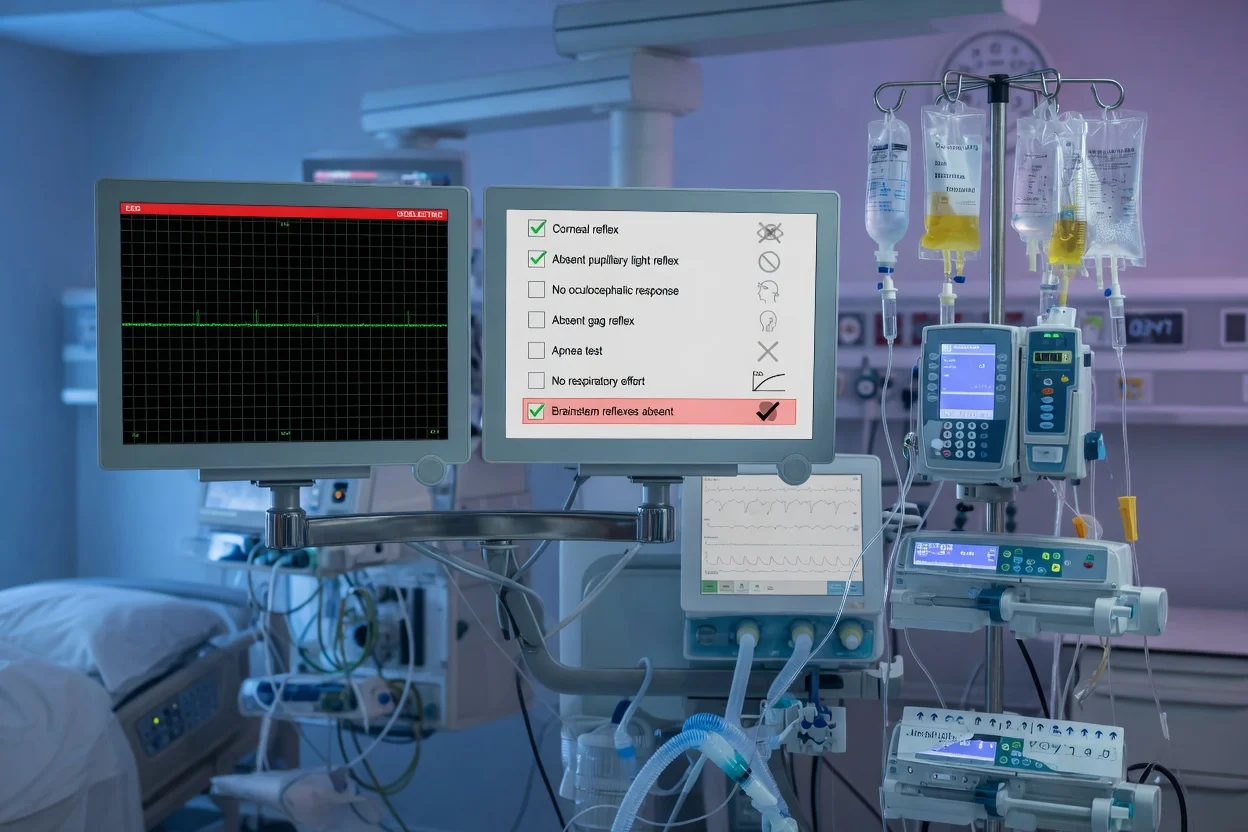
Brain death (DBD) vs circulatory death (DCD) donation
| Feature | DBD (brain death) | DCD (circulatory death) |
|---|---|---|
| Definition | Brain death diagnosed | Death after cardiac arrest (withdrawal of life support) |
| Patient status | Brain DEAD, heart BEATING | Brain intact or damaged, heart STOPPED |
| Organ quality | BETTER (organs perfused until retrieval) | Variable (warm ischaemia time after arrest) |
| Timing of death | Time of brain death declaration | Time of cessation of circulation (2-5 min asystole) |
| Organs available | All (heart, lungs, liver, kidneys, pancreas, intestines) | Most (heart — controversial; lungs, liver, kidneys, pancreas) |
| Consent | Family (or patient registration) | Family (or patient registration) |
| ICU management | Donor optimisation (maintain perfusion until retrieval) | Withdrawal in OR/ICU → observe → death → rapid retrieval |
| Frequency | ~60% of donations | ~40% (increasing) |
Brain death diagnosis and donation process in ICU
- SUSPECT brain death — patient with catastrophic brain injury (TBI, SAH, anoxia), deeply comatose (GCS 3), no brainstem reflexes, on mechanical ventilation
- PRECONDITIONS (must be met before testing): (a) Known cause of irreversible brain damage (CT/MRI evidence). (b) Exclusion of REVERSIBLE causes: hypothermia (core temp >35°C), CNS depressant drugs (check levels — barbiturates, benzodiazepines, opioids), metabolic (severe hyponatraemia, hypoglycaemia, hepatic encephalopathy), neuromuscular blockade (train-of-four). (c) Adequate oxygenation + haemodynamics (PaO2 ≥200 for apnoea test, SBP ≥100)
- CLINICAL EXAMINATION (two doctors, independently): (a) COMA (GCS 3 — no response to pain). (b) ABSENT brainstem reflexes: pupil reflex (fixed, dilated — no response to light), corneal reflex (no blink on touch), oculocephalic (doll's eyes — absent), oculovestibular (caloric — ice water in ear, no eye movement), gag/cough reflex (absent on suctioning). (c) APNOEA TEST: disconnect ventilator, oxygen via catheter at 6 L/min, PaCO2 must rise to ≥60 mmHg (or ≥20 above baseline) with NO respiratory effort
- DOCUMENT — two examinations, two doctors (per jurisdictional requirements). Time of death = time of second examination (or first — varies by jurisdiction). Record all findings
- NOTIFY — organ donation team (IMMEDIATELY after first positive examination — if family supportive or patient registered). Family discussion (trained requestor — separate from end-of-life discussion)
- DONOR MANAGEMENT (if donation proceeding): maintain MAP ≥65 (noradrenaline, vasopressin), normothermia (36-37°C), normoglycaemia, electrolytes, lung protective ventilation (if lung donation), hormone resuscitation (vasopressin, T3/T4, insulin — if haemodynamically unstable)
- ORGAN RETRIEVAL — surgical retrieval in operating theatre. Coordinate with transplant teams
Exam practice — SAQs
SAQ — Brainstem death testing prerequisites after subarachnoid haemorrhage
10 minutes · 10 marks
A 54-year-old man is in ICU on day 3 after an aneurysmal subarachnoid haemorrhage. He is intubated and sedated, GCS 3 with fixed dilated pupils and no cough on tracheal suctioning. The consultant plans to perform brainstem death tests this afternoon. Current observations: core temperature 34.2 degrees C; thiopentone infusion 4 mg/kg/h running for the last 48 hours for intracranial pressure control; noradrenaline 0.3 mcg/kg/min with MAP 72 mmHg; chest X-ray shows right lower lobe consolidation.
SAQ — Donation after circulatory death (DCD) pathway and warm ischaemia time
10 minutes · 10 marks
A 62-year-old man has devastating anoxic brain injury after an out-of-hospital cardiac arrest with a down-time of 35 minutes. After multiple multidisciplinary meetings, the treating team and family have agreed to withdraw life-sustaining therapy on the basis of futility. The family has consented to organ donation. The patient does not fulfil brain death criteria and the agreed pathway is donation after circulatory death (DCD). He is ventilated (FiO2 0.4), on noradrenaline 0.15 mcg/kg/min with MAP 70 mmHg. Creatinine 110 micromol/L; liver function normal.
Clinical pearls
Red flags
Prognosis
Brain death diagnosis and organ donation (ANZICS data)
Brain death determination: clinical exam + apnoea test is STANDARD (no confirmatory test needed in most jurisdictions). Sensitivity ~100%, specificity ~100% (when criteria met correctly). Donor optimisation: vasopressin + T3/T4 + corticosteroids — reduces vasopressor need, improves organ function in some studies (controversial — UNOS trial 2007 showed no mortality benefit for heart transplant recipients from hormone-treated donors). DCD: increasing — now ~40% of donations in Australia/NZ. Outcomes comparable to DBD for kidney, liver, pancreas. Lung: DCD outcomes similar to DBD. Heart: DCD controversial (some centres — emerging, using ex-vivo perfusion). 1 donor: up to 8 organs + 50 tissue grafts. Australia/NZ consent rate: ~60% (family asked). Higher if patient registered (AODR). Waitlist: ~1,400 Australians waiting for transplant (2019). ~50 die waiting each year.
Determination of brain death — preconditions in detail
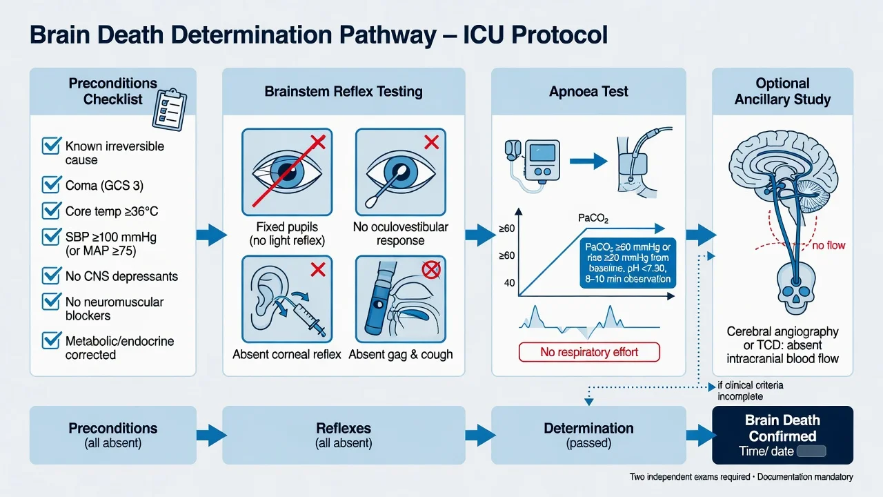
Preconditions that MUST be met before clinical brain death testing
| Domain | Requirement | Threshold / detail |
|---|---|---|
| Known cause | Irreversible catastrophic brain injury established | CT/MRI showing catastrophic injury (massive SAH, herniation, diffuse cerebral oedema, anoxic injury). Cause MUST be known — "unexplained coma" is NOT a basis for diagnosing brain death |
| Core temperature | Normothermia | Core temp ≥36°C (AAN 2010). Hypothermia mimics brain death — rewarm actively (forced warm air, warmed IV fluids, haemofiltration). Below 35°C reflexes are unreliable |
| Neuromuscular blockade | Fully recovered | Train-of-four ≥4/4 (no fade), OR documented time elapsed ≥ 4 half-lives since last dose. Rocuronium/suxamethonium clearance must be confirmed |
| CNS depressant drugs | Sub-therapeutic | Check levels: pentobarbital < 5 mg/L; benzodiazepines (flumazenil 0.2 mg challenge if uncertain); opioids (naloxone 0.4 mg challenge). If therapeutic levels likely — wait ≥ 5 half-lives |
| Severe metabolic/endocrine | Corrected | Na 130-160; glucose 5-15 mmol/L; normal pH/PaCO2; ammonia normal; no severe hepatic/renal encephalopathy |
| Blood pressure | SBP ≥ 100 mmHg, MAP ≥ 75 | Use vasopressors/inotropes to maintain — hypotension abolishes brainstem reflexes. Avoid over-correction (vasopressors may mask mild brain function) |
| Oxygenation | PaO2 ≥ 200 mmHg | Pre-oxygenase before apnoea test to prevent hypoxia during disconnection |
| Acid-base | Normal or pre-existing baseline pH | Chronic CO2 retainers — target PaCO2 rise to ≥ 20 mmHg above their (elevated) baseline |
Brainstem reflex examination — component by component
The six cranial nerve brainstem reflexes tested in brain death determination
| Reflex | Afferent / efferent nerves | Normal response | Brain-dead (absent) | Notes |
|---|---|---|---|---|
| Pupillary light | CN II / III (parasympathetic) | Pupils constrict briskly to bright light | Fixed, dilated (4-9 mm); no constriction | Record size; anisocoria acceptable if both unresponsive. Pre-existing iris surgery / atropine can confound |
| Corneal | CN V / VII (facial) | Blink on corneal touch with cotton wisp | No blink, no globe movement | Touch cornea (not sclera). Avoid trauma — may damage donor corneas for transplantation |
| Oculocephalic (doll's eyes) | CN III, IV, VI + vestibulospinal | Eyes move OPPOSITE to head turn | Eyes remain fixed mid-position (move WITH head) | CONTRAINDICATED if cervical spine not cleared |
| Oculovestibular (cold caloric) | CN VIII / III, IV, VI | Slow tonic deviation TOWARDS irrigated ear, then nystagmus away | No eye movement whatsoever | 50 mL ice-cold water into each external auditory canal after confirming clear tympanic membrane. Wait 60 s between ears |
| Gag | CN IX / X | Gag on posterior pharynx stimulation | Absent | Use suction catheter to posterior pharynx |
| Cough | CN X (vagus) | Cough on tracheal suctioning | Absent | Pass suction catheter to carina and oscillate |
| Facial grimace (CN V/VII) | CN V / VII | Grimace to deep supraorbital pressure | No facial movement | Tests pontine facial pathways |
Apnoea test — the detailed protocol (PaCO2 must reach ≥ 60 mmHg)
- PRE-OXYGENATE — ensure PaO2 ≥ 200 mmHg with FiO2 100% for ≥ 10 min before starting. Prevents hypoxia during disconnection
- CONFIRM preconditions — core temp ≥ 36°C, SBP ≥ 100, Na/glucose normal, no residual sedation/NMB. Document on the form
- BASELINE ABG — record PaO2, PaCO2, pH. Note the starting PaCO2 (critical for the "rise by ≥ 20" threshold if baseline is elevated)
- DISCONNECT from ventilator. Insert an oxygen catheter / T-piece delivering 100% O2 at 6-12 L/min directly into the trachea (apnoeic oxygenation — diffuses across alveoli, prevents desaturation)
- OBSERVE — watch chest/abdomen continuously for ANY respiratory effort for 8-15 minutes
- IF ANY respiratory effort (chest rise, abdominal contraction, accessory muscle use) → TEST NEGATIVE → patient NOT brain dead. Reconnect immediately. Reconsider diagnosis / seek confirmatory test
- IF NO effort — repeat ABG at 8 min. If PaCO2 ≥ 60 mmHg (or ≥ 20 mmHg above baseline) AND pH < 7.30 → TEST POSITIVE (consistent with brain death)
- IF haemodynamic instability (severe hypotension, arrhythmia, desaturation) → ABORT the test, reconnect, treat, and re-attempt later (or proceed to confirmatory test). The test must NEVER cause harm to the donor organs
- DOCUMENT — exact times, baseline/final ABG values, presence/absence of effort, observer names
Confirmatory (ancillary) tests
Confirmatory tests for brain death — when clinical exam is incomplete
| Test | What it shows | Sensitivity | Specificity | Practical notes |
|---|---|---|---|---|
| Cerebral angiography (4-vessel) | Gold standard — no intracranial blood flow above the petrous segment | Very high | Very high | Invasive, requires transfer to angiography suite — impractical in unstable donor. External carotid flow preserved |
| CT angiography (CTA) | No intracranial arterial enhancement (suspended filling of cortical vessels) | ~85-95% | ~95-100% | Fast, widely available. Protocols vary — "7-vessel" and "venous phase" techniques. False positives in high ICP with delayed circulation |
| CT perfusion | No cerebral blood flow | Good | Good | Adjunct to CTA; helpful when CTA equivocal |
| Radionuclide scintigraphy (HMPAO SPECT) | No cerebral / cerebellar / brainstem perfusion / uptake | High | High | Portable gamma camera at bedside. Distinguishes scalp/skull flow (preserved) from cerebral (absent). Useful in ECMO/therapeutic hypothermia |
| EEG | Isoelectric (electrocerebral silence — flat line ≥ 30 min, ≥ 8 electrodes, ≥ 30 µV/mm sensitivity) | High | Lower (false positives) | Detects CORTICAL activity only — does NOT confirm brainstem death. Deep sedation/hypothermia cause false-positive "flat line". Largely superseded in US/Australia; still used in some jurisdictions |
| Transcranial Doppler (TCD) | Oscillating / to-and-fro flow OR systolic spikes with no diastolic flow (high-resistance pattern) | ~90-99% | ~98-100% | Bedside, repeatable, non-invasive. ~10% of patients have no temporal acoustic window. Requires experienced operator |
When to use a confirmatory test (rather than clinical exam alone)
- Inability to complete one or more components of the clinical exam — e.g., severe facial/orbital trauma (cannot test corneal/caloric), otic disruption (cannot caloric), unstable cervical spine (cannot oculocephalic)
- Chronic severe lung disease making the apnoea test unsafe (rapid desaturation, severe acidosis)
- Inability to exclude confounders completely — e.g., prolonged sedative clearance in renal failure, concurrent therapeutic hypothermia protocol
- ECMO — apnoea test PaCO2 measurement unreliable; clinical exam possible but confirmatory test advised
- Paediatric / neonatal cases — some jurisdictions mandate ancillary testing, especially in neonates
- Medico-legal / jurisdictional requirement — several countries (e.g., some European nations, parts of Asia) REQUIRE an ancillary test for legal documentation
- Family or staff disagreement — to provide objective corroborative data (though legal determination remains clinical)
EEG vs cerebral angiography vs TCD — choosing the ancillary test
| Feature | EEG | Cerebral angiography | TCD |
|---|---|---|---|
| What it confirms | Cortical electrical activity | Cerebral blood flow (anatomical gold standard) | Cerebral blood flow (physiological) |
| Detects brainstem death? | NO — cortex only | YES — covers all territories | YES — anterior + posterior circulation |
| Bedside? | Yes | No (angiography suite) | Yes |
| Sensitivity to sedation/hypothermia | HIGH — causes false-positive isoelectric trace | None | Minimal |
| Time | 30 min minimum | 1-2 h + transfer | 15-30 min |
| Current preference | Falling out of favour | When definitive flow documentation needed | Increasing — repeatable, bedside |
DBD vs DCD pathways in detail
Controlled DCD vs uncontrolled DCD vs DBD
| Feature | DBD (brain death) | Controlled DCD (cDCD) | Uncontrolled DCD (uDCD) |
|---|---|---|---|
| Setting | ICU, ventilated patient declared brain dead | Planned withdrawal of life-sustaining therapy (WLST) in ICU/OR | Failed resuscitation in ED/ICU/community (unexpected cardiac arrest) |
| Pre-mortem planning | None (death by neurologic criteria) | Extensive — family discussion, timing, location, retrieval team on standby | None — sudden death; rapid response protocol (preservation within minutes) |
| Circulation at organ retrieval | Heart BEATING — perfused organs | Heart STOPPED after planned withdrawal + stand-off | Heart STOPPED after failed CPR |
| Warm ischaemia time | None | Defined — measured from SBP < 50 to cold perfusion (target < 30 min liver, < 60 min kidney) | Long, uncontrolled — often limits usable organs |
| Organs typically transplantable | All — heart, lungs, liver, kidneys, pancreas, intestine | Kidneys, liver, lungs, pancreas; HEART now possible with ex-vivo perfusion | Mostly kidneys (most ischaemia-tolerant); rarely liver |
| Frequency (ANZ) | ~ 60% of deceased donors | ~ 40% (rising rapidly) | Rare / regional only |
Controlled DCD pathway — step-by-step (the cDCD process after WLST decision)
- DECISION to withdraw life-sustaining therapy (WLST) — made independently by treating team + family, on best-interest / futility grounds. Donation is NEVER a reason to withdraw
- ASSESS DCD ELIGIBILITY — donor team evaluation: age, comorbidities, organ function, projected warm ischaemia time. Consent for donation obtained (separate from WLST decision)
- PRE-MORTEM PLANNING / STAND-BY — coordinate timing between ICU, OR, retrieval surgeons, anaesthetics, transplant teams. Determine location of withdrawal (ICU vs OR — affects warm ischaemia time). Plan cannulation strategy (in-situ cold perfusion via aorta)
- PRE-EMPTIVE PREPARATION (after WLST decision, BEFORE withdrawal) — may include: heparin administration (controversial — not universal), arterial/venous lines for rapid cannulation, family time
- WITHDRAWAL OF LIFE-SUSTAINING THERAPY — extubation (or terminal wean), cessation of vasopressors/inotropes, comfort measures only. Family may be present
- OBSERVATION PERIOD — monitor for circulatory arrest. If asystole / absence of circulation < 60 min from withdrawal → proceed. If > 60-120 min (centre threshold) → DCD abandoned, return to comfort care (organs would sustain excessive warm ischaemia)
- DEATH DECLARATION — after confirmation of absent circulation (no pulse, no heart sounds, asystole on monitor) and a mandatory NO-TOUCH / STAND-OFF PERIOD (5 minutes in Australia/UK; 2-5 minutes US — varies by jurisdiction) to rule out autoresuscitation[4] }
- RAPID RETRIEVAL + COLD PERFUSION — immediate transfer to OR (if withdrawn in ICU) or in-situ cannulation. Cold preservation solution (UW / HTK) flushed via aorta. Document functional warm ischaemia time (from SBP < 50 to perfusion)
- EX-VIVO PERFUSION (modern practice) — DCD hearts/lungs now resuscitated on ex-vivo machine perfusion (Organ Care System / TransMedics) before transplantation — has enabled DCD heart transplantation
Time-of-death determination: DBD vs DCD
| Aspect | DBD | DCD |
|---|---|---|
| Time of death | Time the second brain death examination confirms death (or first, jurisdiction-dependent) | Time of completion of the no-touch observation period after circulatory arrest |
| Definition of death applied | Neurologic criteria (irreversible cessation of all brain function) | Circulatory criteria (irreversible cessation of circulation) |
| Documentation | Two clinician signatures, two examinations, full reflex + apnoea record | Single clinician confirms circulatory arrest; documents no-touch period |
| Permanence | Established by brain death examination | Established by no-touch period (5 min) + absence of autoresuscitation |
Donor management — physiology of the brain-dead donor
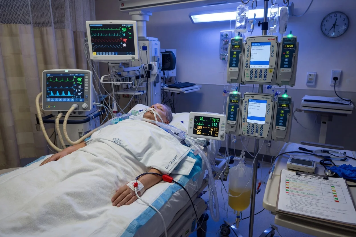
Physiological derangements following brain death — the catecholamine storm and after
| Phase | Time | Pathophysiology | Clinical consequence |
|---|---|---|---|
| Catecholamine storm (autonomic crisis) | 0-30 min post herniation | Massive sympathetic outflow → noradrenaline/adrenaline surge; then abrupt vagal (brainstem) shutdown | Severe hypertension, tachyarrhythmias, vasoconstriction → myocardial injury, pulmonary oedema, capillary leak. Then sudden hypotension |
| Vasoplegic shock | 30 min onward | Loss of sympathetic vasomotor tone + myocardial depression | Hypotension, low SVR, high cardiac output (distributive shock). Often requires multiple vasopressors |
| Endocrine failure | 1-2 h onward | Hypothalamic-pituitary axis ceases — ADH, TSH, ACTH deficiency | Diabetes insipidus (hypernatraemia, hypovolaemia), low T3/T4, low cortisol |
| Coagulopathy | Variable | Release of tissue factor, hypothermia, dilution | DIC-like picture, bleeding at retrieval |
| Hypothermia | Universal | Hypothalamic thermostat loss — poikilothermia | Core temp drifts to ambient; worsens coagulopathy and arrhythmia |
| Metabolic | Progressive | Hyperglycaemia (insulin resistance + deficiency), acidosis, hyperkalaemia | Multi-organ cellular dysfunction |
Haemodynamic donor management
Haemodynamic targets in the brain-dead donor (organ-protective)
- MAP ≥ 65 mmHg (70 mmHg preferred) — noradrenaline first-line for vasoplegia; vasopressin SPARING (reduces catecholamine dose, treats DI)
- SBP ≥ 100 mmHg — avoid hypotension (organ hypoperfusion) AND hypertension (worsens neurogenic pulmonary oedema)
- Heart rate 60-120 bpm — bradycardia suggests high vagal tone / AV block; tachycardia worsens myocardial O2 demand. Persistent bradycardia unresponsive to atropine is itself a sign of brainstem death (vagal nuclei destroyed)
- Cardiac index ≥ 2.5 L/min/m² — if low, add inotrope (adrenaline, milrinone, dobutamine)
- CVP / fluid balance — euvolaemia; titrate to CVP 6-10 mmHg or dynamic indices (PPV, SVV, IVC variability). AVOID fluid overload — damages lungs (degrades PaO2/FiO2) and heart
- Echocardiography — baseline LV function; recheck after resuscitation. LV dysfunction may recover with hormone therapy. LVEF < 45% after optimisation → heart often unsuitable for transplantation
- Arterial line + central access — continuous BP, frequent sampling. Pulmonary artery catheter / TEE in unstable donors
Vasopressor selection in the brain-dead donor
| Agent | Role | Rationale | Cautions |
|---|---|---|---|
| Noradrenaline | First-line vasopressor | α-agonist restores SVR in vasoplegia | High doses > 0.5 mcg/kg/min may worsen organ microcirculation — review |
| Vasopressin | SPARING + DI treatment | Low-dose (0.01-0.04 U/min) restores vascular smooth muscle tone, treats ADH deficiency, halves catecholamine requirements | Avoid overshoot hypertension; reduces need for noradrenaline |
| Adrenaline | Inotrope + vasopressor | If MAP low with bradycardia / myocardial depression | Lactic acidosis at high doses (worsens organ viability); use cautiously |
| Methylprednisolone | Adjunct (15 mg/kg) | Treats relative adrenal insufficiency; downregulates donor immune activation; thought to improve lung / liver yield | Give early once brain death declared |
Electrolyte, metabolic and temperature management
Electrolyte and metabolic targets in the brain-dead donor
| Parameter | Target | Common derangement | Management |
|---|---|---|---|
| Serum Na⁺ | 135-155 mmol/L | Hypernatraemia (DI — water loss) | D5W / 0.45% saline; desmopressin / vasopressin for DI. AVOID Na > 160 — associated with worse liver graft function |
| Serum K⁺ | 3.5-5.5 mmol/L | Hypokalaemia (DI urinary losses, alkalosis) — OR hyperkalaemia (acidosis, cell leak) | Replace carefully; correct acidosis first; avoid over-correction (arrhythmia) |
| Serum Mg²⁺ / PO₄²⁻ | Normal | Hypomagnesaemia / hypophosphataemia common | Replace — prevents arrhythmia, respiratory muscle weakness |
| Glucose | 6-10 mmol/L | Hyperglycaemia (insulin resistance + deficiency) | Insulin infusion titrated; conventional (not tight — risk of hypoglycaemia) |
| pH | 7.35-7.45 | Metabolic acidosis (shock, renal failure) | Treat cause; bicarbonate if pH < 7.20 with metabolic acidosis |
| Hb | ≥ 70-80 g/L | Anaemia (haemodilution, bleeding) | Transfuse to maintain O2 delivery; balance with risk of TRALI |
| Core temp | ≥ 36°C | Hypothermia (poikilothermia) | Forced warm air, warmed IV fluids, haemofiltration circuit. Hypothermia worsens coagulopathy + arrhythmia |
Hormone replacement therapy — vasopressin, T3/T4, corticosteroids
Hormone replacement in the brain-dead donor — what, why, dosing, evidence
| Hormone | Deficiency it treats | Typical dose | Evidence | When to use |
|---|---|---|---|---|
| Vasopressin (low-dose) | ADH deficiency (DI) + catecholamine-sparing in vasoplegia | 0.01-0.04 U/min (1-2.4 U/h) continuous | Strong — reduces noradrenaline requirement, treats DI, well-tolerated | Standard — almost all haemodynamically unstable donors |
| Desmopressin (DDAVP) | ADH deficiency (DI) when vasoplegia absent | 1-4 mcg IV bolus (every 6-12 h) | Effective for DI alone | When DI present but BP stable (no need for vasopressor effect) |
| Triiodothyronine (T3) / Levothyroxine (T4) | Low-T3 syndrome; cardiovascular instability | T3: 4 mcg bolus then 3 mcg/h OR 20 mcg bolus then 10 mcg/h × 24 h. T4: 20 mcg bolus then 10 mcg/h | CONTROVERSIAL — meta-analyses conflicting; landmark randomised UNOS study showed NO survival benefit for heart recipients from hormone-treated donors, but haemodynamics often improve | Use selectively — haemodynamically unstable donors requiring escalating vasopressors (especially for heart/liver procurement) |
| Insulin | Hyperglycaemia + (high-dose) inotropic support | Glucose control: 1-4 U/h titrated. High-dose inotropic: controversial | Standard for glucose control; high-dose protocol unproven | Glucose control routine; high-dose rarely used |
| Methylprednisolone | Relative adrenal insufficiency + dampen donor immune activation | 15 mg/kg IV (1 g typical) every 24 h | Reasonable evidence — reduces inflammatory cytokines, may improve lung / liver yield | Standard once brain death declared, especially for lung procurement |
Hormonal resuscitation protocol (haemodynamically unstable brain-dead donor)
- CONFIRM brain death declared and donation proceeding — do NOT initiate hormone protocol on a patient who is not yet legally dead
- VASOPRESSIN 0.01-0.04 U/min IV continuous — first-line. Aim for urine output 0.5-3 mL/kg/h (treats DI AND vasoplegia). Titrate to MAP and to Na⁺ normalisation
- METHYLPREDNISOLONE 15 mg/kg IV (typically 1 g) — give as a single bolus; repeat q24h. Reduces inflammatory cascade, supports adrenal insufficiency
- T3 (liothyronine) 4 mcg IV bolus then 3 mcg/h infusion — IF haemodynamically unstable on escalating catecholamines OR poor LV function on echo. Reassess at 2-4 h
- INSULIN infusion titrated to glucose 6-10 mmol/L — hyperglycaemia is universal. Some protocols use high-dose insulin (1 U/kg/h) as inotrope — controversial; not routine
- REASSESS haemodynamics every 1-2 h — typically vasopressor requirements FALL after 2-6 h of combined therapy. Recheck echocardiography — LV function may recover sufficiently for heart donation
- DOCUMENT — record baseline and serial vasopressor doses, echo findings, hormone doses — required for organ acceptance decisions by transplant teams
Additional clinical pearls — advanced
More red flags
[1]Key trials and consensus documents
Wijdicks EFM et al, 2010 — AAN Evidence-Based Guideline Update: Determining Brain Death in Adults (Neurology)
Design: Systematic evidence review forming the basis of the American Academy of Neurology guideline update. Findings: No published evidence of neurologic recovery in patients who fulfil AAN brain death criteria. Confirmed the sufficiency of the clinical examination (prerequisites + brainstem areflexia + apnoea test). No evidence that confirmatory tests are routinely needed. Significance: Established the modern standard for adult brain death determination in the US and beyond; basis for most hospital protocols. [1] }
Greer DM et al, 2020 — World Brain Death Project Consensus (JAMA)
Design: International multidisciplinary consensus (AAN, AAP, CNS, SCCM, ESICM, WFSICCM, WFN, ANZICS) — the most comprehensive attempt to harmonise brain death determination globally. Recommendations: Uniform minimum clinical criteria (prerequisites, examination, apnoea test); when ancillary testing is required; special populations (ECMO, paediatric, therapeutic hypothermia). Significance: The 2020 reference document for international practice — cited in most updated hospital policies; promotes standardisation across jurisdictions. [6] }
McKeown DW et al, 2012 — Management of the Heart-Beating Brain-Dead Organ Donor: a Systematic Review and Meta-Analysis (BJA)
Design: Systematic review of donor management interventions. Findings: Hormonal resuscitation (vasopressin + T3/T4 + corticosteroid) — evidence suggests haemodynamic improvement and reduced vasopressor need; outcome benefit for recipients remains controversial. Specific donor-management targets (MAP, Na, fluid balance, lung-protective ventilation) shown to influence organ yield. Significance: Established the evidence base for the structured donor-care bundle used by organ procurement organisations worldwide. [3] }
Manara AR et al, 2023 — Autoresuscitation After Circulatory Arrest: an Updated Systematic Review (CJA)
Design: Updated systematic review examining cases of autoresuscitation (spontaneous return of circulation after cessation of CPR) — directly relevant to the DCD no-touch period. Findings: Across > 600 reported cases, no autoresuscitation occurred beyond ~ 4 min 20 s after cessation of circulation. Supports the 5-minute no-touch period used in DCD as safe. Significance: Provides the empirical basis for the irreversibility criterion in DCD death determination. [4] }
Greer DM et al, 2016 — Variability of Brain Death Policies in the United States (JAMA Neurology)
Design: Survey of brain death determination policies across US hospitals. Findings: Substantial variability in prerequisite thresholds (e.g., apnoea test PaCO2 ≥ 55 vs ≥ 60), number of required examinations, observation intervals, qualifications of examiners, and use of ancillary testing — despite uniform underlying criteria. Significance: Documented the practice variability that motivated the 2020 World Brain Death Project consensus; raised concerns about consistency and medicolegal defensibility. [2] }
Saposnik G et al, 2009 — Spontaneous and Reflex Movements in Brain Death: a Systematic Review (Neurology)
Design: Systematic review of reported motor movements in clinically brain-dead patients. Findings: Movements (finger jerks, toe responses, the Lazarus sign, respiratory-like movements) reported in a substantial minority of brain-dead patients — all attributable to spinal cord reflexes, not brain-mediated activity. Significance: Reassures clinicians and families that spinal reflexes do NOT invalidate a brain death determination; pre-emptive explanation prevents distress. [8] }
Donor management quick-reference bundle
The brain-dead donor care bundle (memorise for exams)
| Domain | Target | Intervention |
|---|---|---|
| MAP | ≥ 65-70 mmHg | Noradrenaline + low-dose vasopressin ± inotrope |
| Heart rate | 60-120 bpm | Treat bradycardia (not atropine-responsive — accept); treat tachyarrhythmia |
| Core temp | ≥ 36°C | Active warming (forced air, warmed fluids) |
| Serum Na | 135-155 mmol/L | D5W + desmopressin / vasopressin for DI |
| Glucose | 6-10 mmol/L | Insulin infusion |
| CVP / volume | Euvolaemia; neutral balance (negative if lung donor) | Goal-directed fluids; avoid overload |
| Ventilation | Vt 6-8 mL/kg, PEEP 5-8, plateau < 30, FiO2 minimised | Lung-protective ventilation (especially if lungs donated) |
| Hormones | Per protocol | Vasopressin 0.01-0.04 U/min + methylprednisolone 15 mg/kg + T3 if unstable |
| Hb | ≥ 70-80 g/L | Transfusion if below |
| Urine output | 0.5-3 mL/kg/h | Treat DI to bring high output down |
The first 60 minutes after brain death declaration — donor resuscitation sequence
- CONFIRM brain death legally declared and documented; family informed; donation team notified
- SECURE invasive monitoring — arterial line, central line (if not present); ensure reliable access
- HAEMODYNAMICS — establish MAP ≥ 65; start noradrenaline; add vasopressin 0.01-0.04 U/min; escalate to adrenaline/inotrope if low cardiac output
- FLUID / VOLUME — assess fluid responsiveness; titrate to euvolaemia (avoid overload, especially for lung donors)
- ELECTROLYTES — check Na, K, Mg, PO4, glucose every 1-2 h; treat DI (desmopressin if BP stable, vasopressin if not); insulin for glucose
- TEMPERATURE — measure core temp; active warming to ≥ 36°C
- VENTILATION — switch to lung-protective settings (Vt 6-8 mL/kg, PEEP 5-8); minimise FiO2; consider recruitment
- HORMONE PANEL — start methylprednisolone 15 mg/kg; add T3 if haemodynamically unstable
- INVESTIGATIONS — baseline echo; CXR; cross-match; serology; cultures; ABG
- DOCUMENT — serial haemodynamics, vasopressor doses, echo findings, electrolytes, hormones — communicated to retrieval team
References
- [1]Wijdicks EFM, Varelas PN, Gronseth GS, Greer DM Evidence-based guideline update: determining brain death in adults: report of the Quality Standards Subcommittee of the American Academy of Neurology Neurology, 2010.PMID 20530327
- [2]Greer DM, Wang HH, Lewis AJ, et al. Variability of Brain Death Policies in the United States JAMA Neurol, 2016.PMID 26719912
- [3]McKeown DW, Bonser RS, Kellum JA Management of the brain-dead organ donor: a systematic review and meta-analysis Transplantation, 2013.PMID 23545508
- [4]Manara AR, Thomas I, Harding R Autoresuscitation after circulatory arrest: an updated systematic review Can J Anaesth, 2023.PMID 37131027
- [5]Rostron A, Farmery AD, Laing HE, et al. Pharmacological normalization of circulation after acute brain death Acta Anaesthesiol Scand, 2012.PMID 22651688
- [6]Greer DM, Shemie SD, Lewis A, et al. Determination of Brain Death/Death by Neurologic Criteria: The World Brain Death Project JAMA, 2020.PMID 32761206
- [7]Russell JA, Epstein LG, Greer DM Apnea testing during brain death assessment: a review of clinical practice and published literature Respir Care, 2013.PMID 22709413
- [8]Saposnik G, Bueri JA, Mauriño J, et al. Movements in brain death: a systematic review Can J Neurol Sci, 2009.PMID 19378707
- [9]Russell JA, Aleccia E Brain Death/Death by Neurologic Criteria Determination Continuum (Minneap Minn), 2021.PMID 34618768
- [10]Nair S, Lingappa L, Rajshekhar V The challenges with brain death determination in adult patients on extracorporeal membrane oxygenation Neurocrit Care, 2011.PMID 21327575