ICU · Physiology / haematology immunology
Blood & Immunology Physiology
Also known as Blood physiology · Immunology physiology · Hematopoiesis · Innate immunity · Adaptive immunity · Complement cascade · Coagulation cascade · Inflammation · Red blood cell physiology · Oxygen-haemoglobin dissociation curve · 2,3-diphosphoglycerate · Bohr effect · Haldane effect · Fibrinolysis · Platelet function · Immunoparalysis · SIRS CARS MARS
Blood and immunology physiology: the hematopoiesis (the pluripotent stem cell → RBC, WBC, platelets). The red blood cell physiology (2,3-DPG, oxygen dissociation curve, Bohr effect, Haldane effect). The innate immunity (the neutrophils, the macrophages, the complement, the natural killer). The adaptive immunity (the T cells — the cellular; the B cells — the humoral antibodies). The complement cascade (the classical, the alternative, the lectin). The coagulation cascade (the intrinsic, the extrinsic, the common — the PT/aPTT/TT). The platelet function (the adhesion — vWF/GPIb; the activation; the aggregation — GPIIb/IIIa/fibrinogen). The fibrinolysis (the plasmin, the D-dimer). The inflammation (the cytokines, the acute-phase response). The immunoparalysis and the SIRS → CARS → MARS paradigm in the critical illness.
On this page & tools
Your progress
Saved locally on this device.
8 MCQs with explanations
Target exams
Overview & definition
The blood and immunology physiology — the hematopoiesis (the blood cell production), the red blood cell physiology (the oxygen transport), the innate and the adaptive immunity, the complement, the coagulation, the fibrinolysis, the platelet function, and the inflammation. All interlinked in the critical illness (the sepsis — the immune dysregulation, the coagulation, the inflammation, the immunoparalysis).[1]
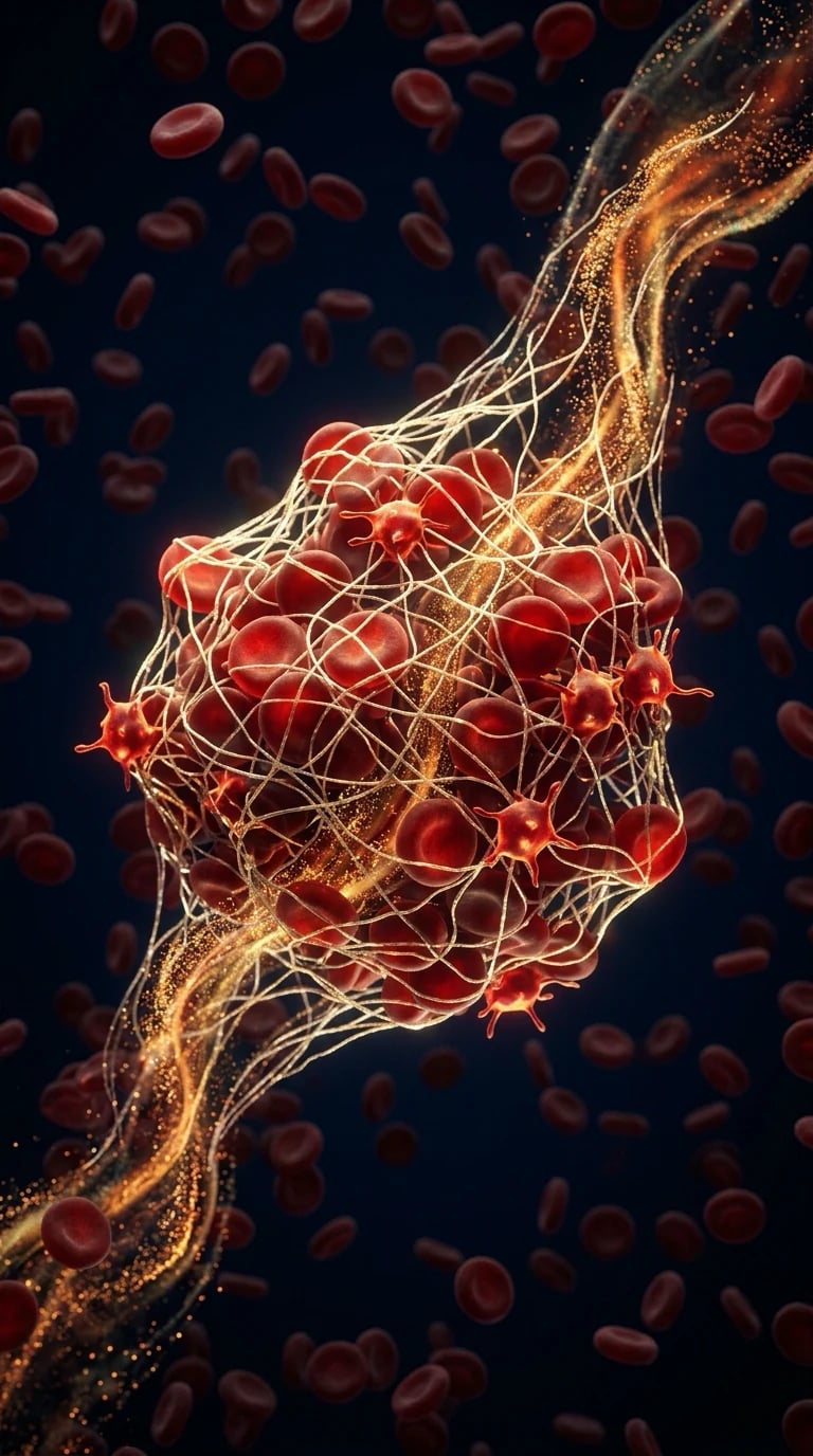
Hematopoiesis
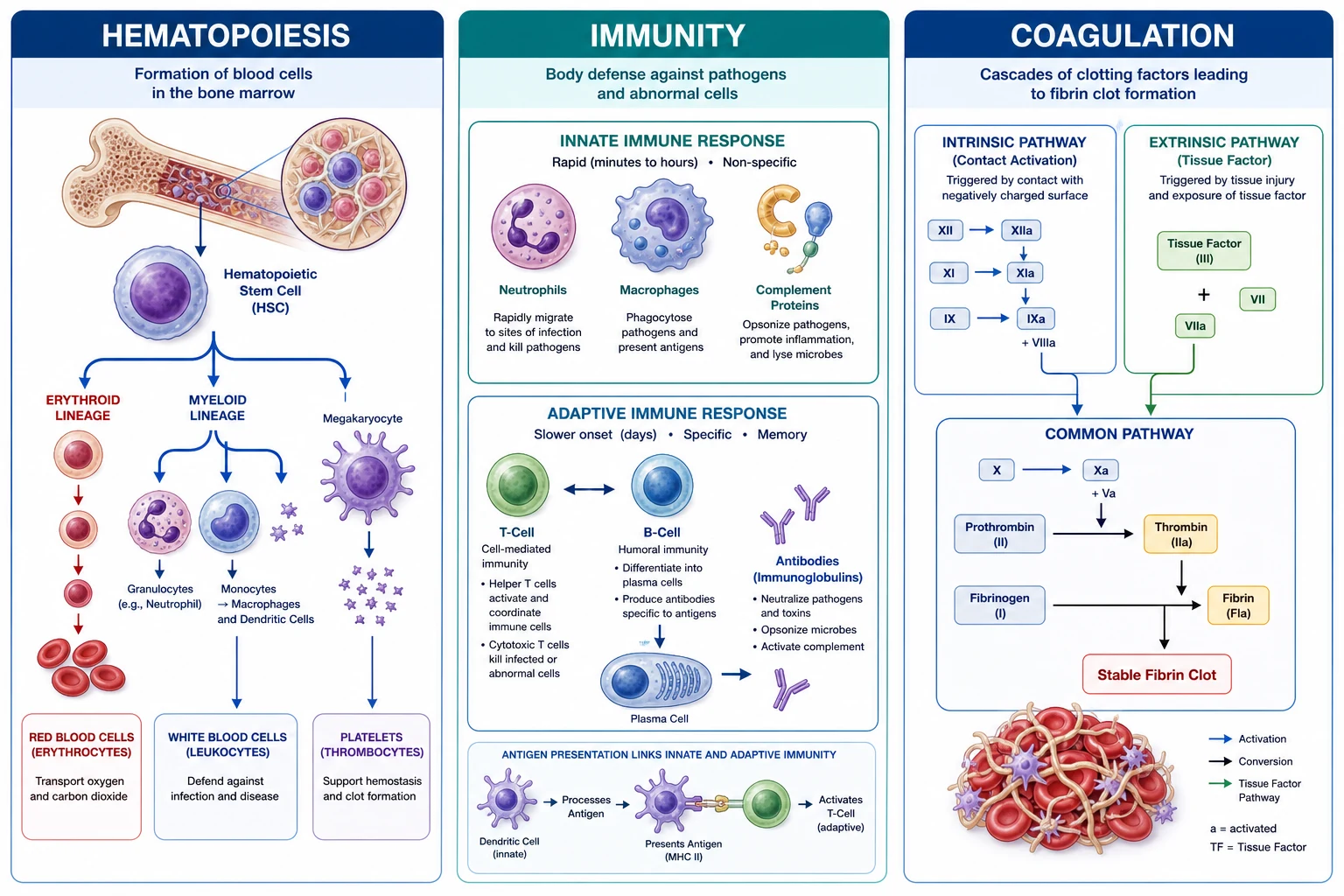
Haematopoiesis is the continuous, regulated production of all blood cells from a small pool of pluripotent haematopoietic stem cells (HSCs) residing in the bone marrow niche. HSCs have two defining properties: self-renewal (asymmetric division producing one stem and one differentiating daughter) and multipotency (the capacity to generate every blood lineage). Their fate is governed by the microenvironment (osteoblasts, endothelial cells, mesenchymal stromal cells, CXCL12-abundant reticular [CAR] cells) and by lineage-specific growth factors (cytokines).[1][1]
The pluripotent stem cell (the bone marrow — CD34+, CD38−, Lin−) → the myeloid (the RBC, the granulocytes — the neutrophils/eosinophils/basophils, the monocytes/macrophages, the platelets) and the lymphoid (the T cells, the B cells, the NK cells).[1][1]
The EPO (the erythropoietin — the kidney interstitial fibroblasts, the peritubular; via HIF-2α when the O2 falls; the RBC production). The TPO (the thrombopoietin — the liver, the constitutive; the platelet production). The G-CSF (the granulocyte colony-stimulating factor — the endothelium/macrophages/fibroblasts; the neutrophil production). The M-CSF (the macrophage colony-stimulating factor; the monocyte/macrophage production). The GM-CSF (the granulocyte-macrophage colony-stimulating factor; the multi-lineage — the neutrophils + the macrophages). The IL-3 (the multi-potential; the early progenitors). The IL-5 (the eosinophil production). The SCF (the stem cell factor; the co-stimulates).[1][1]
Haematopoiesis — the differentiation hierarchy
- Pluripotent HSC (CD34+, CD38−, CD90+, Lin−) — self-renewing, rare (~1 in 10^4 marrow cells), maintained in the quiescent endosteal niche by CXCL12/CXCR4 signalling and Tie2–angiopoietin-1
- Multipotent progenitor (MPP) — the first commitment step, loses self-renewal, retains all lineage potential
- The bifurcation into two main lineage trees:
- COMMON MYELOID PROGENITOR (CMP) — driven by IL-3 and GM-CSF →
- Granulocyte-monocyte progenitor (GMP) → neutrophils (G-CSF is the dominant late driver — basis of filgrastim), monocytes → tissue macrophages (M-CSF — Kupffer cells, alveolar macrophages, microglia), eosinophils (IL-5), basophils/mast cells (IL-3, IL-4)
- Megakaryocyte-erythroid progenitor (MEP) → megakaryocytes → platelets (TPO — basis of eltrombopag/romiplostim), erythrocytes (EPO from kidney — basis of recombinant EPO)
- COMMON LYMPHOID PROGENITOR (CLP) → B cells (mature in bone marrow), T cells (migrate to thymus — positive then negative selection), NK cells (bone marrow)
- COMMON MYELOID PROGENITOR (CMP) — driven by IL-3 and GM-CSF →
- The lineage-specific growth factors determine the OUTPUT of each arm (see table below)
- The mature cells enter the circulation — the granulocytes (the short-lived — the hours-days); the lymphocytes and the memory cells (the long-lived — the years)
Lineage-specific haematopoietic growth factors — what drives what
| Growth factor | Source | Primary target lineage | Dominant product | ICU relevance |
|---|---|---|---|---|
| EPO (erythropoietin) | Kidney (peritubular fibroblasts, via HIF-2α) | CFU-E → proerythroblast | Erythrocytes | Recombinant EPO for anaemia of critical illness (thrombosis risk restricts use); low in CKD |
| G-CSF | Endothelium, macrophages, fibroblasts | GMP → myelocyte → neutrophil | Neutrophils | Filgrastim for chemo-induced neutropenia; mobilises HSCs for harvest |
| M-CSF | Endothelium, fibroblasts | GMP → monocyte | Monocytes/macrophages | Tissue macrophage maintenance |
| IL-5 | Th2 cells, mast cells | Eosinophil lineage | Eosinophils | Mepolizumab (anti-IL-5) for eosinophilic asthma |
| TPO (thrombopoietin) | Liver (constitutive) | Megakaryocyte → platelets | Platelets | TPO mimetics (eltrombopag, romiplostim, avatrombopag) for ITP |
| IL-3 | T cells, mast cells | CMP and early progenitors | Multi-lineage (broad myeloid) | Acts EARLY — broad myeloid expansion |
| GM-CSF | T cells, macrophages, endothelium | CMP/GMP — granulocytes + macrophages | Neutrophils + macrophages | Sargramostim; alveolar macrophage function (deficiency → pulmonary alveolar proteinosis) |
| SCF (stem cell factor) | Bone marrow stroma | HSCs, early progenitors | Multi-lineage support | Co-stimulates with other growth factors |
Red blood cell physiology — oxygen transport and the haemoglobin-oxygen dissociation curve
The red blood cell (erythrocyte) is the oxygen-carrying vehicle: a biconcave, anucleate disc packed with haemoglobin (~280 million molecules per cell) that transports O2 from lungs to tissues and facilitates CO2 return. Haemoglobin is a tetramer of 4 globin chains (adult HbA = 2α + 2β) each cradling a haem group (a porphyrin ring with a central Fe2+ ion). Oxygen binds reversibly to the Fe2+ in a cooperative manner — binding of one O2 molecule increases affinity for the next, producing the characteristic sigmoid (S-shaped) oxygen-haemoglobin dissociation curve (ODC).[1][7]
The oxygen-haemoglobin dissociation curve
The ODC relates the partial pressure of oxygen (PaO2, x-axis) to haemoglobin saturation (SaO2, y-axis). Its sigmoid shape has critical physiological implications: [1]
The oxygen-haemoglobin dissociation curve — the three zones
- The steep lower portion (PaO2 10–40 mmHg) — the tissue-level zone. Small drops in PaO2 here release LARGE amounts of O2 to tissues. At PaO2 = 40 mmHg (mixed venous), SaO2 ≈ 75% — meaning ~25% of O2 has been unloaded. This is the physiological reserve that ensures O2 delivery even when tissue PO2 falls
- The plateau/upper portion (PaO2 60–100 mmHg) — the pulmonary zone. Haemoglobin is >90% saturated above PaO2 60 mmHg. This means PaO2 can fall from 100 to 60 mmHg with only a small drop in saturation — a safety margin. But it ALSO means that raising PaO2 above 100 mmHg adds negligible additional O2 content (dissolved O2 is only 0.003 mL/dL/mmHg)
- The P50 — the PaO2 at which haemoglobin is 50% saturated. The normal P50 = 26.6 mmHg (at pH 7.4, 37°C, PCO2 40 mmHg). P50 is the INDEX of haemoglobin-oxygen affinity: ↑P50 = ↓affinity (right shift, easier unloading); ↓P50 = ↑affinity (left shift, harder unloading)
Factors shifting the oxygen-haemoglobin dissociation curve
| Factor | Direction | Mechanism | Physiological / clinical context |
|---|---|---|---|
| ↑H+ (↓pH) | RIGHT (↑P50, ↓affinity) | Protons stabilise the T (tense) state of Hb | The Bohr effect — facilitates O2 unloading in metabolically active (acidic) tissues |
| ↑PCO2 | RIGHT | CO2 forms carbamino compounds with Hb terminal amines → stabilises T state | Bohr effect component; tissue CO2 high |
| ↑Temperature | RIGHT | Denatures Hb slightly toward T state | Exercising muscle, fever — O2 unloading enhanced |
| ↑2,3-DPG | RIGHT | Binds deoxy-β-chains → stabilises T state | Chronic hypoxia, anaemia, high altitude, chronic lung disease — compensation to improve O2 unloading |
| ↓H+ (↑pH) | LEFT (↓P50, ↑affinity) | Protons leave → R (relaxed) state favoured | Alkalosis — impairs O2 unloading |
| ↓Temperature | LEFT | — | Hypothermia |
| ↓2,3-DPG | LEFT | Less T-state stabilisation | Stored blood (banked RBCs lose 2,3-DPG over days), alkalosis |
| CO (carbon monoxide) | LEFT (and ↓capacity) | Binds Hb at O2 site ~240× more avidly → carboxyhaemoglobin; remaining sites hold O2 tighter | CO poisoning — SaO2 falsely near-normal on pulse oximetry (does not detect COHb) |
| MetHb (methaemoglobin) | LEFT (and ↓capacity) | Fe3+ cannot bind O2; left-shifts remaining sites | Methaemoglobinaemia — chocolate-brown blood; reversal with methylene blue |
| Fetal Hb (HbF) | LEFT | 2α + 2γ chains; γ chains bind 2,3-DPG poorly → higher affinity | Fetus extracts O2 from maternal blood (lower P50 ~19 mmHg) |
The Bohr effect
The Bohr effect (1904, Christian Bohr) describes the RIGHTWARD shift of the ODC caused by increased H+ concentration (decreased pH) and increased PCO2. In metabolically active tissues, CO2 production and lactic acid release lower local pH → protons bind haemoglobin (stabilising the deoxy/T state) → haemoglobin's affinity for O2 DECREASES → more O2 is released to the tissues that need it most. In the lungs, CO2 is blown off, pH rises, protons leave haemoglobin → affinity INCREASES → O2 is loaded. The Bohr effect is thus a local, demand-driven mechanism coupling O2 delivery to metabolic need.[7]
The Haldane effect
The Haldane effect is the mirror image for CO2 transport: deoxygenated haemoglobin carries MORE CO2 than oxygenated haemoglobin. Deoxy-Hb is a weaker acid (better proton acceptor) than oxy-Hb, so it binds more H+ (facilitating the bicarbonate buffer reaction: CO2 + H2O → H+ + HCO3−) and forms more carbamino compounds. Physiologically: in the TISSUES, O2 is released → Hb becomes deoxygenated → its CO2-carrying capacity RISES → CO2 is taken up. In the LUNGS, O2 binds → Hb oxygenates → CO2-carrying capacity FALLS → CO2 is released for exhalation. The Haldane effect accounts for ~50% of total CO2 exchange and doubles the effectiveness of the Bohr effect.[7]
2,3-Diphosphoglycerate (2,3-DPG)
2,3-DPG is an organophosphate produced as a byproduct of glycolysis via the Rapoport-Luebering shunt (a detour in erythrocyte glycolysis unique to RBCs). It binds specifically to the β-chains of DEOXY-haemoglobin (in a central cavity that only exists in the T/tense state) → stabilises the deoxy conformation → REDUCES haemoglobin's O2 affinity → RIGHTWARD shift of the ODC → ENHANCED O2 unloading to tissues. 2,3-DPG is the CHRONIC compensatory mechanism for hypoxia/anaemia: it increases over hours-days in response to chronic hypoxia (high altitude, chronic lung disease, anaemia, high-output cardiac failure) to improve tissue O2 delivery. Stored (banked) blood progressively loses 2,3-DPG — a clinical concern in massive transfusion (the transfused RBCs hold onto their O2 tightly, left shift, poor tissue unloading, recovering over 24-48 hours in vivo).[7]
The oxygen content of blood — the two components
| Component | Amount | Formula | Notes |
|---|---|---|---|
| O2 bound to haemoglobin | ~19.7 mL/dL (dominant) | 1.34 × Hb × SaO2 | 1.34 mL O2 per gram Hb (Hüfner constant); at Hb 15, SaO2 98% → ~19.7 mL/dL |
| O2 dissolved in plasma | ~0.3 mL/dL (minor) | 0.003 × PaO2 | Linear with PaO2; at PaO2 100 → 0.3 mL/dL; only becomes significant under hyperbaric conditions |
| Total CaO2 (arterial O2 content) | ~20 mL/dL | (1.34 × Hb × SaO2) + (0.003 × PaO2) | The Hb-bound fraction is ~99% — hence anaemia or desaturation, not PaO2 per se, drives O2 content |
The innate immunity
Innate immunity is phylogenetically ancient, germ-line encoded (no somatic recombination), responds in minutes to hours, and has no immunological memory. Its cellular effectors are neutrophils, macrophages, dendritic cells, NK cells, mast cells, eosinophils and basophils; its soluble effectors are complement, acute phase proteins, and interferons. Innate cells recognise conserved pathogen motifs (PAMPs — pathogen-associated molecular patterns) and endogenous damage signals (DAMPs — damage-associated, e.g. HMGB1, heat-shock proteins) through pattern recognition receptors (PRRs): Toll-like receptors (TLRs, on surface/endosome), NOD-like receptors (NLRs, cytosolic — drive inflammasome → IL-1β/IL-18), RIG-I-like receptors (RLRs, viral RNA), and C-type lectins.[2]
The neutrophils (the most abundant circulating leucocyte — the first responders; the phagocytosis via Fc/C3b receptors; the oxidative burst via NADPH oxidase → ROS; the myeloperoxidase → hypochlorous acid; the granule enzymes; the NETs — the neutrophil extracellular traps — the extruded chromatin decorated with antimicrobial proteins). The neutropenia → the invasive bacterial/fungal infection. The bandemia/leukocytosis → the infection. The NETs → the thrombosis and the ARDS.[1][2]
The macrophages (the marrow → the blood monocyte → the tissue macrophage — the Kupffer cell [liver], the alveolar macrophage [lung], the microglia [brain]; the phagocytosis + the antigen presentation via MHC II to CD4+ T cells; the cytokine release — the TNF-α, the IL-1, the IL-6; the efferocytosis — the clearance of apoptotic cells).[1][2]
The dendritic cells (the professional antigen-presenting cells — the bridge the innate → the adaptive; the capture antigen in the periphery, the mature, the migrate to lymph node, the present peptide on MHC II [and cross-present on MHC I] to naïve T cells + co-stimulate [CD80/86 → CD28]). The quintessential activator of naïve T cells; without co-stimulation the T cells become anergic.[2]
The natural killer (the NK — the lymphoid lineage; the kill the virus-infected and the tumour cells; the "missing-self" recognition — the inhibited by self MHC I via the killer immunoglobulin-like receptors [KIRs]; the cells that DOWNREGULATE MHC I [virus, tumour] lose the inhibition → the killing via perforin/granzyme + FasL; the ADCC — the CD16 binds IgG-opsonised cells).[2]
Cellular effectors of innate immunity
| Cell | Origin | Key function | Mechanism | ICU relevance |
|---|---|---|---|---|
| Neutrophil | Marrow (G-CSF), most abundant leucocyte | First-responder phagocyte | Phagocytosis, oxidative burst (NADPH oxidase → ROS), myeloperoxidase → hypochlorous acid, NETs | Bandemia/leukocytosis in infection; neutropenia → invasive infection; NETs drive thrombosis/ARDS; CGD (NADPH oxidase defect) |
| Monocyte/Macrophage | Marrow → blood monocyte → tissue macrophage | Phagocytosis + antigen presentation + cytokines | PRR signalling, phagolysosome, efferocytosis of apoptotic cells; TNF-α, IL-1, IL-6 | Alveolar macrophages first lung defence; macrophage activation syndrome (MAS); GM-CSF autoantibody → alveolar proteinosis |
| Dendritic cell | Marrow | Professional APC — bridges innate→adaptive | Captures antigen, matures, presents on MHC II (cross-present on MHC I) + co-stimulates (CD80/86→CD28) | Without co-stimulation → T cell anergy |
| NK cell | Marrow (lymphoid) | Kill virus-infected/tumour cells; ADCC | "Missing-self" via KIRs; cells losing MHC I → killed by perforin/granzyme + FasL; CD16 for ADCC | Important in herpesvirus immunity; ADCC exploited by rituximab |
| Mast cell / basophil | Marrow | Allergy, parasites | FcεRI binds IgE → degranulation (histamine, tryptase, leukotrienes) | Anaphylaxis; elevated tryptase |
| Eosinophil | Marrow (IL-5) | Parasite defence, allergy | Major basic protein, eosinophil peroxidase — toxic to helminths | Asthma, eosinophilic oesophagitis |
The complement system
The complement — the cascade of ~30 plasma and cell-surface proteins → the opsonisation (the C3b tags the pathogens for phagocytosis), the inflammation (the C3a, C4a, C5a — the anaphylatoxins — the recruit and the activate the neutrophils, the mast cell degranulation), the lysis (the membrane attack complex [MAC, C5b-9] — the punches the holes in the membranes), and the immune complex clearance (the C3b-coated complexes bind CR1 on erythrocytes for hepatic/splenic removal).[2][6][9]
The three pathways — the classical (the antibody-antigen; the C1q binds the Fc of IgG/IgM), the alternative (the spontaneous "tick-over" hydrolysis of C3 [C3(H2O)] on any surface), the lectin (the mannose-binding lectin [MBL] + ficolins bind the microbial sugars → activate MASP-1/2). All converge on the C3 → the C3a/C5a (the anaphylatoxins — the vasodilation, the chemotaxis, the mast cell degranulation) + the C3b (the opsonisation) + the MAC (the C5b-9 the membrane attack the complex — the osmotic lysis).[9]
The three complement activation pathways — all converge at C3
| Feature | Classical | Lectin | Alternative |
|---|---|---|---|
| Trigger | Antigen-antibody complex (IgG/IgM); also CRP, apoptotic cells | Mannose/fucose/N-acetylglucosamine on microbial surfaces | Spontaneous "tick-over" hydrolysis of C3 (C3[H2O]) |
| Initiator | C1q binds Fc of IgG/IgM → C1r → C1s | MBL + ficolins → MASP-1/2 | C3(H2O) + factor B → factor D cleaves B → C3 convertase |
| C3 convertase | C4b2a | C4b2a (same, via MASP) | C3bBb (properdin stabilises) |
| C5 convertase | C4b2a3b | C4b2a3b | C3bBb3b |
| Amplification | C3b deposits massively | As classical | The dominant AMPLIFICATION loop for ALL pathways |
| Termination | C5 → C5a + C5b → MAC (C5b6789n) → lysis | Same | Same |
| Regulators | C1-inhibitor (deficiency → hereditary angioedema), factor H/I, DAF, CD59 | Same regulators | Factor H, factor I, properdin (+), DAF |
| Clinical | Low C3/C4 in SLE, post-strep GN; C1-inh deficiency → angioedema | MBL deficiency → recurrent childhood infections | Factor H/I deficiency → atypical HUS; CD59 deficiency → PNH |
The acute-phase response
The acute-phase response — the systemic, liver-driven reaction to the inflammation (chiefly IL-6, also IL-1β, TNF-α) that rewires the plasma protein synthesis within the hours: [1]
Acute phase reactants — what rises and what falls
| Protein | Change | Driver | Role / clinical use |
|---|---|---|---|
| CRP | ↑↑ (up to 1000-fold) | IL-6 | Opsonin; classical complement activator; the workhorse infection/inflammation marker — trends guide antibiotic duration |
| Procalcitonin | ↑↑ | Bacterial infection (IL-1β/TNF) | Rises specifically with bacterial infection; guides antibiotic initiation/cessation |
| Ferritin | ↑↑ | IL-6, iron sequestration | Acute phase AND iron storage; very high in HLH/MAS, COVID cytokine storm; low in iron deficiency |
| Fibrinogen | ↑ | IL-6 | Acute phase reactant AND coagulation factor I; rises in inflammation (hence ESR rises — RBCs aggregate on fibrinogen) |
| Serum amyloid A | ↑↑ | IL-6 | Precursor of AA amyloid in chronic inflammation |
| Haptoglobin | ↑ | — | Binds free Hb; paradoxically LOW in haemolysis (consumed) |
| Albumin / transferrin | ↓ (negative acute phase) | Reprioritised synthesis | Low albumin in inflammation reflects illness severity, not just nutrition |
The adaptive immunity
Adaptive immunity is vertebrate-specific, generated by somatic recombination of antigen-receptor genes (VDJ recombination → >10^9 unique receptors), takes days to weeks to peak, and — uniquely — generates immunological memory. It has two arms: cell-mediated (T cells) and humoral (B cells/antibody). Antigen must first be presented: dendritic cells take up antigen in the periphery, mature, and traffic to lymph nodes where they present peptide on MHC to naïve T cells — providing signal 1 (TCR→MHC-peptide), signal 2 (co-stimulation: CD80/86→CD28), and signal 3 (polarising cytokines). All three required to avoid anergy.[2]
The T cells (the cellular) — the CD4 (the helper — the coordinates the response via cytokines: Th1 [T-bet; IFN-γ, IL-2 — intracellular pathogens, macrophage activation], Th2 [GATA3; IL-4/5/13 — parasites, allergy, IgE class-switch], Th17 [RORγt; IL-17/22 — extracellular bacteria/fungi, mucosal defence, autoimmunity], Tfh [Bcl6; IL-21 — germinal centre B cell help], Treg [FoxP3; IL-10, TGF-β — immune suppression/tolerance]); the CD8 (the cytotoxic — the kill the virus-infected and tumour cells via perforin/granzyme + FasL).[2]
T cell subsets and their cytokine signatures
| Subset | Marker / MHC restriction | Master regulator / cytokine | Function |
|---|---|---|---|
| CD4+ Th1 | CD4, MHC II | T-bet; IFN-γ, IL-2 | Intracellular pathogens (viruses, TB); macrophage activation |
| CD4+ Th2 | CD4, MHC II | GATA3; IL-4, IL-5, IL-13 | Parasites; allergy; IgE class-switching; eosinophil recruitment |
| CD4+ Th17 | CD4, MHC II | RORγt; IL-17, IL-22 | Extracellular bacteria/fungi; mucosal defence; autoimmunity (psoriasis, IBD) |
| Tfh | CD4, CXCR5, Bcl6 | IL-21 | Follicular helper — germinal centre response, affinity maturation |
| Treg | CD4, CD25, FoxP3 | IL-10, TGF-β | Immune SUPPRESSION / tolerance; IPEX syndrome if FoxP3 mutated |
| CD8+ CTL | CD8, MHC I | Perforin, granzyme, FasL | Kill virus-infected and tumour cells presenting endogenous peptide on MHC I |
The B cells and antibodies
The B cells (the humoral) — the mature in the bone marrow (undergo VDJ recombination of the immunoglobulin heavy and light chain genes), and after the antigen encounter (usually T-dependent, in germinal centres) differentiate into plasma cells (antibody factories) and memory B cells. The primary antibody response produces IgM first (the pentamer — the efficient complement activator, the first line), then, under CD4+ T cell help (CD40-CD40L + cytokines), the cell undergoes class-switching to IgG (the opsonisation, the complement, the placental transfer), IgA (the mucosal immunity), or IgE (the parasites/allergy). The affinity maturation (the somatic hypermutation in germinal centres) improves the binding. This order — the IgM then IgG — and the immunological memory it generates are the basis of the vaccination and the secondary (faster, IgG-dominant) response.[2][14]
Immunoglobulin classes
| Class | Structure | Key role | Clinical |
|---|---|---|---|
| IgG | Monomer; crosses placenta (FcRn) | Major secondary response; opsonisation, complement (classical), ADCC, neonatal immunity | Quantitatively dominant; IVIG for immunodeficiency/immune modulation; specific IgG = past infection/vaccination |
| IgM | Pentamer (J chain); first produced | Primary response; potent complement activator (classical); B-cell surface receptor (monomer) | Specific IgM = acute/recent infection |
| IgA | Dimer (secretory piece) at mucosa | Mucosal/secretory immunity (saliva, tears, breast milk, gut) | Selective IgA deficiency = commonest primary immunodeficiency |
| IgE | Monomer; binds mast cell/basophil FcεRI | Parasite defence; immediate hypersensitivity (allergy, anaphylaxis) | Measured in allergy; omalizumab (anti-IgE) for asthma/chronic urticaria |
| IgD | Monomer; B-cell surface | Naïve B-cell receptor (function less clear) | Marker of mature naïve B cells |
Primary haemostasis — the platelet function
Primary haemostasis forms the initial, unstable platelet plug at a site of vascular injury, occurring within seconds. It requires (1) an intact vascular response (vasoconstriction), (2) functioning platelets (adequate number AND function), and (3) von Willebrand factor (vWF). vWF is synthesised by endothelial cells (Weibel-Palade bodies) and megakaryocytes, and circulates bound to factor VIII (stabilising it).[3][13]
Primary haemostasis — the adhesion → the activation → the aggregation
- The vascular injury → the endothelial denudation exposes the subendothelial collagen + the tissue factor; the reflex vasoconstriction (local) slows the flow
- The platelet adhesion: the circulating vWF binds the exposed collagen and undergoes a conformational change → exposes its A1 domain which binds the platelet GPIb-IX-V complex (this tethering is ESSENTIAL at high shear — the deficiency = the Bernard-Soulier syndrome [lack of GPIb]). vWF also carries factor VIII in circulation (stabilising it — vWF deficiency → low VIII)
- The platelet activation: the adhesion triggers the intracellular signalling (the GPVI and α2β1 collagen receptors; the GPVI is the main activator) → the Ca2+ mobilisation → the shape change (disc → sphere with long pseudopodia), the granule release:
- The dense (δ) granules → the ADP, the serotonin, the calcium → recruit and activate the neighbouring platelets
- The α-granules → the vWF, the fibrinogen, the factor V, the P-selectin (the marker of activation), the PF4
- Concurrently, the phospholipase A2 → arachidonic acid → COX-1 → thromboxane A2 (TXA2) is synthesised and released → the potent platelet activator + the vasoconstrictor (this is the target of aspirin)
- The platelet aggregation: the activation flips GPIIb/IIIa (αIIbβ3) into its high-affinity state → binds fibrinogen (and vWF) → the fibrinogen bridges the adjacent platelets → the platelet plug (the deficiency of GPIIb/IIIa = the Glanzmann thrombasthenia)
- The stabilisation: the plug is initially friable; the secondary haemostasis (the coagulation cascade) lays down the fibrin to stabilise it into the definitive clot
Drugs targeting primary haemostasis — the receptor each blocks
| Drug | Target | Mechanism | Notes |
|---|---|---|---|
| Aspirin | COX-1 | Irreversible acetylation → blocks TXA2 synthesis; lasts platelet lifespan (7-10 days) | Antiplatelet; GIT bleeding risk |
| Clopidogrel / prasugrel / ticagrelor | P2Y12 (ADP receptor) | Block ADP-mediated activation (irreversible for clopidogrel/prasugrel; reversible for ticagrelor) | Dual antiplatelet therapy with aspirin post-PCI |
| Abciximab, eptifibatide, tirofiban | GPIIb/IIIa | Block final common aggregation pathway | IV GPIIb/IIIa inhibitors in acute PCI |
| Dipyridamole | Phosphodiesterase → ↑cAMP | Reduces platelet activation | Combined with aspirin for stroke prevention |
The coagulation cascade
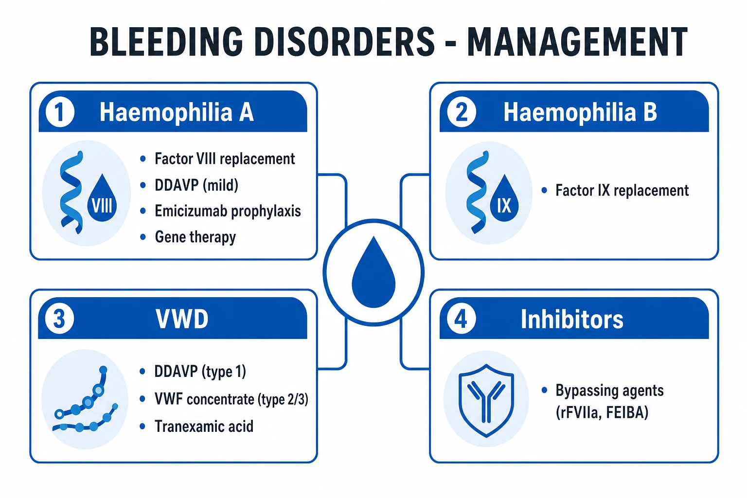
Secondary haemostasis is the cascade of plasma serine protease zymogens that, once activated, generate thrombin — the master enzyme that converts soluble fibrinogen into insoluble, cross-linked fibrin. It is conventionally divided into extrinsic, intrinsic, and common pathways. The classic model remains how the laboratory tests are interpreted: PT/INR reflects the extrinsic + common pathways (VII, X, V, II, I), while aPTT reflects the intrinsic + common pathways (XII, XI, IX, VIII, X, V, II, I). The thrombin time (TT) assesses the final step — the conversion of fibrinogen to fibrin by thrombin — and is prolonged by low fibrinogen, dysfibrinogenaemia, or heparin/DTI contamination.[1][3][4]
Most coagulation factors are serine protease zymogens made in the liver (except factor VIII [endothelium], vWF [endothelium/megakaryocytes]). Factors II, VII, IX, X, Protein C, Protein S are vitamin K-dependent (γ-carboxylation enables calcium binding and membrane anchoring — the basis of warfarin). Factor V and VIII are cofactors (not enzymes); factor XIII is a transglutaminase that cross-links fibrin.[4]
The extrinsic pathway (tissue factor) — the initiation
The extrinsic (the tissue factor pathway — the initiator). The tissue factor (TF, thromboplastin) is exposed by the injury (the subendothelial cells; also inducible on monocytes/endothelium in inflammation) → binds the circulating factor VIIa → the extrinsic tenase (TF-VIIa) → activates factor X → Xa.[3]
Extrinsic (tissue factor) pathway — the initiator
- The vascular injury exposes the TISSUE FACTOR (TF) — the integral membrane protein on subendothelial cells (and inducible on monocytes/endothelium in inflammation)
- TF binds the circulating factor VIIa (small amount circulates pre-activated) → the extrinsic tenase complex (TF-VIIa)
- TF-VIIa activates factor X → Xa (and also IX → IXa, linking to the intrinsic pathway)
- Xa + cofactor Va + phospholipid + Ca2+ = PROTHROMBINASE complex → cleaves the prothrombin (II) → thrombin (IIa) — a small "spark" of thrombin
- This initial thrombin then AMPLIFIES the response: activates platelets, factors V, VIII, XI, XIII, and (bound to thrombomodulin) activates protein C — the switch from initiation to propagation
The intrinsic (contact) pathway — the amplification
The intrinsic (the contact activation pathway — the amplification loop). The factor XII (Hageman) → XIIa → XI → XIa → IX → IXa → IXa + VIIIa (intrinsic tenase) → X → Xa. The deficiencies: XII (no bleeding); XI (mild — haemophilia C); IX (haemophilia B — Christmas disease); VIII (haemophilia A).[1]
Intrinsic (contact activation) pathway — the amplification loop
- The factor XII (Hageman factor) binds negatively charged surfaces (polyphosphate, collagen, DNA, RNA) → autoactivates to XIIa (the basis of the aPTT — surface contact with kaolin/cephalin)
- XIIa → activates XI → XIa
- XIa → activates IX → IXa
- IXa + cofactor VIIIa + phospholipid + Ca2+ = INTRINSIC TENASE complex → activates X → Xa (this is the SUSTAINED, high-output route to Xa — far more efficient than TF-VIIa alone, hence "amplification")
- The deficiencies (XII — no bleeding; XI — mild bleeding [haemophilia C]; IX — haemophilia B "Christmas disease"; VIII — haemophilia A) localise the lesion to the intrinsic pathway: the prolonged APTT with the normal PT
The common pathway — the thrombin generation and the fibrin
The common (the X → the Va → the prothrombinase → the thrombin [IIa] → the fibrinogen (I) → the fibrin → the cross-linked by XIIIa).[1]
Common pathway — from Xa to cross-linked fibrin
- Xa (from either pathway, but sustained supply from the intrinsic tenase) + Va + phospholipid + Ca2+ → PROTHROMBINASE → cleaves prothrombin (II) → thrombin (IIa) in a BURST
- Thrombin (IIa) cleaves fibrinogen (factor I) → releases fibrinopeptides A and B → fibrin MONOMERS → spontaneous polymerisation into fibrin STRANDS (soft clot)
- Thrombin also activates factor XIII → XIIIa (a transglutaminase)
- XIIIa cross-links fibrin (and α2-antiplasmin to fibrin, protecting against premature lysis) → STABLE, insoluble clot — the definitive haemostatic plug
- Thrombin simultaneously: activates platelets (PAR-1/PAR-4), activates V, VIII, XI, XIII (positive feedback), and — when bound to endothelial thrombomodulin — activates protein C (negative feedback / anticoagulant switch)
Coagulation factors — type, site of synthesis, vitamin K-dependence
| Factor | Name | Type | Synthesis | Vitamin K-dependent? |
|---|---|---|---|---|
| I | Fibrinogen | Structural (substrate) | Liver | No |
| II | Prothrombin | Serine protease zymogen | Liver | Yes |
| III | Tissue factor | Cofactor (transmembrane) | Subendothelium, monocytes | No |
| V | Proaccelerin | Cofactor | Liver | No |
| VII | Proconvertin | Serine protease zymogen | Liver | Yes |
| VIII | Antihaemophilic factor | Cofactor | Endothelium (stabilised by vWF) | No |
| IX | Christmas factor | Serine protease zymogen | Liver | Yes |
| X | Stuart-Prower | Serine protease zymogen | Liver | Yes |
| XI | Plasma thromboplastin antecedent | Serine protease zymogen | Liver | No |
| XII | Hageman factor | Serine protease zymogen | Liver | No |
| XIII | Fibrin-stabilising factor | Transglutaminase | Megakaryocytes/macrophages | No |
| — | Protein C | Serine protease zymogen | Liver | Yes |
| — | Protein S | Cofactor | Liver | Yes |
Coagulation laboratory tests — what each pathway measures
| Test | Pathway measured | Factors assessed | Normal range | Prolonged by |
|---|---|---|---|---|
| PT / INR | Extrinsic + common | VII, X, V, II, I | PT 11-14 s; INR ~1.0 | Warfarin, liver disease, vitamin K deficiency, DIC, factor VII deficiency |
| aPTT | Intrinsic + common | XII, XI, IX, VIII, X, V, II, I | 25-35 s | Heparin, haemophilia A/B, vWD, DIC, lupus anticoagulant, factor XII deficiency |
| Thrombin time (TT) | Final common step | Fibrinogen → fibrin (I) | <20 s | Heparin/DTI contamination, low fibrinogen, dysfibrinogenaemia, DIC |
| Fibrinogen (Clauss) | Factor I level | I | 2-4 g/L | DIC, massive transfusion, liver failure |
| D-dimer | Fibrinolysis | Cross-linked fibrin degradation | <0.5 µg/mL | Thrombosis (DVT/PE), DIC, inflammation, malignancy, pregnancy, post-surgery |
| Anti-Xa assay | Heparin effect | Direct measurement of heparin-antithrombin activity | Varies by assay | Confirms heparin levels independent of APTT (useful when APTT unreliable) |
Fibrinolysis — the clot removal
Once the haemostasis is secured, the fibrinolysis dissolves the clot to restore the blood flow and allow the repair. The central enzyme is plasmin, generated from the circulating plasminogen by the tissue plasminogen activator (tPA, released from endothelium) and the urokinase-type PA (uPA). Critically, the tPA is ~500-fold more active when BOUND to fibrin — so the plasmin generation is localised to the clot surface, minimising the systemic fibrinogenolysis. The plasmin cleaves the fibrin (and the fibrinogen) into the degradation products (FDPs) and, specifically for the cross-linked fibrin, D-dimers — a D-dimer therefore indicates that the thrombin (→ cross-linking via XIIIa) AND the plasmin (→ lysis) have both been active, i.e. genuine clot formation and breakdown.[4][8]
The regulators restrain the fibrinolysis to avoid the premature clot lysis or the systemic bleeding: [1]
Fibrinolysis — the pathway and its regulators
- Endothelium releases tPA (and uPA) in response to venous stasis, exercise, hypoxia, and bradykinin
- tPA binds fibrin (the clot surface) → becomes ~500× more active → converts plasminogen → plasmin locally (fibrin-localised → avoids systemic fibrinogenolysis)
- Plasmin cleaves cross-linked fibrin → FDPs (fragments X, Y, D, E) and D-dimer (specific to cross-linked fibrin — requires both thrombin [→ XIIIa cross-linking] AND plasmin [→ lysis])
- PAI-1 (plasminogen activator inhibitor-1) — from endothelium and platelets; the principal inhibitor of tPA/uPA. The elevated PAI-1 (obesity, sepsis, post-surgery) → the hypofibrinolytic, prothrombotic state
- α2-antiplasmin — the primary inhibitor of plasmin in plasma; cross-linked to fibrin by XIIIa (protecting the clot from premature lysis)
- TAFI (thrombin-activatable fibrinolysis inhibitor) — cleaves C-terminal lysines from partially degraded fibrin → removes plasminogen/tPA binding sites → DOWNREGULATES fibrinolysis
Fibrinolytic agents — and their antidotes
| Drug | Mechanism | Antidote / reversal |
|---|---|---|
| Alteplase (tPA) | Direct plasminogen → plasmin on fibrin | No specific antidote; aminocaproic acid/tranexamic acid (anti-fibrinolytic); cryoprecipitate/fibrinogen if bleeding |
| Tenecteplase | Modified tPA (longer half-life, fibrin-specific) | As above |
| Streptokinase | Indirect (forms complex with plasminogen → activates other plasminogen) | As above; antigenic |
| Tranexamic acid / aminocaproic acid | Lysine analogues → block plasminogen binding to fibrin → antifibrinolytic | N/A (they ARE the reversal of fibrinolytics) |
The natural anticoagulants — restraining the cascade
Left unchecked, the thrombin generation would propagate systemically. The three natural anticoagulant systems localise and terminate the coagulation. Their failure causes the thrombophilia; their pharmacological augmentation is the basis of the heparin, the fondaparinux, and (historically) the activated protein C.[4][6]
The protein C/S pathway — the thrombin-thrombomodulin switch
- Once a clot is forming, thrombin escaping into the circulation binds thrombomodulin (a constitutive endothelial transmembrane protein) — this SWITCHES thrombin's function from procoagulant to ANTICOAGULANT
- The thrombin-thrombomodulin complex (with EPCR — endothelial protein C receptor) activates the circulating protein C → activated protein C (APC)
- APC + its cofactor protein S (both vitamin K-dependent) bind the platelet/phospholipid surfaces
- APC proteolytically inactivates factors Va and VIIIa (the cofactors of prothrombinase and intrinsic tenase) → shuts down further thrombin generation → localises the clot
- Deficiency of protein C or S (or factor V Leiden → APC resistance) → failure to inactivate Va/VIIIa → a PROTHROMBOTIC (thrombophilic) state. Homozygous protein C deficiency → neonatal purpura fulminans (massive thrombosis); warfarin reduces protein C first (shortest half-life of vitamin K-dependent factors) → transient prothrombotic state → warfarin-induced skin necrosis (hence "bridge" with heparin)
The three natural anticoagulant systems
| System | Mechanism | Deficiency → | Pharmacological exploitation |
|---|---|---|---|
| Antithrombin (serpin) | Neutralises thrombin (IIa), Xa, IXa, XIa, XIIa by forming irreversible 1:1 complexes | Inherited AT deficiency (AD) or consumption in sepsis/DIC → VTE, heparin resistance | HEPARIN (UFH and LMWH) and fondaparinux potentiate antithrombin ~1000-fold — the entire basis of heparin anticoagulation |
| Protein C / S | APC + protein S inactivate Va and VIIIa | Protein C/S deficiency, factor V Leiden (APC resistance) → thrombophilia; warfarin-induced skin necrosis | Activated protein C (drotrecogin alfa) was used in severe sepsis (withdrawn — PROWESS-SHOCK negative) |
| TFPI (tissue factor pathway inhibitor) | Binds and inhibits the TF-VIIa complex in a Xa-dependent manner (feedback: requires Xa generation first) → limits the extrinsic pathway | Rare thrombosis | Recombinant TFPI (tifacogin) studied in sepsis (negative) |
The SIRS → CARS → MARS paradigm — the immune dysregulation in critical illness
The inflammatory response in critical illness is not simply "too much inflammation" — it is a dynamic, biphasic (sometimes multiphasic) dysregulation that Bone (1996) framed as the SIRS → CARS → MARS continuum. This paradigm remains the conceptual framework for understanding why septic patients die equally from hyperinflammation (early) and immunosuppression (late), and why immune-modulating trials (anti-TNF, IL-1 receptor antagonist) that targeted only SIRS failed.[10][11][12]
SIRS — Systemic Inflammatory Response Syndrome
SIRS is the exaggerated, uncontrolled systemic pro-inflammatory response to a trigger (infection, trauma, burns, pancreatitis, haemorrhage, ischaemia-reperfusion). The macrophages, monocytes, and neutrophils, activated by PAMPs/DAMPs via TLRs, release a cytokine storm — TNF-α, IL-1β, IL-6, IL-8, IFN-γ → fever/ hypothermia, tachycardia, tachypnoea, leukocytosis/leukopenia (the original SIRS criteria: ≥2 of these four). The endothelial activation → capillary leak, vasodilation, hypotension, microvascular thrombosis. The complement and coagulation cascades co-activate (DIC). The SIRS is the HYPERINFLAMMATORY phase — potentially lethal if unchecked, but also the appropriate initial response to clear the infection. Sepsis-3 (2016) subsumed SIRS into the new sepsis definition (organ dysfunction from dysregulated host response to infection) because SIRS was too sensitive and non-specific — but the SIRS concept of systemic hyperinflammation remains physiologically valid.[10][11]
CARS — Compensatory Anti-inflammatory Response Syndrome
CARS is the endogenous counter-regulatory response that follows (or accompanies) SIRS — an anti-inflammatory surge designed to prevent the inflammatory response from destroying the host. The monocytes and Tregs release IL-10, TGF-β, soluble TNF receptors, IL-1 receptor antagonist (IL-1ra) → downregulate the pro-inflammatory response. The T cells shift from Th1 (pro-inflammatory, IFN-γ) to Th2 (anti-inflammatory, IL-4/IL-10). The monocyte HLA-DR expression FALLS (a marker of monocyte deactivation — see immunoparalysis). When CARS overshoots, the patient becomes immunoparalysed/immunosuppressed → vulnerable to nosocomial infection, viral reactivation (CMV, HSV), and unable to clear the primary infection. The early deaths in sepsis are often SIRS-dominant (refractory vasodilatory shock, multi-organ failure); the late deaths (beyond day 3-7) are often CARS-dominant (secondary infections, immunoparalysis).[10][12]
MARS — Mixed Antagonist Response Syndrome
MARS is the state where SIRS and CARS coexist or oscillate simultaneously — the patient has features of both hyperinflammation (fever, vasoplegia, high CRP) AND immunosuppression (low monocyte HLA-DR, opportunistic infection, viral reactivation). MARS is the most common real-world presentation in prolonged critical illness: the inflammatory and anti-inflammatory responses are not sequential but overlapping and chaotic. This explains why single-target immunomodulation (blocking TNF, or boosting IL-1ra) has failed in trials — the patient's immune state is dynamic, mixed, and not predictable by a single biomarker. The concept of MARS underpins the modern search for immune phenotyping (monocyte HLA-DR, IL-6, CRP trends, cytokine profiling) to stratify patients for immunostimulatory (IFN-γ, GM-CSF, IL-7) versus anti-inflammatory (steroids, anti-IL-6) therapy.[10][12]
The SIRS → CARS → MARS continuum — the immune phases of critical illness
| Feature | SIRS (hyperinflammation) | CARS (immunoparalysis) | MARS (mixed) |
|---|---|---|---|
| Dominant response | Pro-inflammatory (TNF-α, IL-1β, IL-6, IFN-γ) | Anti-inflammatory (IL-10, TGF-β, IL-1ra) | Both simultaneously/oscillating |
| Timing | Early (hours-days) | Late (days-weeks) | Variable — often prolonged ICU stay |
| Monocyte HLA-DR | Normal or ↑ | ↓↓ (<8000 molecules/cell = immunoparalysis) | Variable |
| Clinical phenotype | Refractory vasodilatory shock, fever, high CRP, multi-organ failure | Nosocomial infection, viral reactivation (CMV/HSV), unable to clear primary infection | Both features — unpredictable, dynamic |
| T cell profile | Th1-dominant (IFN-γ) | Th2-dominant (IL-4, IL-10); lymphopenia | Mixed; T cell exhaustion (PD-1↑) |
| Therapeutic implication | Anti-inflammatory (steroids, anti-IL-6) MIGHT help if early | Immunostimulation (IFN-γ, GM-CSF, IL-7, PD-1 blockade) MIGHT help | Needs immune phenotyping first — no single therapy fits all |
| Mortality driver | Refractory shock, MOF | Secondary infection, immunoparalysis | Both — the deadliest combination |
Immunoparalysis in critical illness
Immunoparalysis (also called sepsis-induced immunosuppression, or injury-induced immunosuppression) is the functionally impaired innate and adaptive immune state that develops in a large fraction of critically ill patients — particularly those with prolonged sepsis, major trauma, burns, pancreatitis, or post-cardiac arrest. It is the CARS/MARS phenotype made concrete: the immune system is not exhausted of cells but the REMAINING cells are functionally silenced.[12]
The hallmarks of immunoparalysis: [1]
The mechanisms of immunoparalysis in critical illness
- Monocyte deactivation — the hallmark biomarker. Monocyte HLA-DR expression falls (measured by flow cytometry; <8000 molecules/cell = immunoparalysis). This reduces antigen presentation → impaired adaptive immune activation. Low HLA-DR correlates with nosocomial infection, viral reactivation, and mortality
- Lymphopenia and T cell exhaustion — sepsis causes profound apoptosis of CD4+, CD8+, and B cells (the immune system "turns off" by cell death). The surviving T cells upregulate inhibitory receptors (PD-1, CTLA-4, LAG-3, TIM-3) → "exhaustion" → impaired cytokine production and cytotoxicity. PD-1 expression on T cells correlates with secondary infection and mortality
- Treg expansion — regulatory T cells (CD4+, CD25+, FoxP3+) expand in sepsis → actively suppress effector T cells via IL-10 and TGF-β
- Th2 polarisation — the cytokine milieu shifts from Th1 (IFN-γ, pro-inflammatory) to Th2 (IL-4, IL-10, anti-inflammatory) → impaired intracellular pathogen clearance
- Neutrophil dysfunction — despite leukocytosis, neutrophils show impaired chemotaxis, phagocytosis, and oxidative burst → "immunoparalysed neutrophils" that cannot clear infection
- Endotoxin tolerance — repeated LPS exposure reprograms monocytes/macrophages to produce LESS TNF-α/IL-1β on rechallenge (a protective adaptation that overshoots into immunosuppression)
Biomarkers of immunoparalysis and their clinical use
| Biomarker | What it measures | Threshold for immunoparalysis | Clinical use |
|---|---|---|---|
| Monocyte HLA-DR (flow cytometry) | Monocyte antigen-presenting capacity | <8000 molecules/cell (or <80% of monocytes positive) | The gold standard; predicts nosocomial infection, viral reactivation, mortality; guides immunostimulation |
| PD-1 on T cells | T cell exhaustion | ↑ expression on CD4+/CD8+ | Correlates with secondary infection and mortality in sepsis |
| IL-10 / TNF-α ratio | Anti-inflammatory/pro-inflammatory balance | High ratio = CARS phenotype | Research/prognostic |
| Lymphocyte count | T cell apoptosis | Lymphopenia (<1000/µL) | Simple, readily available; persistent lymphopenia = poor prognosis |
| mHLA-DR kinetics | Recovery of immune function | Rising mHLA-DR = immune reconstitution | Serial monitoring — rising trend may support de-escalation of immunostimulation |
Short-answer questions — fellowship exam practice
SAQ — Late sepsis immunoparalysis with nosocomial infection and CMV reactivation
10 minutes · 10 marks
A 64-year-old man is day 12 of his ICU admission for pneumococcal pneumonia complicated by septic shock and ARDS. He was weaned from noradrenaline on day 4 and from the ventilator on day 8. He is now febrile (38.8°C) with purulent endotracheal secretions and new bilateral infiltrates. Laboratory tests: WCC 4.2, lymphocyte count 0.6 × 10⁹/L, CRP 140 (down from 320 a week ago, now rising again). CMV PCR has turned positive at 18,000 IU/mL. Blood cultures grow Candida albicans. The consultant asks you to outline the immune dysregulation underlying his deterioration and a management approach.
SAQ — Restrictive versus liberal transfusion in septic shock (TRISS)
10 minutes · 10 marks
A 58-year-old woman (weight 70 kg) is on day 3 of ICU admission for septic shock from a pyelonephritis with Escherichia coli bacteraemia. She has been weaned to noradrenaline 0.08 mcg/kg/min with a MAP of 72. She has no acute coronary syndrome. Serial haemoglobin has fallen from 128 to 74 g/L. Lactate is 1.6 mmol/L and she is making 45 mL/h of urine. The registrar asks whether she should be transfused to maintain haemoglobin above 90 g/L for tissue oxygen delivery. You are asked to justify your transfusion strategy with reference to the trial evidence.
Clinical pearls
Red flags
Key trials and evidence
Bone 1996 — SIRS, CARS, and the Newtonian metaphor (PMID 8674323)
Source
Critical Care Medicine 1996;24:1125-1128 — the editorial that framed the SIRS/CARS paradigm
Key contribution
Proposed that critical illness is a dynamic balance (Newton's third law) between SIRS (pro-inflammatory) and CARS (anti-inflammatory/compensatory) — and that MORS (mixed) is the common real-world state. Predicted that anti-inflammatory monotherapy would fail because the immune phase varies by patient and time
Key finding
The early deaths in sepsis are SIRS-dominant (hyperinflammation); the late deaths are CARS-dominant (immunoparalysis/secondary infection). The therapeutic target depends on which phase dominates — a concept vindicated by the failure of subsequent anti-TNF/IL-1ra trials
Clinical bottom line
The SIRS/CARS/MARS paradigm remains the conceptual framework for sepsis immunology — and the rationale for immune phenotyping and personalised immunomodulation (immunostimulation in CARS, anti-inflammatory in SIRS)
Singer 2016 — Sepsis-3 definitions (PMID 26903338)
Source
JAMA 2016;315:801-810 — the Third International Consensus Definitions for Sepsis and Septic Shock
Key contribution
Retired the SIRS criteria from the definition of sepsis (too sensitive, non-specific) and redefined sepsis as life-threatening organ dysfunction caused by a dysregulated host response to infection (SOFA ≥2). Introduced qSOFA (altered mentation, systolic ≤100, resp rate ≥22) for bedside screening
Key finding
Sepsis is fundamentally about DYSREGULATION — not just infection plus inflammation. This encompasses both the SIRS (hyperinflammatory) and CARS (immunosuppressive) ends of the spectrum, and the coagulation/endothelial dysfunction (DIC) that accompanies both
Clinical bottom line
Sepsis-3 reframed sepsis as organ dysfunction from a dysregulated host response — the SIRS criteria remain useful for detecting infection/inflammation but no longer define sepsis
Esmon 2005 — Inflammation and coagulation crosstalk (PMID 16281932)
Source
British Journal of Haematology 2005;131:417-430 — the definitive review of coagulation-inflammation crosstalk
Key contribution
Established the molecular links by which inflammation activates coagulation (TF expression on monocytes/endothelium, cytokine-driven thrombin generation) and coagulation feeds back to inflammation (thrombin-PAR signalling, protein C pathway)
Key finding
The protein C pathway (thrombin-thrombomodulin → APC → inactivates Va/VIIIa) is both anticoagulant and anti-inflammatory; its failure (sepsis) drives microvascular thrombosis
Clinical bottom line
Sepsis is a thrombo-inflammatory disease — coagulopathy (DIC) and inflammation are inseparable, explaining why anticoagulant strategies were trialled (and why activated protein C once seemed promising)
Mackman 2009 — Tissue factor and the extrinsic pathway (PMID 19372318)
Source
Anesthesia and Analgesia 2009;108:1447-1452 — the tissue factor/factor VIIa pathway review
Key contribution
Defined tissue factor as the principal initiator of coagulation in vivo (not the contact/intrinsic pathway), reframing the extrinsic pathway as physiological haemostasis
Key finding
TF is constitutively expressed on subendothelial cells and inducible on monocytes/endothelium by cytokines (sepsis) and cancer — explaining infection- and malignancy-associated thrombosis
Clinical bottom line
The TF-VIIa pathway is the therapeutic target of recombinant factor VIIa (rFVIIa) used off-label for refractory massive haemorrhage, and explains the prothrombotic phenotype of sepsis and cancer
Jensen 2004 — RBC pH, Bohr effect, and oxygenation-linked phenomena (PMID 15491402)
Source
Acta Physiologica Scandinavica 2004;182:215-227 — the integrative review of RBC O2/CO2 transport
Key contribution
Unified the Bohr effect, Haldane effect, 2,3-DPG regulation, and the role of RBC membrane transporters (band 3, AE1) in coupling O2 and CO2 transport to acid-base balance
Key finding
Oxygenation-linked proton and chloride binding to haemoglobin (via the band 3/anion exchanger) is the molecular basis of the Bohr effect — and 2,3-DPG modulates this by stabilising the deoxy (T) state
Clinical bottom line
P50 = 26.6 mmHg is the quantitative anchor; 2,3-DPG and pH are the chief physiological rightward shifters. Stored blood loses 2,3-DPG — a practical concern in massive transfusion
Prognosis
The prognosis of the disordered blood/immune physiology in the ICU tracks the underlying derangement. DIC carries a mortality of 40-80% in sepsis, driven by the precipitating cause — survival hinges on source control and organ support, not on correcting numbers in isolation. HIT, if unrecognised, has a 30-50% thrombosis rate and up to 5-10% mortality; prompt cessation of heparin and alternative anticoagulation substantially reduce amputation and death. Massive transfusion / trauma-induced coagulopathy mortality is determined by the lethal triad — early, ratio-based blood product resuscitation, tranexamic acid within 3 h, and correction of acidosis/hypothermia/hypocalcaemia are survival-defining. Immunoparalysis in prolonged critical illness carries a substantial mortality from secondary nosocomial infection and viral reactivation — the patient who survives the initial cytokine storm but dies of VAP or candidemia at day 14. The prognosis improves with immune phenotyping (monocyte HLA-DR) and targeted immunostimulation in selected patients. Inherited thrombophilia (protein C/S deficiency, factor V Leiden, antithrombin deficiency) confers a lifelong VTE risk and informs duration of anticoagulation. Haematopoiesis failure (marrow failure, severe sepsis, post-chemo neutropenia) prognosis follows the reversibility of the marrow insult; growth factor support (G-CSF, EPO, TPO-mimetics) and antimicrobial prophylaxis are temporising. Across all, the integrated view — that haematopoiesis, RBC physiology, immunity, coagulation, and fibrinolysis are one continuously regulated system that fails together in critical illness — is the exam-grade and bedside-grade principle.[1][4][10][12]
References
- [1]Orkin SH, Zon LI Hematopoiesis: an evolving paradigm for stem cell biology Cell, 2008.PMID 18295580
- [2]Chaplin DD Overview of the immune response J Allergy Clin Immunol, 2010.PMID 20176265
- [3]Mackman N The role of tissue factor and factor VIIa in hemostasis Anesth Analg, 2009.PMID 19372318
- [4]Esmon CT The interactions between inflammation and coagulation Br J Haematol, 2005.PMID 16281932
- [5]Levi M, Schultz M Coagulopathy and platelet disorders in critically ill patients Minerva Anestesiol, 2010.PMID 20935621
- [6]Conway EM Thrombomodulin and its role in inflammation Semin Immunopathol, 2012.PMID 21805323
- [7]Jensen FB Red blood cell pH, the Bohr effect, and other oxygenation-linked phenomena in blood O2 and CO2 transport Acta Physiol Scand, 2004.PMID 15491402
- [8]Cesarman-Maus G, Hajjar KA Molecular mechanisms of fibrinolysis Br J Haematol, 2005.PMID 15842654
- [9]Morgan BP Regulation of the complement membrane attack pathway Crit Rev Immunol, 1999.PMID 10422598
- [10]Bone RC Sir Isaac Newton, sepsis, SIRS, and CARS Crit Care Med, 1996.PMID 8674323
- [11]Singer M, Deutschman CS, Seymour CW, et al The Third International Consensus Definitions for Sepsis and Septic Shock (Sepsis-3) JAMA, 2016.PMID 26903338
- [12]Muszynski JA, Thakkar R, Hall MW Inflammation and innate immune function in critical illness Curr Opin Pediatr, 2016.PMID 27043087
- [13]Ruggeri ZM, Mendolicchio GL Interaction of von Willebrand factor with platelets and the vessel wall Hamostaseologie, 2015.PMID 25612915
- [14]Schroeder HW Jr, Cavacini L Structure and function of immunoglobulins J Allergy Clin Immunol, 2010.PMID 20176268