ICU · first-part-physiology
Blood and Immunology Physiology — Comprehensive
Also known as Haematopoiesis · Haematopoietic stem cell · Innate immunity · Adaptive immunity · Complement system · Coagulation cascade · Haemostasis · Primary haemostasis · Secondary haemostasis · Fibrinolysis · Antithrombin · Protein C pathway · Tissue factor pathway · Natural anticoagulants
Blood and immunology physiology — the integrated biology of haematopoiesis, innate and adaptive immunity, haemostasis (primary + secondary), fibrinolysis, and the natural anticoagulant regulatory systems. HAEMATOPOIESIS: pluripotent haematopoietic stem cell (HSC) in bone marrow → self-renewal + differentiation down two lineages. MYELOID lineage → erythrocytes (EPO-driven), neutrophils (G-CSF), monocytes/macrophages (M-CSF), eosinophils (IL-5), basophils, and megakaryocytes/platelets (TPO). LYMPHOID lineage → T cells (thymus), B cells (marrow), NK cells. Common progenitors: CMP (common myeloid progenitor, driven by IL-3/GM-CSF) and CLP (common lymphoid progenitor). INNATE IMMUNITY (minutes-hours): neutrophils (phagocytosis + neutrophil extracellular traps [NETs]), macrophages (phagocytosis + antigen presentation via MHC II), dendritic cells (professional antigen presenters bridging innate→adaptive), NK cells (kill virus-infected/tumour cells lacking MHC I, regulated by activating/inhibitory receptors), complement (classical [antibody-triggered, C1q], alternative [spontaneous hydrolysis of C3, amplification], lectin [mannose-binding lectin → MASP] pathways → converge at C3 convertase → C5 → membrane attack complex [MAC, C5b-9] → osmotic lysis + opsonisation [C3b] + inflammation [C3a/C5a]), acute phase proteins (CRP, procalcitonin, ferritin — produced by liver under IL-6 drive). ADAPTIVE IMMUNITY (days-weeks): T cells (CD4+ helper — coordinates response via cytokines, Th1/Th2/Th17/Tfh/Treg; CD8+ cytotoxic — kills virus-infected cells via perforin/granzyme + FasL), B cells (plasma cells produce antibody; IgM first then class-switching to IgG/IgA/IgE; memory B cells) → immunological memory. PRIMARY HAEMOSTASIS: platelet adhesion (vWF binds exposed subendothelial collagen + platelet GPIb-IX-V) → activation (shape change, granule release: ADP, serotonin, TXA2 from COX/thromboxane synthase; GPVI signals) → aggregation (GPIIb/IIIa [αIIbβ3] binds fibrinogen → platelet plug). SECONDARY HAEMOSTASIS: EXTRINSIC pathway (tissue factor + VIIa → activates X → tenase), INTRINSIC pathway (XII → XI → IX + VIIIa → activates X — amplification loop), COMMON pathway (X → Va → prothrombinase → thrombin [IIa] → fibrinogen → fibrin → cross-linked by XIIIa). FIBRINOLYSIS: plasminogen → plasmin (via tPA from endothelium; uPA) → degrades fibrin → D-dimer (and FDPs); regulated by PAI-1 (inhibits tPA) and α2-antiplasmin. REGULATION (natural anticoagulants): ANTITHROMBIN (serpin — inhibits thrombin, IXa, Xa, XIa, XIIa — potentiated ~1000-fold by heparin via conformational activation of its reactive site and the heparin-binding lysine); PROTEIN C/S (thrombin-thrombomodulin on endothelium activates protein C → activated protein C [APC] + protein S as cofactor → inactivates Va and VIIIa by proteolysis; deficiency → thrombophilia); TFPI (tissue factor pathway inhibitor — inhibits VIIa-TF complex after Xa generation, feedback inhibition of extrinsic pathway). The endothelial glycocalyx + thrombomodulin + heparan sulfates + tPA + ADAMTS13 maintain a constitutive anti-thrombotic vessel surface.
On this page & tools
Your progress
Saved locally on this device.
7 MCQs with explanations
Target exams
Red flags
Overview
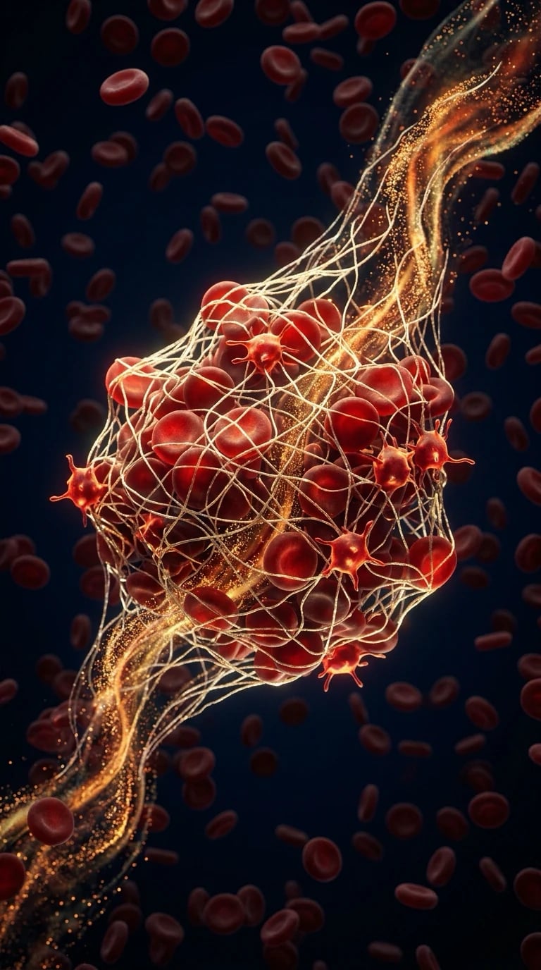
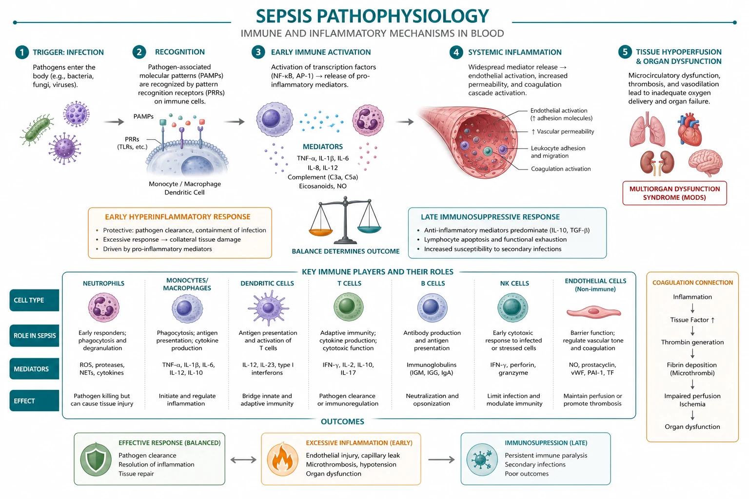
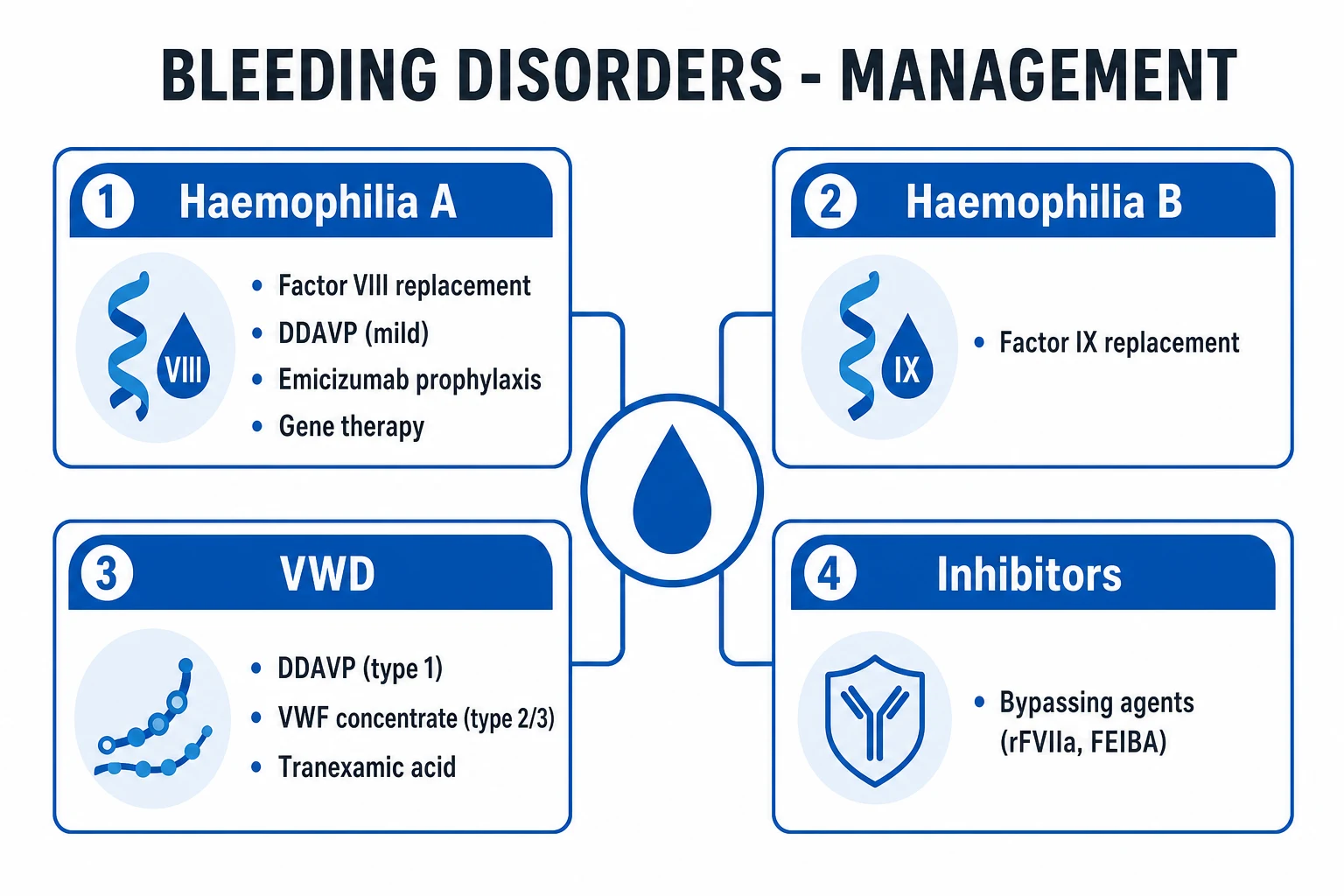
Haematopoiesis — the hierarchy of blood cell production [1]
Haematopoiesis is the continuous, regulated production of all blood cells from a small pool of pluripotent haematopoietic stem cells (HSCs) residing in the bone marrow niche. HSCs have two defining properties: self-renewal (asymmetric division producing one stem and one differentiating daughter) and multipotency (the capacity to generate every blood lineage). Their fate is governed by the microenvironment (osteoblasts, endothelial cells, mesenchymal stromal cells, CXCL12-abundant reticular [CAR] cells) and by lineage-specific growth factors (cytokines).[1]
Commitment proceeds through progressively restricted progenitors: [1]
Haematopoiesis — the differentiation hierarchy
- Pluripotent HSC (CD34+, CD38−, CD90+, Lin−) — self-renewing, rare (~1 in 10^4 marrow cells), maintained in quiescent endosteal niche by CXCL12/CXCR4 signalling and Tie2–angiopoietin-1
- Multipotent progenitor (MPP) — first commitment step, loses self-renewal, retains all lineage potential
- Bifurcation into two main lineage trees:
- COMMON MYELOID PROGENITOR (CMP) — driven by IL-3 and GM-CSF (act on multiple myeloid lineages), then:
- Granulocyte-monocyte progenitor (GMP) → neutrophils (G-CSF is the dominant late driver — basis of filgrastim), monocytes → tissue macrophages (M-CSF), eosinophils (IL-5), basophils/mast cells (IL-3, IL-4)
- Megakaryocyte-erythroid progenitor (MEP) → megakaryocytes → platelets (TPO [thrombopoietin] from liver — basis of eltrombopag/romiplostim), erythrocytes (EPO from the kidney in response to HIF-2α — basis of recombinant EPO)
- COMMON LYMPHOID PROGENITOR (CLP) → B cells (mature in bone marrow), T cells (migrate to and mature in the thymus — positive then negative selection), NK cells (bone marrow)
- COMMON MYELOID PROGENITOR (CMP) — driven by IL-3 and GM-CSF (act on multiple myeloid lineages), then:
- Lineage-specific growth factors determine the OUTPUT of each arm — see the comparison table below
- Mature cells enter circulation — granulocytes (neutrophils, eosinophils, basophils) are short-lived (hours-days), while lymphocytes and memory cells can persist for years
Lineage-specific haematopoietic growth factors — what drives what
| Growth factor | Source | Primary target lineage | Dominant product | ICU relevance |
|---|---|---|---|---|
| EPO (erythropoietin) | Kidney interstitial fibroblasts (peritubular), via HIF-2α when O2 falls | CFU-E → proerythroblast | Erythrocytes | Recombinant EPO for anaemia of critical illness (use restricted — thrombosis risk); EPO low in CKD |
| G-CSF (granulocyte colony-stimulating factor) | Endothelium, macrophages, fibroblasts | GMP → myelocyte → neutrophil | Neutrophils | Filgrastim for chemo-induced neutropenia, mobilises HSCs for harvest |
| M-CSF | Endothelium, fibroblasts | GMP → monocyte | Monocytes/macrophages | — |
| IL-5 | Th2 cells, mast cells | Eosinophil lineage | Eosinophils | Mepolizumab (anti-IL-5) for eosinophilic asthma |
| TPO (thrombopoietin) | Liver (constitutive) | Megakaryocyte → platelets | Platelets | TPO mimetics (eltrombopag, romiplostim, avatrombopag) for ITP/chronic liver disease platelet boost |
| IL-3 | T cells, mast cells | CMP and multipotent progenitors | Multi-lineage (myeloid broad) | Acts EARLY — broad myeloid expansion |
| GM-CSF | T cells, macrophages, endothelium | CMP/GMP — granulocytes + macrophages | Neutrophils + macrophages | Sargramostim; also drives alveolar macrophage function (deficiency → pulmonary alveolar proteinosis) |
| SCF (stem cell factor) | Bone marrow stroma | HSCs, early progenitors | Multi-lineage support | Co-stimulates with other growth factors |
Innate immunity — the first, fast, non-specific response
Innate immunity is phylogenetically ancient, germ-line encoded (no somatic recombination), responds in minutes to hours, and has no immunological memory. Its cellular effectors are neutrophils, macrophages, dendritic cells, NK cells, mast cells, eosinophils and basophils; its soluble effectors are complement, acute phase proteins, and interferons. Innate cells recognise conserved pathogen motifs (PAMPs — pathogen-associated molecular patterns) and endogenous damage signals (DAMPs — damage-associated, e.g. HMGB1, heat-shock proteins) through pattern recognition receptors (PRRs): Toll-like receptors (TLRs, on surface/endosome), NOD-like receptors (NLRs, cytosolic — drive inflammasome → IL-1β/IL-18), RIG-I-like receptors (RLRs, viral RNA), and C-type lectins.[2]
Cellular effectors of innate immunity
| Cell | Origin | Key function | Mechanism | Clinical/ICU relevance |
|---|---|---|---|---|
| Neutrophil | Marrow (G-CSF), most abundant circulating leucocyte | First-responder phagocyte | Phagocytosis (opsonin Fc/C3b receptors), oxidative burst (NADPH oxidase → ROS), granule enzymes (myeloperoxidase → hypochlorous acid), NETs (neutrophil extracellular traps — extruded chromatin decorated with antimicrobial proteins) | Bandemia/l leukocytosis in infection; neutropenia → invasive bacterial/fungal infection; NETs drive thrombosis and ARDS; CGD (NADPH oxidase defect) → recurrent catalase-positive infections |
| Monocyte/Macrophage | Marrow → blood monocyte → tissue macrophage (Kupffer cell, alveolar macrophage, microglia) | Phagocytosis + antigen presentation (MHC II to CD4+ T cells) + cytokine release (TNF-α, IL-1, IL-6) | PRR signalling, phagolysosome fusion; macrophages also clear apoptotic cells (efferocytosis) | Tissue macrophages drive chronic inflammation; alveolar macrophages first lung defence; macrophage activation syndrome (MAS); GM-CSF autoantibody → alveolar proteinosis |
| Dendritic cell | Marrow | Professional antigen-presenting cell — bridges innate → adaptive | Capture antigen in periphery, mature, migrate to lymph node, present peptide on MHC II (and cross-present on MHC I) to naïve T cells + co-stimulate (CD80/86 → CD28) | The quintessential activator of naïve T cells; without co-stimulation T cells become anergic |
| NK cell | Marrow (lymphoid lineage) | Kill virus-infected and tumour cells, antibody-dependent cellular cytotoxicity (ADCC) | "Missing-self" recognition: inhibited by self MHC I via killer immunoglobulin-like receptors (KIRs); cells that DOWNREGULATE MHC I (virus, tumour) lose inhibition → killing via perforin/granzyme + FasL. CD16 binds IgG-opsonised cells (ADCC) | Important in viral immunity (herpesviruses); ADCC exploited by therapeutic antibodies (rituximab) |
| Mast cell / basophil | Marrow | Allergy, parasites | FcεRI binds IgE → degranulation (histamine, tryptase, leukotrienes) | Anaphylaxis, urticaria; elevated tryptase in anaphylaxis/mastocytosis |
| Eosinophil | Marrow (IL-5) | Parasite defence, allergy | Major basic protein, eosinophil peroxidase — toxic to helminths | Asthma, eosinophilic oesophagitis, drug hypersensitivity |
The complement system
Complement is a cascade of ~30 plasma and cell-surface proteins that, once activated, generates: opsonisation (C3b tags pathogens for phagocytosis), inflammation (C3a, C4a, C5a are anaphylatoxins — recruit and activate neutrophils, cause mast cell degranulation), lysis (the membrane attack complex [MAC], C5b-9, punches holes in membranes), and immune complex clearance (C3b-coated complexes bind CR1 on erythrocytes for hepatic/splenic removal).[2][6]
Three activation pathways converge on a single effector mechanism: [1]
The three complement activation pathways — all converge at C3
| Feature | Classical | Lectin | Alternative |
|---|---|---|---|
| Trigger | Antigen-antibody complex (IgG/IgM bound to pathogen); also CRP, apoptotic cells | Mannose/fucose/N-acetylglucosamine on microbial surfaces (absent on vertebrates) | Spontaneous "tick-over" hydrolysis of C3 (C3[H2O]) on any surface |
| Initiator | C1q binds Fc of IgG/IgM → activates C1r → C1s | MBL (mannose-binding lectin) + ficolins bind sugar → activate MASP-1/2 | C3(H2O) binds factor B → factor D cleaves B → Ba + Bb → fluid-phase C3 convertase |
| C3 convertase | C4b2a (C1s cleaves C4 → C4a+C4b; C4b2a; then cleaves C2 → C2a joins) | C4b2a (same as classical, via MASP cleavage of C4/C2) | C3bBb (properdin stabilises) |
| Amplification | C3b deposits massively; joins convertase → C5 convertase (C4b2a3b / C3bBb3b) | As classical | The dominant AMPLIFICATION loop for ALL pathways — any C3b generated feeds the alternative loop |
| Termination | C5 convertase cleaves C5 → C5a (anaphylatoxin) + C5b → assembles MAC (C5b6789n) → osmotic lysis | Same | Same |
| Regulators | C1-inhibitor (deficiency → hereditary angioedema), factor H/I, DAF, CD59 (protectin, inhibits MAC on self cells) | Same regulators | Factor H, factor I, properdin (+), DAF |
| Clinical | Low C3/C4 in immune-complex disease (SLE, post-strep GN); C1-inh deficiency → angioedema | MBL deficiency → recurrent infections in children | Factor H/I deficiency → atypical HUS; C3 deficiency → recurrent pyogenic infection; CD59 deficiency → PNH (paroxysmal nocturnal haemoglobinuria) |
Acute phase proteins
The acute phase response is a systemic, liver-driven reaction to inflammation (chiefly IL-6, also IL-1β, TNF-α) that rewires plasma protein synthesis within hours: [1]
Acute phase reactants — what rises and what falls
| Protein | Change | Driver | Role / clinical use |
|---|---|---|---|
| CRP (C-reactive protein) | ↑↑ (can rise 1000-fold) | IL-6 | Opsonin; classical complement activator; the workhorse infection/inflammation marker — trends guide antibiotic duration |
| Procalcitonin | ↑↑ | Bacterial infection, IL-1β/TNF | Calcitonin propeptide from extra-thyroid tissues; rises specifically with bacterial infection (and severe systemic inflammation); used to guide antibiotic initiation/cessation (lower in viral infection) |
| Ferritin | ↑↑ | IL-6, iron sequestration | Acute phase reactant AND iron storage; very high in adult Still's, HLH/MAS, COVID cytokine storm; low in iron deficiency |
| Fibrinogen | ↑ | IL-6 | Both acute phase reactant AND coagulation factor (factor I); rises in inflammation (hence ESR rises — RBCs aggregate on fibrinogen) |
| Serum amyloid A | ↑↑ | IL-6 | Replaces HDL apolipoprotein in inflammation; precursor of AA amyloid in chronic inflammation |
| Haptoglobin | ↑ | — | Binds free haemoglobin; paradoxically LOW in haemolysis (consumed) |
| Albumin / transferrin | ↓ (negative acute phase) | Reprioritised synthesis | "Reverse" markers; low albumin in inflammation reflects illness severity, not just nutrition |
Adaptive immunity — specificity and memory (days to weeks)
Adaptive immunity is vertebrate-specific, generated by somatic recombination of antigen-receptor genes (VDJ recombination — yields a repertoire of >10^9 unique receptors), takes days to weeks to peak, and — uniquely — generates immunological memory (faster, stronger response on re-exposure). It has two arms: cell-mediated (T cells) and humoral (B cells/antibody). Antigen must first be presented: dendritic cells take up antigen in the periphery, mature, and traffic to lymph nodes where they present peptide on MHC to naïve T cells, providing signal 1 (TCR→MHC-peptide), signal 2 (co-stimulation: CD80/86→CD28), and signal 3 (polarising cytokines) — all three required to avoid anergy.[2]
T cell subsets and their cytokine signatures
| Subset | Marker / MHC restriction | Master regulator / cytokine | Function |
|---|---|---|---|
| CD4+ Th1 | CD4, MHC II | T-bet; IFN-γ, IL-2 | Intracellular pathogens (viruses, intracellular bacteria, TB); macrophage activation |
| CD4+ Th2 | CD4, MHC II | GATA3; IL-4, IL-5, IL-13 | Extracellular parasites; allergy; B cell class-switching to IgE; eosinophil recruitment |
| CD4+ Th17 | CD4, MHC II | RORγt; IL-17, IL-22 | Extracellular bacteria/fungi; neutrophil recruitment; mucosal defence; autoimmunity (psoriasis, IBD) |
| Tfh | CD4, CXCR5, Bcl6 | IL-21 | Follicular helper — drives B cell germinal centre response and affinity maturation |
| Treg | CD4, CD25, FoxP3 | IL-10, TGF-β | Immune SUPPRESSION / tolerance; prevents autoimmunity; IPEX syndrome if FoxP3 mutated |
| CD8+ cytotoxic T lymphocyte (CTL) | CD8, MHC I | Perforin, granzyme, FasL | Kill virus-infected and tumour cells presenting endogenous peptide on MHC I |
B cells and antibodies
B cells mature in the bone marrow (undergo VDJ recombination of the immunoglobulin heavy and light chain genes), and after antigen encounter (usually T-dependent, in germinal centres) differentiate into plasma cells (antibody factories) and memory B cells. The primary antibody response produces IgM first (pentamer — efficient complement activator, first line), then, under CD4+ T cell help (CD40-CD40L + cytokines), the cell undergoes class-switching to IgG (opsonisation, complement, placental transfer), IgA (mucosal immunity), or IgE (parasites/allergy). Affinity maturation (somatic hypermutation in germinal centres) improves binding. This order — IgM then IgG — and the immunological memory it generates are the basis of vaccination and the secondary (faster, IgG-dominant) response.[2]
Immunoglobulin classes
| Class | Structure | Key role | Clinical |
|---|---|---|---|
| IgG | Monomer; crosses placenta (FcRn) | Major secondary response; opsonisation, complement (classical), ADCC, neonatal immunity | Quantitatively dominant; IVIG for immunodeficiency/immune modulation; specific IgG = past infection/vaccination |
| IgM | Pentamer (J chain); first produced | Primary response; potent complement activator (classical); B-cell surface receptor (monomer) | Specific IgM = acute/recent infection |
| IgA | Dimer (secretory piece) at mucosa | Mucosal/secretory immunity (saliva, tears, breast milk, gut) | Selective IgA deficiency = commonest primary immunodeficiency |
| IgE | Monomer; binds mast cell/basophil FcεRI | Parasite defence; immediate hypersensitivity (allergy, anaphylaxis) | Measured in allergy; omalizumab (anti-IgE) for asthma/chronic urticaria |
| IgD | Monomer; B-cell surface | Naïve B-cell receptor (function less clear) | Marker of mature naïve B cells |
Primary haemostasis — building the platelet plug
Primary haemostasis forms the initial, unstable platelet plug at a site of vascular injury, occurring within seconds. It requires (1) an intact vascular response (vasoconstriction), (2) functioning platelets (adequate number AND function), and (3) von Willebrand factor (vWF). vWF is synthesised by endothelial cells (Weibel-Palade bodies) and megakaryocytes, and circulates bound to factor VIII (stabilising it).[3]
Primary haemostasis — adhesion → activation → aggregation
- VASCULAR INJURY → endothelial denudation exposes subendothelial collagen + tissue factor; reflex vasoconstriction (local) slows flow
- PLATELET ADHESION: circulating vWF binds exposed collagen and undergoes a conformational change → exposes its A1 domain which binds platelet GPIb-IX-V complex (this tethering is ESSENTIAL at high shear — deficiency = Bernard-Soulier syndrome [lack of GPIb])
- PLATELET ACTIVATION: adhesion triggers intracellular signalling (GPIVI and α2β1 collagen receptors; GPVI is the main activator) → Ca2+ mobilisation → shape change (disc → sphere with long pseudopodia), granule release:
- Dense (δ) granules → ADP, serotonin, calcium → recruit and activate neighbouring platelets
- α-granules → vWF, fibrinogen, factor V, P-selectin (marker of activation), PF4
- Concurrently, phospholipase A2 → arachidonic acid → COX-1 → thromboxane A2 (TXA2) is synthesised and released → potent platelet activator + vasoconstrictor (this is the target of aspirin)
- PLATELET AGGREGATION: activation flips GPIIb/IIIa (αIIbβ3) into its high-affinity state → binds fibrinogen (and vWF) → fibrinogen bridges adjacent platelets → platelet plug (deficiency of GPIIb/IIIa = Glanzmann thrombasthenia)
- STABILISATION: the plug is initially friable; secondary haemostasis (coagulation cascade) lays down fibrin to stabilise it into a definitive clot
Drugs targeting primary haemostasis — the receptor each blocks
| Drug | Target | Mechanism | Notes |
|---|---|---|---|
| Aspirin | COX-1 | Irreversible acetylation → blocks TXA2 synthesis; lasts platelet lifespan (7-10 days) | Antiplatelet; GIT bleeding risk |
| Clopidogrel / prasugrel / ticagrelor | P2Y12 (ADP receptor) | Block ADP-mediated activation (irreversible for clopidogrel/prasugrel; reversible for ticagrelor) | Dual antiplatelet therapy with aspirin post-PCI |
| Abciximab, eptifibatide, tirofiban | GPIIb/IIIa | Block final common aggregation pathway | IV GPIIb/IIIa inhibitors in acute PCI |
| Dipyridamole | Phosphodiesterase → ↑cAMP | Reduces platelet activation | Combined with aspirin for stroke prevention |
Secondary haemostasis — the coagulation cascade
Secondary haemostasis is the cascade of plasma serine protease zymogens that, once activated, generate thrombin — the master enzyme that converts soluble fibrinogen into insoluble, cross-linked fibrin. It is conventionally divided into extrinsic, intrinsic, and common pathways, though the modern cell-based model (initiation on tissue-factor-bearing cells → amplification on platelets → propagation) better reflects physiology. The classic model remains how the laboratory tests are interpreted: PT/INR reflects the extrinsic + common pathways (VII, X, V, II, I), while APTT reflects the intrinsic + common pathways (XII, XI, IX, VIII, X, V, II, I).[3][4]
Most coagulation factors are serine protease zymogens made in the liver (except factor VIII [endothelium], vWF [endothelium/megakaryocytes]). Factors II, VII, IX, X, Protein C, Protein S are vitamin K-dependent (γ-carboxylation enables calcium binding and membrane anchoring — the basis of warfarin). Factor V and VIII are cofactors (not enzymes); factor XIII is a transglutaminase that cross-links fibrin.[5]
The extrinsic pathway (tissue factor pathway) — initiation
Extrinsic (tissue factor) pathway — the initiator
- Vascular injury exposes TISSUE FACTOR (TF, thromboplastin) — an integral membrane protein on subendothelial cells (and inducible on monocytes/endothelium in inflammation)
- TF binds circulating factor VIIa (small amount circulates pre-activated) → forms the extrinsic tenase complex (TF-VIIa)
- TF-VIIa activates factor X → Xa (and also IX → IXa, linking to the intrinsic pathway)
- Xa + cofactor Va + phospholipid + Ca2+ = PROTHROMBINASE complex → cleaves prothrombin (II) → thrombin (IIa) — a small "spark" of thrombin
- This initial thrombin then AMPLIFIES the response: it activates platelets, factors V, VIII, XI, and XIII, and (bound to thrombomodulin) activates protein C — the switch from initiation to propagation
The intrinsic (contact) pathway — amplification
Intrinsic (contact activation) pathway — the amplification loop
- Factor XII (Hageman factor) binds negatively charged surfaces (polyphosphate, collagen, DNA, RNA, inorganic polyP) → autoactivates to XIIa (the basis of the aPTT — surface contact with kaolin/cephalin)
- XIIa → activates XI → XIa
- XIa → activates IX → IXa
- IXa + cofactor VIIIa + phospholipid + Ca2+ = INTRINSIC TENASE complex → activates X → Xa (this is the SUSTAINED, high-output route to Xa — far more efficient than TF-VIIa alone, hence "amplification")
- Deficiencies (XII — no bleeding; XI — mild bleeding [haemophilia C]; IX — haemophilia B "Christmas disease"; VIII — haemophilia A) localise the lesion to the intrinsic pathway: prolonged APTT with normal PT
The common pathway — thrombin generation and fibrin
Common pathway — from Xa to cross-linked fibrin
- Xa (from either pathway, but sustained supply from the intrinsic tenase) + Va + phospholipid + Ca2+ → PROTHROMBINASE → cleaves prothrombin (II) → thrombin (IIa) in a BURST
- Thrombin (IIa) cleaves fibrinogen (factor I) → releases fibrinopeptides A and B → fibrin MONOMERS → spontaneous polymerisation into fibrin STRANDS (soft clot)
- Thrombin also activates factor XIII → XIIIa (a transglutaminase)
- XIIIa cross-links fibrin (and α2-antiplasmin to fibrin, protecting against premature lysis) → STABLE, insoluble clot — the definitive haemostatic plug
- Thrombin simultaneously: activates platelets (PAR-1/PAR-4), activates V, VIII, XI, XIII (positive feedback), and — when bound to endothelial thrombomodulin — activates protein C (negative feedback / anticoagulant switch)
Coagulation factors — type, site of synthesis, vitamin K-dependence
| Factor | Name | Type | Synthesis | Vitamin K-dependent? |
|---|---|---|---|---|
| I | Fibrinogen | Structural (substrate) | Liver | No |
| II | Prothrombin | Serine protease zymogen | Liver | Yes |
| III | Tissue factor | Cofactor (transmembrane) | Subendothelium, monocytes | No |
| V | Proaccelerin | Cofactor | Liver | No |
| VII | Proconvertin | Serine protease zymogen | Liver | Yes |
| VIII | Antihaemophilic factor | Cofactor | Endothelium (stabilised by vWF) | No |
| IX | Christmas factor | Serine protease zymogen | Liver | Yes |
| X | Stuart-Prower | Serine protease zymogen | Liver | Yes |
| XI | Plasma thromboplastin antecedent | Serine protease zymogen | Liver | No |
| XII | Hageman factor | Serine protease zymogen | Liver | No |
| XIII | Fibrin-stabilising factor | Transglutaminase | Megakaryocytes/macrophages | No |
| — | Protein C | Serine protease zymogen | Liver | Yes |
| — | Protein S | Cofactor | Liver | Yes |
Fibrinolysis — removing the clot
Once haemostasis is secured, fibrinolysis dissolves the clot to restore blood flow and allow repair. The central enzyme is plasmin, generated from circulating plasminogen by tissue plasminogen activator (tPA, released from endothelium) and urokinase-type PA (uPA). Critically, tPA is ~500-fold more active when BOUND to fibrin — so plasmin generation is localised to the clot surface, minimising systemic fibrinogenolysis. Plasmin cleaves fibrin (and fibrinogen) into degradation products (FDPs) and, specifically for cross-linked fibrin, D-dimers — a D-dimer therefore indicates that thrombin (→ cross-linking via XIIIa) AND plasmin (→ lysis) have both been active, i.e. genuine clot formation and breakdown.[5]
Regulators restrain fibrinolysis to avoid premature clot lysis or systemic bleeding:
- PAI-1 (plasminogen activator inhibitor-1) — from endothelium and platelets; the principal inhibitor of tPA/uPA. Elevated PAI-1 (in obesity, sepsis, post-surgery) → a hypofibrinolytic, prothrombotic state.
- α2-antiplasmin — the main circulating inhibitor of plasmin; cross-linked into the clot by XIIIa.
- TAFI (thrombin-activatable fibrinolysis inhibitor / carboxypeptidase B) — activated by the thrombin-thrombomodulin complex; clips C-terminal lysines from partially degraded fibrin, removing tPA/plasminogen binding sites → clot stabilisation. [1]
Fibrinolytic agents — and their antidotes
| Drug | Mechanism | Antidote / reversal |
|---|---|---|
| Alteplase (tPA) | Direct plasminogen → plasmin on fibrin | No specific antidote; aminocaproic acid/tranexamic acid (anti-fibrinolytic); cryoprecipitate/fibrinogen if bleeding |
| Tenecteplase | Modified tPA (longer half-life, fibrin-specific) | As above |
| Streptokinase | Indirect (forms complex with plasminogen → activates other plasminogen) | As above; antigenic |
| Tranexamic acid / aminocaproic acid | Lysine analogues → block plasminogen binding to fibrin → antifibrinolytic | N/A (they ARE the reversal of fibrinolytics) |
Natural anticoagulants — restraining the cascade
Left unchecked, thrombin generation would propagate systemically. Three natural anticoagulant systems localise and terminate coagulation. Their failure causes thrombophilia; their pharmacological augmentation is the basis of heparin, fondaparinux, and (historically) activated protein C.[4][6]
The protein C/S pathway — the thrombin-thrombomodulin switch
- Once a clot is forming, thrombin escaping into the circulation binds thrombomodulin (a constitutive endothelial transmembrane protein) — this SWITCHES thrombin's function from procoagulant to ANTICOAGULANT
- The thrombin-thrombomodulin complex (with endothelial protein C receptor, EPCR) activates circulating protein C → activated protein C (APC)
- APC + its cofactor protein S (both vitamin K-dependent) bind platelet/phospholipid surfaces
- APC proteolytically inactivates factors Va and VIIIa (the cofactors of prothrombinase and intrinsic tenase) → shuts down further thrombin generation → localises the clot
- Deficiency of protein C or S (or factor V Leiden → APC resistance) → failure to inactivate Va/VIIIa → a PROTHROMBOTIC (thrombophilic) state. Homozygous protein C deficiency → neonatal purpura fulminans (massive thrombosis); warfarin reduces protein C first (shortest half-life of vitamin K-dependent factors) → transient prothrombotic state → warfarin-induced skin necrosis (hence "bridge" with heparin, especially in known protein C deficiency)
The three natural anticoagulant systems
| System | Mechanism | Deficiency → | Pharmacological exploitation |
|---|---|---|---|
| Antithrombin (serpin) | Neutralises thrombin (IIa), Xa, IXa, XIa, XIIa by forming irreversible 1:1 complexes via its reactive site (Arg393-Ser394) | Inherited AT deficiency (AD) or consumption in sepsis/DIC → VTE, heparin resistance | HEPARIN (UFH and LMWH) and fondaparinux potentiate antithrombin ~1000-fold — the entire basis of heparin anticoagulation |
| Protein C / S | APC + protein S inactivate Va and VIIIa | Protein C/S deficiency, factor V Leiden (APC resistance) → thrombophilia; warfarin-induced skin necrosis | Activated protein C (drotrecogin alfa) was used in severe sepsis (withdrawn — PROWESS-SHOCK negative) |
| TFPI (tissue factor pathway inhibitor) | Binds and inhibits the TF-VIIa complex in a Xa-dependent manner (feedback: requires Xa generation first) → limits the extrinsic pathway | Rare thrombosis | Pharmacological recombinant TFPI (tifacogin) studied in sepsis (negative) |
Clinical pearls
Red flags
Key trials and evidence
Esmon 2005 — Inflammation and coagulation crosstalk (PMID 16281932)
Source
British Journal of Haematology 2005;131:417-430 — the definitive review of coagulation-inflammation crosstalk
Key contribution
Established the molecular links by which inflammation activates coagulation (TF expression on monocytes/endothelium, cytokine-driven thrombin generation) and coagulation feeds back to inflammation (thrombin-PAR signalling, protein C pathway)
Key finding
The protein C pathway (thrombin-thrombomodulin → APC → inactivates Va/VIIIa) is both anticoagulant and anti-inflammatory; its failure (sepsis) drives microvascular thrombosis
Clinical bottom line
Sepsis is a thrombo-inflammatory disease — coagulopathy (DIC) and inflammation are inseparable, explaining why anticoagulant strategies were trialled (and why activated protein C once seemed promising)
Levi 2010 — Coagulopathy in the critically ill (PMID 20935621)
Source
Minerva Anestesiologica 2010;76:851-857 — practical review of coagulation disorders in ICU
Key contribution
Systematised the differential of acquired coagulopathy in critical illness — DIC, massive blood loss/dilutional, anticoagulant-related, liver failure, vitamin K deficiency
Key finding
Standard coagulation tests (PT, APTT, platelets, fibrinogen, D-dimer) remain the backbone, but viscoelastic testing (TEG/ROTEM) better reflects whole-clot physiology and guides goal-directed haemostatic therapy
Clinical bottom line
In ICU bleeding, distinguish consumption (DIC) from dilution (trauma) from anticoagulation (drug) from synthesis failure (liver) — each demands a different transfusion/antidote strategy
Mackman 2009 — Tissue factor and the extrinsic pathway (PMID 19372318)
Source
Anesthesia and Analgesia 2009;108:1447-1452 — the tissue factor/factor VIIa pathway review
Key contribution
Defined tissue factor as the principal initiator of coagulation in vivo (not the contact/intrinsic pathway), reframing the extrinsic pathway as physiological haemostasis
Key finding
TF is constitutively expressed on subendothelial cells and inducible on monocytes/endothelium by cytokines (sepsis) and cancer — explaining infection- and malignancy-associated thrombosis
Clinical bottom line
The TF-VIIa pathway is the therapeutic target of recombinant factor VIIa (rFVIIa) used off-label for refractory massive haemorrhage, and explains the prothrombotic phenotype of sepsis and cancer
Prognosis
The prognosis of disordered blood/immune physiology in the ICU tracks the underlying derangement. DIC carries a mortality of 40-80% in sepsis, driven by the precipitating cause — survival hinges on source control and organ support, not on correcting numbers in isolation. HIT, if unrecognised, has a 30-50% thrombosis rate and up to 5-10% mortality; prompt cessation of heparin and alternative anticoagulation substantially reduce amputation and death. Thrombotic microangiopathy (TTP) is universally fatal within days without urgent plasma exchange (caplacizumab now adjunctive); HUS and complement-mediated TMA benefit from eculizumab. Massive transfusion / traumainduced coagulopathy mortality is determined by the lethal triad — early, ratio-based blood product resuscitation (PROPPR), tranexamic acid within 3 h (CRASH-2), and correction of acidosis/hypothermia/hypocalcaemia are survival-defining. Inherited thrombophilia (protein C/S deficiency, factor V Leiden, antithrombin deficiency) confers a lifelong VTE risk and informs duration of anticoagulation. Haematopoiesis failure (marrow failure, severe sepsis, post-chemo neutropenia) prognosis follows the reversibility of the marrow insult; growth factor support (G-CSF, EPO, TPO-mimetics) and antimicrobial prophylaxis are temporising. Across all, the integrated view — that haematopoiesis, immunity, coagulation, and fibrinolysis are one continuously regulated system that fails together in critical illness — is the exam-grade and bedside-grade principle.[1][4][5][6]
Blood and immunology physiology SAQ
10 minutes · 10 marks
A 68-year-old with septic shock has Hb 78 g/L, PaO2 60 mmHg on FiO2 0.5, temperature 39 C, and pH 7.25. Explain oxygen carriage and unloading, then outline coagulation/innate immune principles relevant to his ICU care.
References
- [1]Orkin SH, Zon LI Hematopoiesis: an evolving paradigm for stem cell biology Cell, 2008.PMID 18295580
- [2]Chaplin DD Overview of the immune response J Allergy Clin Immunol, 2010.PMID 20176265
- [3]Mackman N The role of tissue factor and factor VIIa in hemostasis Anesth Analg, 2009.PMID 19372318
- [4]Esmon CT The interactions between inflammation and coagulation Br J Haematol, 2005.PMID 16281932
- [5]Levi M Coagulopathy and platelet disorders in critically ill patients Minerva Anestesiol, 2010.PMID 20935621
- [6]Conway EM Thrombomodulin and its role in inflammation Semin Immunopathol, 2012.PMID 21805323