ICU · Physiology / cellular
Cellular Physiology & Membrane Potentials
Also known as Membrane potential · Resting membrane potential · Action potential · Na/K ATPase · Nernst equation · Goldman equation · Ion channels · Excitation-contraction coupling · Oxidative phosphorylation · Cytopathic hypoxia · HIF-1 alpha · Apoptosis versus necrosis
Cellular physiology and membrane potentials: the resting membrane potential (minus 70 mV; the Na/K ATPase — 3 Na out, 2 K in; the K leak the dominant). The Nernst equation (the equilibrium potential). The Goldman equation (the permeability-weighted). The action potential (the depolarisation Na influx, the repolarisation K efflux, the refractory). The cell membrane (the phospholipid bilayer, the channels, the receptors). Excitation-contraction coupling (the skeletal, the cardiac, the smooth). Cellular metabolism (glycolysis, Krebs cycle, oxidative phosphorylation, the ATP). Oxygen sensing (the HIF-1 alpha, the mitochondrial oxygen utilisation). Cellular hypoxia (the hypoxic, the stagnant, the anemic, the histotoxic; the cytopathic in sepsis). The cell death (the apoptosis, the necrosis, the necroptosis). The reactive oxygen species and the antioxidant defenses. The clinical correlations (the hyperkalaemia, the hypocalcaemia, the local anaesthetics, the channelopathies).
On this page & tools
Your progress
Saved locally on this device.
8 MCQs with explanations
Target exams
Overview & definition
The cellular physiology — the membrane potential (the electrical difference across the cell membrane). The resting potential (minus 70 mV). The action potential. The Na/K ATPase. The Nernst and the Goldman. The clinical correlations.[1]
Cellular physiology is the biophysical and biochemical basis of every phenomenon intensivists manipulate: the electrical excitability that antiarrhythmics and neuromuscular blockers target, the excitation-contraction coupling that inotropes and calcium-channel blockers alter, and the cellular energetics (glycolysis, the Krebs cycle, oxidative phosphorylation) whose failure produces the lactataemia, cytopathic hypoxia and multiorgan dysfunction of sepsis. This topic draws together membrane structure, bioelectricity, muscle coupling, metabolism, oxygen sensing, and the modes of cell death — the cellular substrate of critical illness. [1]
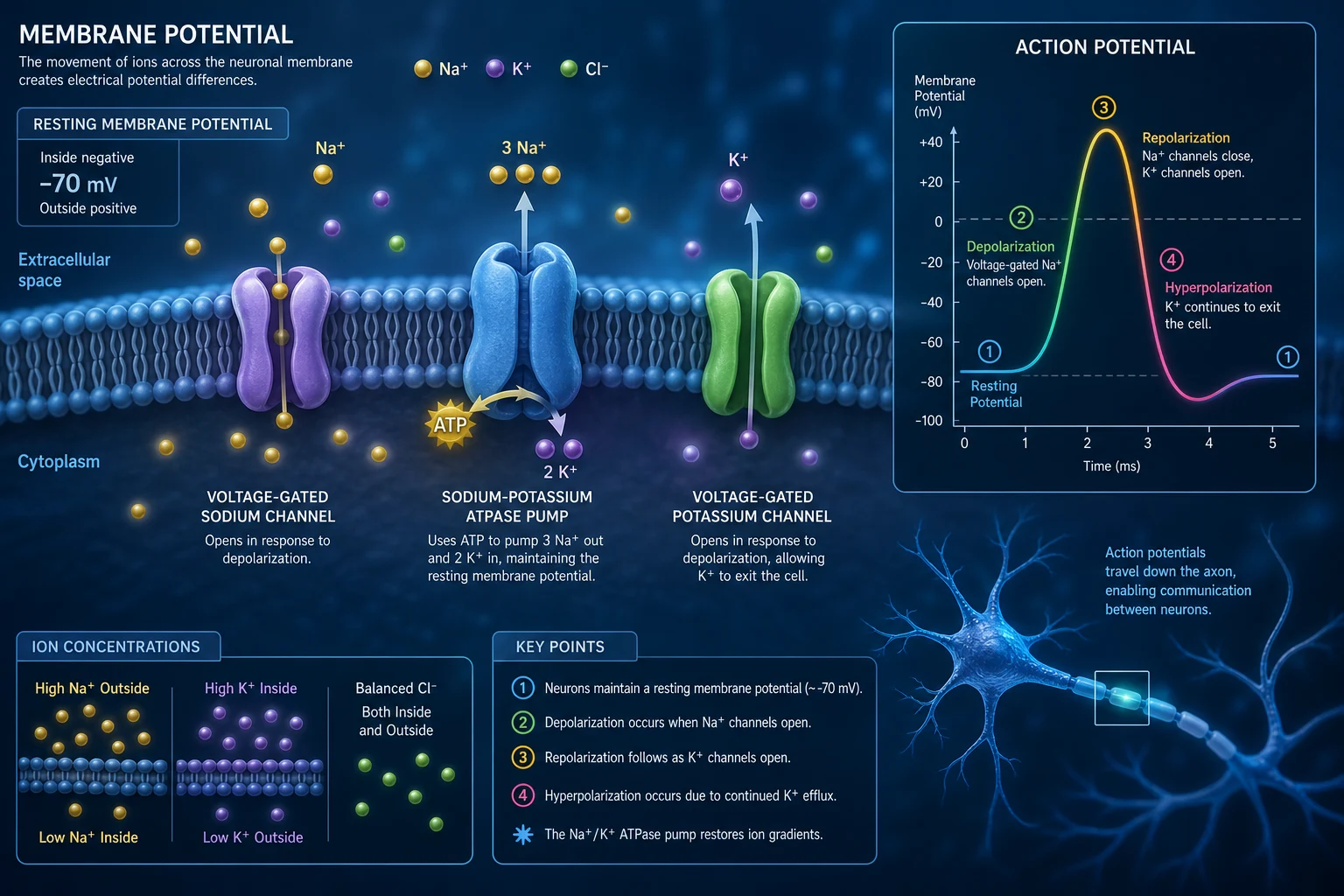
Cell membrane structure — the phospholipid bilayer
Every cell is bounded by the plasma membrane, a 5–8 nm phospholipid bilayer that is the substrate for the membrane potential, for signalling and for transport. The membrane is NOT a passive barrier: it is a dynamic, selectively permeable structure whose protein complement determines the electrophysiology of the cell. [1]
The lipid bilayer
- Phospholipid molecules are amphipathic — a hydrophilic phosphate head and two hydrophobic fatty-acid tails. In water they self-assemble tail-to-tail into a bilayer, with the polar heads facing the aqueous extracellular and intracellular compartments.
- The hydrophobic core is essentially impermeable to ions (Na+, K+, Ca2+, Cl−), to large polar molecules (glucose, amino acids) and to proteins — all of which require specific transport proteins. It is permeable to lipid-soluble substances (O2, CO2, steroid hormones, volatile anaesthetics) which diffuse down their concentration gradient directly through the lipid.
- Cholesterol is intercalated between phospholipids (roughly 1 cholesterol per phospholipid) — it modulates membrane fluidity (rigidifies at high temperature, prevents crystallisation at low temperature) and decreases water permeability.
- Asymmetric distribution: the outer (extracellular) leaflet is enriched in phosphatidylcholine and sphingomyelin; the inner (cytoplasmic) leaflet in phosphatidylethanolamine and phosphatidylserine. Phosphatidylserine externalisation to the outer leaflet is an "eat me" signal for apoptosis — the basis of the Annexin V assay. [1]
Membrane proteins — channels, carriers, receptors, pumps
The functional specificity of the membrane resides in its proteins, classified by function: [1]
Membrane proteins — the four functional classes
| Class | Mechanism | Energetics | Examples (ICU-relevant) |
|---|---|---|---|
| Ion channels | Aqueous pore; ions flow down electrochemical gradient | Passive (no ATP); gated (voltage, ligand, mechanosensitive) | Voltage-gated Na+ (Phase 0), K+ (repolarisation), Ca2+ (L-type, Phase 2); nAChR (NMJ); KATP (pancreatic beta cell) |
| Carrier / transporter | Binds substrate, undergoes conformational change | Facilitated diffusion (passive) OR active | GLUT (glucose), Na+/K+/2Cl− (furosemide target), Na+/Ca2+ exchanger |
| Ion pumps (ATPases) | Uses ATP hydrolysis to move ions AGAINST gradient | Primary active | Na+/K+ ATPase (the pump), Ca2+ ATPase (SERCA, PMCA), H+/K+ ATPase (gastric, PPI target) |
| Receptors | Bind ligand → intracellular signal | GPCR, RTK, ionotropic | Beta-1 adrenoceptor (Gs/cAMP), insulin receptor (RTK), nAChR (ion channel) |
Gating mechanisms — how channels open
- Voltage-gated channels open in response to a change in membrane potential (a charged S4 transmembrane segment — the "voltage sensor" — moves outward as the membrane depolarises). Examples: voltage-gated Na+, K+, Ca2+ channels — the channels of the action potential.
- Ligand-gated channels open when a chemical ligand binds an extracellular or intracellular site. Examples: nicotinic ACh receptor (ACh), GABA-A receptor (GABA, benzodiazepines), NMDA receptor (glutamate).
- Mechanically-gated channels open with physical deformation (stretch, pressure). Examples: cochlear hair cells, vascular baroreceptors.
- Second-messenger-gated channels open in response to an intracellular messenger (cAMP, cGMP, IP3, Ca2+). Example: cGMP-gated channels of photoreceptors; IP3 receptors on the sarcoplasmic reticulum. [1]
Resting membrane potential
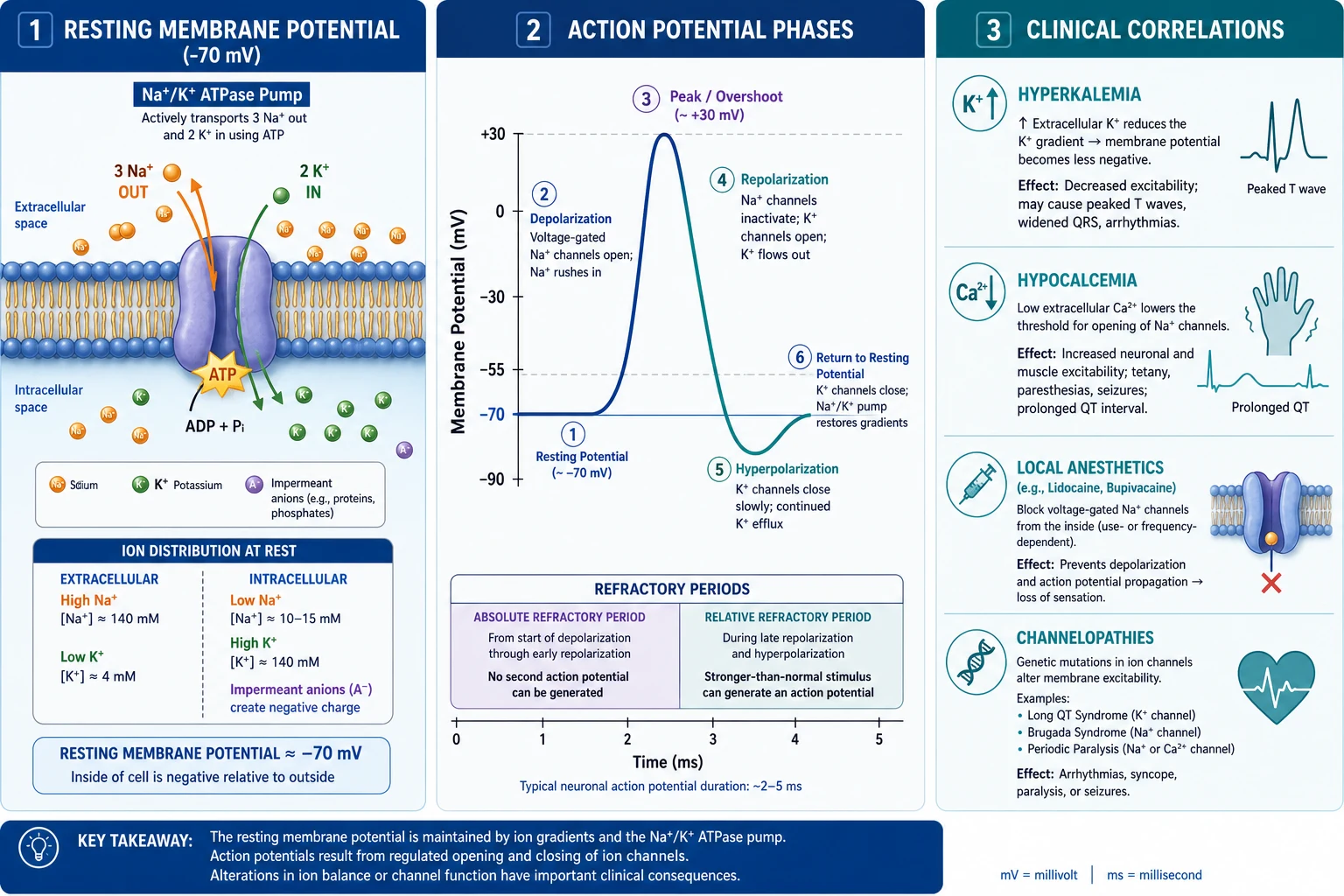
- Minus 70 mV (inside negative).[1]
- Na/K ATPase — 3 Na out, 2 K in (electrogenic).[1]
- K leak — the dominant determinant (K diffuses out; the membrane most permeable to K).[1]
- Nernst — E(K) = minus 90 mV (close to resting = K dominant).[1]
- Goldman — the permeability-weighted average of all ions.[1]
Why the resting potential exists — three determinants
The resting membrane potential (RMP) — typically minus 70 to minus 90 mV in excitable cells (inside negative relative to outside) — is generated by three factors operating together: [1]
- Concentration gradients established and maintained by the Na+/K+ ATPase. Intracellular K+ is high (approx 140 mmol/L) while extracellular K+ is low (approx 4 mmol/L); intracellular Na+ is low (approx 10–15 mmol/L) while extracellular Na+ is high (approx 140 mmol/L). The pump moves 3 Na+ out and 2 K+ in per ATP hydrolysed — it is electrogenic (net one positive charge out per cycle, contributing minus 4 to minus 10 mV to the RMP directly).
- Selective membrane permeability. At rest the membrane is 25–100 times more permeable to K+ than to Na+ (because of constitutively open K+ "leak" / two-pore-domain K2P channels). K+ therefore diffuses OUT down its concentration gradient, carrying positive charge out and leaving the inside negative. Na+ cannot follow to any significant extent (the membrane resists it), so the negative charge accumulates.
- The Donnan effect / fixed intracellular anions. Large impermeant intracellular anions (proteins, organic phosphates) cannot leave the cell, contributing a small sustained negative intracellular charge. [1]
The RMP therefore sits CLOSE to the potassium equilibrium potential EK, because K+ is the dominant permeant ion at rest. [1]
The Nernst equation — equilibrium potential for a SINGLE ion
The Nernst equation gives the membrane potential at which a single ion is at electrochemical equilibrium (no net flux — the electrical force exactly balances the chemical/concentration force): [1]
E(ion) = (RT / zF) × ln( [ion]outside / [ion]inside ) [1]
Where R is the gas constant, T the absolute temperature (310 K at 37 °C), z the ion valence, and F the Faraday constant. At 37 °C the term RT/F × ln(10) evaluates to 61.5 mV, giving the practical logarithmic form: [1]
E(ion) = (61.5 / z) × log10( [ion]outside / [ion]inside ) [1]
Worked examples (typical mammalian myocyte concentrations): [1]
| Ion | z | [outside] (mM) | [inside] (mM) | Equilibrium potential E(ion) |
|---|---|---|---|---|
| K+ | +1 | 4 | 140 | EK = 61.5 × log10(4/140) = minus 95 mV |
| Na+ | +1 | 145 | 15 | ENa = 61.5 × log10(145/15) = +60 mV |
| Ca2+ | +2 | 2.5 | 0.0001 | ECa = (61.5/2) × log10(2.5/0.0001) = +130 mV |
| Cl− | −1 | 110 | 4 | ECl = −61.5 × log10(110/4) = minus 89 mV |
The RMP (minus 70 to minus 90 mV) sits near EK (minus 95 mV) — confirming that K+ is the dominant determinant of the resting potential. Note Ca2+ has the largest driving force (its equilibrium potential is very positive, so it always wants to ENTER the cell whenever a Ca2+ channel opens — the basis of excitation-contraction coupling and neurotransmitter release). [1]
The Goldman-Hodgkin-Katz (Goldman) equation — all permeant ions together
Real membranes are simultaneously permeable to several ions. The Goldman equation (Goldman 1943; Hodgkin and Katz 1949) generalises the Nernst equation into a permeability-weighted average under the constant-field assumption (the electric field across the ~5 nm bilayer is assumed uniform). For monovalent ions: [1]
Vm = (RT/F) × ln( (PK·[K+]o + PNa·[Na+]o + PCl·[Cl−]i) / (PK·[K+]i + PNa·[Na+]i + PCl·[Cl−]o) ) [1]
Note the Cl− terms are inverted (intracellular in the numerator, extracellular in the denominator) because Cl− is an anion — the opposite sign convention. The permeability ratio PK : PNa : PCl at rest is approximately 1 : 0.04 : 0.45 — K+ dominates. Worked example at rest (Vm = 61.5 × log10(11.6 / 190.1) = minus 74.5 mV). During the action-potential upstroke PNa rises ~500-fold and Vm swings towards ENa (+60 mV) — exactly as observed. The equation reduces to the Nernst equation if only one ion is permeant. [1]
Key teaching point the equation encodes: the membrane potential is pulled towards whichever ion currently has the highest relative permeability. At rest, K+ dominates (Vm near EK); during Phase 0, Na+ dominates (Vm swings towards ENa). [1]
Action potential generation
- Threshold (minus 55 mV — voltage-gated Na channels open).[1]
- Depolarisation — Na influx (positive feedback — more depolarisation opens more channels).[1]
- Repolarisation — Na channels inactivate; K channels open; K efflux.[1]
- Hyperpolarisation — K channels remain open briefly (overshoot the resting).[1]
- Refractory — absolute (Na channels inactivated); relative (membrane hyperpolarised).[1]
The molecular events underlying each phase
The action potential is generated by sequential opening and closing of voltage-gated ion channels, each with distinct gating states: [1]
- Voltage-gated Na+ channels have three functional states: RESTING (closed, available — the gate is the activation gate; requires a negative RMP to be available), OPEN (activated — both gates open, Na+ rushes in), and INACTIVATED (the ball-and-chain inactivation gate plugs the pore; the channel cannot reopen until the membrane repolarises and the inactivation gate is removed). This inactivation is the molecular basis of the refractory period.
- Depolarisation (the upstroke). At threshold (about minus 55 mV), voltage-gated Na+ channels open → Na+ rushes in down its electrochemical gradient (ENa = +60 mV, so a huge inward driving force) → the inside becomes more positive → more Na+ channels open → a regenerative positive feedback → the upstroke overshoots towards +30 to +40 mV. The dV/dt is set by the number of available Na+ channels — which is why hyperkalaemia (which inactivates Na+ channels by depolarising the RMP) slows the upstroke and widens the QRS.
- Repolarisation. Na+ channels inactivate (inactivation gate closes); delayed-rectifier voltage-gated K+ channels open (slowly, because their activation gate is voltage-sensitive but slow) → K+ efflux carries positive charge out → the membrane repolarises back towards EK.
- Afterhyperpolarisation. K+ channels close slowly, so they remain open briefly past the resting potential → the membrane transiently hyperpolarises towards EK (minus 95 mV) before the K+ leak equilibrium re-establishes minus 70 to minus 90 mV. [1]
Refractory periods — why impulses travel one way
- Absolute refractory period — no stimulus, however large, can generate a second action potential. It corresponds to the period when most Na+ channels are in the INACTIVATED state (the inactivation gate is shut). This is ESSENTIAL — it prevents the action potential from propagating backwards, and it limits the maximum firing frequency of a neuron or muscle cell.
- Relative refractory period — a supranormal (larger-than-usual) stimulus is required. It corresponds to the afterhyperpolarisation (the membrane is closer to EK, further from threshold) PLUS partially recovered Na+ channels. The relative refractory period is the basis of the phenomenon that repeated stimuli are harder to transmit at high frequency (relevant to the decrement in myasthenia gravis). [1]
Propagation of the action potential
Local current flows from the depolarised (active) region into the adjacent resting region → bring it to threshold → it fires → the wave propagates. In myelinated axons, conduction is saltatory — the impulse "jumps" between nodes of Ranvier (where Na+ channels are concentrated), giving high conduction velocity (up to 120 m/s) with low energy cost. Local anaesthetics block propagation by physically plugging the inner pore of voltage-gated Na+ channels from the intracellular side (they must cross the membrane in uncharged form first — which is why inflamed, acidic tissue [pH 6 — the protonated form cannot cross] is relatively resistant to local anaesthesia, "the infected tooth that won't freeze"). [1]
Excitation-contraction coupling — skeletal, cardiac and smooth muscle
Excitation-contraction (E-C) coupling is the process by which an electrical action potential is converted into a mechanical contraction. All three muscle types share the SAME contractile machinery (actin and myosin, regulated by Ca2+) but use RADICALLY different strategies to deliver Ca2+ to the contractile apparatus. [1]
The common contractile mechanism
In ALL muscle types, contraction occurs by the sliding-filament / cross-bridge cycle:
- Ca2+ binds the regulatory protein (troponin C in striated muscle; calmodulin in smooth muscle).
- This exposes actin binding sites for the myosin head.
- The myosin head (carrying ADP + Pi) binds actin → power stroke (Pi release → myosin pivots → pulls the actin filament towards the centre of the sarcomere) → ADP release.
- A new ATP binds → myosin detaches from actin → ATP hydrolysis → myosin head re-cocks → the cycle repeats as long as Ca2+ is bound and ATP is available.
- Relaxation requires both Ca2+ removal (back into the SR / out of the cell) AND ATP (to detach the myosin head — in ATP depletion the muscle stiffens in the "rigor" state, the basis of rigor mortis). [1]
Excitation-contraction coupling (cardiac) — the calcium-induced calcium release (CICR) mechanism
- ACTION POTENTIAL arrives at the sarcolemma and spreads into the T-tubules.
- Voltage-gated L-type Ca2+ channels (Cav1.2) in the T-tubule membrane open → a SMALL amount of Ca2+ ENTERS the cell from the extracellular fluid ("trigger calcium").
- Calcium-induced calcium release (CICR): the trigger Ca2+ binds and opens the ryanodine receptor 2 (RyR2) on the sarcoplasmic reticulum (SR) → MASSIVE Ca2+ release from the SR (cytoplasmic Ca2+ rises from 0.1 to 10 micromol/L — a 100-fold jump).
- Ca2+ binds troponin C → tropomyosin shifts → exposes the actin binding site → myosin cross-bridge cycling → CONTRACTION.
- RELAXATION — Ca2+ is removed from the cytoplasm by: (a) SERCA2a pumps it back into the SR (70–80% of removal — the dominant mechanism); (b) the Na+/Ca2+ exchanger (NCX) extrudes it (20–25%); (c) the plasma-membrane Ca2+ ATPase (PMCA, 1–5%). As Ca2+ falls it dissociates from troponin → relaxation.
Skeletal vs cardiac vs smooth muscle — the three coupling strategies
Excitation-contraction coupling — skeletal vs cardiac vs smooth muscle
| Feature | SKELETAL muscle | CARDIAC muscle | SMOOTH muscle |
|---|---|---|---|
| RMP | minus 90 mV (stable) | minus 90 mV (stable working; unstable in nodal cells) | minus 50 to minus 60 mV (unstable — slow waves) |
| AP duration | 2–5 ms (brief, allows tetany) | 200–300 ms (long plateau — prevents tetany) | 50–100 ms (variable, may be sustained) |
| Voltage sensor / coupling | DHPR (Cav1.1) acts as a VOLTAGE SENSOR — mechanical coupling | L-type Ca2+ channel (Cav1.2) acts as an ION CHANNEL — CICR | No T-tubules; Ca2+ entry via L-type + receptor-operated channels |
| Ca2+ release channel | RyR1 | RyR2 | IP3 receptor + RyR |
| Source of activator Ca2+ | Almost entirely SR (RyR1) — extracellular Ca2+ NOT required per beat | SR (RyR2) + extracellular (L-type trigger) — DEPENDENT on extracellular Ca2+ | SR + extracellular Ca2+ |
| Mechano-coupling | DIRECT physical DHPR-to-RyR1 contact (no Ca2+ entry needed) | Ca2+ ENTRY is the trigger for CICR | Pharmacomechanical — agonist (e.g. noradrenaline) binds GPCR → IP3 → SR release |
| Regulation of force | Motor-unit RECRUITMENT + stimulus FREQUENCY (tetany) | GRADED — Ca2+ influx (length-tension, frequency-force, beta-adrenergic) | Latch-bridge — sustained tone at LOW ATP cost |
| Contractile protein regulation | Troponin C binds Ca2+ | Troponin C binds Ca2+ | Calmodulin binds Ca2+ → activates myosin light-chain kinase (MLCK) → phosphorylates myosin light chain |
| Can tetanise? | YES | NO (long refractory = long AP) | YES (sustained tone) |
| Speed | Very fast (less than 5 ms) | Slow (tens of ms) | Very slow (sustained) |
Why skeletal and cardiac muscle differ — the molecular basis
- Skeletal muscle (RyR1 + DHPR mechanical coupling). The DHPR in the T-tubule physically "bumps" RyR1 through a direct foot-process interaction → RyR1 opens → Ca2+ release. Because coupling is mechanical and direct, every action potential releases a near-fixed quantum of Ca2+ (all-or-none within a sarcomere). Skeletal muscle contracts for HOURS in zero-Ca2+ solution — the SR store is self-sufficient.[4]
- Cardiac muscle (RyR2 + L-type Ca2+ channel CICR). Ca2+ ENTRY through the L-type channel is the trigger; without extracellular Ca2+ the heart CANNOT contract. This is why hypocalcaemia and calcium-channel-blocker overdose cause cardiac (but not skeletal-muscle) failure, and why calcium gluconate is the specific antidote.[1]
- Smooth muscle (pharmacomechanical coupling). There are no T-tubules. Ca2+ enters via L-type and receptor-operated channels AND is released from the SR via IP3 receptors (IP3 generated when an agonist binds a Gq-coupled GPCR, e.g. noradrenaline on vascular alpha-1 receptors). Ca2+ binds calmodulin → activates MLCK → phosphorylates the myosin regulatory light chain → cross-bridge cycling. Relaxation requires myosin light-chain PHOSPHATASE (dephosphorylation). This is the target of nitric oxide (→ cGMP → MLCK inhibition → vasodilation) and of beta-2 agonists in bronchial smooth muscle.
Calcium handling in cardiac muscle — the targets of inotropes
Force of cardiac contraction is GRADED by cytoplasmic Ca2+, which is regulated at four nodes — every one a drug target in ICU: [1]
- Beta-1 adrenergic stimulation (adrenaline, noradrenaline, dobutamine, dopamine): binds beta-1 GPCR → Gs → adenylyl cyclase → cAMP → PKA → phosphorylates (a) L-type Ca2+ channel (more Ca2+ entry, more trigger, more SR release → positive inotropy), (b) phospholamban (relieves inhibition of SERCA2a → faster reuptake → positive lusitropy AND more SR Ca2+ for next beat → positive inotropy), (c) troponin I (faster Ca2+ dissociation → faster relaxation), (d) the funny current (positive chronotropy).[1][2]
- Phosphodiesterase-3 inhibitors (milrinone, enoximone): prevent cAMP breakdown → raise cAMP WITHOUT beta-receptor stimulation → positive inotropy + vasodilation ("inodilator"). Used when beta-receptors are downregulated (chronic heart failure).
- Digoxin: inhibits the Na+/K+ ATPase → intracellular Na+ rises → Na+/Ca2+ exchanger activity falls → intracellular Ca2+ rises → positive inotropy (but with a narrow therapeutic index and toxicity in hypokalaemia).
- Calcium-channel blockers (verapamil, diltiazem): block L-type Ca2+ channels → negative inotropy, negative chronotropy, negative dromotropy.
The neuromuscular junction — quantal release and safety factor
The neuromuscular junction (NMJ) is the archetypal chemical synapse and the target of every neuromuscular blocking agent and anticholinesterase. [1]
Neuromuscular transmission — molecular steps
- Action potential arrives at the motor nerve terminal → depolarisation opens P/Q-type voltage-gated Ca2+ channels (Cav2.1) → Ca2+ enters the terminal.
- Vesicle fusion — Ca2+ binds synaptotagmin → triggers the SNARE complex (synaptobrevin, SNAP-25, syntaxin) to zipper → synaptic vesicle fuses → exocytosis of acetylcholine.
- Quantal release — each vesicle contains a FIXED quantum of about 5000–10000 ACh molecules. A single action potential releases 50–300 quanta.
- Postsynaptic action — ACh diffuses across the cleft → binds the nicotinic ACh receptor (a ligand-gated cation channel) on the motor endplate → Na+ influx → local depolarisation (the endplate potential).
- Muscle action potential — the endplate potential depolarises adjacent sarcolemma to threshold → regenerative muscle action potential → excitation-contraction coupling → contraction.
- Termination — ACh is hydrolysed within 1 ms by acetylcholinesterase in the cleft → choline is recycled.
The endplate potential is typically 3–4 times LARGER than needed to reach threshold — the safety factor. This redundancy ensures that virtually every nerve action potential produces a muscle action potential. Clinical consequence: in myasthenia gravis the safety factor falls to about 1× (antibodies reduce receptor number) → at high firing rates the endplate potential no longer reaches threshold → fatigable weakness (the decremental response on repetitive stimulation). In Lambert-Eaton (antibodies against presynaptic Ca2+ channels → fewer quanta released) the weakness IMPROVES with use (incremental response).[5]
Cellular metabolism — glycolysis, Krebs cycle, oxidative phosphorylation
The cell extracts energy from nutrients to regenerate ATP — the universal energy currency. The complete oxidation of one molecule of glucose yields about 30–32 ATP (modern, lower-than-textbook figures account for the proton leak and transport costs) through three linked pathways. [1]
Glycolysis (cytoplasm, anaerobic-capable)
Glycolysis splits one glucose (6 carbons) into two pyruvate (3 carbons), with a net gain of 2 ATP (substrate-level phosphorylation) and 2 NADH. It does NOT require oxygen — which is why it is the SOLE energy source for the red blood cell (which has no mitochondria) and why it can sustain anaerobic metabolism in ischaemic or exercising muscle (pyruvate → lactate, regenerating NAD+ so glycolysis can continue). [1]
- Rate-limiting step: phosphofructokinase-1 (PFK-1), inhibited by ATP and citrate (signals of energy sufficiency), activated by AMP and fructose-2,6-bisphosphate (insulin signal).
- Fates of pyruvate: AEROBIC → enters mitochondria → acetyl-CoA (via pyruvate dehydrogenase) → Krebs cycle; ANAEROBIC → lactate (via lactate dehydrogenase, regenerating NAD+). [1]
The Krebs cycle (mitochondrial matrix, aerobic)
The Krebs (citric acid / tricarboxylic acid, TCA) cycle oxidises acetyl-CoA (2 carbons, from pyruvate, fatty-acid beta-oxidation or ketone bodies) to CO2, yielding per acetyl-CoA: 3 NADH, 1 FADH2, 1 GTP (= ATP), 2 CO2. The cycle does NOT directly use O2, but it REQUIRES O2 indirectly (to re-oxidise NADH and FADH2 via the electron transport chain — without O2 as the final electron acceptor, NADH accumulates and the cycle stops). Rate-limiting enzyme: isocitrate dehydrogenase (activated by ADP, inhibited by ATP and NADH). [1]
Oxidative phosphorylation (inner mitochondrial membrane — the electron transport chain)
Oxidative phosphorylation (OXPHOS) is the major ATP producer: about 26–28 of the 30–32 ATP per glucose come from here. The electron transport chain (ETC) consists of four multisubunit complexes (I, II, III, IV) embedded in the inner mitochondrial membrane, plus two mobile carriers (coenzyme Q / ubiquinone, and cytochrome c). [1]
Oxidative phosphorylation — the electron transport chain and ATP synthase
- NADH and FADH2 deliver electrons. NADH (from glycolysis, pyruvate dehydrogenase and the Krebs cycle) donates electrons to Complex I (NADH dehydrogenase). FADH2 (from the Krebs cycle and beta-oxidation, via succinate dehydrogenase) donates electrons to Complex II — entering LOWER in the chain (so FADH2 pumps fewer protons and yields less ATP, about 1.5 ATP vs 2.5 ATP per NADH).
- Electrons pass down the chain — I → coenzyme Q → III → cytochrome c → IV. At each transfer the electrons lose free energy, which is captured to pump protons (H+) from the matrix into the intermembrane space. Complexes I, III and IV are proton pumps; Complex II is not.
- A proton-motive force is generated — the proton gradient (chemical + electrical, about 180 mV total) across the inner mitochondrial membrane. This is the chemiosmotic coupling (Mitchell, Nobel 1978): the energy of substrate oxidation is stored as a proton electrochemical gradient.
- OXYGEN IS THE FINAL ELECTRON ACCEPTOR. At Complex IV (cytochrome c oxidase), electrons reduce O2 to water (O2 + 4e− + 4H+ → 2H2O). WITHOUT O2, the entire chain backs up, the proton gradient collapses, and ATP synthesis STOPS — this is the biochemical definition of hypoxia at the mitochondrial level.
- ATP synthase (Complex V) — protons flow BACK into the matrix through ATP synthase, driving the rotation of its rotor (the F0 portion embedded in the membrane) → conformational change in the F1 catalytic domain → ADP + Pi → ATP. About 3–4 protons per ATP synthesised, plus 1 for the ATP-ADP translocator.
- ATP-ADP translocase exports ATP to the cytoplasm in exchange for ADP — maintaining the cytoplasmic ATP/ADP ratio.
The ATP yield — accounting the 30–32 ATP per glucose
ATP accounting — the complete oxidation of one glucose
| Stage | Location | Products (per glucose) | ATP equivalent |
|---|---|---|---|
| Glycolysis (substrate-level) | Cytoplasm | 2 ATP (net) | 2 |
| Glycolysis (NADH shuttle) | Cytoplasm → matrix | 2 NADH (malate-aspartate shuttle = 2.5 each; glycerol-3-phosphate shuttle = 1.5 each) | 3–5 |
| Pyruvate dehydrogenase | Matrix | 2 NADH | 5 |
| Krebs cycle (substrate-level) | Matrix | 2 GTP | 2 |
| Krebs cycle (reducing equivalents) | Matrix | 6 NADH + 2 FADH2 | 18 + 3 = 21 |
| TOTAL (oxidative) | — | — | 30–32 ATP |
Why this matters in ICU: the brain, heart and kidney are OBLIGATE aerobic organs — they cannot sustain glycolysis alone and die within minutes of losing O2 (hence the cardiac arrest timeline). The red blood cell (no mitochondria) and the renal medulla rely entirely on glycolysis. Sepsis shifts cells towards aerobic glycolysis (the "Warburg-like" phenotype) — pyruvate is preferentially converted to lactate even when O2 is adequate, contributing to septic lactataemia that does NOT simply reflect tissue hypoxia. [1]
Oxygen sensing at the cellular level — HIF-1 alpha and mitochondrial oxygen utilisation
Cells must sense and adapt to changes in oxygen availability. Two molecular systems dominate: the hypoxia-inducible factor (HIF) transcription factor system (genomic, slow, adaptive) and the mitochondrial oxygen utilisation system (acute, metabolic, the actual sink for O2). [1]
HIF-1 alpha — the master oxygen sensor
Hypoxia-inducible factor 1 (HIF-1) is a heterodimeric transcription factor (HIF-1 alpha + HIF-1 beta / ARNT subunits) that is the MASTER REGULATOR of the transcriptional response to hypoxia. Its elegance lies in an oxygen-dependent degradation switch: [1]
- In NORMOXIA: HIF-1 alpha is hydroxylated on specific proline residues by prolyl hydroxylase domain (PHD) enzymes. This hydroxylation REQUIRES molecular O2 (PHDs are 2-oxoglutarate-dependent dioxygenases — they consume O2). Hydroxylated HIF-1 alpha is recognised by the von Hippel-Lindau (VHL) E3 ubiquitin ligase → polyubiquitinated → degraded by the proteasome. HIF-1 alpha is therefore undetectable in normoxia.
- In HYPOXIA: PHD activity falls (no O2 substrate) → HIF-1 alpha is NOT hydroxylated → NOT recognised by VHL → NOT degraded → it ACCUMULATES → translocates to the nucleus → dimerises with HIF-1 beta → binds hypoxia-response elements (HREs) in target genes → upregulates hundreds of adaptive genes. [1]
HIF-1 alpha target genes orchestrate a coordinated adaptation to hypoxia: erythropoietin (increased red cell mass), vascular endothelial growth factor (VEGF) (angiogenesis), glycolytic enzymes (shift to anaerobic ATP production), glucose transporter GLUT-1 (increased glucose uptake), iNOS (vasodilation), and iron metabolism genes (transferrin, ceruloplasmin).[9][10]
Clinical correlate — HIF stabilisers: roxadustat, daprodustat and other PHD inhibitors (used for renal anaemia) deliberately block PHD → stabilise HIF-1 alpha → mimic hypoxia → stimulate erythropoietin and iron mobilisation. Conversely, VHL tumour-suppressor loss (von Hippel-Lindau disease) constitutively stabilises HIF → haemangioblastomas, renal cell carcinoma, polycythaemia. [1]
Mitochondrial oxygen utilisation — the actual O2 sink
The cell's overwhelming consumer of O2 is cytochrome c oxidase (Complex IV) of the electron transport chain, which reduces O2 to water. Tissue O2 partial pressure at the mitochondrion is therefore the relevant variable for cellular energetics. Critical points: [1]
- O2 extraction is flow-limited then diffusion-limited. As blood passes through a capillary, O2 is extracted; the venous O2 reflects what was NOT extracted. In shock, low flow maximises extraction (low SvO2); in sepsis, impaired extraction (cytopathic hypoxia) leaves venous O2 paradoxically HIGH.
- The P50 of cytochrome c oxidase for O2 is extremely low (about 0.1 mmHg) — meaning mitochondria can keep making ATP until tissue PO2 is vanishingly small. Therefore cellular hypoxia (ATP depletion) is a LATE event — it requires profound tissue O2 depletion. This is why a normal arterial PaO2 does NOT guarantee adequate cellular oxygenation in sepsis (cytopathic hypoxia — see below).[13]
Cellular hypoxia — the four classical types
Hypoxia is defined as insufficient O2 delivery to / utilisation by the cell to sustain oxidative phosphorylation. Four mechanisms are classically distinguished — and identifying which type is present changes management entirely: [1]
The four types of cellular hypoxia
| Type | Mechanism | PaO2 | Oxygen content | SvO2 | Lactate | Examples |
|---|---|---|---|---|---|---|
| Hypoxic (hypoxaemic) | Low arterial O2 content (low PaO2) | LOW | LOW | LOW | High | High altitude, hypoventilation, severe pneumonia, ARDS, CO poisoning (functional anaemia) |
| Stagnant (circulatory) | Inadequate tissue PERFUSION (low flow) | NORMAL | NORMAL | LOW (high extraction) | High | Cardiogenic shock, haemorrhagic shock, hypovolaemia, tamponade, PE |
| Anaemic | Low O2-CARRYING CAPACITY (low Hb) | NORMAL | LOW | NORMAL or LOW | Variable (late) | Acute haemorrhage, haemolysis, severe anaemia, methaemoglobinaemia, CO poisoning |
| Histotoxic (cytopathic) | Cells CANNOT UTILISE O2 (mitochondria poisoned) | NORMAL | NORMAL | HIGH (O2 not extracted) | High | Cyanide (Complex IV poisoning), carbon monoxide (competes with O2), severe sepsis (cytopathic) |
Clinical fingerprints that distinguish the types
- Hypoxic hypoxia: LOW PaO2 + low SpO2. Correct with supplemental O2, ventilation, PEEP.
- Stagnant hypoxia: NORMAL PaO2 but LOW mixed venous O2 saturation (SvO2, measured on a pulmonary artery catheter — reflects high extraction as the body tries to compensate). Cold peripheries, oliguria, mottled skin. Correct with FLUIDS, inotropes, vasoactive drugs — restore perfusion, NOT more O2.
- Anaemic hypoxia: NORMAL PaO2 + NORMAL SvO2 early, but LOW total O2 content. The body compensates by raising cardiac output; decompensation occurs below about 70 g/L Hb (faster if acutely lost). Correct with TRANSFUSION.
- Histotoxic hypoxia: NORMAL PaO2 but HIGH SvO2 (the hallmark — cells cannot extract O2, so venous blood remains oxygenated) + severe LACTIC ACIDOSIS + a normal-to-high cardiac output. The classical cause is cyanide (Complex IV poisoning — the antidotes are hydroxocobalamin and sodium thiosiosulphate). The sepsis variant is cytopathic hypoxia (see below). [1]
Cytopathic hypoxia in sepsis
In sepsis, lactataemia and organ dysfunction can occur DESPITE adequate (even supranormal) O2 delivery, normal PaO2, normal or high Hb, and high SvO2. Fink and colleagues proposed the term cytopathic hypoxia to describe this: the cells cannot UTILISE the oxygen being delivered, because the mitochondria themselves are dysfunctional.[7][13]
Mechanisms of cytopathic hypoxia
- Inhibition of pyruvate dehydrogenase (PDH). Inflammatory mediators (notably TNF-alpha) and endotoxin inhibit PDH → pyruvate cannot enter the Krebs cycle → it is shunted to lactate instead. This produces lactataemia WITHOUT tissue hypoxia (the "aerobic glycolysis" / Warburg-like phenotype of sepsis). Thiamine is a PDH cofactor — thiamine deficiency (common in critically ill, alcoholics, malnourished) worsens this and is a reversible cause of septic lactataemia.[15]
- Nitric oxide and peroxynitrite damage to the electron transport chain. Inducible NOS (iNOS) generates high NO in sepsis; NO and its product peroxynitrite (ONOO−, formed from NO + superoxide) inhibit Complexes I and II → reduced electron transport → reduced O2 utilisation. This is partly REVERSIBLE early, becoming irreversible as proteins are nitrated.
- Poly(ADP-ribose) polymerase (PARP) activation. Oxidative and nitrosative stress damages DNA → PARP is activated to repair it → PARP depletes NAD+ (the substrate for many reactions, and a cofactor whose depletion cripples glycolysis too) → catastrophic energy failure. This is the "suicide hypothesis" of cellular bioenergetic failure.
- Mitochondrial swelling and permeability transition. Sustained Ca2+ overload and oxidative stress open the mitochondrial permeability transition pore (mPTP) → mitochondrial depolarisation → uncoupling of OXPHOS → ATP synthase runs BACKWARDS, hydrolysing ATP rather than making it → a positive feedback bioenergetic catastrophe (see Cell Death below).
The evidence — Brealey 2002
Brealey and colleagues (2002) demonstrated, in skeletal-muscle biopsies of patients with septic shock, that mitochondrial complex I activity was REDUCED in proportion to the severity of shock, and that reduced complex activity was ASSOCIATED with mortality — even though oxygen delivery was maintained. This was the first direct human evidence that mitochondrial dysfunction (not oxygen delivery) is the rate-limiting step in septic organ failure.[6]
Clinical implications of cytopathic hypoxia
- Resuscitation to supranormal O2 delivery is futile (and harmful). The landmark goal-directed therapy trials (Shoemaker, then the EGDT era — Rivers 2001, then PROCESS, ARISE, ProMISe disproving benefit) assumed the problem was O2 DELIVERY. In cytopathic hypoxia the cells cannot USE the extra O2 — so aggressive O2 delivery targets do not help and may harm (fluid overload, hyperoxia-induced ROS).[8]
- Lactate is a marker, not always of hypoxia. In sepsis, a falling lactate with adequate perfusion suggests improving mitochondrial function; a persistently high lactate may reflect ongoing cytopathic hypoxia OR impaired hepatic clearance OR beta-2-agonist-induced glycolysis (salbutamol) — not necessarily tissue hypoxia.
- Mitochondrial rescue is a therapeutic frontier. Strategies investigated include exogenous cytochrome c (Fink), succinate (Protti — a Complex II substrate that bypasses the damaged Complex I), melatonin (antioxidant), and NAD+ precursors. None yet proven in large trials.[13][14]
Mitochondrial dysfunction in critical illness
Beyond sepsis, mitochondrial dysfunction is the common pathway of organ failure across critical illness — it explains why multiple organs fail SIMULTANEOUSLY in multiorgan failure (rather than the heart failing first, then the kidneys, etc.). [1]
- Post-cardiac-arrest syndrome: the ischaemia-reperfusion injury generates a burst of mitochondrial ROS at reperfusion → opening of the mPTP → mitochondrial uncoupling → secondary energy failure. Targeted temperature management and avoidance of hyperoxia aim to limit this mitochondrial injury.
- Acute kidney injury: the renal tubular cells are mitochondria-rich and exquisitely O2-sensitive. Ischaemia → mitochondrial fragmentation → tubular cell injury. Recovery requires mitochondrial biogenesis (PGC-1alpha driven).
- ICU-acquired weakness: critical illness myopathy and polyneuropathy are associated with mitochondrial dysfunction in skeletal muscle and nerve — contributing to the prolonged weakness and delayed weaning seen in long-stay ICU patients.
- Singer's hypothesis (2004): multiorgan failure is an EVOLUTIONARILY CONSERVED, ADAPTIVE, metabolically-mediated response to overwhelming inflammation — cells DELIBERATELY enter a hibernation-like state (reduced mitochondrial activity, reduced O2 consumption) to weather the storm, and most patients RECOVER organ function if they survive. This re-frames multiorgan failure not as catastrophic damage but as a protective "shut-down" that we should support, not over-stimulate. It explains the otherwise-puzzling near-complete recovery of organ function in survivors.[8]
Cell death — apoptosis versus necrosis versus necroptosis
Cell death is not a single event — there are distinct, regulated programmes with different morphologies, immunological consequences, and clinical implications. The Nomenclature Committee on Cell Death (2018) codified these.[12]
The three modes of cell death
| Feature | APOPTOSIS | NECROSIS | NECROPTOSIS |
|---|---|---|---|
| Nature | PROGRAMMED, regulated (active, energy-requiring) | UNprogrammed, accidental (passive) | PROGRAMMED, regulated form of necrosis |
| Morphology | Cell SHRINKS; chromatin CONDENSES; membrane BLEBS; nucleus fragments | Cell SWELLS; membrane RUPTURES; organelles disrupt | Cell SWELLS; membrane RUPTURES (necrosis-like) |
| Membrane integrity | PRESERVED (intact) → no inflammation | LOST (ruptured) → severe inflammation | LOST (ruptured) → severe inflammation |
| ATP requirement | REQUIRES ATP (active process) | No (energy failure CAUSES necrosis) | Active, requires the kinase machinery |
| Molecular trigger | Intrinsic (mitochondrial — cytochrome c release, caspase-9) or extrinsic (death receptor — Fas, TNF, caspase-8) pathway → executioner caspases (3, 6, 7) | Overwhelming injury (ischaemia, toxins, trauma, complement) → ATP depletion → membrane failure | Death receptor (TNF, Fas) WHEN caspase-8 is INHIBITED → RIPK1/RIPK3/MLKL cascade |
| Nuclear fragmentation | Yes (apoptotic bodies) | No (lysis, karyorrhexis) | No |
| Inflammatory response | MINIMAL ("silent" — apoptotic bodies phagocytosed) | MARKED (DAMPs released → inflammation) | MARKED (DAMPs released → inflammation) |
| ICU relevance | Lymphocyte apoptosis in sepsis → immunoparalysis; cardiomyocyte apoptosis in reperfusion | Infarct core (MI, stroke) — the dead tissue | Reperfusion injury, neurodegeneration; implicated in organ injury |
The intrinsic (mitochondrial) apoptotic pathway
Cell stress (DNA damage, oxidative stress, growth-factor withdrawal, ER stress) → BH3-only proteins (PUMA, NOXA, BID) activated → they neutralise the anti-apoptotic proteins (Bcl-2, Bcl-xL) → Bax and Bak oligomerise on the outer mitochondrial membrane → mitochondrial outer membrane permeabilisation (MOMP) → cytochrome c (normally resident in the intermembrane space) is released into the cytoplasm → binds APAF-1 → forms the apoptosome → activates caspase-9 → activates executioner caspases (3, 6, 7) → ordered cell dismantling. This is why cytochrome c release is BOTH the apoptotic signal AND (in cytopathic hypoxia) a marker of catastrophic electron-transport disruption. [1]
The mitochondrial permeability transition pore (mPTP) — the catastrophic switch
Sustained mitochondrial Ca2+ overload, oxidative stress, or phosphate accumulation open the mitochondrial permeability transition pore (mPTP) — a non-specific high-conductance channel in the inner membrane. Opening collapses the proton gradient (uncoupling) → mitochondrial SWELLING (water enters osmotically) → outer membrane RUPTURE → cytochrome c release → if ATP is still available, APOPTOSIS; if ATP is depleted (e.g. ischaemia), NECROSIS. This single switch explains why ischaemia-reperfusion produces both apoptotic (penumbra) and necrotic (core) death in the same tissue. Cyclosporin A inhibits mPTP opening (by binding cyclophilin D) — investigated as cardioprotection in reperfusion (the CIRCUS trial was negative, but the concept stands). [1]
Why lymphocyte apoptosis matters in sepsis
Hotchkiss and colleagues showed that in fatal sepsis there is a marked INCREASE in lymphocyte apoptosis (the spleen and lymph nodes are depleted of lymphocytes at autopsy), contributing to the immunoparalysis / immunosuppression that characterises the later phase of sepsis and predisposes to nosocomial infection. This is why sepsis is biphasic — an early hyperinflammatory phase ("cytokine storm") followed by a late immunosuppressive phase — and why strategies to reverse immunosuppression (interleukin-7, PD-1 blockade) have been investigated.[8]
Reactive oxygen species and antioxidant defenses
Reactive oxygen species (ROS) are partially reduced oxygen intermediates that are more reactive than ground-state O2. They are produced NATURALLY (predominantly by mitochondria) as by-products of oxidative phosphorylation, and are also generated in large quantities by inflammatory cells (neutrophil NADPH oxidase — the "respiratory burst") as a microbial killing mechanism. At low levels ROS serve as SIGNALLING molecules; at high levels they damage lipids, proteins and DNA (oxidative stress). [1]
The ROS species — a hierarchy of reactivity
| ROS species | Source | Reactivity |
|---|---|---|
| Superoxide (O2·−) | Electron leak at Complexes I and III of the ETC; NADPH oxidase (neutrophils); xanthine oxidase (reperfusion) | Moderate — the "parent" ROS |
| Hydrogen peroxide (H2O2) | Dismutation of superoxide by superoxide dismutase (SOD) | Lower reactivity, but DIFFUSES across membranes (signalling role) |
| Hydroxyl radical (·OH) | Fenton reaction (H2O2 + Fe2+ → ·OH + OH−); Haber-Weiss | EXTREMELY reactive — the most damaging ROS, reacts with the first molecule it meets |
| Hypochlorous acid (HOCl) | Myeloperoxidase (neutrophils) from H2O2 + Cl− | Potent microbicidal agent (the active ingredient of bleach) |
| Peroxynitrite (ONOO−) | NO + superoxide (rapid, diffusion-limited reaction) | Potent oxidant and nitrating agent — damages ETC in sepsis |
The PRIMARY site of physiological ROS production is Complex III of the electron transport chain, where electrons can "leak" directly onto O2 to form superoxide — Murphy (2009) estimates about 0.1–2% of consumed O2 becomes superoxide even in health.[11]
Antioxidant defenses — the protective systems
Cells maintain a layered antioxidant defense to keep ROS in check: [1]
Antioxidant defense systems
| Defense | Location | Substrate / mechanism | Clinical note |
|---|---|---|---|
| Superoxide dismutase (SOD) | Cytosolic (Cu/Zn-SOD), mitochondrial (Mn-SOD) | O2·− + 2H+ → H2O2 + O2 (the first line — converts superoxide to the less reactive H2O2) | Mitochondrial Mn-SOD is essential — knockout is neonatal lethal |
| Catalase | Peroxisomes | H2O2 → H2O + O2 (2 molecules of H2O2 → 2 H2O + O2) | High capacity, lower affinity |
| Glutathione peroxidase (GPx) | Cytosol + mitochondria | H2O2 + 2 GSH → 2 H2O + GSSG (reduced glutathione → oxidised) | SELENIUM-dependent — selenium deficiency impairs GPx and is common in critical illness |
| Glutathione (GSH) | Cytosol | The major intracellular reductant; regenerated from GSSG by glutathione reductase (NADPH-dependent) | Depleted in critical illness; N-acetylcysteine (NAC) replenishes GSH |
| Thioredoxin system | Cytosol + mitochondria | Reduces protein disulphides; NADPH-dependent | Important in redox signalling |
| Vitamin E (alpha-tocopherol) | Membranes | Lipid-soluble — breaks lipid peroxidation chain reactions in membranes | Deficiency → haemolytic anaemia, neuropathy |
| Vitamin C (ascorbate) | Cytosol | Water-soluble — scavenges ROS, regenerates vitamin E | High-dose vitamin C investigated in sepsis (CITRIS-ALI, LOVIT — mixed/negative) |
| Uric acid | Plasma | A major plasma antioxidant (scavenges peroxynitrite) | High in gout, but protective against oxidative stress |
Oxidative stress in critical illness
An imbalance between ROS production and antioxidant defense — oxidative stress — is implicated in the pathogenesis of ARDS, sepsis, reperfusion injury, traumatic brain injury, and neurodegeneration. Mechanisms of injury: [1]
- Lipid peroxidation — ROS attack polyunsaturated fatty acids in membranes → loss of membrane integrity → cell lysis. Measured as malondialdehyde (MDA).
- Protein oxidation and nitration — ROS/RNS damage enzyme active sites → loss of function; nitrated proteins accumulate (e.g. nitrated alpha-synuclein in Parkinson's).
- DNA damage — ROS cause base modifications (8-oxo-guanine) and strand breaks → PARP activation → NAD+ depletion → bioenergetic failure (the suicide hypothesis, above).
- Mitochondrial ROS amplification — ROS damage the ETC → MORE electron leak → MORE ROS → a vicious cycle. Damaged mitochondria are the SOURCE as well as the TARGET of ROS. [1]
Clinical correlations
- Hyperkalaemia — E(K) shifts positive; resting depolarises; the ECG changes (peaked T, wide QRS).[1]
- Hypocalcaemia — the threshold shifts; the neuronal excitability increases.[1]
- Local anaesthetics — Na channel block (prevent depolarisation).[1]
- Channelopathies — the genetic ion channel disorders.[1]
These classical correlations extend to the whole of critical-care pharmacology. Every inotrope (adrenaline, milrinone, digoxin, levosimendan), every antiarrhythmic (Class I Na-channel block, Class III K-channel block, Class IV Ca-channel block), every neuromuscular blocker and reversal agent (suxamethonium, rocuronium, sugammadex, neostigmine), every antiepileptic and anaesthetic acts at one of the channels, pumps, receptors or enzymes described above. Mastering cellular physiology is therefore not optional for the intensivist — it is the single framework that unifies the pharmacology. [1]
[1]Exam practice — SAQs
SAQ — Cytopathic hypoxia in septic shock: rising lactate despite a high SvO2
10 minutes · 10 marks
A 62-year-old man (weight 80 kg) with septic shock from an obstructed, infected urinary tract is on noradrenaline 0.35 mcg/kg/min, MAP 68, central venous oxygen saturation (ScvO2) 78 per cent, urine output 25 mL/h, core temperature 38.6 degrees C. He has received 30 mL/kg crystalloid and is ventilated for ARDS (P/F 180, FiO2 0.6). Over six hours his lactate has risen from 3.2 to 5.8 mmol/L despite an apparently adequate oxygen delivery; haemoglobin 102 g/L, mixed venous SvO2 82 per cent. The registrar proposes pushing the MAP target to 85 mmHg and adding dobutamine to further augment global oxygen delivery. You are asked to advise.
SAQ — Cell membrane physiology and channelopathies: hypokalaemic periodic paralysis
10 minutes · 10 marks
A 24-year-old man (weight 75 kg) presents to the emergency department at 02:00 with flaccid, areflexic quadriparesis that began three hours after a large carbohydrate meal and an evening of strenuous football. He has had four similar episodes in two years, each resolving spontaneously over six to twelve hours. His father has had identical attacks since his twenties. On examination: afebrile, HR 56, BP 124/72, RR 16, SpO2 98 per cent on room air; power 1/5 in all four limbs, spared respiratory and cranial nerves. Serum potassium 2.4 mmol/L (T waves flattened, prominent U waves, corrected QT 500 ms on ECG). Venous pH 7.42. CK 180 U/L. You are asked to explain the cellular physiology and to manage the acute episode.
Clinical pearls
[1]Red flags
[1] [1] [1] [1]Key trials and evidence
Bers 2002 — Cardiac excitation-contraction coupling (PMID 11805843)
Source
Nature — the definitive molecular description of cardiac E-C coupling
Key contribution
Described the molecular mechanism of calcium-induced calcium release (CICR) via RyR2 in cardiac myocytes
Key finding
The L-type Ca2+ channel (Cav1.2) provides 'trigger calcium' that activates RyR2 → massive SR Ca2+ release; Ca2+ removal by SERCA2a, NCX and PMCA
Clinical bottom line
The molecular framework for understanding all cardiac physiology — from digoxin and beta-agonist inotropy to calcium-channel-blocker toxicity and dantrolene
Rios & Pizarro 1991 — Voltage sensor of E-C coupling in skeletal muscle (PMID 2057528)
Source
Physiological Reviews — seminal review of skeletal-muscle E-C coupling
Key contribution
Established the MECHANICAL (direct DHPR-to-RyR1) coupling model in skeletal muscle, contrasting with CICR in cardiac muscle
Key finding
Skeletal muscle does NOT require extracellular Ca2+ entry for contraction; cardiac muscle DOES
Clinical bottom line
Explains why cardiac (not skeletal) muscle fails in hypocalcaemia and calcium-channel-blocker overdose, and why dantrolene (RyR1-specific) treats malignant hyperthermia
MacLennan & Kranias 2003 — Phospholamban as regulator of cardiac contractility (PMID 12838339)
Source
Nature Reviews Molecular Cell Biology — landmark review of the SERCA2a-phospholamban axis
Key contribution
Established phospholamban as the reversible inhibitor of SERCA2a; PKA/CaMKII phosphorylation relieves inhibition → faster Ca2+ reuptake (lusitropy) and greater SR load (inotropy)
Clinical bottom line
The molecular basis for beta-adrenergic lusitropy, PDE3-inhibitor action, and heart-failure gene therapy targeting SERCA2a/phospholamban
DiFrancesco 2010 — The funny current (If) and cardiac pacemaking (PMID 20167941)
Source
Circulation Research — definitive review of HCN-channel pacemaker physiology
Key contribution
Established If (via HCN channels) as the primary determinant of diastolic depolarisation and heart rate, directly modulated by cAMP
Key finding
Ivabradine selectively blocks If → pure heart-rate reduction without negative inotropy
Clinical bottom line
Explains sympathetic/vagal chronotropy and the therapeutic niche of ivabradine in HFrEF and inappropriate sinus tachycardia
Brealey 2002 — Mitochondrial dysfunction in septic shock (PMID 12133657)
Source
Lancet — first direct human evidence of mitochondrial dysfunction in sepsis
Key contribution
Measured skeletal-muscle mitochondrial complex I activity in septic-shock patients at the bedside
Key finding
Complex I activity was REDUCED in proportion to shock severity, and reduced activity was ASSOCIATED WITH MORTALITY — despite maintained O2 delivery
Clinical bottom line
Direct human evidence that mitochondrial dysfunction (cytopathic hypoxia), not oxygen delivery, is the rate-limiting step in septic organ failure
Singer 2004 — Multiorgan failure as an adaptive metabolic response (PMID 15302200)
Source
Lancet — hypothesis paper reframing multiorgan failure
Key contribution
Proposed that multiorgan failure is an evolutionarily-conserved, endocrine-mediated, metabolic (hibernation-like) response to overwhelming inflammation — cells deliberately 'shut down' to survive
Key finding
Explains the otherwise-puzzling near-complete RECOVERY of organ function in sepsis survivors — the shutdown is reversible, not destructive
Clinical bottom line
Re-frames resuscitation: support the protective shutdown, do not over-stimulate with supranormal O2 delivery targets or excessive inotropes
Fink 2015 — Cytopathic hypoxia in sepsis (PMID 25560289)
Source
Pediatric Critical Care Medicine — the definitive review of the cytopathic hypoxia concept
Key contribution
Synthesised the mechanisms of cytopathic hypoxia: PDH inhibition, NO/peroxynitrite ETC damage, PARP activation, mPTP opening
Key finding
Sepsis produces a state where cells cannot utilise O2 despite adequate delivery — the basis for the failure of supranormal-O2-delivery resuscitation strategies
Clinical bottom line
The conceptual basis for mitochondrial-protective therapies (thiamine, cytochrome c, succinate, NAD+ precursors) in sepsis
Semenza 2000 — HIF-1 as the master hypoxia sensor (PMID 10749844)
Source
Journal of Applied Physiology — the defining review of HIF-1 biology
Key contribution
Established HIF-1 alpha as the master transcriptional regulator of the hypoxic response, acting via the PHD/VHL oxygen-dependent degradation switch
Key finding
HIF-1 alpha upregulates hundreds of adaptive genes (erythropoietin, VEGF, glycolytic enzymes, GLUT-1) in hypoxia
Clinical bottom line
The basis for HIF-stabiliser drugs (roxadustat, daprodustat) in renal anaemia, and for understanding the cellular hypoxic response in ischaemia and tumour biology
Murphy 2009 — How mitochondria produce reactive oxygen species (PMID 19061483)
Source
Biochemical Journal — the definitive mechanistic review of mitochondrial ROS
Key contribution
Mapped the precise sites of superoxide production in the electron transport chain (predominantly Complexes I and III)
Key finding
About 0.1–2% of consumed O2 becomes superoxide even in health; this rises dramatically when the ETC is damaged (reperfusion, sepsis)
Clinical bottom line
The mechanistic basis for ischaemia-reperfusion injury, mitochondrial ROS amplification, and antioxidant therapy
Galluzzi 2018 — Molecular mechanisms of cell death (PMID 29362479)
Source
Cell Death and Differentiation — the Nomenclature Committee on Cell Death 2018 guidelines
Key contribution
Codified the essential, unequivocal regulators of the major cell death modalities (apoptosis, necroptosis, pyroptosis, ferroptosis)
Key finding
Defined the molecular boundaries distinguishing apoptosis (caspase-dependent, silent), necroptosis (RIPK1/RIPK3/MLKL, inflammatory) and pyroptosis (gasdermin, inflammatory)
Clinical bottom line
The standard reference for the modes of cell death seen in critical illness (lymphocyte apoptosis in sepsis, reperfusion necrosis, inflammasome-driven pyroptosis)
Wood & Slater 2001 — Safety factor at the neuromuscular junction (PMID 11275359)
Source
Progress in Neurobiology — quantitative review of NMJ reliability
Key contribution
Defined the safety factor (about 3–4x) by which the endplate potential exceeds threshold, ensuring reliable neuromuscular transmission
Key finding
Reduction of the safety factor to about 1x underlies myasthenia gravis (decrement on repetitive stimulation); reduced quantal content underlies Lambert-Eaton (increment)
Clinical bottom line
Explains the bedside signatures of NMJ disorders, train-of-four monitoring, and the pharmacology of neuromuscular blockers and reversal agents
Protti 2007 — Succinate recovers mitochondrial respiration in septic muscle (PMID 17855829)
Source
Critical Care Medicine — experimental study of a Complex II substrate
Key contribution
Showed that succinate (a Complex II substrate that BYPASSES the damaged Complex I) restored mitochondrial oxygen consumption in septic rat skeletal muscle
Key finding
Sepsis-induced mitochondrial dysfunction is at least PARTLY at Complex I, and is partly reversible by providing electrons downstream of the block
Clinical bottom line
Proof-of-concept for mitochondrial-substrate therapy (succinate, exogenous cytochrome c, NAD+ precursors) as a future treatment for cytopathic hypoxia
Synthesis — cross-cutting cellular mechanisms
The integrative framework — how cellular physiology explains critical-care pharmacology
| Drug / toxin | Cellular target | Mechanism | Effect |
|---|---|---|---|
| Adrenaline, dobutamine | Beta-1 GPCR → Gs → cAMP → PKA | Phosphorylates L-type Ca2+ channel + phospholamban + troponin I + If | Positive inotropy, lusitropy, chronotropy |
| Milrinone | PDE3 | Prevents cAMP breakdown | Inodilation (inotropy + vasodilation) |
| Digoxin | Na+/K+ ATPase | Inhibition → ↑ intracellular Na → ↓ NCX → ↑ intracellular Ca | Positive inotropy (narrow therapeutic index) |
| Verapamil, diltiazem | L-type Ca2+ channel (Class IV) | Block Phase 2 Ca2+ influx | Negative inotropy / chronotropy / dromotropy |
| Amiodarone, sotalol | K+ channel (Class III) | Block delayed rectifier K+ (IKr/IKs) | Prolong AP → prolonged QT |
| Lidocaine, local anaesthetics | Voltage-gated Na+ channel | Plug channel from inside | Block Phase 0 → no conduction |
| Suxamethonium | nAChR (agonist) | Sustained depolarisation → Na channel inactivation | Paralysis + hyperkalaemia (denervated muscle) |
| Rocuronium | nAChR (competitive antagonist) | Block ACh binding | Paralysis (reversible with sugammadex) |
| Dantrolene | RyR1 | Blocks SR Ca2+ release in skeletal muscle | Treats malignant hyperthermia |
| Hydroxocobalamin | Binds cyanide | Removes cyanide from cytochrome c oxidase (Complex IV) | Antidote for cyanide (histotoxic hypoxia) |
| Thiamine | Pyruvate dehydrogenase cofactor | Restores pyruvate entry to Krebs cycle | Reverses septic/Wernicke lactataemia |
| N-acetylcysteine | Glutathione precursor | Replenishes GSH | Antidote for paracetamol (glutathione-depleting) hepatotoxicity |
| Ivabradine | HCN (If) channel | Blocks the funny current | Pure heart-rate reduction (no negative inotropy) |
References
- [1]Bers DM Cardiac excitation-contraction coupling Nature, 2002.PMID 11805843
- [2]MacLennan DH, Kranias EG Phospholamban: a crucial regulator of cardiac contractility Nat Rev Mol Cell Biol, 2003.PMID 12838339
- [3]DiFrancesco D The role of the funny current in pacemaker activity Circ Res, 2010.PMID 20167941
- [4]Rios E, Pizarro G Voltage sensor of excitation-contraction coupling in skeletal muscle Physiol Rev, 1991.PMID 2057528
- [5]Wood SJ, Slater CR Safety factor at the neuromuscular junction Prog Neurobiol, 2001.PMID 11275359
- [6]Brealey D, Brand M, Hargreaves I, et al Association between mitochondrial dysfunction and severity and outcome of septic shock Lancet, 2002.PMID 12133657
- [7]Fink MP Cytopathic hypoxia and sepsis: is mitochondrial dysfunction pathophysiologically important or just an epiphenomenon Pediatr Crit Care Med, 2015.PMID 25560289
- [8]Singer M, De Santis V, Vitale D, Jeffcoate W Multiorgan failure is an adaptive, endocrine-mediated, metabolic response to overwhelming systemic inflammation Lancet, 2004.PMID 15302200
- [9]Semenza GL HIF-1: mediator of physiological and pathophysiological responses to hypoxia J Appl Physiol (1985), 2000.PMID 10749844
- [10]Semenza GL O2-regulated gene expression: transcriptional control of cardiorespiratory physiology by HIF-1 J Appl Physiol (1985), 2004.PMID 14766767
- [11]Murphy MP How mitochondria produce reactive oxygen species Biochem J, 2009.PMID 19061483
- [12]Galluzzi L, Bravo-San Pedro JM, Vitale I, et al Molecular mechanisms of cell death: recommendations of the Nomenclature Committee on Cell Death 2018 Cell Death Differ, 2018.PMID 29362479
- [13]Fink MP Administration of exogenous cytochrome c as a novel approach for the treatment of cytopathic hypoxia Crit Care Med, 2007.PMID 17713377
- [14]Protti A, Carr J, Moran J, et al Succinate recovers mitochondrial oxygen consumption in septic rat skeletal muscle Crit Care Med, 2007.PMID 17855829
- [15]Ji R, Li D, Liu Q, et al The Warburg Effect Promotes Mitochondrial Injury Regulated by Uncoupling Protein-2 in Septic Acute Kidney Injury Shock, 2021.PMID 32496419
- [16]Brooks GA, Arevalo JA, Osmond AD, et al The tortuous path of lactate shuttle discovery: From cinders and boards to the lab and ICU J Sport Health Sci, 2020.PMID 32444344