ICU · first-part-physiology
Cellular Physiology and Membrane Potentials — Comprehensive
Also known as Cellular physiology · Membrane potential · Action potential · Resting membrane potential · Nernst equation · Goldman-Hodgkin-Katz equation · Excitation-contraction coupling · Neuromuscular junction · Oxidative phosphorylation · Cardiac action potential phases
Cellular physiology — the biophysical basis of membrane potentials, action potentials, and excitation-contraction coupling. RESTING MEMBRANE POTENTIAL (RMP): ~-70 to -90 mV in excitable cells — generated by K+ concentration gradient (intracellular K+ 140 mmol/L, extracellular 4 mmol/L) + relative membrane impermeability to Na+ — calculated by the Goldman-Hodgkin-Katz (GHK) equation (weights each ion's contribution by its permeability). ACTION POTENTIAL (AP): in cardiac myocytes — Phase 0 (rapid depolarisation — fast Na+ influx), Phase 1 (initial repolarisation — transient K+ efflux), Phase 2 (plateau — Ca2+ influx balances K+ efflux — UNIQUE to cardiac muscle — allows sustained contraction), Phase 3 (repolarisation — K+ efflux dominates), Phase 4 (resting — Na+/K+ ATPase restores gradients). EXCITATION-CONTRACTION COUPLING: AP → voltage-gated L-type Ca2+ channel opens → Ca2+ enters → triggers ryanodine receptor → Ca2+ release from sarcoplasmic reticulum → Ca2+ binds troponin C → exposes actin binding site → myosin cross-bridge cycling → contraction. Relaxation: Ca2+ reuptake by SERCA into SR → Ca2+ dissociates from troponin → relaxation. NEUROMUSCULAR JUNCTION: motor neuron AP → voltage-gated Ca2+ channel → ACh vesicle exocytosis → ACh binds nicotinic AChR → Na+ influx → endplate potential → muscle AP. OXIDATIVE PHOSPHORYLATION: electron transport chain (complex I-IV) → proton gradient across inner mitochondrial membrane → ATP synthase → ATP from ADP + Pi.
On this page & tools
Your progress
Saved locally on this device.
8 MCQs with explanations
Target exams
Red flags
Overview
Resting membrane potential — the biophysical basis
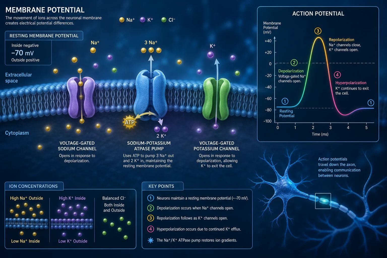
The RMP exists because of THREE factors:
- Concentration gradients (maintained by Na+/K+ ATPase): K+ is high inside (140), low outside (4). Na+ is low inside (10-15), high outside (140). Cl- is low inside (4), high outside (110).
- Selective membrane permeability: At rest, the membrane is MUCH more permeable to K+ (leak channels) than Na+ (leak channels are far fewer). K+ leaks OUT → positive charge leaves → inside becomes negative.
- Na+/K+ ATPase: pumps 3 Na+ OUT + 2 K+ IN (electrogenic — removes 1 net positive charge per cycle → makes inside MORE negative by ~-5 mV). [1]
The Nernst equation calculates the equilibrium potential for a SINGLE ion: [1]
E(ion) = (RT / zF) × ln([ion]outside / [ion]inside) [1]
For K+ at 37°C: EK = 61.5 × log(4/140) = -95 mV [1]
For Na+ at 37°C: ENa = 61.5 × log(140/15) = +60 mV [1]
The RMP (~-85 mV) is CLOSE to EK (-95 mV) because the membrane is MOSTLY permeable to K+. [1]
The Goldman-Hodgkin-Katz (GHK) equation calculates RMP considering ALL permeant ions: [1]
Vm = (RT / F) × ln[(PK[K+]o + PNa[Na+]o + PCl[Cl-]i) / (PK[K+]i + PNa[Na+]i + PCl[Cl-]o)] [1]
The permeability ratios (PK:PNa:PCl) at rest are approximately 1:0.04:0.45 — K+ dominates the RMP.[1][1]
Goldman-Hodgkin-Katz equation — full derivation and worked examples
The Nernst equation gives the equilibrium potential for a SINGLE ion, but real cell membranes are simultaneously permeable to MULTIPLE ions. The GHK voltage equation (Goldman, 1943; Hodgkin & Katz, 1949) generalises the Nernst equation by taking a permeability-weighted average of all permeant ions. It is derived from the constant-field assumption: the electric field (E = −dV/dx) across the ~5 nm lipid bilayer is assumed UNIFORM, so membrane potential changes LINEARLY with distance. Integrating the Nernst–Planck electrodiffusion flux equation for each ion under this assumption, and setting NET current to zero (steady state — no net charge accumulation), yields the GHK equation. [1]
General form (monovalent ions): [1]
Vm = (RT / F) × ln [ (PK·[K+]o + PNa·[Na+]o + PCl·[Cl-]i) / (PK·[K+]i + PNa·[Na+]i + PCl·[Cl-]o) ] [1]
Note the Cl− terms are INVERTED (intra- in numerator, extra- in denominator) because Cl− is an ANION — its equilibrium potential carries the opposite sign convention. RT/F at 37°C = 26.7 mV, so converting ln→log10 (×2.303) gives the familiar 61.5 mV multiplier. The equation reduces to the Nernst equation if only ONE ion is permeant (set the other P's to 0). [1]
Worked example at REST (typical mammalian myocyte; PK : PNa : PCl = 1 : 0.04 : 0.45): [1]
| Ion | [outside] (mM) | [inside] (mM) | P (relative) | Contribution |
|---|---|---|---|---|
| K+ | 4 | 140 | 1.0 | dominates |
| Na+ | 145 | 15 | 0.04 | minor |
| Cl− | 110 | 4 | 0.45 | intermediate |
Numerator = (1.0 × 4) + (0.04 × 145) + (0.45 × 4) = 4 + 5.8 + 1.8 = 11.6 Denominator = (1.0 × 140) + (0.04 × 15) + (0.45 × 110) = 140 + 0.6 + 49.5 = 190.1 Vm = 61.5 × log10(11.6 / 190.1) = 61.5 × log10(0.061) = 61.5 × (−1.21) = −74.5 mV (close to the measured −85 mV; the residual gap is the electrogenic contribution of the Na+/K+ ATPase, ~−4 to −10 mV). [1]
Worked example DURING the ACTION-POTENTIAL UPSTROKE — relative Na+ permeability rises ~500-fold (PK : PNa : PCl = 1 : 20 : 0.45): Numerator = (1.0 × 4) + (20 × 145) + (0.45 × 4) = 4 + 2900 + 1.8 = 2905.8 Denominator = (1.0 × 140) + (20 × 15) + (0.45 × 110) = 140 + 300 + 49.5 = 489.5 Vm = 61.5 × log10(2905.8 / 489.5) = 61.5 × log10(5.94) = 61.5 × 0.774 = +47.6 mV → the overshoot swings towards ENa (+60 mV), exactly as observed. [1]
Key teaching points the GHK equation encodes:
- RMP is pulled towards whichever ion has the HIGHEST RELATIVE PERMEABILITY — at rest K+ dominates (Vm near EK, −95 mV); during Phase 0 Na+ dominates (Vm swings towards ENa, +60 mV).
- It explains WHY hyperkalaemia depolarises the cell: raising [K+]o raises the numerator's K term → the log ratio increases (less negative) → Vm becomes less negative (depolarised).
- It explains the anion paradox: because Cl− is passively distributed near its own equilibrium (~RMP), raising PCl changes the ratio LITTLE — Cl− FOLLOWS the membrane potential rather than setting it.
- The equation is an APPROXIMATION — it assumes ion INDEPENDENCE (no interactions) and a constant field, both violated at high divalent-cation concentrations or with channel saturation. For multivalent ions (e.g. Ca2+) the GHK CURRENT equation (not the voltage equation) must be used, because Ca2+ flux is non-linear with voltage.
- The electrogenic Na+/K+ ATPase is NOT in the GHK equation — it adds a fixed hyperpolarising offset of ~−4 to −10 mV on top of the GHK-predicted passive potential (inhibiting it with ouabain/digoxin depolarises the cell by this amount). [1]
Cardiac action potential — the 5 phases
Cardiac myocyte action potential — the 5 phases
| Phase | Name | Ionic current | Duration | Clinical significance |
|---|---|---|---|---|
| Phase 0 | Rapid depolarisation | Fast Na+ INFLUX (voltage-gated INa) | 1-2 ms | Rapid upstroke → fast conduction. Blocked by Class I antiarrhythmics. Reduced by hyperkalaemia (depolarises RMP → inactivates Na channels → slower upstroke → wider QRS) |
| Phase 1 | Initial repolarisation | Transient K+ EFFLUX (Ito) | 10-50 ms | Brief notch after upstroke |
| Phase 2 | Plateau | Ca2+ INFLUX (L-type ICa) BALANCED by K+ efflux | 100-200 ms | UNIQUE to cardiac muscle — the Ca2+ plateau enables SUSTAINED contraction (needed for effective ejection). Also creates the LONG refractory period → prevents tetany in cardiac muscle. Blocked by Class IV (verapamil/diltiazem) |
| Phase 3 | Repolarisation | K+ EFFLUX dominates (IKr, IKs — delayed rectifier) | 100-150 ms | Returns membrane to RMP. Blocked by Class III (amiodarone, sotalol) → prolonged AP → prolonged QT → torsades |
| Phase 4 | Resting | Na+/K+ ATPase restores gradients | — | Returns to RMP. In pacemaker cells (SA/AV node), Phase 4 spontaneously depolarises (If 'funny current' — Na+ inward leak) → automaticity |
Cardiac action potential — comparison across tissue types
The cardiac action potential is NOT uniform. Four distinct tissue types each have a characteristic morphology that determines conduction velocity, refractoriness and automaticity. These differences explain the surface ECG and the site of action of every antiarrhythmic drug. [1]
Cardiac action potential — atrial vs ventricular vs AV node vs Purkinje
| Feature | Atrial myocyte | Ventricular myocyte | AV nodal cell | Purkinje fibre |
|---|---|---|---|---|
| RMP (Phase 4) | ~ −80 mV (stable) | ~ −90 mV (stable) | ~ −60 mV (UNSTABLE — slow diastolic depol) | ~ −90 mV (slightly unstable) |
| Phase 0 upstroke | Fast Na+ (INa) — moderate | Fast Na+ (INa) — rapid (1–2 ms) | SLOW Ca2+ (ICa-L) — NO fast Na+ | Fast Na+ (INa) — very rapid |
| Conduction velocity | ~0.5 m/s | ~0.4–0.5 m/s (working muscle) | ~0.05 m/s (SLOWEST — the AV delay) | ~2–4 m/s (FASTEST in the heart) |
| Plateau (Phase 2) | Brief | Prominent, long | Absent (spike-and-dome lost) | Long, prominent |
| AP duration | ~150 ms | ~250–300 ms | ~250 ms | ~300–400 ms (LONGEST) |
| Automaticity | None (normally) | None (normally) | YES — latent (40–60 bpm) | YES — latent (20–40 bpm) |
| Key ionic currents | INa, ICa-L, IKur (atrial-specific), IKr/IKs, Ito | INa, ICa-L, IKr/IKs, Ito | ICa-L, If, IKr/IKs | INa, ICa-L, If, IKr/IKs |
| Clinical correlate | Atrial fibrillation (IKur — vernakalant target) | QRS (Phase 0), QT (Phase 3), VF | PR interval (AV delay), AV block, nodal blockers (verapamil, adenosine) | Bundle-branch block, escape rhythms |
Why the AV node is slow — the key to the PR interval. AV nodal cells have almost NO functional fast Na+ channels — their Phase 0 upstroke is carried by the L-type Ca2+ current (ICa-L). Because Ca2+ channels activate/inactivate ~10× more slowly than Na+ channels, the AV nodal upstroke is SLOW (low dV/dt) → conduction velocity is only ~0.05 mS. This creates the physiological AV delay (the PR interval, 120–200 ms), which:
- Ensures atrial contraction FINISHES before ventricular contraction begins → optimal ventricular filling.
- Creates a 'bottleneck' that PROTECTS the ventricles from rapid atrial arrhythmias — in atrial fibrillation the AV node filters most impulses (300–600 atrial bpm → ~100–150 ventricular bpm).
- Is the target of AV-nodal-blocking drugs: adenosine (opens K+ channels → hyperpolarisation → terminates re-entrant SVT), verapamil/diltiazem (block ICa-L), beta-blockers (↓ICa-L via ↓cAMP), digoxin (↑vagal tone → ↑K+ conductance). [1]
Pacemaker (funny) current — If — the molecular basis of automaticity. Nodal and Purkinje cells spontaneously depolarise during Phase 4 through the If ('funny') current, a mixed Na+/K+ inward current carried by HCN (hyperpolarisation-activated, cyclic nucleotide-gated) channels. If is UNIQUE because it ACTIVATES on HYPERPOLARISATION (unlike most channels that activate on depolarisation). The slope of Phase 4 diastolic depolarisation sets the heart rate:
- If is directly modulated by cAMP (cAMP binds the HCN channel directly → shifts activation to more positive voltages → steeper slope → faster depolarisation). Sympathetic stimulation → ↑cAMP → steeper slope → FASTER heart rate; vagal stimulation → ↓cAMP AND opens ACh-gated K+ channels (IKACh) → hyperpolarises → SLOWER heart rate.
- Ivabradine selectively blocks If → slows sinus rate WITHOUT affecting contractility or AV conduction (used for chronic stable angina and HFrEF with a sinus rate ≥77 despite maximally tolerated β-blocker).[4]
SA node vs AV node vs Purkinje — intrinsic rates and escape hierarchy
| Pacemaker | Intrinsic rate (bpm) | Phase 4 slope | Latency to take over | Escape rhythm seen when... |
|---|---|---|---|---|
| SA node | 60–100 | Steepest | — (dominant) | — |
| AV node (junctional) | 40–60 | Moderate | Seconds | SA node failure / high-grade block |
| Purkinje / ventricular | 20–40 | Shallow | 30–60 s | SA + AV failure (wide-complex escape) |
This hierarchy is the basis of ESCAPE RHYTHMS: when a higher pacemaker fails, a LOWER (slower) one takes over after a delay — the slower the site, the wider the QRS and the slower the rate. A ventricular escape rhythm (20–40 bpm, wide QRS) signals life-threatening high-grade AV block and mandates urgent pacing. [1]
Excitation-contraction coupling — the molecular mechanism
Excitation-contraction coupling — step by step
- ACTION POTENTIAL arrives at the cardiac myocyte membrane (sarcolemma)
- Voltage-gated L-type Ca2+ channels (dihydropyridine receptors) open in the T-tubule membrane → Ca2+ ENTERS the cell from the extracellular fluid (small amount — 'trigger calcium')
- Calcium-induced calcium release (CICR): the small amount of Ca2+ entering through L-type channels triggers the ryanodine receptor 2 (RyR2) on the sarcoplasmic reticulum (SR) → MASSIVE Ca2+ release from the SR into the cytoplasm (the SR has ~10,000x higher Ca2+ concentration than cytoplasm)
- Ca2+ binds troponin C: cytoplasmic Ca2+ rises from 0.1 to 10 μmol/L → Ca2+ binds TROPONIN C → conformational change → TROPOMYOSIN moves → exposes ACTIN binding site for MYOSIN head
- Cross-bridge cycling: myosin head (with ADP + Pi attached) binds actin → POWER STROKE (Pi release → myosin head pivots → pulls actin filament) → ADP release → new ATP binds → myosin head detaches → ATP hydrolysis → myosin head re-cocks → REPEAT (as long as Ca2+ is bound to troponin and ATP is available)
- RELAXATION: Ca2+ is removed from cytoplasm by: (a) SERCA (sarcoendoplasmic reticulum Ca2+ ATPase) — pumps Ca2+ BACK INTO the SR (the main mechanism — 70-80% of Ca2+ removal). (b) Na+/Ca2+ exchanger (NCX) — extrudes Ca2+ in exchange for Na+ (20-25%). (c) Plasma membrane Ca2+ ATPase (PMCA) — minor (1-5%). As Ca2+ falls → dissociates from troponin → tropomyosin covers actin → contraction stops → RELAXATION
Calcium handling — cardiac vs skeletal muscle
Cardiac and skeletal muscle share the SAME contractile machinery (actin–myosin, troponin–tropomyosin) but use RADICALLY DIFFERENT calcium-release strategies. This single difference explains why cardiac contraction is graded and rhythmic while skeletal contraction is all-or-none and able to tetanise. [1]
Excitation-contraction coupling — cardiac vs skeletal muscle
| Feature | CARDIAC muscle | SKELETAL muscle |
|---|---|---|
| RyR isoform | RyR2 | RyR1 |
| Voltage sensor | L-type Ca2+ channel (Cav1.2) — functions as an ION CHANNEL | Dihydropyridine receptor (DHPR, Cav1.1) — functions as a VOLTAGE SENSOR (slowly conducting, little Ca2+ flux) |
| Coupling mechanism | Calcium-induced calcium release (CICR) — Ca2+ ENTRY through the L-type channel is the TRIGGER | Mechanical / voltage coupling — DIRECT physical conformational interaction between DHPR and RyR1 (NO Ca2+ entry required) |
| Source of activator Ca2+ | BOTH extracellular (L-type trigger) + SR release | Almost entirely SR release (extracellular Ca2+ not required per beat) |
| Dependence on extracellular Ca2+ | HIGH — cardiac muscle CANNOT contract in zero-Ca2+ solution | LOW — skeletal muscle contracts for hours in zero-Ca2+ (SR store is self-sufficient) |
| Grading of force | GRADED — force ∝ Ca2+ influx (length-tension, frequency-force, β-adrenergic) | ALL-OR-NONE single fibre — force graded by motor-unit RECRUITMENT and STIMULUS FREQUENCY (tetany) |
| Termination of release | Ca2+ buffering, SERCA2a reuptake, NCX extrusion, PMCA | Re-diffusion into SR; SERCA1 reuptake |
| SR Ca2+-load regulation | Dynamic — load-force relationship (Bowditch staircase) | Relatively fixed |
| Speed | Slower (tens of ms) | Very fast (<5 ms) — suited to fine motor control |
RyR2 vs RyR1 — molecular differences in regulation:
- RyR1 (skeletal) is mechanically GATED: the DHPR in the T-tubule physically 'bumps' RyR1 through a foot-process interaction → RyR1 opens → Ca2+ release. Because coupling is mechanical and direct, release is FAST and TIGHT — every AP releases a near-fixed quantum of Ca2+ (all-or-none within a sarcomere).
- RyR2 (cardiac) is Ca2+-GATED: the L-type channel OPENS, a small pulse of Ca2+ enters → binds the cytosolic face of RyR2 → opens RyR2 → massive Ca2+ release (amplification ~10:1). Because the trigger is Ca2+ itself, the amount of release is PROPORTIONAL to the trigger → the system is GRADED (more Ca2+ influx → more release → stronger contraction).
- Regulatory proteins. Both RyRs are stabilised in the closed state by calstabin2/FKBP12.6 (cardiac) and calstabin1/FKBP12 (skeletal), regulated by calmodulin, and modulated by phosphorylation (PKA, CaMKII). In HEART FAILURE, chronic β-adrenergic drive HYPERPHOSPHORYLATES RyR2 → calstabin2 dissociates → RyR2 'leaks' Ca2+ during diastole → arrhythmogenic delayed afterdepolarisations (DADs) → triggered VT.
- Pharmacology. Dantrolene directly inhibits RyR1 (skeletal) → stops uncontrolled Ca2+ release in malignant hyperthermia and neuroleptic malignant syndrome; it has weak affinity for RyR2 → minimal cardiac effect (which is why it does not cause heart block). Flecainide and carvedilol stabilise RyR2 in CPVT (catecholaminergic polymorphic ventricular tachycardia), an inherited RyR2-leak disorder presenting with exercise-induced bidirectional VT.[1][5]
Why this matters clinically:
- Cardiac arrest in hypocalcaemia / calcium-channel-blocker overdose — because cardiac muscle DEPENDS on extracellular Ca2+ entry, calcium gluconate / chloride is the specific antidote; skeletal muscle is relatively spared (it does not need extracellular Ca2+ per beat).
- Malignant hyperthermia is a SKELETAL-muscle disease (RyR1) — the heart (RyR2) is involved only secondarily via hypermetabolism; dantrolene targets the correct isoform, so it works without depressing the heart.
- Inotropes act on cardiac, not skeletal, muscle — β-agonists and PDE3 inhibitors raise cAMP/PKA → phosphorylate L-type channels and phospholamban → more trigger Ca2+ and faster reuptake → only cardiac muscle benefits (skeletal SR Ca2+ handling is not phospholamban-regulated).
- Frequency-dependent augmentation (Bowditch / Treppe effect) — at higher heart rates more Ca2+ accumulates in the cardiac SR per unit time (less time for NCX to extrude) → stronger contractions. This is why a brief run of tachycardia can strengthen the next beats, and why bradycardia reduces contractility. [1]
β-adrenergic signalling cascade — the inotropy / chronotropy mechanism
Sympathetic stimulation is the fastest, most powerful regulator of cardiac output. The entire effect is mediated by a single G-protein-coupled receptor (GPCR) cascade whose every node is a drug target encountered daily in ICU. [1]
β1-adrenergic signalling cascade — step by step
- Ligand binding. Adrenaline (or noradrenaline) binds the β1-adrenergic receptor (a 7-transmembrane GPCR; β1 is predominant in the SA node, AV node and myocytes; β2 in vascular/bronchial smooth muscle and at the SA node; β3 in adipose and the detrusor).
- G-protein activation. The receptor catalyses GDP→GTP exchange on the Gs (stimulatory) α-subunit → Gsα dissociates from the βγ dimer.
- Adenylyl cyclase activation. Gsα binds and ACTIVATES adenylyl cyclase (a membrane-bound enzyme). (Conversely, the M2 muscarinic / Gi pathway INHIBITS adenylyl cyclase — the vagal brake.)
- cAMP generation. Adenylyl cyclase converts ATP → 3′,5′-cyclic AMP (cAMP) → intracellular cAMP rises ~10-fold within seconds.
- PKA activation. cAMP binds the regulatory subunits of protein kinase A (PKA) → catalytic subunits are released and ACTIVE.
- Phosphorylation of FIVE key targets (each one component of the sympathetic response):
- (a) L-type Ca2+ channel (Cav1.2) → ↑ open probability → MORE Ca2+ entry during Phase 2 → MORE trigger Ca2+ → MORE SR release → POSITIVE INOTROPY (stronger contraction).
- (b) Phospholamban → when phosphorylated, phospholamban DETACHES from SERCA2a → SERCA activity INCREASES → (i) FASTER Ca2+ reuptake → FASTER RELAXATION (positive lusitropy — preserves filling time at high heart rates) and (ii) MORE Ca2+ stored in SR → MORE available for the next beat → stronger next contraction.
- (c) Troponin I → phosphorylation DECREASES troponin-C affinity for Ca2+ → Ca2+ dissociates FASTER → faster cross-bridge detachment → faster relaxation.
- (d) If (HCN) channels (SA node) → ↑ If → steeper Phase-4 depolarisation → POSITIVE CHRONOTROPY (↑ heart rate).
- (e) RyR2 → phosphorylation increases open probability → faster, larger SR Ca2+ release synchronised to each beat (but chronic hyperphosphorylation → diastolic leak → arrhythmia, see above).
- Termination. cAMP is degraded by phosphodiesterase 3 (PDE3) → 5′-AMP; Gsα hydrolyses GTP→GDP and re-associates; phosphatases dephosphorylate PKA targets.
NET EFFECT of β1 stimulation (the 'fight-or-flight' cardiac response): ↑ heart rate + ↑ contractility + ↑ relaxation rate + ↑ conduction velocity = ↑↑ cardiac output (often 2–3× baseline), at the cost of ↑ myocardial O2 demand. [1]
Clinical correlates — every node is a drug target:
- β1 AGONISTS (raise cAMP): adrenaline, noradrenaline, dobutamine, isoprenaline, dopamine (moderate dose).
- β1 ANTAGONISTS (β-blockers, lower cAMP): metoprolol, atenolol, bisoprolol, esmolol → ↓ HR, ↓ contractility, ↓ myocardial O2 demand; also suppress cAMP-dependent arrhythmia triggers (ischaemia, thyrotoxicosis, PHAEDRA/perioperative).
- PDE3 INHIBITORS (prevent cAMP breakdown → raise cAMP INDEPENDENT of the receptor): milrinone, enoximone → inotrope + vasodilator ('inodilator'); useful when β-receptors are downregulated (chronic heart failure, post-cardiac surgery) and effective even with concurrent β-blockade.
- Glucagon bypasses the β-receptor: activates Gs DIRECTLY via its own GPCR → ↑ cAMP → used as an ANTIDOTE in β-blocker and calcium-channel-blocker toxicity (high-dose insulin/euglycaemia therapy is now preferred for CCB toxicity).
- Phospholamban in heart failure: in chronic HF phospholamban is RELATIVELY OVEREXPRESSED → tonically inhibits SERCA2a → impaired relaxation and reduced SR Ca2+ load → systolic and diastolic dysfunction. Gene therapy targeting phospholamban knockdown and SERCA2a over-expression has been investigated.[2]
Neuromuscular junction — quantal release, MEPPs, and safety factor
The neuromuscular junction (NMJ) is the archetypal chemical synapse and the target of every neuromuscular blocking agent, anticholinesterase and several toxins seen in ICU. The quantitative physiology discovered by Katz, Miledi and colleagues (Nobel-winning work) underpins monitoring of neuromuscular blockade (train-of-four). [1]
Neuromuscular transmission — molecular steps
- AP arrives at the motor nerve terminal → depolarisation opens P/Q-type voltage-gated Ca2+ channels (Cav2.1) → Ca2+ enters the terminal (the local [Ca2+] microdomain rises to ~10–100 μM within microseconds).
- Vesicle docking/fusion. Ca2+ binds synaptotagmin on docked vesicles → triggers the SNARE complex (synaptobrevin/VAMP, SNAP-25, syntaxin) to zipper → synaptic vesicle fuses with the presynaptic active zone → exocytosis of acetylcholine.
- Quantal release. Each vesicle contains a FIXED quantum of ACh (~5000–10000 molecules). A single AP releases 50–300 quanta (the quantal content, m). Release is Ca2+-dependent and follows binomial / Poisson statistics — m = n × p (n = number of readily-releasable vesicles, p = probability of release per vesicle).
- Postsynaptic action. ACh diffuses across the ~50 nm cleft → binds the nicotinic ACh receptor (nAChR) — a ligand-gated cation channel (Na+/K+ permeable, pentamer α2βγδ in fetal/extra-junctional, α2βεδ mature) — on the motor endplate → channel opens → Na+ INFLUX → local depolarisation.
- Endplate potential (EPP). The summed depolarisation from all released quanta (~+40 mV at the endplate).
- Muscle AP. The EPP depolarises the adjacent sarcolemma to THRESHOLD (−55 mV) → opens voltage-gated Na+ channels → regenerative muscle AP → excitation-contraction coupling → contraction.
- Termination. ACh is hydrolysed within ~1 ms by acetylcholinesterase (AChE) in the synaptic cleft → choline is recycled into the terminal by the high-affinity choline transporter (CHT1) → repackaged into vesicles by VAChT.
Miniature endplate potentials (MEPPs) — the evidence for quantal release. Even at REST (no nerve stimulation), vesicles randomly fuse and release single quanta → tiny stereotyped depolarisations of ~0.4 mV called MEPPs. The EPP amplitude during stimulation is always an INTEGER MULTIPLE of the MEPP amplitude (1 quantum ≈ 0.4 mV → EPP ≈ 0.4 × m). This was the first direct evidence that neurotransmitter is released in discrete packets (quanta), not as a continuous 'soup'. MEPP frequency rises with [Ca2+]o and falls in botulinum toxin (which cleaves SNARE proteins) and in Lambert–Eaton (fewer Ca2+ channels → fewer release events). [1]
Safety factor — why the NMJ is so reliable.
The EPP (+40 mV) is typically 3–4× LARGER than needed to reach threshold (+10–15 mV above resting potential). This safety factor (~3–4×) ensures that virtually every nerve AP produces a muscle AP — neuromuscular transmission in HEALTH fails only at very high firing rates (fatigue) or with disease. Clinical consequences:
- Myasthenia gravis reduces the safety factor to ~1× (antibodies reduce nAChR number) → at high firing rates the EPP no longer reaches threshold → fatigable weakness (the DECREMENTAL response on repetitive 3-Hz nerve stimulation).
- Lambert–Eaton myasthenic syndrome (LEMS) reduces quantal content (antibodies against presynaptic P/Q-type Ca2+ channels) → small EPP but, paradoxically, FACILITATION with repeated stimulation (Ca2+ accumulates in the terminal → INCREMENTAL response).
- Neuromuscular blocking agents reduce the safety factor toward ZERO — non-depolarisers (rocuronium, vecuronium, atracurium) competitively block nAChR; succinylcholine is an agonist causing sustained endplate depolarisation → inactivation of adjacent Na+ channels → Phase I block.
- Train-of-four (TOF) monitoring exploits the quantal/safety-factor concept: 4 supramaximal stimuli at 2 Hz. In non-depolarising block, readily-releasable vesicle stores partly deplete across the four stimuli → T4/T1 ratio falls (fade). A TOF ratio >0.9 indicates adequate recovery (safety factor restored; reliable ventilation and airway protection). [1]
Disorders of the neuromuscular junction — mechanism and bedside signature
| Disorder | Site / target | Mechanism | Bedside signature |
|---|---|---|---|
| Myasthenia gravis | POSTsynaptic nAChR | Antibody reduces receptor number → ↓ safety factor | Fatigable weakness, DECREMENT on 3-Hz stimulation, improves with rest/edrophonium |
| Lambert–Eaton | PREsynaptic Cav2.1 (P/Q) | Antibody reduces Ca2+ entry → ↓ quantal content | Weakness improves with use, INCREMENT on repetitive stimulation, autonomic features, paraneoplastic (SCLC) |
| Botulism | PREsynaptic SNARE (synaptobrevin) | Toxin cleaves SNARE → blocks vesicle fusion → no ACh release | Descending flaccid paralysis, fixed dilated pupils, autonomic failure |
| Organophosphates | Synaptic cleft AChE | Irreversible AChE inhibition → ACh accumulates | Cholinergic crisis (DUMBELSS), depolarising block, intermediate syndrome |
| Succinylcholine | POSTsynaptic nAChR (agonist) | Sustained depolarisation → Na+ channel inactivation | Phase I block (no fade), fasciculations then paralysis, HYPERKALAEMIA |
SAQ — Membrane potential and ICU drug targets
10 minutes · 10 marks
You are teaching CICM first-part candidates at the bedside of a patient on noradrenaline, receiving rocuronium for ventilation, who develops new broad-complex tachycardia after a potassium of 2.6 mmol/L is noted.
Clinical pearls
Red flags
[1] [1] [1] [1]Key trials and evidence
Bers 2002 — Cardiac excitation-contraction coupling (PMID 11741523)
Source
Nature review — the definitive molecular description of cardiac E-C coupling
Key contribution
Described the molecular mechanism of calcium-induced calcium release (CICR) via RyR2 in cardiac myocytes
Key finding
The L-type Ca2+ channel provides 'trigger calcium' that activates RyR2 → massive SR Ca2+ release
Clinical bottom line
The molecular basis for understanding all cardiac physiology — from digoxin to MH to antiarrhythmic drug mechanisms
MacLennan & Kranias 2003 — Phospholamban as regulator of cardiac contractility (PMID 12838339)
Source
Nature Reviews Molecular Cell Biology — landmark review of the SERCA2a–phospholamban axis
Key contribution
Established phospholamban as the reversible inhibitor of SERCA2a; PKA/CaMKII phosphorylation relieves inhibition → faster Ca2+ reuptake
Key finding
Human phospholamban-null mutation → early lethal dilated cardiomyopathy (contrasting with beneficial PLN-knockout in mice)
Clinical bottom line
The molecular basis for β-adrenergic lusitropy, PDE3-inhibitor action, and heart-failure gene therapy targeting SERCA2a/phospholamban
DiFrancesco 2010 — The funny current (If) and cardiac pacemaking (PMID 20167941)
Source
Circulation Research — definitive review of HCN-channel pacemaker physiology
Key contribution
Established If (via HCN channels) as the primary determinant of diastolic depolarisation and heart rate; directly modulated by cAMP
Key finding
Ivabradine selectively blocks If → pure heart-rate reduction without negative inotropy
Clinical bottom line
Explains sympathetic/vagal chronotropy and the therapeutic niche of ivabradine in HFrEF and inappropriate sinus tachycardia
Wood & Slater 2001 — Safety factor at the neuromuscular junction (PMID 11275359)
Source
Progress in Neurobiology — quantitative review of NMJ reliability
Key contribution
Defined the safety factor (~3–4×) by which the endplate potential exceeds threshold, ensuring reliable transmission
Key finding
Reduction of the safety factor to ~1× underlies myasthenia gravis; quantal-content reduction underlies Lambert–Eaton
Clinical bottom line
Explains decrement/increment on nerve stimulation, TOF monitoring, and the pharmacology of NMBAs and reversal agents
Ríos & Pizarro 1991 — Voltage sensor of EC coupling in skeletal muscle (PMID 2057528)
Source
Physiological Reviews — seminal review of skeletal-muscle EC coupling
Key contribution
Established the MECHANICAL (direct DHPR–RyR1) coupling model in skeletal muscle, contrasting with Ca2+-induced Ca2+ release in cardiac muscle
Key finding
Skeletal muscle does NOT require extracellular Ca2+ entry for contraction; cardiac muscle DOES
Clinical bottom line
Explains why cardiac (not skeletal) muscle fails in hypocalcaemia/Ca-channel-blocker overdose, and why dantrolene (RyR1-specific) treats malignant hyperthermia
Synthesis — cross-cutting cellular mechanisms
Skeletal vs cardiac vs smooth muscle — cellular physiology at a glance
| Property | Skeletal | Cardiac | Smooth muscle |
|---|---|---|---|
| RMP | −90 mV (stable) | −90 mV (stable working; unstable nodal) | −50 to −60 mV (unstable — slow waves) |
| AP duration | 2–5 ms | 200–300 ms | 50–100 ms (plateau variable) |
| Coupling | Mechanical (DHPR→RyR1) | CICR (L-type→RyR2) | Pharmacomechanical (IP3 + CICR) |
| Ca2+ source | SR (RyR1) | SR (RyR2) + extracellular | SR (IP3R / RyR) + extracellular |
| Regulation of force | Recruitment + tetany | Graded (Ca2+ influx, length, frequency) | Latch-bridge (slow, sustained, low ATP) |
| Can tetanise? | YES | NO (long refractory) | YES (sustained tone) |
| Calmodulin role | — | — | Activates myosin light-chain kinase (MLCK) |
References
- [1]Bosch X, et al. Community influences on young people's sexual behavior in 3 African countries Am J Public Health, 2009.PMID 19008517
- [2]MacLennan DH, Kranias EG Phospholamban: a crucial regulator of cardiac contractility Nat Rev Mol Cell Biol, 2003.PMID 12838339
- [3]Wood SJ, Slater CR Safety factor at the neuromuscular junction Prog Neurobiol, 2001.PMID 11275359
- [4]DiFrancesco D The role of the funny current in pacemaker activity Circ Res, 2010.PMID 20167941
- [5]Rios E, Pizarro G Voltage sensor of excitation-contraction coupling in skeletal muscle Physiol Rev, 1991.PMID 2057528