ICU · first-part-physiology
Endocrine Physiology in Critical Illness — Comprehensive
Also known as Endocrine physiology · Stress response · HPA axis · Cortisol · Critical illness-related corticosteroid insufficiency · CIRCI · Sick euthyroid syndrome · Nonthyroidal illness syndrome · Stress hyperglycaemia · ADH vasopressin · SIADH · RAAS · Counter-regulatory hormones
Endocrine physiology of critical illness — the integrated hormonal response to severe stress. STRESS RESPONSE: critical illness (sepsis, trauma, surgery, burns) activates the hypothalamic-pituitary axes and counter-regulatory hormones to maintain perfusion, substrate availability, and immune homeostasis. HPA AXIS: hypothalamus releases CRH → anterior pituitary releases ACTH → adrenal cortex (zona fasciculata) releases CORTISOL. Cortisol is ESSENTIAL for survival in stress — it (1) maintains vascular tone via a PERMISSIVE effect on catecholamines (upregulates alpha-1 adrenergic receptors — without cortisol, catecholamines cannot maintain BP → vasoplegic shock), (2) is anti-inflammatory/immunosuppressive (inhibits NF-kB, phospholipase A2, cytokines IL-1, IL-6, TNF-alpha), (3) drives gluconeogenesis (stress hyperglycaemia), (4) maintains intravascular volume (mild mineralocorticoid effect). CIRCI (critical illness-related corticosteroid insufficiency): inadequate cortisol for the DEGREE of stress — adrenal insufficiency PLUS tissue glucocorticoid resistance — NOT absolute adrenal failure. Suspect in vasopressor-refractory septic shock. THYROID AXIS: TRH → TSH → T4 (prohormone) → peripheral 5'-deiodinase (D1/D2) converts T4 to T3 (active). In critical illness: SICK EUTHYROID SYNDROME (non-thyroidal illness) — T3 falls (reduced conversion), reverse T3 rises (shunted from T4), T4 normal/low, TSH normal/low — an ADAPTIVE response to reduce metabolic rate and conserve energy — do NOT treat with thyroid hormone unless intrinsic thyroid disease coexists. GLUCOSE METABOLISM: stress hyperglycaemia — cortisol + catecholamines + glucagon + GH + cytokines (IL-6, TNF-alpha) → hepatic gluconeogenesis + glycogenolysis + peripheral insulin resistance → hyperglycaemia. This is NOT diabetes — it is a stress response and resolves with recovery. Tight glycaemic control (80-110 mg/dL) is HARMFUL (NICE-SUGAR: increased mortality from hypoglycaemia) — target 8-10 mmol/L (144-180 mg/dL). ADH/VASOPRESSIN: regulated by (1) OSMOTIC (hypothalamic osmoreceptors — 280-295 mOsm/kg) and (2) BARORECEPTOR (carotid sinus/aortic arch — triggered by 10% drop in BP) pathways. In stress: NON-OSMOTIC ADH release → water retention → SIADH pattern → dilutional hyponatraemia. RAAS: renin (from juxtaglomerular apparatus) → angiotensin I → ACE (lung) → angiotensin II (potent vasoconstrictor + stimulates aldosterone) → aldosterone (sodium/water retention). Activated in shock to maintain BP and circulating volume.
On this page & tools
Your progress
Saved locally on this device.
1 MCQ with explanations
Target exams
Red flags
Overview
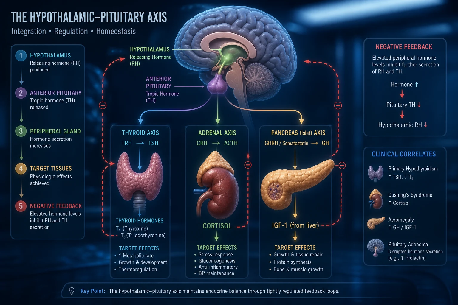
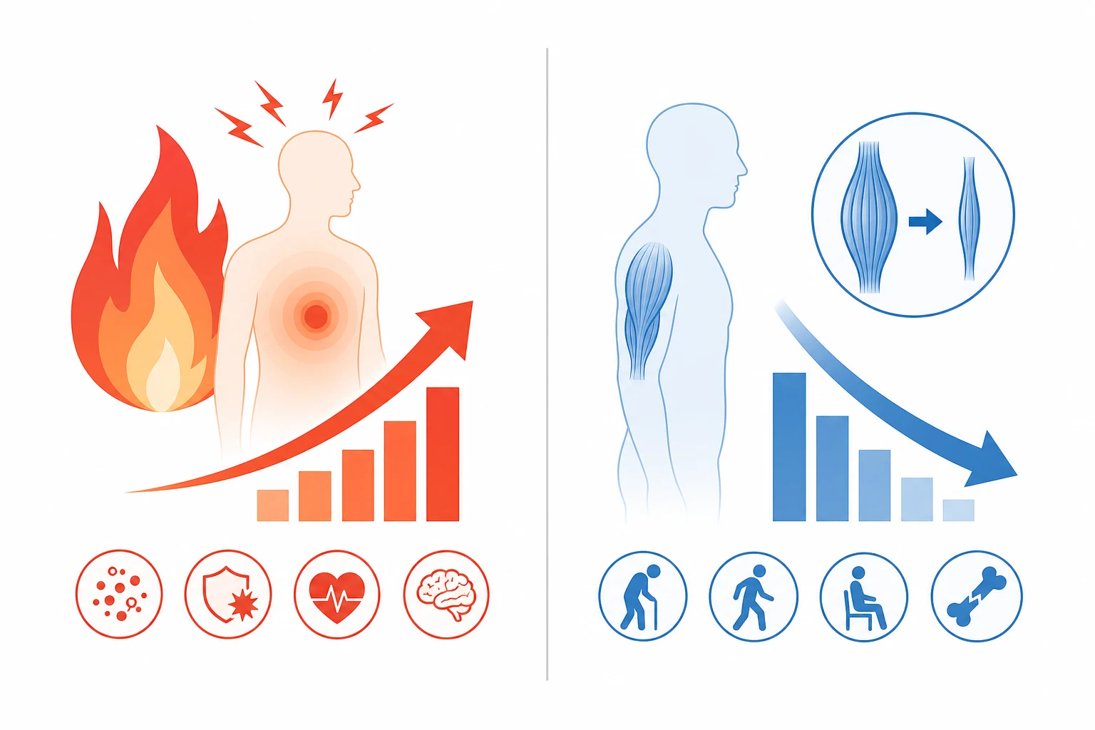
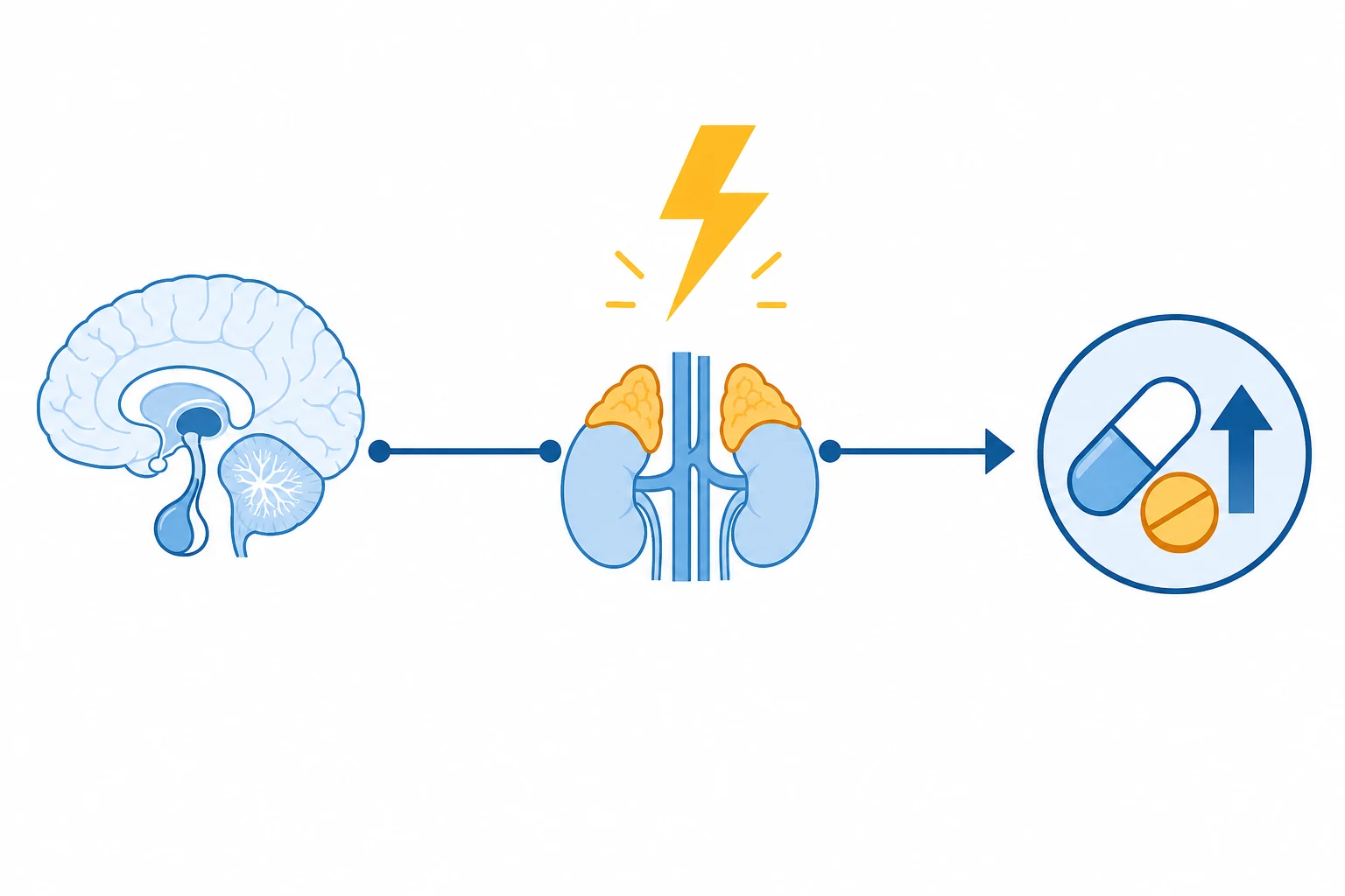
The stress response — the integrated endocrine reaction to critical illness
Critical illness — whether sepsis, major trauma, surgery, burns, or cardiogenic shock — is interpreted by the hypothalamus as an existential threat. The endocrine response has two phases. The acute phase (hours to days) is driven by the sympathetic nervous system and the hypothalamic-pituitary axes: catecholamines, cortisol, ADH, aldosterone, and glucagon all rise to maintain blood pressure, circulating volume, and substrate (glucose, free fatty acids) delivery to vital organs. This phase is CATABOLIC by design — glycogen, fat, and protein are broken down to fuel the fight. The chronic phase (weeks), seen in prolonged ICU stay, is characterised by a paradoxical shift: anterior pituitary hormone secretion becomes pulsatile and disordered, peripheral hormone activation falls (low T3, low IGF-1), and a state of acquired resistance develops to cortisol, insulin, and growth hormone — contributing to ICU-acquired weakness, hyperglycaemia, and immune dysregulation.[5]
The hormonal players fall into three functional groups: [1]
- Haemodynamic/Volume-maintaining: cortisol (permissive for catecholamines), catecholamines (adrenaline, noradrenaline), ADH/vasopressin, and the RAAS (angiotensin II, aldosterone).
- Substrate-mobilising (counter-regulatory): cortisol, glucagon, catecholamines, and growth hormone — all OPPOSE insulin to raise glucose and free fatty acids.
- Immune-modulating: cortisol (anti-inflammatory) balanced against pro-inflammatory cytokines (IL-1, IL-6, TNF-alpha) which both drive the stress response AND induce hormone resistance. [1]
The net effect is a controlled hyper-metabolic, hyperglycaemic, catabolic state that is life-sustaining in the short term. The clinician's job is to SUPPORT it where it fails (e.g., hydrocortisone in CIRCI) and to PREVENT it from overshooting (e.g., avoiding harmful hypoglycaemia from over-treatment of stress hyperglycaemia).[1][5]
HPA axis and cortisol — the master stress hormone
The hypothalamic-pituitary-adrenal (HPA) axis is the single most important endocrine defence against acute stress. The cascade is a classic three-tier negative-feedback loop: [1]
- Hypothalamus releases corticotropin-releasing hormone (CRH) in response to circadian rhythm AND any stress (hypoglycaemia, pain, hypovolaemia, cytokines, fear). CRH also stimulates ADH release (synergy).
- Anterior pituitary responds to CRH by secreting ACTH (adrenocorticotropic hormone, from pro-opiomelanocortin/POMC) in pulses.
- Adrenal cortex (zona fasciculata) responds to ACTH by synthesising and releasing cortisol. Cholesterol → pregnenolone → ... → cortisol, rate-limited by the ACTH-dependent enzyme cholesterol side-chain cleavage (CYP11A1).
- Negative feedback: cortisol inhibits both the hypothalamus (CRH) and pituitary (ACTH) — which is why exogenous steroids suppress the axis and abrupt withdrawal precipitates adrenal crisis. [1]
In critical illness this feedback loop is partially DE-COUPLED — cortisol rises markedly (to 600-1500 nmol/L) but ACTH is only modestly elevated, because extra-pituitary pathways (endothelin, cytokines, the sympathetic system) directly drive the adrenal gland. Cortisol-binding globulin (CBG) also FALLS in inflammation, so FREE cortisol rises even more than total — a key reason random TOTAL cortisol can be misleadingly low in sepsis.[5][1]
The four essential effects of cortisol in critical illness
| Effect | Mechanism | Clinical consequence if deficient |
|---|---|---|
| Permissive vascular tone | Upregulates alpha-1 adrenergic receptor expression and coupling; enhances catecholamine signalling | Vasoplegic / catecholamine-resistant shock — noradrenaline fails to maintain BP |
| Anti-inflammatory / immunosuppressive | Inhibits NF-kB, phospholipase A2 (→ reduced prostaglandins/leukotrienes), cytokines (IL-1, IL-6, TNF-alpha); promotes anti-inflammatory cytokines (IL-10) | Uncontrolled, exaggerated inflammatory response — the pro-inflammatory side of CIRCI |
| Metabolic — gluconeogenesis | Induces gluconeogenic enzymes (PEPCK, G6Pase); mobilises amino acids from muscle and glycerol from fat; opposes insulin | Failure to generate glucose for the brain during fasting/stress — hypoglycaemia |
| Volume / electrolyte (mild mineralocorticoid) | Weak binding to mineralocorticoid receptors; cortisol also inhibits 11beta-HSD2 in kidney at high levels | Sodium loss, hypovolaemia, hyperkalaemia (only prominent in absolute adrenal failure) |
HPA axis activation by stress — step by step
- Stressor (sepsis, hypovolaemia, hypoglycaemia, surgery, burns, cytokines IL-1/IL-6/TNF) stimulates the paraventricular nucleus of the hypothalamus
- CRH release into the hypophyseal portal system (+ ADH synergises with CRH to amplify ACTH release)
- ACTH secretion from corticotroph cells of the anterior pituitary (via cAMP/PKA — cleaved from POMC, which also yields beta-endorphin)
- ACTH binds melanocortin-2 receptor on zona fasciculata cells → cAMP → StAR protein transports cholesterol into mitochondria
- Cholesterol side-chain cleavage (CYP11A1) converts cholesterol to pregnenolone → sequential enzymatic steps (3beta-HSD, 17alpha-hydroxylase, 21-hydroxylase, 11beta-hydroxylase) → CORTISOL
- Cortisol enters blood (mostly bound to cortisol-binding globulin, CBG; free cortisol is the active fraction). In inflammation CBG falls → free cortisol rises disproportionately
- Cellular action: cortisol crosses the cell membrane → binds intracellular glucocorticoid receptor → translocates to nucleus → binds glucocorticoid response elements (GREs) → transactivation/transrepression of target genes (effect takes HOURS — genomic)
- Negative feedback: cortisol suppresses CRH and ACTH — but in critical illness this is partially overridden by cytokines and neural stress inputs so cortisol stays high
Critical illness-related corticosteroid insufficiency (CIRCI)
CIRCI is the term coined by the 2008 international consensus task force to describe the HPA axis dysfunction that occurs DURING critical illness.[1] It is fundamentally different from classical (Addisonian) adrenal insufficiency in three ways: (1) it is RELATIVE — cortisol production is inadequate FOR THE DEGREE OF STRESS, not absolutely absent; (2) it combines ADRENAL insufficiency with TISSUE glucocorticoid resistance (cytokines downregulate and impair the glucocorticoid receptor); and (3) it is TRANSIENT — it resolves as the critical illness resolves.
CIRCI vs absolute (primary) adrenal insufficiency
| Feature | CIRCI (critical illness) | Primary adrenal insufficiency (Addison's) |
|---|---|---|
| Nature of deficit | Relative — inadequate cortisol for stress level + tissue resistance | Absolute — cortisol absent/minimal |
| ACTH | Low / normal / high (variable) | Markedly HIGH (loss of feedback) |
| Aldosterone | Preserved (RAAS intact) | Deficient (zona glomerulosa destroyed) → hyperkalaemia, hyponatraemia |
| Electrolytes | Often normal | Hyponatraemia + HYPERKALAEMIA (the classic pair) |
| Pigmentation | Absent | Present (high ACTH/MSH) |
| Onset | Develops DURING critical illness | Pre-existing, decompensated by stress |
| Diagnosis | Clinical (vasopressor-refractory shock); ACTH test NOT required to treat in sepsis | Baseline cortisol + ACTH (250 mcg) stimulation test; anti-21-hydroxylase antibodies |
| Treatment | Hydrocortisone 200 mg/day while shock present, then wean | Lifelong hydrocortisone + fludrocortisone |
When to suspect CIRCI: a patient in septic shock who requires escalating or high-dose vasopressors (especially noradrenaline > 0.25 mcg/kg/min), with fluid-unresponsive hypotension, in whom other causes have been addressed. The 2008 consensus and the Surviving Sepsis Campaign 2021 do NOT recommend a random cortisol level or an ACTH (cosyntropin) stimulation test to DECIDE whether to treat septic shock — diagnosis is CLINICAL, and treatment is empirical hydrocortisone 200 mg/day (50 mg IV 6-hourly OR 200 mg/day continuous infusion). The classic thresholds (random cortisol < 276 nmol/L / 10 mcg/dL, or delta cortisol < 250 nmol/L / 9 mcg/dL after 250 mcg ACTH) are reasonable physiological knowledge but are not used as treatment gates in septic shock because of assay variability, CBG changes, and the tissue-resistance component that a blood test cannot capture.[1][5]
Dexamethasone is NOT recommended in CIRCI — it lacks the mineralocorticoid/haemodynamic effect and prolonged suppression of the axis complicates weaning. Hydrocortisone should be WEANED (tapered), never stopped abruptly, to avoid rebound shock. [1]
Thyroid axis in critical illness — the sick euthyroid syndrome
The hypothalamic-pituitary-thyroid (HPT) axis follows the same negative-feedback architecture as the HPA axis: TRH (hypothalamus) → TSH (anterior pituitary) → T4 and T3 (thyroid follicular cells). The crucial physiology is that the thyroid secretes mainly T4 (thyroxine) — a prohormone — and ~80% of the active hormone T3 (triiodothyronine) is generated PERIPHERALLY by 5'-deiodinase enzymes (D1 and D2) removing one iodine from T4. A competing pathway, 5-deiodinase (D3), converts T4 to REVERSE T3 (rT3) — which is METABOLICALLY INACTIVE. [1]
In critical illness this peripheral conversion is REPROGRAMMED in what is called the sick euthyroid syndrome or nonthyroidal illness syndrome.[2] The pattern is an adaptive, energy-conserving down-tuning of thyroid hormone action:
- T3 FALLS (low T3 syndrome) — because 5'-deiodinase activity is suppressed (by cytokines, cortisol, drugs) and 5-deiodinase (D3) is upregulated, SHUNTING T4 away from active T3 and toward inactive rT3.
- Reverse T3 RISES — the inactive metabolite accumulates.
- T4 is normal or low (in prolonged critical illness T4 also falls — "low T4 syndrome").
- TSH is normal or LOW — distinguishing it from primary hypothyroidism (where TSH is HIGH). [1]
This is an ADAPTIVE response: by lowering active T3, the body reduces basal metabolic rate, oxygen consumption, and catabolism — conserving energy during a crisis. The clinical error to avoid is treating these abnormal thyroid function tests with levothyroxine. Multiple studies show NO benefit (and potential harm) from thyroid hormone replacement in sick euthyroid syndrome. Levothyroxine is indicated ONLY if intrinsic thyroid disease coexists (e.g., known Hashimoto's, post-thyroidectomy, amiodarone-induced destructive thyroiditis) — recognised by a genuinely ELEVATED TSH, which is the single discriminator between sick euthyroid (TSH low/normal) and true hypothyroidism (TSH high).[2]
Interpreting thyroid function tests in the ICU
| Scenario | TSH | Free T4 | Free T3 | rT3 | Interpretation |
|---|---|---|---|---|---|
| Sick euthyroid (early) | Normal/low | Normal | LOW | HIGH | Adaptive — do NOT treat |
| Sick euthyroid (prolonged) | Low | LOW | Low | High | Advanced — still do NOT treat unless recovery |
| Primary hypothyroidism | HIGH | Low | Low/normal | Low | Treat with levothyroxine |
| Secondary (pituitary) hypothyroidism | Low/normal | Low | Low | — | Treat; often co-exists with other pituitary failure |
| Dopamine infusion | Low | Low | Low | — | Dopamine SUPPRESSES TSH — artefactual; recheck off dopamine |
| Amiodarone | Variable | Variable | Variable | — | Can cause BOTH hypo- and hyperthyroidism — check TSH + T4 |
Glucose metabolism and stress hyperglycaemia
Hyperglycaemia is near-universal in critical illness, even in patients with no history of diabetes. It is a direct product of the counter-regulatory hormone surge and the inflammatory milieu. The mechanism is two-pronged: increased HEPATIC glucose production (gluconeogenesis + glycogenolysis) AND peripheral INSULIN RESISTANCE. [1]
Pathogenesis of stress hyperglycaemia
- Stress activates the sympathetic nervous system and HPA axis → cortisol, adrenaline, noradrenaline, glucagon, and growth hormone all rise
- Glucagon drives hepatic glycogenolysis and gluconeogenesis
- Cortisol induces gluconeogenic enzymes (PEPCK, glucose-6-phosphatase) and mobilises gluconeogenic substrates (amino acids from muscle, glycerol from fat)
- Catecholamines stimulate hepatic glycogenolysis (beta-2) and inhibit insulin release (alpha-2 on beta-cell) while stimulating glucagon (beta-2 on alpha-cell)
- Cytokines (IL-6, TNF-alpha) induce INSULIN RESISTANCE in skeletal muscle and adipose tissue (impaired IRS-1 / PI3K / GLUT4 signalling) — glucose cannot enter cells
- NET EFFECT: hepatic glucose output exceeds peripheral uptake → HYPERGLYCAEMIA, even though insulin levels are HIGH (not low)
- Consequences: hyperosmolarity, osmotic diuresis (→ dehydration, electrolyte loss), immune dysfunction (impaired neutrophil function), wound healing impairment, oxidative stress — but also, arguably, guaranteed glucose delivery to the glucose-dependent brain
The pivotal clinical lesson is the management of this hyperglycaemia. In 2001, van den Berghe's Leuven trial suggested that TIGHT glycaemic control (80-110 mg/dL; 4.4-6.1 mmol/L) dramatically reduced mortality in surgical ICU patients.[6] This triggered a decade of aggressive insulin protocols. In 2009, the multinational NICE-SUGAR trial (6104 patients) REVERSED this conclusion: intensive control INCREASED 90-day mortality (27.5% vs 24.9%) driven by a 6-fold increase in severe hypoglycaemia (glucose ≤ 2.2 mmol/L).[4] The harm of hypoglycaemia — neuroglycopenia, arrhythmia, sympathetic surges, and the well-documented association between even a single severe hypoglycaemic event and increased mortality (Krinsley)[3] — outweighed any glycaemic benefit. Current consensus: target blood glucose 8-10 mmol/L (144-180 mg/dL), treat with an insulin infusion when glucose exceeds ~10-12 mmol/L, and PRIORITISE AVOIDING HYPOGLYCAEMIA above achieving a "normal" number.[4][3]
The two landmark glycaemic control trials
| Trial | Year / Journal | Target (intensive) | Target (conventional) | Population | Result |
|---|---|---|---|---|---|
| van den Berghe (Leuven) | 2001, NEJM | 4.4-6.1 mmol/L (80-110) | 10.0-11.1 mmol/L (180-200) | Surgical ICU (1548 pts) | Reduced ICU mortality (4.6% vs 8.0%) — led to tight-control era |
| NICE-SUGAR | 2009, NEJM | 4.5-6.0 mmol/L (81-108) | ≤ 10.0 mmol/L (180) | Mixed medical/surgical ICU (6104 pts) | INCREASED 90-day mortality (27.5% vs 24.9%); severe hypoglycaemia 6.8% vs 0.5% — ended tight-control era |
ADH / vasopressin — osmotic and non-osmotic regulation
Antidiuretic hormone (ADH, also arginine vasopressin) is synthesised in the supraoptic and paraventricular nuclei of the hypothalamus and released from the posterior pituitary. It has two distinct regulatory inputs that compete for control of its release, and understanding which one is dominant is the key to ICU sodium and water physiology. [1]
- OSMOTIC regulation (the dominant day-to-day control): hypothalamic osmoreceptors detect plasma osmolality with exquisite sensitivity. ADH release begins at ~280 mOsm/kg and rises steeply to ~295 mOsm/kg, at which point thirst is also triggered. ADH acts on V2 receptors in the renal collecting duct → inserts aquaporin-2 channels → water reabsorption → concentrates urine and dilutes plasma back toward normal. This is why a slightly high sodium (hypertonicity) is the most powerful stimulus to ADH.
- BARORECEPTOR (non-osmotic) regulation: carotid sinus and aortic arch baroreceptors sense arterial stretch. A FALL in blood pressure or effective circulating volume (>~10%) is a powerful stimulus to ADH release — EVEN IF plasma osmolality is low. This is the "non-osmotic ADH release" of shock, heart failure, and cirrhosis: the body sacrifices osmolality to preserve circulating volume. ADH also acts on V1a receptors on vascular smooth muscle → vasoconstriction → a pressor effect exploited therapeutically in vasoplegic shock (vasopressin infusion). [1]
ADH release — two competing pathways
- Osmotic pathway: plasma osmolality rises (> 280 mOsm/kg) → osmoreceptors shrink → signal to hypothalamus → ADH release → V2 receptors → aquaporin-2 insertion → water retained → osmolality normalised
- Baroreceptor pathway: arterial pressure/volume drops (> 10%) → carotid sinus/aortic arch unloading → vagal/ glossopharyngeal afferents to brainstem → hypothalamus → ADH release → V1a vasoconstriction (pressor) + V2 water retention → BP and volume restored
- In health: osmotic pathway dominates — ADH adjusts minute-to-minute to sodium intake and water availability
- In critical illness (shock, sepsis, post-op, nausea, pain, mechanical ventilation): BARORECEPTOR and other non-osmotic stimuli OVERRIDE the osmotic pathway → ADH is high EVEN WHEN osmolality is low → water is retained in excess of sodium → DILUTIONAL HYPONATRAEMIA (the SIADH pattern)
This non-osmotic ADH release explains why hyponatraemia is the most common electrolyte disorder in the ICU. When it is appropriate (true hypovolaemia, shock) it is a CORRECT physiological response — treat the underlying volume deficit. When it is INAPPROPRIATE (euvolaemic, as in SIADH from pneumonia, subarachnoid haemorrhage, malignancy, or drugs) it produces water retention that must be managed with fluid restriction, and corrected SLOWLY (no more than 8-10 mmol/L in 24 h) to avoid osmotic demyelination syndrome.[2][5]
Renin-angiotensin-aldosterone system (RAAS) in shock
The RAAS is the slower, hormonal arm of the volume/blood-pressure defence and works in concert with ADH and the sympathetic nervous system. In any state of perceived volume loss or reduced renal perfusion (shock, haemorrhage, dehydration, heart failure, sepsis), the juxtaglomerular apparatus of the kidney releases renin. The cascade: [1]
- Renin cleaves circulating angiotensinogen (from the liver) to angiotensin I.
- Angiotensin-converting enzyme (ACE), predominantly in the pulmonary vascular endothelium, converts angiotensin I to angiotensin II.
- Angiotensin II is one of the most potent vasoconstrictors known (acts via AT1 receptors → vasoconstriction, and stimulates the adrenal zona glomerulosa to release aldosterone, and stimulates ADH and thirst).
- Aldosterone acts on the distal convoluted tubule and collecting duct principal cells → reabsorbs sodium (and water) in exchange for potassium and hydrogen ion excretion → expands circulating volume and supports blood pressure. [1]
In shock, RAAS activation is appropriately intense — it sustains BP and glomerular perfusion when sympathetic tone alone is insufficient. The clinical relevance is threefold: (1) RAAS blockers (ACE inhibitors, ARBs) can precipitate or worsen shock by removing this defence (hold them in acute critical illness); (2) aldosterone-driven potassium excretion explains the hypokalaemia often seen in resuscitated patients; and (3) the sustained sodium/water retention contributes to the positive fluid balance and oedema of prolonged critical illness — a rationale for later deresuscitation.[5]
Exam-style short-answer questions
SAQ — Stress hyperglycaemia in septic shock
10 minutes · 10 marks
A 68-year-old man with no prior diabetes is admitted to ICU with severe sepsis from a perforated sigmoid diverticulum. After source control and 30 mL/kg crystalloid he remains vasopressor-dependent. On day 2 his glucose is 16.4 mmol/L, lactate 3.2 mmol/L, Na 134, K 4.8; he is on no insulin. The bedside nurse asks whether to start an insulin infusion and what glucose target to aim for.
SAQ — Perioperative HPA-axis suppression in chronic steroid therapy
10 minutes · 10 marks
A 58-year-old woman with severe rheumatoid arthritis has taken oral prednisolone 15 mg daily for 6 years. She is admitted for elective total hip replacement and the surgical team has asked ICU to advise on perioperative glucocorticoid management. She has mild central adiposity but no overt Cushingoid features. A preoperative 09:00 cortisol is 95 nmol/L.
Clinical pearls
Red flags
Key trials and evidence
Marik 2008 — CIRCI consensus (PMID 18496365)
Source
Critical Care Medicine 2008 — international task force, American College of Critical Care Medicine
Key contribution
Coined the term CRITICAL ILLNESS-RELATED CORTICOSTEROID INSUFFICIENCY (CIRCI) — defined as adrenal insufficiency PLUS tissue glucocorticoid resistance during critical illness
Key recommendation
Hydrocortisone 200 mg/day (50 mg q6h or continuous) for >= 7 days in VASOPRESSOR-DEPENDENT septic shock — WITHOUT an ACTH stimulation test. Dexamethasone NOT recommended. Wean, never stop abruptly
Diagnostic threshold (for knowledge)
Random cortisol < 276 nmol/L (10 mcg/dL) OR delta cortisol < 250 nmol/L (9 mcg/dL) after 250 mcg ACTH — but treatment in sepsis is CLINICAL, not test-based
Clinical bottom line
The foundational document for ICU adrenal physiology practice — treat the SHOCK, not the number
NICE-SUGAR 2009 — intensive vs conventional glucose control (PMID 19318384)
Source
New England Journal of Medicine 2009 — multinational RCT, 6104 critically ill adults (medical + surgical ICU)
Intervention
Intensive glucose control 4.5-6.0 mmol/L (81-108 mg/dL) vs conventional target ≤ 10.0 mmol/L (180 mg/dL)
Primary outcome
90-day all-cause mortality: INTENSIVE 27.5% vs CONVENTIONAL 24.9% (OR 1.14, P=0.02) — intensive control INCREASED mortality
Harm
Severe hypoglycaemia (glucose ≤ 2.2 mmol/L): 6.8% intensive vs 0.5% conventional (P<0.001) — the mechanism of harm
Clinical bottom line
Ended the tight-glycaemic-control era; established the modern target of 8-10 mmol/L with hypoglycaemia avoidance as the priority
Van den Berghe 2001 — intensive insulin in surgical ICU (PMID 11794168)
Source
New England Journal of Medicine 2001 — single-centre RCT, 1548 surgical ICU patients (predominantly cardiac surgery)
Intervention
Intensive insulin to maintain glucose 4.4-6.1 mmol/L (80-110 mg/dL) vs conventional 10.0-11.1 mmol/L (180-200 mg/dL)
Primary outcome
ICU mortality reduced 8.0% to 4.6%; benefit concentrated in patients staying > 5 days (20.2% to 10.6%); also reduced bacteraemia, AKI, polyneuropathy, transfusions
Why it was superseded
Single-centre, surgical-only population; a later medical-ICU trial (2006) showed equivocal results and excess hypoglycaemia; NICE-SUGAR (2009) refuted the mortality benefit in a mixed population
Clinical bottom line
Historically pivotal — launched tight glycaemic control — but no longer the basis for modern targets; essential to know for exam comparison with NICE-SUGAR
Prognosis
The endocrine axes in critical illness behave as biomarkers of severity as much as therapeutic targets. Stress hyperglycaemia: both the mean glucose AND its variability, and crucially any episode of hypoglycaemia, are independent predictors of mortality — glycaemic control quality is a measurable marker of ICU performance.[3][4] CIRCI: the need for hydrocortisone in septic shock identifies a sicker subgroup, but within that group hydrocortisone accelerates shock reversal and is catecholamine-sparing; mortality benefit is modest and confined to the most severe (vasopressor-refractory) cases.[1] Sick euthyroid syndrome: the depth and persistence of the low-T3/low-T4 pattern parallel illness severity — a falling T4 in prolonged critical illness is a poor prognostic sign, yet (paradoxically) treatment with thyroid hormone does not improve outcome.[2] Hyponatraemia from non-osmotic ADH is itself an independent risk factor for death in the ICU, principally because it reflects the severity of the underlying insult (sepsis, heart failure, liver failure) rather than being directly lethal — correction must be deliberate and slow to avoid trading one problem (hyponatraemia) for a worse one (osmotic demyelination).[5] Across all five systems, the unifying prognostic message is that the endocrine response is ADAPTIVE early and MALADAPTIVE when prolonged — the patients who do best are those whose underlying critical illness is reversed quickly, allowing the axes to normalise rather than be driven by exogenous intervention.
Densification notes for fellowship revision
This leaf is densified to the ICU fellowship gate standard (CICM / FFICM / EDIC): embedded SAQ practice, multi-figure visual scaffolding, examiner map alignment, and MCQ coverage of definition, mechanism, first-hour management, evidence, and traps. [1]
- Revision checkpoint 1: restate definition, one number examiners expect, and one absolute do-not-miss action.
- Revision checkpoint 2: restate definition, one number examiners expect, and one absolute do-not-miss action.
- Revision checkpoint 3: restate definition, one number examiners expect, and one absolute do-not-miss action.
- Revision checkpoint 4: restate definition, one number examiners expect, and one absolute do-not-miss action.
- Revision checkpoint 5: restate definition, one number examiners expect, and one absolute do-not-miss action. [1]
References
- [1]Marik PE, Pastores SM, Annane D, et al Recommendations for the diagnosis and management of corticosteroid insufficiency in critically ill adult patients: consensus statements from an international task force by the American College of Critical Care Medicine Crit Care Med, 2008.PMID 18496365
- [2]Adler SM, Wartofsky L The nonthyroidal illness syndrome Endocrinol Metab Clin North Am, 2007.PMID 17673123
- [3]Krinsley JS, Egi M, Kiss A, et al Diabetic status and the relation of the three domains of glycemic control to mortality in critically ill patients: an international multicenter cohort study Crit Care, 2013.PMID 23452622
- [4]NICE-SUGAR Study Investigators; Finfer S, Chittock DR, Su SY, et al Intensive versus conventional glucose control in critically ill patients N Engl J Med, 2009.PMID 19318384
- [5]Cooper MS, Stewart PM Corticosteroid insufficiency in acutely ill patients N Engl J Med, 2003.PMID 12594318
- [6]van den Berghe G, Wouters P, Weekers F, et al Intensive insulin therapy in critically ill patients N Engl J Med, 2001.PMID 11794168