ICU · first-part-physiology
GI and Hepatic Physiology — Comprehensive (Splanchnic Circulation, Liver Metabolism, Gut Barrier, Bile)
Also known as GI physiology · Hepatic physiology · Splanchnic circulation · Liver metabolism · Gut barrier · Bacterial translocation · Bilirubin metabolism · Bile formation · Gut origin of MODS · Hepatic drug metabolism · Enterohepatic circulation · Gut microbiome
GI and hepatic physiology — the integrated splanchnic circulation, liver metabolic functions, gut barrier, and bile physiology that underpin multiple organ dysfunction in critical illness. SPLANCHNIC CIRCULATION: receives ~25% of cardiac output — mesenteric arteries (coeliac, SMA, IMA) supply the gut → gut capillaries → PORTAL VEIN (75% of liver blood flow, oxygen-poor, nutrient-rich) + HEPATIC ARTERY (25%, oxygen-rich) form the liver's DUAL blood supply → hepatic sinusoids → central vein → hepatic vein → IVC. The gut is a LOW-FLOW vulnerable organ — in shock (any type) splanchnic vasoconstriction (alpha-1, angiotensin II, vasopressin) diverts flow to heart/brain → mesenteric ischaemia → mucosal injury → bacterial/endotoxin translocation across the disrupted gut barrier → portal bacteremia → Kupffer cell activation → cytokine storm (TNF-alpha, IL-1, IL-6) → systemic inflammatory response → multiple organ dysfunction syndrome (MODS) — the 'gut motor' of MODS. LIVER METABOLIC FUNCTIONS: (1) GLUCOSE HOMEOSTASIS — glycogen storage (100 g), glycogenolysis (glycogen → glucose-1-phosphate → glucose), gluconeogenesis (lactate, amino acids, glycerol → glucose); the liver is the glycostat — maintains plasma glucose 4-7 mmol/L. (2) PROTEIN SYNTHESIS — albumin (oncotic pressure, drug binding), ALL clotting factors EXCEPT von Willebrand factor (made by endothelium) and factor VIII (mostly endothelial) — factors II, VII, IX, X (vitamin K-dependent), I (fibrinogen), V, XI, XII, XIII; complement proteins; acute-phase proteins (CRP, ferritin). (3) DRUG METABOLISM — PHASE I: cytochrome P450 (CYP3A4, CYP2D6, CYP1A2, CYP2C9) — oxidation/reduction/hydrolysis (adds or exposes a functional group, often makes drug MORE active or reactive); PHASE II: conjugation (glucuronidation, sulphation, acetylation, glutathione conjugation) — attaches a polar group → water-soluble metabolite for renal/biliary excretion. BOTH phases are IMPAIRED in cirrhosis (reduced CYP activity + portosystemic shunting reduces first-pass metabolism → drug accumulation). (4) BILIRUBIN METABOLISM — senescent RBCs (reticuloendothelial system) → haem oxygenase cleaves haem → biliverdin → biliverdin reductase → UNCONJUGATED (indirect) bilirubin — FAT-SOLUBLE, albumin-bound, cannot be excreted in urine → hepatocyte uptake → conjugated with GLUCURONIC ACID (UGT1A1) → CONJUGATED (direct) bilirubin — WATER-SOLUBLE → excreted in bile → gut bacteria deconjugate → UROBILINOGEN → most excreted in stool (STERCOBILIN = brown colour); ~10% reabsorbed (enterohepatic circulation) → re-excreted in bile OR urine as urobilinogen. (5) AMMONIA METABOLISM — gut bacteria produce NH3 from protein/urea → portal blood → hepatocyte UREA CYCLE (carbamoyl phosphate synthetase-I → ornithine cycle) → urea → kidney excretion. In liver failure the urea cycle FAILS → NH3 accumulates → crosses blood-brain barrier → astrocyte glutamine synthesis (glutamine synthetase) → osmotic astrocyte swelling → cerebral oedema + hepatic encephalopathy. GUT BARRIER: the single-cell-thick intestinal epithelium + mucus layer (goblet cells) + tight junctions (claudins, occludin, zonula occludens) + antimicrobial peptides (defensins, RegIIIgamma) + gut-associated lymphoid tissue (GALT — Peyer's patches, lamina propria lymphocytes, secretory IgA) + commensal microbiome (10^13 organisms) — DISRUPTED in critical illness by ischaemia/reperfusion, antibiotics (dysbiosis), proton pump inhibitors (bacterial overgrowth), altered motility (ileus), parenteral nutrition → bacterial translocation → MODS. BILE: hepatocytes synthesise primary bile acids (cholic, chenodeoxycholic acid) from cholesterol → conjugated with glycine/taurine → secreted into canaliculi → gallbladder storage → released post-prandially (CCK) → emulsify dietary fat (form mixed micelles) → 95% reabsorbed in terminal ileum (enterohepatic circulation, 6-10 cycles/day) → also the excretory route for conjugated bilirubin, cholesterol, and lipophilic drug metabolites. CHOLESTASIS (intrahepatic [sepsis, drugs, PBC] or extrahepatic [gallstones, stricture, tumour]) → conjugated bilirubin back-up → jaundice + pruritus (bile acid skin deposition) + malabsorption of fat-soluble vitamins (A, D, E, K).
On this page & tools
Your progress
Saved locally on this device.
Target exams
Red flags
Overview
Splanchnic circulation — the blood supply of the gut and liver
The splanchnic circulation is the largest regional vascular bed and the only one with two capillary beds in series connected by a portal vein — an arrangement that is both metabolically essential and haemodynamically perilous in shock. [1]
The arterial supply to the gut
Three major arteries arise from the abdominal aorta to supply the gastrointestinal tract: [1]
- Coeliac trunk (T12) — supplies foregut: stomach, proximal duodenum, liver, spleen, pancreas (via left gastric, common hepatic, splenic arteries).
- Superior mesenteric artery (SMA) (L1) — supplies midgut: distal duodenum, jejunum, ileum, ascending and proximal two-thirds of transverse colon. The SMA territory is the most vulnerable to EMBOLIC occlusion (the SMA leaves the aorta at a narrow acute angle — emboli preferentially lodge here, classically at the origin, sparing the proximal jejunal and right colic branches → 'sparse abdominal film' with normal proximal bowel).
- Inferior mesenteric artery (IMA) (L3) — supplies hindgut: distal transverse, descending and sigmoid colon, upper rectum. [1]
Extensive collateral anastomoses protect the gut: the marginal artery of Drummond and the arc of Riolan connect SMA and IMA; the pancreaticoduodenal arcade connects coeliac and SMA. The colonic watershed areas — splenic flexure (SMA/IMA junction, Griffith's point) and rectosigmoid junction (Sudeck's point) — have the poorest collaterals and are the most susceptible to ISCHAEMIC colitis in low-flow states. [1]
The portal system — the first capillary bed drains into the second
The gut capillaries (the first capillary bed) absorb nutrients and drain into the portal vein, which delivers blood to a SECOND capillary bed — the hepatic sinusoids. This is the portal system (a vein interposed between two capillary beds). [1]
The liver's dual blood supply
| Feature | Portal vein (~75% of flow) | Hepatic artery (~25% of flow) |
|---|---|---|
| Origin | Confluence of superior mesenteric + splenic veins (drains gut, spleen, pancreas, gallbladder) | Branch of the coeliac trunk (common hepatic → proper hepatic → right/left hepatic) |
| Oxygen content | Partially deoxygenated (nutrient-rich) | Fully oxygenated (oxygen-rich) |
| Oxygen contribution | ~50-60% of hepatic O2 supply (despite low saturation — large flow) | ~40-50% of hepatic O2 supply (high saturation — smaller flow) |
| Flow regulation | Passive — follows gut blood flow and splanchnic arterial inflow | Autoregulated (arteriolar resistance adjusts to maintain total hepatic flow); has a hepatic arterial buffer response (HABR) — adenosine washout mechanism: if portal flow FALLS, hepatic arterial flow RISES to compensate |
| Pressure | Low (~5-10 mmHg) | Systemic arterial (~90 mmHg at origin) |
| Clinical significance | Portal hypertension (cirrhosis) → ascites, varices, splenomegaly; portal vein thrombosis → intestinal congestion | Hepatic artery thrombosis (post-transplant) → graft loss; hepatic artery chemoembolisation; the HABR protects the liver when portal flow falls |
Blood from both sources mixes in the hepatic sinusoids — fenestrated, discontinuous capillaries lined by specialised endothelial cells and flanked by Kupffer cells (resident macrophages) and hepatic stellate (Ito) cells (vitamin A storage; when activated, the source of fibrosis/cirrhosis). Blood then drains to the central vein of each lobule → hepatic veins → IVC → right heart. Bile flows in the OPPOSITE direction (hepatocyte → bile canaliculus → bile ductules → hepatic duct), the classic countercurrent arrangement of the liver acinus. [1]
Splanchnic circulation in shock — the 'canary' organ
The splanchnic bed behaves as a sacrificial circulation: in any low-flow or low-pressure state, neurohumoral vasoconstrictors (sympathetic noradrenaline at alpha-1 receptors, angiotensin II, vasopressin V1, endothelin-1) preferentially constrict the splanchnic arterioles to divert cardiac output toward the heart and brain. The gut therefore becomes ischaemic EARLY and remains so LONG after systemic perfusion is restored (splanchnic vasoconstriction is the last to reverse — the splanchnic debt repaid last).[2]
The consequences form the chain that links shock to MODS: [1]
From shock to MODS — the gut-origin ('gut motor') pathway
- SHOCK (any type) → falling cardiac output / blood pressure → sympathetic, RAAS, vasopressin surge → intense splanchnic vasoconstriction → mesenteric hypoperfusion
- MESENTERIC ISCHAEMIA → the gut mucosa (the most metabolically active, fastest-turnover epithelium in the body) becomes hypoxic → switch to anaerobic metabolism → mucosal acidosis → epithelial cell apoptosis and shedding → villus shortening and denudation
- GUT BARRIER BREAKDOWN → loss of mucus layer, disruption of tight junctions (claudin/occludin internalisation), collapse of the unstirred water layer → the barrier that normally confines 10^13 luminal organisms becomes leaky
- BACTERIAL/ENDOTOXIN TRANSLOCATION → viable bacteria (especially Gram-negatives), bacterial products (endotoxin/LPS, peptidoglycan, bacterial DNA), and inflammatory mediators produced in the ischaemic gut itself cross into the lamina propria and enter the PORTAL VEIN and mesenteric LYMPHATICS
- KUPFFER CELL ACTIVATION → portal endotoxin and the lipid-rich mesenteric lymph activate the resident hepatic macrophages (Kupffer cells, ~80% of body fixed macrophages) → they generate a cytokine cascade (TNF-alpha, IL-1beta, IL-6, IL-8, platelet-activating factor, thromboxane A2) and reactive oxygen/nitrogen species
- SYSTEMIC INFLAMMATORY RESPONSE → cytokines (and any endotoxin that overwhelms hepatic clearance) spill into the systemic circulation → endothelial activation, neutrophil sequestration (especially in the lungs), microvascular thrombosis, increased capillary permeability → SIRS
- MULTIPLE ORGAN DYSFUNCTION SYNDROME (MODS) → progressive, self-sustaining dysfunction of distant organs (lung → ARDS; kidney → AKI; heart → myocardial depression; liver → hepatocellular failure; brain → septic encephalopathy) — even AFTER the original insult is controlled, because the gut continues to fuel the inflammatory fire
The pivotal insight — articulated by Deitch and others — is that the gut is not merely a VICTIM of shock but a DRIVER of distant organ failure: the 'gut motor' of MODS. Two routes carry gut-derived noxious material systemically: the portal route (directly to the liver → Kupffer cell activation) and the mesenteric lymph route (lymph drains into the cisterna chyli → thoracic duct → systemic venous circulation, bypassing the hepatic 'first-pass' detoxification). The lymph route explains why factors produced in the ischaemic gut injure distant organs (lungs, heart) even when portal bacteremia is not detected — mesenteric lymph from shocked animals is itself toxic to endothelium and myocardium.[1][4]
Liver metabolic functions — the body's metabolic factory
The liver weighs ~1.5 kg and performs >500 functions. For the intensivist, five are central: glucose, protein, drug, bilirubin, and ammonia metabolism. [1]
1. Glucose homeostasis — the liver as glycostat
The liver maintains plasma glucose within the narrow 4-7 mmol/L range by balancing storage, mobilisation, and new synthesis: [1]
Hepatic glucose handling — fed vs fasted state
| Process | Fed state (insulin dominant) | Fasted / stressed state (glucagon, catecholamines, cortisol dominant) |
|---|---|---|
| Glycogenesis | Glucose → glycogen (stimulated by insulin; glycogen synthase ACTIVE) | Suppressed |
| Glycogenolysis | Suppressed | Glycogen → glucose-1-phosphate → glucose (glycogen phosphorylase ACTIVE) — provides glucose for ~12-24 h; stores (100 g) then exhausted |
| Gluconeogenesis | Suppressed | NEW glucose from lactate (Cori cycle), glycerol, and amino acids (alanine → pyruvate via alanine aminotransferase) — the MAIN source of glucose in prolonged fasting and critical illness; driven by glucagon (↑ PEPCK, fructose-1,6-bisphosphatase, glucose-6-phosphatase) |
| Net effect | Glucose UPTAKE and storage | Glucose OUTPUT to feed the brain (obligate glucose consumer, 120 g/day) |
In critical illness the liver is pushed to maximal gluconeogenesis by the counter-regulatory surge (the stress hyperglycaemia of sepsis/trauma/burns), driven by the massive substrate supply (lactate from hypoperfused tissues, amino acids from muscle catabolism, glycerol from lipolysis) and hormonal override of insulin. Two failure modes matter in ICU: (a) hypoglycaemia in acute liver failure (exhausted glycogen + failed gluconeogenesis + hyperinsulinaemia from impaired insulin clearance) — check glucose hourly, it is masked by encephalopathy; (b) refeeding — when nutrition restarts after starvation, the insulin surge drives phosphate/magnesium/potassium intracellularly for glycolysis and ATP phosphorylation → refeeding syndrome. [1]
2. Protein synthesis — the liver makes most plasma proteins
The liver synthesises the majority of circulating proteins. The clotting factors are the most exam-relevant: [1]
Hepatic protein synthesis — the clinically critical products
| Protein | Function | Half-life | Clinical relevance in liver failure |
|---|---|---|---|
| Albumin | Oncotic pressure (75% of colloid oncotic), drug/bilirubin binding, antioxidant, endothelial stabiliser | ~20 days | Low albumin (cirrhosis, critical illness) → oedema, ascites, altered drug binding (↑ free fraction of highly-bound drugs). LONG half-life → albumin is a LATE marker of synthetic failure |
| Clotting factors II, VII, IX, X | Coagulation (COMMON + extrinsic + intrinsic pathways). II, VII, IX, X are vitamin K-dependent (gamma-carboxylation required for activity) | Factor VII ~6 h (SHORTEST) | The EARLIEST marker of synthetic failure because factor VII has the shortest half-life → PT/INR rises FIRST (the basis of INR in King's College Criteria). Corrects with vitamin K (if cholestatic) but NOT in hepatocellular failure (no functional hepatocytes to carboxylate) |
| Factor V | Common pathway cofactor (NOT vitamin-K-dependent) | ~12-15 h | Falls in pure hepatocellular failure; factor V level distinguishes hepatocellular from cholestatic coagulopathy (factor V preserved in cholestasis/vit-K deficiency, falls in hepatocellular failure) |
| Fibrinogen (factor I) | Acute-phase reactant; final substrate of coagulation | ~4 days | Often NORMAL or RAISED in acute illness (acute-phase response); a LOW fibrinogen in liver disease implies decompensation or DIC |
| Factors VIII, vWF | Intrinsic pathway / platelet adhesion | Variable | Paradoxically NORMAL or RAISED in liver failure (made by endothelium, not hepatocytes) → explains the 'rebalanced haemostasis' of cirrhosis |
| Complement (C3, C4) | Innate immunity, opsonisation | — | Reduced in cirrhosis → impaired opsonisation → susceptibility to infection (esp. spontaneous bacterial peritonitis) |
| Acute-phase proteins (CRP, ferritin, fibrinogen) | Inflammatory response | — | Synthesised as part of the acute-phase response (IL-6 driven) — a normal CRP does NOT exclude infection in advanced cirrhosis |
The synthesis of ALL clotting factors EXCEPT factor VIII and von Willebrand factor (both endothelial) is hepatic. This is the classic exam point: in liver failure, factors II/V/VII/IX/X fall but factor VIII is preserved or elevated (endothelial origin, and cleared less), producing a rebalanced — not simply hypocoagulable — haemostatic state (hence the risk of portal vein thrombosis in cirrhosis despite a high INR). [1]
3. Drug metabolism — Phase I and Phase II
The liver is the principal organ of xenobiotic biotransformation, converting lipophilic drugs (which would accumulate in fat and resist renal excretion) into polar, water-soluble metabolites for elimination. This occurs in two phases: [1]
Hepatic drug metabolism — Phase I vs Phase II
| Feature | Phase I | Phase II |
|---|---|---|
| Reaction | Oxidation, reduction, hydrolysis — modifies the molecule by adding or exposing a functional group (-OH, -NH2, -SH, -COOH) | Conjugation — attaches a bulky polar endogenous moiety to the functional group |
| Enzyme system | Cytochrome P450 superfamily (haem-thiolate enzymes in smooth ER) — CYP3A4 (~50% of drugs), CYP2D6, CYP1A2, CYP2C9, CYP2E1 | Transferases — UGT (glucuronidation), sulfotransferases (sulphation), N-acetyltransferases, glutathione-S-transferases |
| Cofactors / requirement | NADPH, O2 | Activated donor substrates (UDP-glucuronic acid, PAPS, acetyl-CoA, glutathione) |
| Effect on drug | Often ACTIVATES (prodrug → active drug: codeine → morphine via CYP2D6) or generates REACTIVE/toxic intermediates (paracetamol → NAPQI via CYP2E1) | Almost always INACTIVATES and increases water solubility for renal/biliary excretion |
| Effect of cirrhosis | MARKEDLY impaired — reduced CYP content + portosystemic shunting bypasses first-pass metabolism → drug accumulation (e.g., reduced midazolam clearance, prolonged sedation) | Relatively PRESERVED early (glucuronidation is less affected than oxidation), but eventually impaired in advanced disease — the basis for preferring lorazepam [glucuronidated] over diazepam [oxidised] in hepatic failure |
| Genetic polymorphism | CYP2D6 (poor vs extensive metabolisers — codeine response), CYP2C19 (clopidogrel) | UGT1A1 (Gilbert syndrome — mild unconjugated hyperbilirubinaemia), NAT2 (isoniazid fast/slow acetylation) |
| Inducers | Phenobarbitone, rifampicin, carbamazepine, phenytoin, alcohol, St John's wort → ↑ metabolism → therapeutic failure | Less commonly induced |
| Inhibitors | Macrolides (erythromycin/clarithromycin), azole antifungals, grapefruit juice, amiodarone, cimetidine → ↓ metabolism → toxicity | Competitive substrates |
First-pass (presystemic) metabolism: orally administered drugs absorbed from the gut pass through the portal vein into the liver BEFORE reaching the systemic circulation — a fraction is metabolised before it ever reaches its target, the first-pass effect (bioavailability < 100%). In cirrhosis, portosystemic shunts (collaterals + surgically created shunts) BYPASS the liver → first-pass metabolism is LOST → bioavailability of high-extraction drugs (morphine, propranolol, verapamil) rises dramatically → a normal oral dose causes overdose. [1]
The clinical rule in liver failure: BOTH phases are impaired, but Phase I (oxidation) falls earlier and faster than Phase II (conjugation). Prefer drugs cleared by conjugation, start LOW and go SLOW, and anticipate prolonged drug effects. [1]
4. Bilirubin metabolism — haem to stercobilin
Bilirubin is the yellow-orange end-product of haem catabolism. Its metabolism is a classic examination cascade, and derangements at each step produce a recognisable clinical-biochemical pattern. [1]
Bilirubin metabolism — step by step
- HAEM CATABOLISM (reticuloendothelial system — spleen, liver Kupffer cells, bone marrow): senescent erythrocytes are phagocytosed; haem oxygenase cleaves the porphyrin ring of haem (from haemoglobin, myoglobin, cytochromes, ineffective erythropoiesis) → releases iron (recycled), carbon monoxide (excreted by lung — measurable as exhaled CO, a marker of haem turnover) and biliverdin (green). ~250-300 mg bilirubin produced daily; ~80% from senescent RBCs, ~20% from non-RBC haem proteins and ineffective erythropoiesis.
- BILIVERDIN → BILIRUBIN: biliverdin reductase reduces biliverdin to unconjugated bilirubin (UCB). UCB is FAT-SOLUBLE (non-polar — internal hydrogen bonding hides the polar groups), so it is transported in plasma bound to ALBUMIN (high affinity) and CANNOT cross the glomerulus → NOT excreted in urine (hence 'indirect' bilirubin — needs alcohol to dissociate from albumin for the van den Bergh reaction). UCB DOES cross the blood-brain barrier and the immature neonatal barrier → kernicterus.
- HEPATOCYTE UPTAKE: UCB dissociates from albumin and is taken up at the hepatocyte sinusoidal membrane by OATP transporters (and facilitated bilirubin transporters) → binds intracellular ligandin (Y protein, GSTA) / Z protein for solubilisation in the cytosol.
- CONJUGATION (the key hepatic step): in the smooth ER, UDP-glucuronosyltransferase (UGT1A1) conjugates UCB with TWO molecules of glucuronic acid → bilirubin diglucuronide (conjugated bilirubin, CB). CB is WATER-SOLUBLE (polar — hydrogen bonds broken, polar glucuronic acids exposed) → freely filtered at the glomerulus → excreted in urine (hence 'direct' bilirubin — reacts directly in the van den Bergh test). This is the step that FAILS in Gilbert and Crigler-Najjar (UGT1A1 deficiency → unconjugated jaundice) and is overwhelmed in massive haemolysis.
- BILIARY EXCRETION: CB is actively secreted across the canalicular membrane into bile by MRP2 (ABCC2) — the rate-limiting, energy-requiring step and the site of failure in Dubin-Johnson syndrome. CB gives bile its yellow-green colour.
- INTESTINAL FATE — UROBILINOGEN and STERCOBILIN: bile reaches the duodenum; colonic BACTERIA deconjugate CB back to UCB and reduce it to urobilinogen (colourless). Most urobilinogen is further reduced to stercobilin (brown) and excreted in FAECES (giving stool its normal brown colour). ~10% of urobilinogen is REABSORBED (the enterohepatic circulation) → returned to the liver via the portal vein → re-excreted in bile OR escapes hepatic uptake and is excreted in URINE (as urobilin, yellow).
Patterns of jaundice — using the bilirubin cascade
| Pattern | Site of lesion | Serum bilirubin | Urine | Stool | Cause |
|---|---|---|---|---|---|
| Pre-hepatic (haemolytic) | Haem catabolism overwhelmed | ↑ UNCONJUGATED | Normal (no CB in urine) — but ↑ urobilinogen | Dark (↑ stercobilin) | Haemolysis, reabsorption of a haematoma, ineffective erythropoiesis |
| Hepatic (hepatocellular) | Uptake/conjugation/excretion impaired | ↑ MIXED (both) | CB present (dark urine); ↑ urobilinogen | Variable (pale if cholestatic element) | Hepatitis, cirrhosis, sepsis, drugs, Gilbert/Crigler-Najjar (isolated UCB) |
| Post-hepatic (obstructive/cholestatic) | Excretion into bile blocked | ↑ CONJUGATED | Dark (CB in urine — 'bilirubinuria', often the FIRST sign) | Pale / clay-coloured ('acholic' — no stercobilin); pruritus | Gallstones, pancreatic cancer, cholangiocarcinoma, primary sclerosing cholangitis; intrahepatic cholestasis (sepsis, drugs) |
The two exam pearls: (1) bilirubinuria (dark urine with conjugated hyperbilirubinaemia) precedes clinical jaundice because CB is water-soluble and filtered before the skin threshold is reached — useful for early detection; (2) pale stool + dark urine = obstructive cholestasis (the conjugated bilirubin cannot reach the gut → no stercobilin → pale stool — and backs up into blood → spills into urine → dark urine). [1]
5. Ammonia metabolism — the urea cycle and hepatic encephalopathy
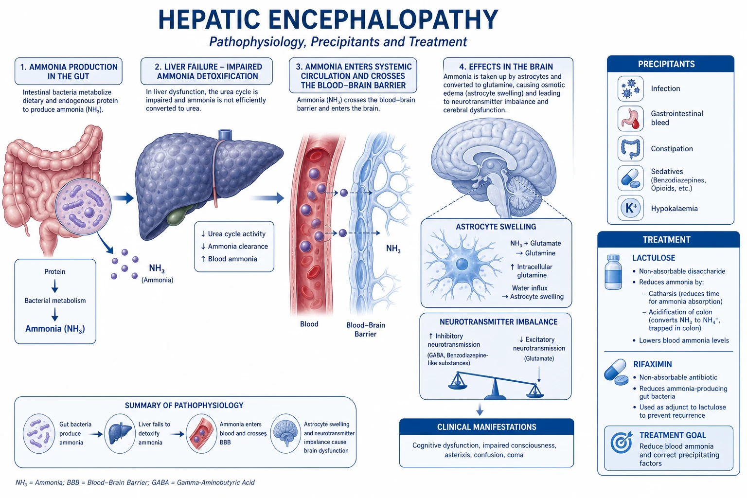
Ammonia (NH3) is a neurotoxic by-product of nitrogen metabolism. The gut generates most of it (bacterial deamination of dietary protein and urease splitting of urea), and the liver detoxifies it via the urea cycle (Krebs-Henseleit cycle) — the reason portal blood ammonia is high but systemic ammonia is low. [1]
Ammonia handling and the urea cycle
- AMMONIA PRODUCTION — primarily in the GUT: bacterial urease splits urea (from the blood/saliva) → NH3 + CO2; bacterial deamination of dietary amino acids → NH3. The kidney (proximal tubule, from glutamine via glutaminase) and muscle (from amino acid transamination) are minor sources. In critical illness and catabolism, muscle becomes a NET producer.
- PORTAL DELIVERY — gut-derived NH3 enters the portal vein (portal [NH3] >> systemic [NH3]).
- HEPATIC DETOXIFICATION via the UREA CYCLE (hepatocyte mitochondria + cytosol): NH3 + CO2 + 2 ATP → carbamoyl phosphate (via carbamoyl phosphate synthetase-I, CPS-I — the rate-limiting enzyme, requires N-acetylglutamate as an obligate activator) → carbamoyl phosphate + ornithine → citrulline (mitochondria) → citrulline + aspartate → argininosuccinate → arginine → arginine + H2O (arginase) → UREA + ornithine (regenerated). The urea cycle converts 2 NH3 + CO2 → urea (2 nitrogen atoms per urea).
- UREA EXCRETION — urea is water-soluble, leaves the liver in systemic blood → filtered by the kidney → excreted in urine (blood urea / BUN / serum urea). A fraction is recycled to the gut (split by bacterial urease) — the urea salvage cycle.
- FAILURE → HYPERAMMONAEMIA — when the liver fails (acute or chronic), or when portal blood is shunted past the liver (portosystemic shunts, spontaneous or TIPSS), NH3 accumulates in systemic blood.
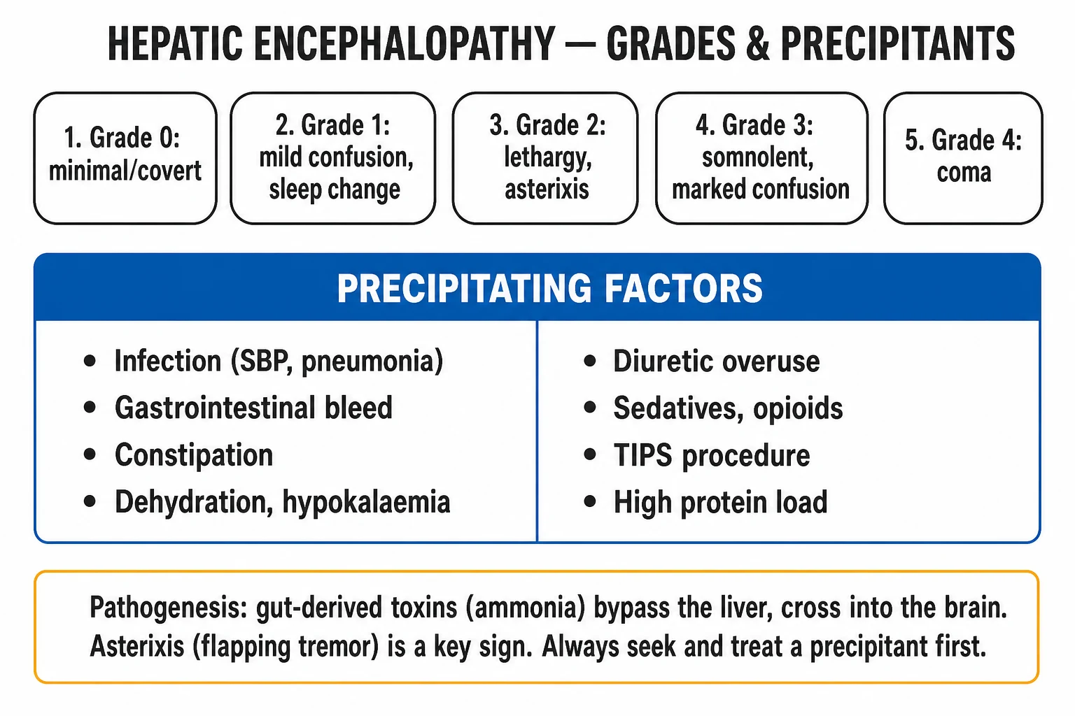
- NEUROTOXICITY → HEPATIC ENCEPHALOPATHY — NH3 crosses the blood-brain barrier → taken up by ASTROCYTES (the only brain cell expressing glutamine synthetase) → NH3 + glutamate → glutamine (via glutamine synthetase). Glutamine is osmotically active → astrocyte SWELLING → cerebral oedema (in acute liver failure, potentially fatal intracranial hypertension/herniation) and astrocyte dysfunction (the Alzheimer type II change) → impaired neurotransmission (glutamate/GABA balance) → hepatic encephalopathy (confusion → asterixis → somnolence → coma).[3]
The ammonia-glutamine-cerebral-oedema link explains why arterial ammonia >150 umol/L in acute liver failure predicts cerebral oedema and herniation (a key prognostic threshold), and why ammonia-lowering therapy (lactulose acidifying the colon to trap NH4+, rifaximin suppressing urease-producing bacteria) is the backbone of hepatic encephalopathy management. Ammonia is NOT the sole toxin (inflammation, benzodiazepine-like compounds, manganese, mercaptans all contribute), but it is the most quantifiable and the most clinically actionable.[3]
Gut barrier function — the gatekeeper that fails in critical illness
The gut barrier is the single largest interface between the body and the external environment (~300 m^2 — the surface area of a tennis court). It must simultaneously ABSORB nutrients and WATER while EXCLUDING 10^13 microorganisms and their products. It has four integrated layers: [1]
The four layers of the gut barrier
| Layer | Components | Function | What disrupts it in ICU |
|---|---|---|---|
| Physical — mucus | Mucus bilayer (goblet cell mucin-2), the unstirred water layer, secretory IgA (plasma-cell derived, transported by the polymeric immunoglobulin receptor) | Lubrication; traps bacteria and endotoxin; secretory IgA agglutinates bacteria and prevents epithelial adherence | Dehydration, reduced mucus production (mucosal ischaemia), PPIs (reduce the mucus-pH gradient) |
| Physical — epithelium + tight junctions | Single layer of columnar epithelial cells (enterocytes, goblet, Paneth, enteroendocrine) joined by TIGHT JUNCTIONS (claudins, occludin, zonula occludens-1/2) and ADHERENS junctions; turnover every 3-5 days (the fastest-renewing epithelium) | Selective permeability — absorbs nutrients/water, excludes bacteria/macromolecules; Paneth cells secrete antimicrobial peptides (defensins/alpha-defensins, RegIIIgamma, cathelicidins) | Ischaemia-reperfusion (the #1 cause — rapid epithelial sloughing), chemotherapy, radiation |
| Immunological — GALT | Gut-associated lymphoid tissue: Peyer's patches (ileum), lamina propria lymphocytes (T cells, plasma cells), intraepithelial lymphocytes, mesenteric lymph nodes — the largest immune organ (~70% of body immune cells) | Immune sampling (M cells over Peyer's patches), antigen presentation, secretory IgA, oral tolerance, innate immune signalling (TLR/NOD) | Immunosuppression, malnutrition, lymphocyte apoptosis (corticosteroids, sepsis) |
| Microbiological — the microbiome | ~10^13 commensal organisms (10x the number of human cells), >1000 species, dominated by Bacteroidetes and Firmicutes; anaerobes >> aerobes (1000:1) | Colonisation resistance (occupy niches, consume nutrients, produce short-chain fatty acids [butyrate — fuel for colonocytes, anti-inflammatory], bacteriocins) that SUPPRESS pathogens; educate the immune system; metabolise bile acids, drugs, fibre | BROAD-SPECTRUM ANTIBIOTICS (the #1 cause of dysbiosis → overgrowth of pathogens/C. difficile), PPIs (gastric pH rise → bacterial overgrowth), parenteral nutrition (no luminal fuel → mucosal atrophy), ileus/altered motility |
Bacterial translocation is the passage of viable bacteria and/or their products (endotoxin, peptidoglycan, bacterial DNA) from the gut lumen to normally sterile extraintestinal sites (mesenteric lymph nodes, portal blood, systemic circulation). Three mechanisms are recognised: (a) transcellular (through the enterocyte), (b) paracellular (between cells — through disrupted tight junctions, the dominant route in ischaemia), and (c) dendritic cell-mediated (sampling by GALT that becomes pathological when the barrier is overwhelmed). Translocation is normally a low-level, immunologically-tolerised event (constant antigen sampling maintains immunity); it becomes pathogenic when the barrier is broken AND the organism is virulent/abundant AND hepatic/immune clearance is overwhelmed.[4][5]
The cycle of critical illness → barrier failure → systemic inflammation is self-perpetuating: gut ischaemia → barrier leak → translocation → Kupffer/immune activation → cytokines → further mucosal injury (cytokines damage tight junctions) → more leak. This is why restoring gut perfusion EARLY and protecting the barrier (early enteral nutrition to maintain mucosal integrity and trophic flow, avoiding unnecessary antibiotics/PPIs, selective decontamination where appropriate) is a legitimate MODS-prevention strategy, and why the gut is both a target and a driver in the critically ill.[1][5]
Bile formation and cholestasis
Bile — synthesis, enterohepatic circulation, function
Bile is a complex aqueous secretion with two functions: DIGESTION (emulsification of dietary fat) and EXCRETION (the route for bilirubin, cholesterol, and lipophilic xenobiotics). Hepatocytes synthesise the primary bile acids — cholic acid and chenodeoxycholic acid — from CHOLESTEROL (via 7alpha-hydroxylase, CYP7A1, the rate-limiting enzyme; bile acid synthesis is the major route of cholesterol catabolism). They are conjugated with glycine or taurine (lowering pKa → soluble at intestinal pH) → secreted into bile canaliculi by BSEP (ABCB11). Bile flows through canaliculi → ductules → hepatic/common bile duct → stored and concentrated in the gallbladder → released into the duodenum post-prandially when cholecystokinin (CCK) (released by I-cells in response to luminal fat/amino acids) contracts the gallbladder and relaxes the sphincter of Oddi.[6]
In the duodenum, bile salts act as DETERGENTS: their amphipathic structure (hydrophobic steroid nucleus + hydrophilic side chain) emulsifies dietary triglycerides into smaller droplets, then forms mixed micelles (bile salts + fatty acids + monoglycerides + fat-soluble vitamins) that deliver lipids to the enterocyte brush border for absorption. 95% of bile acids are reabsorbed — actively in the TERMINAL ILEUM (via ASBT, the apical sodium-dependent bile salt transporter) and passively throughout the gut — return to the liver via the portal vein, are re-secreted (the enterohepatic circulation, 6-10 cycles/day, with a total bile acid pool of ~3 g recycled against a daily synthesis of only ~0.5 g). Gut bacteria deconjugate and dehydroxylate primary bile acids → secondary bile acids (deoxycholic, lithocholic).[6]
Terminal ileal disease/resection (Crohn's, surgical) disrupts reabsorption → bile acid loss in stool → depletion of the pool → fat malabsorption (steatorrhoea) and gallstone formation (cholesterol supersaturation from a depleted bile acid pool). [1]
Cholestasis — intrahepatic and extrahepatic
Cholestasis is impaired bile formation or flow. The defining biochemical signature is a CONJUGATED (direct) hyperbilirubinaemia with raised alkaline phosphatase and gamma-GT (the canalicular enzymes), because conjugated bilirubin that cannot be excreted backs up into blood. [1]
Intrahepatic vs extrahepatic cholestasis
| Feature | Intrahepatic | Extrahepatic |
|---|---|---|
| Site | Hepatocyte / canalicular / ductule (within the liver) | Large bile ducts (outside the liver) |
| Mechanism | Impaired bile salt transport, canalicular contraction failure, bile ductule injury | Mechanical obstruction to bile flow |
| Causes | Sepsis (the #1 ICU cause — cytokines downregulate BSEP/MRP2 transporters), drugs (anaesthetic agents, antibiotics, amoxicillin-clavulanate, statins), total parenteral nutrition, viral/alcoholic hepatitis, primary biliary cholangitis, intrahepatic cholestasis of pregnancy | Choledocholithiasis (gallstones), pancreatic cancer (head), cholangiocarcinoma, primary sclerosing cholangitis, biliary stricture, pancreatitis |
| Imaging | Normal ducts (ultrasound/MRCP) | DILATED intra- and extra-hepatic ducts ± a visible mass/stone |
| Clinical clues | Systemic illness, drug exposure, transaminitis | Painless jaundice + palpable gallbladder (Courvoisier's law — a palpable gallbladder in PAINLESS jaundice is unlikely to be stones), pale stool, dark urine, pruritus |
The consequences of cholestasis relevant to ICU:
- Jaundice and pruritus — conjugated bilirubin and bile acids deposit in skin (pruritus, often worse at night, refractory to antihistamines — treat with cholestyramine/bile acid sequestrants, rifampicin, ondansetron; intractable in cirrhosis → albumin dialysis [MARS]).
- Fat-soluble vitamin malabsorption (A, D, E, K) — because bile is absent to form micelles. Vitamin K malabsorption → coagulopathy (corrects with parenteral vitamin K, distinguishing it from hepatocellular failure); vitamin D → osteomalacia/bone disease (a long-term problem in chronic cholestasis).
- Steatorrhoea and, in prolonged cholestasis, malnutrition.
- Sepsis-induced intrahepatic cholestasis is so common in ICU that an isolated raised bilirubin (conjugated) + ALP in a septic patient is often diagnosed on clinical grounds, resolving as sepsis resolves. [1]
Exam practice — SAQs
SAQ — Splanchnic circulation in shock: a persistent lactate and the ischaemic gut
10 minutes · 10 marks
A 68-year-old man is admitted to ICU 12 hours after an emergency Hartmann procedure for a perforated sigmoid diverticulum. He remains in septic shock despite 30 mL/kg crystalloid: MAP 62 on noradrenaline 0.35 mcg/kg/min, lactate risen from 3.2 to 5.8 mmol/L, urine output 10 mL/h. He is intubated and ventilated (FiO2 0.6) with cool peripheries, a distended silent abdomen and coffee-ground nasogastric aspirate. AST 480, ALT 410 (baseline normal), INR 2.1, bilirubin 52. The registrar asks why the lactate keeps climbing when the blood pressure is acceptable.
Clinical pearls
Red flags
[1]Prognosis
Outcomes and physiological determinants in gut/hepatic dysfunction
| Scenario | Mechanism / Outcome | Notes |
|---|---|---|
| Mesenteric ischaemia (any shock) | Mortality 50-80% if transmural infarction; the gut is the 'canary' of the splanchnic bed | Restoration of perfusion EARLY is the only effective therapy; lactate is the sentinel marker of ongoing splanchnic hypoperfusion |
| Gut barrier disruption → translocation | Drives MODS mortality; the 'second hit' phenomenon — a subsequent insult (infection, surgery) in a primed host produces exaggerated inflammation | Prevent: restore perfusion, early enteral nutrition, antibiotic/PPI stewardship |
| Acute liver failure — cerebral oedema | Leading cause of death; arterial ammonia >150 + grade III-IV encephalopathy = high risk | King's College criteria trigger transplant referral; mortality ~30-50% without transplant, ~20% with |
| Cirrhosis — drug handling | Accumulation of sedatives/opioids → oversedation, hypotension, encephalopathy precipitation | Both Phase I and Phase II impaired; first-pass lost; start LOW, go SLOW |
| Cholestasis | Reversible if cause removed (sepsis, drugs, obstruction relieved) | Fat-soluble vitamin malabsorption → coagulopathy (vit K), bone disease (vit D); pruritus often the most distressing symptom |
| Sepsis-induced intrahepatic cholestasis | Functional, reversible as sepsis resolves; isolated conjugated bilirubin + raised ALP/GGT | Cytokine-mediated downregulation of BSEP/MRP2 — no obstruction, no necrosis |
The unifying physiological principle is that the gut-liver axis is BOTH a target of, and a driver of, critical illness. In shock, the sacrificed splanchnic circulation becomes ischaemic, the gut barrier fails, and bacterial/endotoxin translocation activates the Kupffer-cell cytokine cascade that propagates inflammation to distant organs — the 'gut motor' of MODS. The failing liver loses its synthetic (clotting factors, albumin), metabolic (glucose, ammonia, drug clearance), and excretory (bilirubin, bile) functions in a predictable sequence — clotting (factor VII, half-life 6h) first, then ammonia/encephalopathy, then albumin (half-life 20 days) last. Understanding this physiology — the dual blood supply, the phases of drug metabolism, the bilirubin cascade, the urea cycle, the gut barrier layers, the enterohepatic circulation — is the foundation for every ICU intervention from early enteral feeding and antibiotic stewardship to ammonia-lowering therapy, neuroprotection in liver failure, and cholestasis management.[1][3][4]
Key trials and evidence
Deitch 2012 — Gut-origin sepsis: evolution of a concept (Surgeon; PMID 22534256)
Source
Seminal review by Edwin Deitch (the leading proponent of the gut-origin theory), synthesising four decades of work
Key contribution
Articulated the 'gut as the motor of MODS' — that the ischaemic, barrier-disrupted gut in shock is not merely a victim but a DRIVER of distant organ failure via bacterial/endotoxin translocation
Key finding
Two routes carry gut-derived noxious material systemically: the PORTAL route (gut -> portal vein -> Kupffer cells -> cytokines) and the MESENTERIC LYMPH route (gut -> thoracic duct -> systemic circulation, bypassing the liver); mesenteric lymph from shocked hosts is itself cytotoxic to distant organs
Clinical bottom line
The physiological rationale for protecting the gut in critical illness — early perfusion, early enteral nutrition, antibiotic/PPI stewardship — as a MODS-prevention strategy
Landow & Andersen 1994 — Splanchnic ischaemia and multiple organ failure (Acta Anaesthesiol Scand; PMID 7839769)
Source
Comprehensive review of splanchnic blood flow physiology and its role in MODS
Key contribution
Established the haemodynamic basis for gut vulnerability — the splanchnic bed is sacrificed FIRST (intense alpha-1/angiotensin II/vasopressin vasoconstriction) and reperfused LAST in any shock state
Key finding
Persistent splanchnic hypoperfusion (the 'splanchnic debt') drives mucosal ischaemia, barrier failure and translocation even after systemic blood pressure is restored — the basis for the persistent lactate that signals ongoing occult gut hypoperfusion
Clinical bottom line
Gastric tonometry/PCO2 monitoring (historical) and lactate clearance are clinical windows onto splanchnic perfusion; the gut is the 'canary' organ of the circulation
Balzan et al 2007 — Bacterial translocation: overview of mechanisms and clinical impact (J Gastroenterol Hepatol; PMID 17376034)
Source
Authoritative review of the mechanisms and clinical consequences of bacterial translocation
Key contribution
Defined the three mechanisms (transcellular, paracellular through disrupted tight junctions, dendritic-cell-mediated) and the three conditions for pathogenic translocation (barrier disruption AND virulent/abundant organisms AND overwhelmed clearance)
Key finding
Translocation is normally a low-level, immunologically-tolerised event (constant antigen sampling maintains immunity); it becomes pathogenic only when the barrier is broken — explaining why gut ischaemia (not baseline physiology) is the key driver in critical illness
Clinical bottom line
The mechanistic link between the disrupted gut barrier and systemic infection/MODS — supporting barrier-protective strategies (enteral nutrition, perfusion, selective decontamination)
References
- [1]Deitch EA Gut-origin sepsis: evolution of a concept Surgeon, 2012.PMID 22534256
- [2]Landow L, Andersen LW Splanchnic ischaemia and its role in multiple organ failure Acta Anaesthesiol Scand, 1994.PMID 7839769
- [3]Blei AT Brain edema in acute liver failure Crit Care Clin, 2008.PMID 18241781
- [4]Balzan S, de Almeida Quadros C, de Cleva R, Zilberstein B, Cecconello I Bacterial translocation: overview of mechanisms and clinical impact J Gastroenterol Hepatol, 2007.PMID 17376034
- [5]Quigley EM, Abu-Shanab A Small intestinal bacterial overgrowth Infect Dis Clin North Am, 2010.PMID 20937459
- [6]Amin SB, Bhutani VK, Watchko JF Apnea in acute bilirubin encephalopathy Semin Perinatol, 2014.PMID 25239473