ICU · Physiology / thermoregulation
Thermoregulation
Also known as Thermoregulation · Body temperature regulation · Hypothalamic set point · Preoptic anterior hypothalamus · Fever · Hyperthermia · Malignant hyperthermia · Heat stroke · Heat exhaustion · Hypothermia · Swiss staging · Shivering · Non-shivering thermogenesis · Brown adipose tissue · Targeted temperature management · Therapeutic hypothermia · Neuroleptic malignant syndrome · Serotonin syndrome · Poikilothermia
Thermoregulation — the hypothalamic set point (37 degrees C). The heat production (the shivering, the metabolism, the thyroxine, the brown fat / non-shivering thermogenesis). The heat loss (the sweating, the vasodilation, the radiation, the convection, the conduction, the respiration). The fever (the prostaglandin E2 — the pyrogen raises the set point). The clinical: the malignant hyperthermia (the ryanodine receptor), the heat stroke, the hypothermia, the neuroleptic malignant syndrome.
On this page & tools
Your progress
Saved locally on this device.
8 MCQs with explanations
Target exams
Overview & definition
The thermoregulation — the hypothalamic set point (37 degrees C). The balance the heat production and the heat loss. The fever (the pyrogen raises the set point). The clinical disorders (the malignant hyperthermia, the heat stroke, the hypothermia, the NMS).[1]
Thermoregulation is the active, neurally-mediated maintenance of core body temperature within a narrow defended range (36-37.5 degrees C) despite wide variation in environmental temperature and metabolic heat load. It is a classical negative-feedback control loop: a sensor (temperature receptors), an integrator with a defended set point (the preoptic area of the anterior hypothalamus, PO/AH), and effectors (behavioural and autonomic heat-production / heat-loss mechanisms). The clinical importance in intensive care is twofold — first, the fever versus hyperthermia distinction dictates entirely different management (antipyretics vs active cooling); second, induced hypothermia (targeted temperature management, TTM) after cardiac arrest is a deliberate, protocolised manipulation of the set point–defended variable to produce neuroprotection, while accidental hypothermia, heat illness, malignant hyperthermia and the neuroleptic malignant syndrome are all disorders of this system encountered in critically ill patients.[1][2]
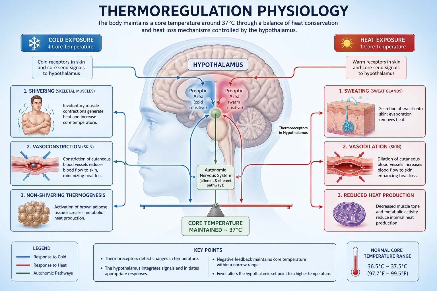
The mechanism
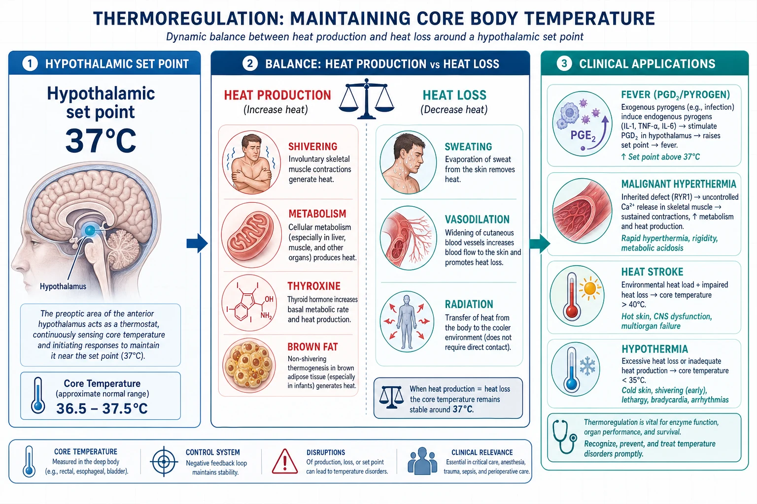
Heat production
- The shivering (the skeletal muscle — the - the - the).[1]
- The metabolism (the basal metabolic rate; the thyroxine; the - the - the).[1]
- The brown fat / the non-shivering thermogenesis (the newborn; the - the - the).[1]
Heat loss
- The sweating (the evaporative).[1]
- The vasodilation (the skin — the radiation).[1]
- The radiation, convection, conduction, respiration.[1]
The fever
- The pyrogen (the exogenous — the bacterial LPS; the endogenous — the IL-1, the IL-6, the TNF) → the prostaglandin E2 (the PGE2) in the hypothalamus → the set point the rises.[1]
- The - the - the (the - the - the).[1]
- The antipyretics (the paracetamol, the NSAIDs) — the block the PGE2 (the COX the inhibition).[1]
The clinical
- The malignant hyperthermia (the ryanodine receptor — the calcium the release; the volatile anaesthetics, the suxamethonium; the dantrolene).[1]
- The heat stroke (the environmental — the core over 40 degrees C; the CNS the dysfunction; the - the - the).[1]
- The hypothermia (the core under 35 degrees C; the - the - the).[1]
- The neuroleptic malignant syndrome (the NMS — the dopamine the blockade; the D2 the antagonists; the - the - the).[1]
Normal thermoregulation — the defended set point and its effectors
The hypothalamic integrator and the set point (36.5-37.5 degrees C)
The defended normal core temperature is 36.5-37.5 degrees C (commonly cited as a set point of ~37 degrees C). The integrator that generates and defends this set point is the preoptic area of the anterior hypothalamus (PO/AH). The PO/AH is itself intrinsically thermosensitive: a high density of warm-sensitive neurons increase their firing rate as local blood/brain temperature rises, and this intrinsic signal is the primary feedback that the integrator compares against the defended set point. Peripheral (skin) thermoreceptors add a feed-forward "anticipatory" input so the body can begin to mount a response (e.g., shivering) before core temperature has actually fallen.[2]
The set point is not fixed — it is physiologically modulated:
- Circadian rhythm: ~0.5-1.0 degrees C oscillation, nadir ~04:00-06:00, peak ~16:00-18:00. This is why a "fever" of 37.8 degrees C in the evening may be normal, and why night-shift ICU temperatures must be interpreted against the circadian nadir.
- Ovarian cycle: luteal-phase rise of ~0.3-0.5 degrees C (progesterone).
- Exercise: transient rise in the effective set point.
- Fever: a regulated, PGE2-mediated upward shift of the set point itself (see below).
- Age: the elderly have a lower basal temperature (~0.5 degrees C) and a blunted febrile response (impaired PGE2 signalling and reduced thermogenic reserve), so a "normal" temperature in a septic 85-year-old does not exclude sepsis.[1]
The thermoregulatory control loop — the four components
| Component | Structure / mechanism | Key point |
|---|---|---|
| Sensor (afferent) | Central warm-sensitive neurons in PO/AH (sense brain/blood temp); peripheral cold/warm receptors in skin (via spinothalamic tract + trigeminal afferents) | The hypothalamus senses ITS OWN temperature — this is the dominant feedback |
| Integrator | Preoptic area of the anterior hypothalamus (PO/AH); secondary nodes dorsomedial hypothalamus (DMH) and raphe pallidus | Houses the defended set point (~37 degrees C); fuses central + peripheral signals; generates the error signal |
| Set point | The defended reference temperature (~36.5-37.5 degrees C) | Raised by PGE2 (fever); modulated by circadian rhythm, hormones, age |
| Effector (efferent) | Autonomic (sympathetic outflow → skin vessels, sweat glands, brown fat, shivering) + behavioural (cortex → clothing, shelter, activity) | Behaviour is the MOST powerful defence in the conscious patient; lost under anaesthesia/sedation |
The thermoregulatory feedback loop — step by step
- SENSE: central warm-sensitive neurons in the PO/AH detect core (blood/brain) temperature; peripheral skin thermoreceptors detect ambient challenge. Afferents travel via spinothalamic tract and trigeminal nerve to thalamus then PO/AH.
- INTEGRATE: the PO/AH fuses central and peripheral inputs and compares the integrated temperature with the defended set point. The magnitude of the error (set point minus actual) determines the intensity of the response.
- DECIDE: if core is BELOW set point (cold challenge), recruit heat-conservation then heat-production pathways in order of metabolic cost. If ABOVE set point (heat challenge), recruit heat-loss pathways.
- EFFERENT — autonomic: sympathetic outflow drives (a) skin blood flow through arteriovenous anastomoses of fingers/toes/ears (constrict to conserve, dilate to lose), (b) eccrine sweat glands, (c) brown adipose tissue (beta-3 adrenergic), (d) shivering via reticulospinal tract to anterior horn cells.
- EFFERENT — behavioural: the cortex is engaged (clothing, shade, shelter, altering activity). In conscious humans this is the MOST powerful defence; it is abolished by general anaesthesia, sedation, dementia and brain injury — which is why anaesthetised patients become poikilothermic.
- EFFECT: core temperature returns towards the set point; the error signal shrinks; the response is gated off (negative feedback completes the loop).
Heat production — mechanisms and magnitude
Resting heat production is ~80 W and comes overwhelmingly from basal metabolic rate (BMR) — the viscera (liver, brain, heart, kidneys) and obligatory ion-pump activity (Na+/K+ ATPase consumes ~20-30% of resting ATP). When heat is lost faster than BMR replaces it, four additional calorigenic mechanisms are recruited: [1]
- Cutaneous vasoconstriction — reduces heat LOSS (not production), the first and cheapest response to cold; closes arteriovenous anastomoses, cuts heat loss by up to ~50%.
- Non-shivering thermogenesis — brown adipose tissue (BAT) burns lipid via uncoupling protein 1 (UCP1 / thermogenin), which short-circuits the mitochondrial proton gradient so oxidation releases heat rather than synthesising ATP. Dominant in neonates (who cannot shiver effectively) and reactivated in adult humans.
- Shivering thermogenesis — asynchronous skeletal muscle contractions driven via the reticulospinal tract; can raise metabolic rate up to ~5 times BMR. Metabolically expensive and dangerous during TTM (see below).
- Hormonal calorigenesis — thyroid hormone (T3/T4) up-regulates tissue metabolic rate and Na+/K+ ATPase density (slow, genomic, hours-days); catecholamines raise metabolism rapidly via beta-adrenergic stimulation (minutes). The thyroid–catecholamine synergy explains the extreme hypermetabolism of thyroid storm and the hypothermia of myxoedema coma. [1]
Heat production mechanisms — relative magnitude and onset
| Mechanism | Magnitude (vs BMR) | Onset | Key physiology | Clinical relevance |
|---|---|---|---|---|
| BMR | 1.0x (baseline) | Constant | Viscera + Na+/K+ ATPase; set by thyroid | Hypothyroid → hypothermia; hyperthyroid → heat intolerance |
| Cutaneous vasoconstriction | Reduces loss ~50% | Seconds | Sympathetic — closes AV anastomoses | First cold response; basis of "cold peripheries" in shock |
| Non-shivering (BAT/UCP1) | ~up to 2-2.5x in neonates | Minutes | UCP1 uncouples oxidative phosphorylation | Neonatal cold stress; reactivated in adults |
| Shivering | up to ~5x | Seconds-minutes | Skeletal muscle, reticulospinal-driven | Dangerous during TTM — must be suppressed |
| Hormonal (T3/T4) | raises BMR set-point | Hours-days (genomic) | Na+/K+ ATPase, beta-receptor density | Thyroid storm hypermetabolism; myxoedema coma hypothermia |
| Hormonal (catecholamines) | ~1.3-1.5x rapid | Minutes | Beta-adrenergic glycogenolysis/lipolysis | Phaeochromocytoma, NMS, serotonin syndrome heat |
| Exercise | up to 10-15x (brief) | Seconds | Voluntary muscle contraction | Exertional heat stroke |
Heat loss — the four physical mechanisms
Heat is lost from the body-shell interface to the environment by four physical processes. The proportions are for a resting, clothed person in a temperate room (~22 degrees C): [1]
Heat loss mechanisms — proportion and physiology
| Mechanism | Proportion | Physics | Amplified by | Clinical exploitation |
|---|---|---|---|---|
| Radiation | ~60% | Infrared electromagnetic transfer from warmer body to cooler surfaces — does NOT require contact or air movement | Cold surrounding surfaces (windows, walls) | Reflective "space" blankets reduce radiative loss; radiant warmers add heat |
| Evaporation | ~25% | Latent heat of vaporisation — 0.58 kcal (2.4 kJ) lost per gram of water vaporised from skin or respiratory tract | High ambient temperature, low humidity, air movement | Wet sheet + fan for heat stroke; alcohol sponging |
| Convection | ~12% | Heat transferred to air at skin surface then carried away by air currents; depends on gradient and air velocity | Wind, forced air | Forced-air cooling; "wind chill"; Bair Hugger (adds warm air) |
| Conduction | ~3% | Direct molecular transfer to cooler object in contact; proportional to gradient and thermal conductivity | Water (conducts ~25x better than air); metal | Cooling blankets, ice-water immersion, water mattress |
| Respiration | (part of evaporation) | Warming + humidifying inspired gas; vaporisation of water from airways | Cold/dry inspired gas, high minute volume | HME (heat-moisture exchanger) filters conserve this loss in ventilated patients |
Behavioural vs autonomic responses — the two efferent channels
Humans defend temperature through two efferent channels. Behavioural thermoregulation (the cortex-driven, conscious response — putting on clothing, seeking shade, altering activity) is the MOST powerful defence in the intact, conscious patient. Autonomic (involuntary) thermoregulation (cutaneous vasomotor tone, sweating, shivering, non-shivering thermogenesis) operates continuously below conscious awareness and is the only channel available when behaviour is abolished. [1]
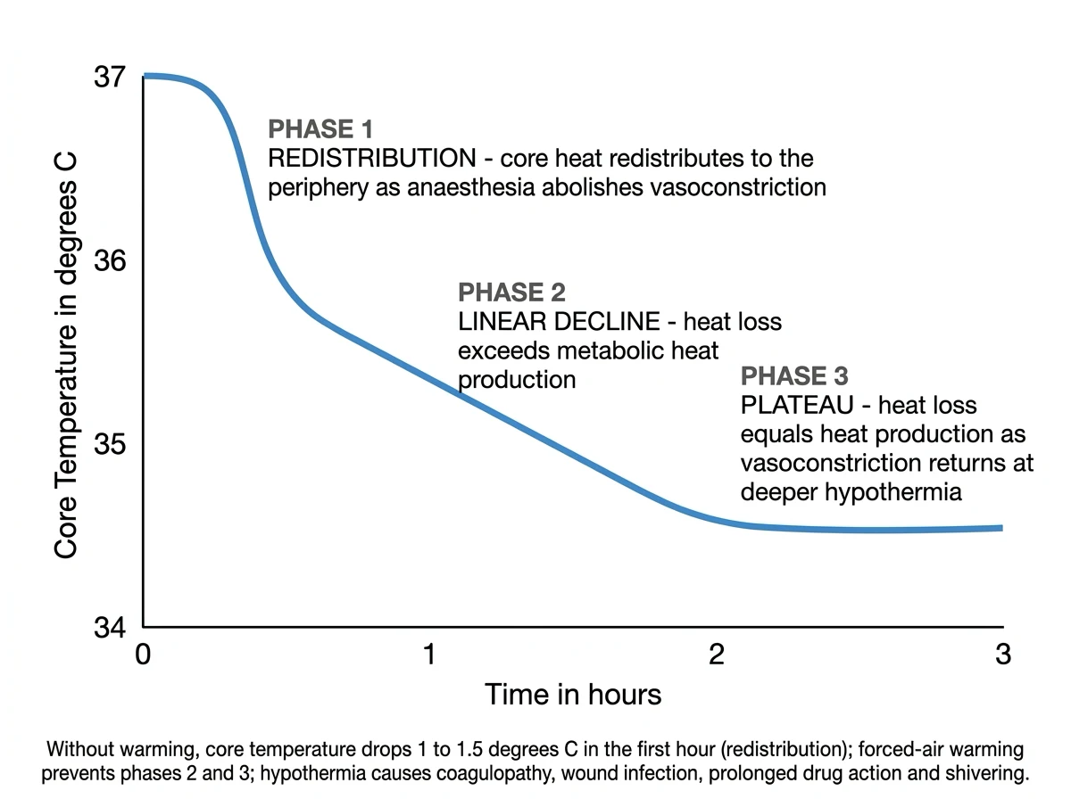
This distinction is exam-critical and explains most perioperative and ICU temperature problems: general anaesthesia abolishes behavioural thermoregulation and blunts the autonomic threshold, so the anaesthetised patient behaves like a poikilotherm and drifts towards ambient temperature unless actively warmed — the basis of inadvertent perioperative hypothermia (a 1-1.5 degrees C drop in the first 30-60 minutes from redistribution of warm core blood to the vasodilated periphery, then a slow linear fall from heat loss exceeding production).[4]
Response to COLD vs response to HEAT — the autonomic recruitment ladder
| Response to COLD (recruited in order, cheapest first) | Response to HEAT (recruited in order) |
|---|---|
| 1. Behavioural (clothing, shelter) | 1. Behavioural (shade, rest, fanning) |
| 2. Cutaneous vasoconstriction (sympathetic — closes AV anastomoses; reduces shell conductance; can cut heat loss ~50%) | 2. Cutaneous vasodilation (active vasodilation — cholinergic co-transmission + nitric oxide; opens AV anastomoses; skin blood flow can rise from ~0.5 to ~7 L/min) |
| 3. Non-shivering thermogenesis (BAT/UCP1) | 3. Sweating (eccrine glands, sympathetic cholinergic; up to ~1-2 L/h acutely) |
| 4. Shivering (skeletal muscle, up to 5x BMR — costly) | 4. (No further autonomic reserve — decompensation = heat stroke) |
| 5. Hormonal (TSH/thyroid up-regulation, catecholamines — chronic) | |
| 6. Piloerection (vestigial in humans — trapped air layer in fur) |
Fever vs hyperthermia — the pivotal exam distinction
This single distinction is examined relentlessly because it changes management. Both present with a high body temperature, but the underlying pathophysiology is opposite. [1]
Fever — the set point is RAISED
In fever, exogenous pyrogens (bacterial endotoxin/LPS, viral particles) and endogenous pyrogens (IL-1, IL-6, TNF-alpha) act on the organum vasculosum of the lamina terminalis (OVLT) — a circumventricular organ with a permeable blood-brain barrier that allows circulating cytokines to reach the brain. This activates phospholipase A2 and cyclo-oxygenase (COX) to convert arachidonic acid to prostaglandin E2 (PGE2) in the PO/AH. PGE2 RAISES the defended set point (e.g., from 37 to 39 degrees C).[2]
The hypothalamus now perceives the body as too COLD relative to the new set point and mounts a cold response: the patient feels cold and shivers ("rigors"), and cuts heat loss by cutaneous vasoconstriction (pale, cold peripheries), until core temperature climbs to the new set point. Once reached, the temperature plateaus and the peripheries vasodilate ("the fever breaks"). When the pyrogen signal resolves — or an antipyretic is given — PGE2 falls, the set point drops back to 37, and the patient now feels hot and sweats/vasodilates to cool down to the new (normal) set point. Antipyretics work by BLOCKING PGE2 synthesis (paracetamol via central/putative COX-3 inhibition; NSAIDs and aspirin via peripheral COX-1/COX-2 inhibition). They therefore work ONLY when the set point is elevated (fever) and have NO effect when the set point is normal (hyperthermia). [1]
Hyperthermia — the set point is NORMAL
In hyperthermia, the set point remains at ~37 degrees C but heat load or heat production exceeds the maximum capacity for heat loss. The hypothalamus is already driving maximal heat-loss responses (sweating, vasodilation) but is overwhelmed. Causes include environmental heat stroke, uncontrolled muscle activity (malignant hyperthermia, NMS, serotonin syndrome, status epilepticus), exaggerated metabolic rate (thyroid storm, salicylate uncoupling of oxidative phosphorylation), and exertional heat generation. Antipyretics are INEFFECTIVE in hyperthermia because the set point is not elevated; treatment is active cooling plus, where relevant, specific antidotes (dantrolene for MH; bromocriptine/dantrolene for NMS; cyproheptadine for serotonin syndrome).[1][14]
Fever vs hyperthermia — the decision table
| Feature | FEVER | HYPERTHERMIA |
|---|---|---|
| Hypothalamic set point | RAISED (by PGE2) | NORMAL |
| Mediator | PGE2 (via COX, driven by IL-1/IL-6/TNF-alpha on OVLT) | None (set point intact) |
| Patient feels | Cold during the rise — rigors, pale peripheries | Hot, flushed, sweating (maximal heat-loss attempt) |
| Skin | Cold, pale, constricted during rise | Hot, flushed, vasodilated |
| Response to antipyretics | YES (block PGE2 → lower set point) | NO (set point not elevated) |
| Rate of rise | Gradual, plateaus at new set point | Can be explosive (MH: up to 1 degrees C every 5 min) |
| Treatment | Treat cause + antipyretics | ACTIVE COOLING + specific antidote + treat cause |
| Examples | Sepsis, infection, inflammation, malignancy, most drug fevers | Heat stroke, MH, NMS, serotonin syndrome, thyroid storm, salicylate toxicity, exertional |
ICU temperature management — targeted temperature management (TTM)
After cardiac arrest, controlled mild hypothermia — now termed targeted temperature management (TTM) — improves neurological outcome. Current ILCOR practice targets 32-36 degrees C for at least 24 hours in comatose adults after cardiac arrest, with active prevention of fever (temperature >=37.5 degrees C) for at least 72 hours. The two landmark RCTs (HACA and Bernard, both 2002) established 32-34 degrees C; the TTM trial (Nielsen 2013) showed equivalence of 33 and 36 degrees C targets; and the TTM2 trial (Dankiewicz 2021) showed that hypothermia at 33 degrees C was no better than normothermia with active fever prevention at 37.5 degrees C — shifting modern practice toward normothermia/fever prevention as the default, with 33 degrees C reserved for selected patients.[5][6][7][8][12]
Mechanism of neuroprotection — cerebral metabolic rate falls 6-7% per degrees C
The physiological rationale rests on the van 't Hoff / Q10 effect on metabolism: lowering temperature reduces the rate of all temperature-dependent biochemical reactions. The clinically taught figure is that cerebral metabolic rate falls by approximately 6-7% for every 1 degrees C reduction in temperature. Cooling from 37 to 33 degrees C (a 4 degrees C drop) therefore reduces cerebral oxygen consumption by roughly 25-30%, buying time for the injured, energy-depleted brain during the vulnerable early reperfusion window. Beyond simple metabolic suppression, hypothermia also:[5]
Mechanisms of TTM neuroprotection — step by step
- Reduced cerebral metabolic rate / O2 demand — 6-7% per degrees C; the injured brain with impaired mitochondrial ATP production is protected against demand ischaemia.
- Reduced reperfusion injury — suppression of the injurious cascade triggered by reperfusion (free-radical / reactive oxygen species generation, lipid peroxidation, blood-brain barrier disruption, vasogenic oedema).
- Reduced excitotoxicity — lower glutamate release and NMDA-mediated calcium influx, reducing neuronal apoptosis and necrosis.
- Reduced seizure activity — hypothermia raises seizure threshold; post-arrest seizures independently worsen outcome and consume cerebral O2.
- Reduced intracranial pressure — reduced cerebral blood volume and oedema; hypothermia is an ICP-lowering therapy in its own right.
- Suppressed inflammation / apoptosis — modulation of the post-arrest SIRS-like inflammatory response and caspase-mediated programmed cell death.
The hazard of shivering during TTM
Because the PO/AH set point is normal (~37 degrees C) in a post-arrest patient, cooling to 33 degrees C creates a 4-degree error signal that the hypothalamus fights with its most powerful weapon: shivering, which can raise metabolic rate up to 5x and cerebral O2 consumption dramatically — directly opposing the neuroprotective goal. Shivering must therefore be suppressed: sedation, opiates (especially meperidine via a central shivering-receptor action), buspirone, skin-surface counter-warming (warming face/limbs reduces the peripheral cold afferent), magnesium, and — if refractory — neuromuscular blockade with continuous EEG monitoring (since the paralysed, cooled patient cannot be clinically assessed for seizures). The Bedside Shivering Assessment Scale (BSAS) is used to titrate this suppression.[5][9]
Complications of TTM — predictable physiological effects
Hypothermia is not benign — every degree of cooling carries predictable physiological effects that must be anticipated and managed: [1]
Predictable physiological effects of TTM (hypothermia 32-36 degrees C)
| System | Effect of hypothermia | Management implication |
|---|---|---|
| Cardiovascular | Initial hypertension/tachycardia (shivering, sympathetic surge); then bradycardia (slowed SA node), prolonged QT → torsades risk; VF below ~30 degrees C | Continuous cardiac monitoring; correct K+/Mg2+; rewarm if unstable arrhythmia |
| Coagulation | Reversible platelet dysfunction + impaired clotting factor activity → bleeding tendency | Monitor for occult bleeding; avoid routine deep procedures |
| Metabolic/enzyme | Reduced insulin secretion + sensitivity → hyperglycaemia; cold-induced diuresis → hypokalaemia; on rewarming intracellular shift reverses → rebound hyperkalaemia | Tight glucose control; check electrolytes q4-6h; anticipate K+ shifts during rewarming |
| Immune | Leucocyte dysfunction → infection risk (pneumonia, line sepsis) | Surveillance cultures; strict line care |
| Pharmacokinetics | Reduced hepatic/renal clearance → drug accumulation (sedatives, analgesics, NMBAs, vasopressors) | Dose reduction; monitor depth of sedation; prolonged recovery |
| Rewarming | Must be SLOW and controlled (~0.25-0.5 degrees C/h) to avoid rebound cerebral oedema, hypotension (vasodilation), electrolyte shift and seizures | Controlled rewarming protocol; continue sedation through rewarming |
Accidental hypothermia — Swiss staging (HT I-IV)
Hypothermia is defined as a core temperature below 35 degrees C. The Swiss staging system (HT stages) classifies accidental hypothermia by clinical severity rather than measured temperature, because field measurement is unreliable and the core/shell gradient is large in severe cases. It correlates closely with outcome and guides the aggressiveness of rewarming, including the decision to institute extracorporeal membrane oxygenation (ECMO) for HT IV.[10][11]
The Swiss staging of accidental hypothermia (HT I-IV)
| Stage | Clinical state | Core temperature (approx.) | Rewarming strategy |
|---|---|---|---|
| HT I | Alert, conscious, shivering | 35-32 degrees C | Active external (forced warm air, blankets); shivering provides endogenous heat |
| HT II | Altered consciousness / drowsy, NOT shivering | 32-28 degrees C | Active external; shivering is lost — exogenous heat now required; handle gently (arrhythmia risk) |
| HT III | Unconscious, vital signs present | 28-24 degrees C | Active internal (warmed IV fluids, warmed humidified gases, bladder/gastric lavage); high arrhythmia risk; minimal handling |
| HT IV | Unconscious, no vital signs (apparent death) — cardiac arrest | <24 degrees C | Full cardiopulmonary bypass / VA-ECMO; "no one is dead until warm and dead" — continue resuscitation to >=32 degrees C before declaring death |
Severity of hypothermia by temperature and the physiological thresholds
| Threshold | Core temperature | Physiological change | Clinical significance |
|---|---|---|---|
| Mild | 35-32 degrees C | Maximal shivering, tachycardia, tachypnoea, impaired judgement | HT I — alert and shivering |
| Moderate | 32-28 degrees C | Shivering ceases, bradycardia, slowed respiration, confusion/somnolence, J (Osborn) waves on ECG | HT II — altered consciousness |
| Severe | 28-24 degrees C | Coma, hypotension, atrial then ventricular arrhythmia, fixed/dilated pupils (hypothermia itself) | HT III — unconscious with vital signs |
| Profound | <24 degrees C | Cardiac arrest (asystole or VF), absent reflexes, apnoea | HT IV — apparent death |
Heat illness — heat exhaustion vs heat stroke
Heat illness is a spectrum from minor cramps through heat exhaustion to life-threatening heat stroke. The crucial distinction is the presence of central nervous system dysfunction and a core temperature over 40 degrees C, which defines heat stroke — a true hyperthermic emergency in which the set point is NORMAL (so antipyretics are useless) and the treatment is rapid active cooling.[1]
Heat exhaustion vs heat stroke — the clinical distinction
| Feature | HEAT EXHAUSTION | HEAT STROKE |
|---|---|---|
| Core temperature | Usually 38-40 degrees C | >40 degrees C (may be much higher in exertional) |
| CNS / mental state | Normal (may be fatigued, headache, irritable) — NO major CNS dysfunction | Marked CNS dysfunction — confusion, agitation, seizures, coma |
| Sweating | Usually present (profuse) | May be present (exertional) or absent/anhidrotic (classic) — anhidrosis is NOT required for diagnosis |
| Haemodynamics | Tachycardia, may be orthostatic; BP usually maintained | Hypotension, shock, distributive physiology |
| End-organ injury | Absent | AKI, hepatocellular injury, rhabdomyolysis, DIC, ARDS |
| Pathophysiology | Volume depletion + salt loss exceeding intake; thermoregulation INTACT | Thermoregulatory FAILURE; systemic inflammatory response + end-organ injury |
| Management | Rest, cool environment, oral/IV rehydration, electrolyte replacement; observe | EMERGENCY — rapid active cooling to <39 degrees C; ICU; treat end-organ failure |
| Outcome | Good with rehydration | Mortality up to 50-80% if cooling delayed; <5% with immediate immersion cooling |
Management of exertional heat stroke — cool first, transport second
- RECOGNISE: collapse with CNS dysfunction + exertion in the heat; core temperature >40 degrees C. Do NOT wait for anhidrosis (sweating may be present).
- COOL IMMEDIATELY — on the field, before transfer: the determinant of survival is the DURATION of hyperthermia. Cold-water / ice-water immersion is the gold standard for exertional heat stroke (cooling rates up to 0.15-0.35 degrees C/min). If immersion unavailable: continuous cold-water-soaked towels rotated every 2-3 min, evaporative (mist + fan), or dousing with iced water + fanning.
- STOP cooling at ~39 degrees C to avoid overshoot hypothermia.
- SUPPORT: airway/oxygen/IV access; treat seizures; IV fluids for hypovolaemia but avoid overload (pulmonary oedema risk); monitor for rhabdomyolysis (creatinine kinase, myoglobinuria), AKI, DIC, hepatic injury, ARDS.
- DO NOT give antipyretics — the set point is normal; paracetamol/NSAIDs are ineffective and may worsen hepatic/coagulation failure. Dantrolene has NO proven role in heat stroke.[1]
Malignant hyperthermia (MH) — ryanodine receptor and dantrolene
Malignant hyperthermia is a pharmacogenetic syndrome of uncontrolled skeletal-muscle calcium release triggered by volatile anaesthetic agents (halothane, sevoflurane, isoflurane, desflurane) and succinylcholine. The underlying defect is a mutation in the ryanodine receptor type 1 (RYR1) — the calcium-release channel of the skeletal-muscle sarcoplasmic reticulum — or the associated dihydropyridine receptor (CACNA1S). When triggered, the mutated RYR1 channel opens uncontrollably, releasing a flood of Ca2+ into the myoplasm. Sustained actin-myosin cross-bridging produces rigidity and a massive increase in muscle metabolism; calcium reuptake (SERCA) burns ATP, generating heat; aerobic metabolism runs to anaerobic, producing CO2, lactate and potassium.[13]
Malignant hyperthermia — pathophysiology step by step
- TRIGGER: exposure to a volatile anaesthetic or succinylcholine in a genetically susceptible individual (autosomal dominant RYR1 / CACNA1S mutation).
- UNCONTROLLED Ca2+ RELEASE: the mutated RYR1 channel releases Ca2+ from the sarcoplasmic reticulum and cannot re-sequester it; intracellular calcium rises massively.
- SUSTAINED CONTRACTION: actin-myosin cross-bridges cannot release → muscle rigidity (especially masseter rigidity); heat production soars.
- METABOLIC STORM: SERCA pumps frantically re-sequester Ca2+ (consuming huge ATP); glycolysis and oxidative phosphorylation accelerate → rising end-tidal CO2 unresponsive to increased ventilation, lactate, hyperkalaemia, acidosis, and a rapidly rising temperature (up to 1 degrees C every 5 minutes).
- CONSEQUENCE: hyperkalaemic arrhythmia, rhabdomyolysis (creatine kinase, myoglobinuria → AKI), disseminated intravascular coagulation, multi-organ failure and death if untreated.
Malignant hyperthermia — early vs late signs
| Early signs (subtle) | Late signs (overt) |
|---|---|
| Rising end-tidal CO2 unresponsive to increased minute ventilation (the earliest and most specific sign) | Marked hyperthermia (up to 1 degrees C every 5 min) — a LATE sign |
| Tachycardia, tachypnoea (if spontaneously breathing) | Generalised muscle rigidity |
| Masseter (jaw) rigidity after succinylcholine | Severe hyperkalaemia, metabolic + respiratory acidosis |
| Sweating, mottling, arrhythmia | Rhabdomyolysis, myoglobinuria, DIC, cardiac arrest |
Management of malignant hyperthermia — the crisis algorithm
- CALL FOR HELP and get the MH kit / dantrolene.
- STOP THE TRIGGER: discontinue ALL volatile agents; switch the anaesthetic machine to a vapour-free circuit; substitute with total intravenous anaesthesia (TIVA). Avoid ALL succinylcholine.
- GIVE DANTROLENE 2.5 mg/kg IV immediately, repeat every 5-10 minutes to a total of 10 mg/kg (or until the episode terminates — some cases need more). Dantrolene is a RYR1 receptor antagonist that blocks calcium release from the sarcoplasmic reticulum — the specific antidote.
- HYPERVENTILATE with 100% oxygen at 2-3x normal minute volume to wash out CO2.
- TREAT ACIDOSIS with sodium bicarbonate; TREAT HYPERKALAEMIA with calcium chloride, insulin/dextrose; ACTIVELY COOL (cold IV fluids, ice, cooling blanket — stop at 38.5 degrees C).
- MAINTAIN URINE OUTPUT (mannitol, already in some dantrolene formulations; fluids) to protect against myoglobinuric AKI; treat arrhythmia (avoid calcium-channel blockers — dangerous interaction with dantrolene → hyperkalaemia/cardiovascular collapse).
- REFER for confirmatory in-vitro contracture testing (IVCT) of the patient and first-degree relatives; counsel on MH susceptibility.[13]
Neuroleptic malignant syndrome (NMS) and serotonin syndrome
Neuroleptic malignant syndrome
NMS is an idiosyncratic, life-threatening reaction to dopamine D2 receptor blockade (neuroleptics — typicals like haloperidol, chlorpromazine; atypicals like risperidone, olanzapine, clozapine; also antiemetics metoclopramide, prochlorperazine; and rapid withdrawal of dopamine agonists in Parkinson's disease). Dopamine receptor blockade in the hypothalamus disrupts thermoregulation, producing a hyperthermia with a (functionally) normal set point — antipyretics are ineffective. The classic tetrad is hyperthermia, generalised "lead-pipe" muscle rigidity, altered mental status, and autonomic instability (labile hypertension, tachycardia, diaphoresis, incontinence), with elevated creatine kinase (rhabdomyolysis), leucocytosis and a low serum iron. Onset is over days to weeks (slow, unlike the explosive onset of MH or serotonin syndrome).[15]
Management: stop the neuroleptic, aggressive active cooling, supportive ICU care (fluids, treat rhabdomyolysis/AKI), and specific pharmacotherapy — bromocriptine (a dopamine agonist, 2.5 mg PO/NG escalating) and/or dantrolene (for severe rigidity/hyperthermia, as it reduces muscle tone). Benzodiazepines help agitation and rigidity. Mortality is ~5-20%; death is from renal failure (rhabdomyolysis), respiratory failure, arrhythmia or thromboembolism. [1]
Serotonin syndrome — the dopaminergic comparator
Serotonin syndrome is the dopaminergic/serotonergic comparator that is most often confused with NMS. It results from excessive central serotonergic activity (SSRIs/SNRIs, MAOIs, tramadol, linezolid, fentanyl, illicit drugs; classically the combination of an MAOI with an SSRI/meperidine). The key distinguishing features are its rapid onset (within hours), neuromuscular hyper-reactivity (clonus, hyperreflexia — especially lower limbs, myoclonus) rather than the lead-pipe rigidity of NMS, hyperthermia and autonomic instability (mydriasis, diarrhoea, tachycardia, agitation). Treatment is stop serotonergic agents, cyproheptadine (a 5-HT2A antagonist), benzodiazepines for agitation and to reduce muscle activity, and active cooling.[14]
Malignant hyperthermia vs NMS vs serotonin syndrome — the three hyperthermia syndromes
| Feature | MALIGNANT HYPERTHERMIA | NEUROPTIC MALIGNANT SYNDROME | SEROTONIN SYNDROME |
|---|---|---|---|
| Mechanism | RYR1 mutation → uncontrolled SR Ca2+ release | Dopamine D2 blockade (hypothalamic) | Excess serotonergic activity |
| Trigger | Volatile anaesthetics + suxamethonium | Neuroleptics / dopamine antagonists (or dopamine-agonist withdrawal) | Serotonergic drugs (SSRI + MAOI classic) |
| Onset | Minutes (intraoperative) | Days-weeks | Hours |
| Neuromuscular | Generalised + masseter rigidity | Lead-pipe rigidity (generalised) | Clonus, hyperreflexia, myoclonus (lower limbs) |
| Temperature | Rapidly rising (1 degrees C/5 min) | Moderate-high | Moderate-high |
| Other | Rising ETCO2, hyperkalaemia, acidosis | CK raised, low serum iron, leucocytosis | Mydriasis, diarrhoea, agitation |
| Specific treatment | Dantrolene + stop trigger | Bromocriptine, dantrolene, cooling | Cyproheptadine, benzodiazepines, cooling |
| Set point | Normal | Normal | Normal |
Poikilothermia — loss of the hypothalamic loop
Poikilothermia is the loss of active thermoregulation such that body temperature drifts passively towards ambient (like a reptile). It occurs when the hypothalamic control loop is interrupted at any point:[2][3]
- High (cervical) spinal cord injury: the hypothalamus (PO/AH) is intact and senses temperature, but descending sympathetic and somatic efferents cannot reach the heart, vasculature, brown fat or skeletal muscle below the lesion. Cutaneous vasomotor control and shivering below the lesion are lost; the patient becomes poikilothermic — hypothermic in a cold ICU, hyperthermic in a warm room. Limited residual control above the lesion (facial/suprapubic sweating, arm shivering) persists.
- Brain death: destruction of the PO/AH integrator abolishes the set point and all autonomic thermoregulation; the body drifts to ambient. (Poikilothermia is one of the clinical signs supporting the diagnosis of brain death.)
- Severe hypothermia itself: below ~30 degrees C the hypothalamic integrator is suppressed and shivering ceases — a vicious cycle.
- General anaesthesia and deep sedation: abolish behavioural thermoregulation and widen autonomic thresholds, producing a functional poikilothermia (the basis of inadvertent perioperative hypothermia). [1]
Exam practice — SAQs
SAQ — Thermoregulation in sepsis: fever, antipyresis and the hypothermic patient
10 minutes · 10 marks
A 72-year-old woman is admitted to ICU with urosepsis and Escherichia coli bacteraemia in septic shock: T 39.4 degrees C, HR 128, BP 82/48 on noradrenaline 0.3 mcg/kg/min (MAP 62 after 30 mL/kg crystalloid), RR 30, SpO2 94 per cent on FiO2 0.5, lactate 4.2. Six hours in, despite 1 g IV paracetamol, her temperature is 39.1 degrees C and the nurse asks whether she should be actively cooled. On the same round, a second patient — a 58-year-old man with pneumococcal pneumonia of comparable severity — is hypothermic at 34.8 degrees C.
Clinical pearls
Red flags
Key trials and evidence
Hypothermia after Cardiac Arrest (HACA) Study Group, 2002 — NEJM (PMID 11856793)
Design
Multicentre RCT; 273 comatose survivors of witnessed out-of-hospital VF cardiac arrest. Mild therapeutic hypothermia (32-34 degrees C for 24 h) vs standard normothermia.
Primary outcome
Favourable neurological outcome (CPC 1-2) at 6 months.
Key result
Favourable outcome 55% (hypothermia) vs 39% (normothermia) — NNT ~6. Mortality 41% vs 55% (NNT ~7 for survival).
Significance
Landmark RCT (with Bernard 2002) establishing therapeutic hypothermia as standard after cardiac arrest — the clinical proof of the 6-7% per degrees C metabolic-suppression concept.
Clinical bottom line
Mild hypothermia after VF cardiac arrest roughly doubles the chance of good neurological recovery — the evidence base for modern TTM (32-36 degrees C).
Bernard et al, 2002 — NEJM (PMID 11856794)
Design
Single-centre RCT; 77 comatose survivors of out-of-hospital VF arrest. Hypothermia (33 degrees C for 12 h, induced in ambulance with cold packs) vs normothermia.
Primary outcome
Favourable neurological outcome (Good Recovery / Moderate Disability / Severe Disability) — dichotomised to discharge home or to rehabilitation vs nursing home/death.
Key result
Favourable outcome 49% (hypothermia) vs 26% (normothermia), p=0.046.
Significance
The companion landmark RCT to HACA — together they established therapeutic hypothermia (32-34 degrees C) as standard post-arrest care in the early 2000s.
Clinical bottom line
Prehospital induction of hypothermia after VF arrest improved neurological recovery — practice-defining alongside HACA.
Nielsen et al (TTM Trial), 2013 — NEJM (PMID 24237006)
Design
Multicentre international RCT; 939 comatose survivors of out-of-hospital cardiac arrest (any rhythm). Targeted temperature 33 degrees C vs 36 degrees C for 36 h.
Primary outcome
All-cause mortality at end of trial; composite of poor neurologic function (CPC 3-5) or death at 180 days.
Key result
No difference in mortality (50% vs 48%) or poor neurological outcome (54% vs 52%) between 33 and 36 degrees C targets.
Significance
Shifted practice: the precise target (33 vs 36 degrees C) matters less than protocolised temperature control and fever prevention — modern TTM became "32-36 degrees C, fever-prevent."
Clinical bottom line
33 and 36 degrees C targets are equivalent after cardiac arrest; what matters is active temperature management and prevention of fever/hyperthermia.
Dankiewicz et al (TTM2 Trial), 2021 — NEJM (PMID 34133859)
Design
International multicentre RCT; 1850 comatose survivors of out-of-hospital cardiac arrest. Hypothermia at 33 degrees C vs normothermia with active fever prevention (<=37.5 degrees C) for 36 h.
Primary outcome
Death from any cause at 180 days.
Key result
No difference in 180-day mortality (50% hypothermia vs 48% normothermia) or in functional outcome at 6 months.
Significance
Reframed modern practice: induced hypothermia at 33 degrees C is NOT superior to normothermia with rigorous fever prevention — shifting the default toward normothermia/fever control, reserving 33 degrees C for selected patients.
Clinical bottom line
After cardiac arrest, active prevention of fever (<=37.5 degrees C for >=72 h) is the defensible default; 33 degrees C hypothermia offers no overall benefit over normothermia.
Arrich et al, 2016 — Cochrane (PMID 26878327)
Design
Systematic review and meta-analysis of RCTs of hypothermia (any method) for neuroprotection in adults after cardiopulmonary resuscitation.
Key finding
Hypothermia improved survival to hospital discharge and favourable neurological outcome vs no hypothermia; the magnitude of benefit narrowed as trial quality improved. No clear difference between 33 and 36 degrees C targets (consistent with TTM).
Significance
The definitive Cochrane synthesis — supports TTM as beneficial while demonstrating that protocolised care and fever prevention matter more than the precise target.
Clinical bottom line
TTM is beneficial after cardiac arrest; both 33 and 36 degrees C are defensible targets provided fever is prevented and shivering suppressed.
Bouchama & Knochel, 2002 — NEJM (PMID 12075060)
Type
Definitive clinical review of heat stroke — the pathophysiology and management reference.
Key concepts
Heat stroke = core temperature >40 degrees C with CNS dysfunction, from failed thermoregulation (set point normal — antipyretics ineffective). Classic (passive environmental) vs exertional (exercise-driven heat production). Cooling must be rapid: ice-water immersion (exertional) or evaporative (classic); delay is the main determinant of mortality.
Clinical bottom line
The archetype of hyperthermia with a normal set point — distinguished from fever, treated by active cooling, the basis for understanding all antipyretic-resistant hyperthermic syndromes in ICU.
Rosenberg et al, 2015 — Orphanet Journal of Rare Diseases (PMID 26238698)
Type
Comprehensive review of malignant hyperthermia — pathophysiology, diagnosis, management.
Key concepts
MH is a pharmacogenetic disorder of the RYR1 (and CACNA1S) calcium-release channel triggered by volatiles and succinylcholine → uncontrolled SR calcium release → rigidity, hypermetabolism, hyperthermia. Diagnosis by in-vitro contracture testing; management is stop trigger + dantrolene (RYR1 antagonist) 2.5 mg/kg IV.
Clinical bottom line
The reference for MH pathophysiology and the dantrolene-first management algorithm; the model RYR1-calcium-release disorder.
Boyer & Shannon, 2005 — NEJM (PMID 15784664)
Type
Definitive clinical review of the serotonin syndrome.
Key concepts
Serotonin syndrome = excess central serotonergic activity (classic: MAOI + SSRI/meperidine) → clonus, hyperreflexia, myoclonus, autonomic instability, hyperthermia. Distinguished from NMS by rapid onset and clonus (not rigidity). Treatment: stop serotonergic agents, cyproheptadine (5-HT2A antagonist), benzodiazepines, active cooling.
Clinical bottom line
The reference for distinguishing serotonin syndrome from NMS and MH in the hyperthermic ICU patient — clonus and rapid onset are the keys.
Prognosis
Prognosis in thermoregulatory disorders is dictated by the underlying cause, the magnitude and DURATION of temperature deviation, and the speed of correction: [1]
- Heat stroke: mortality correlates directly with the duration and peak of hyperthermia — cooling delayed beyond ~30 minutes carries mortality up to 50-80%; rapid ice-water immersion (exertional) drops mortality to <5%. End-organ injury (AKI, liver failure, DIC, rhabdomyolysis, ARDS) drives outcome.[1]
- Cardiac arrest + TTM: the neurological outcome is determined by the depth and duration of the initial ischaemia and the quality of post-arrest care (TTM/fever prevention, haemodynamics, oxygenation); TTM/fever prevention improves favourable-outcome rates to ~50% in witnessed VF arrest.[5][8]
- Accidental hypothermia: outcome is excellent for HT I-II with appropriate rewarming; HT III-IV carry significant mortality, but HT IV (arrest) can recover fully with VA-ECMO/cardiopulmonary bypass rewarming — outcome correlates with serum potassium (a potassium >8-10 mmol/L in HT IV suggests hypoxia-asphyxia preceding the hypothermia and predicts non-survival).[11]
- Malignant hyperthermia: with prompt dantrolene and protocolised management, mortality has fallen from ~70% to <5%; delays in recognition and treatment remain the main determinant of death.[13]
- NMS: mortality ~5-20%; death is from renal failure (rhabdomyolysis), respiratory failure, arrhythmia or thromboembolism; early recognition and bromocriptine/dantrolene improve outcome.[15]
- Serotonin syndrome: usually resolves within 24-72 h of stopping the offending agent and supportive care; severe cases with hyperthermia can be fatal.[14]
Key thermoregulation outcomes
References
- [1]Bouchama A, Knochel JP Heat stroke N Engl J Med, 2002.PMID 12075060
- [2]Romanovsky AA Thermoregulation: some concepts have changed. Functional architecture of the thermoregulatory system Am J Physiol Regul Integr Comp Physiol, 2007.PMID 17008453
- [3]DiMicco JA, Zaretsky DV The dorsomedial hypothalamus: a new player in thermoregulation Am J Physiol Regul Integr Comp Physiol, 2007.PMID 16959861
- [4]Lenhardt R The effect of anesthesia on body temperature control Front Biosci (Schol Ed), 2010.PMID 20515846
- [5]Hypothermia after Cardiac Arrest Study Group Mild therapeutic hypothermia to improve the neurologic outcome after cardiac arrest N Engl J Med, 2002.PMID 11856793
- [6]Bernard SA, Gray TW, Buist MD, et al Treatment of comatose survivors of out-of-hospital cardiac arrest with induced hypothermia N Engl J Med, 2002.PMID 11856794
- [7]Nielsen N, Wetterslev J, Cronberg T, et al (TTM Trial) Targeted temperature management at 33°C versus 36°C after cardiac arrest N Engl J Med, 2013.PMID 24237006
- [8]Dankiewicz J, Cronberg T, Lilja G, et al (TTM2 Trial) Hypothermia versus Normothermia after Out-of-Hospital Cardiac Arrest N Engl J Med, 2021.PMID 34133859
- [9]Arrich J, Holzer M, Havel C, Mullner M, Herkner H Hypothermia for neuroprotection in adults after cardiopulmonary resuscitation Cochrane Database Syst Rev, 2016.PMID 26878327
- [10]Durrer B, Brugger H, Syme D (ICAR-MEDCOM) The medical on-site treatment of hypothermia: ICAR-MEDCOM recommendation High Alt Med Biol, 2003.PMID 12713717
- [11]Brugger H, Durrer B, Elsensohn F, et al (ICAR-MEDCOM) Resuscitation of avalanche victims: Evidence-based guidelines of the international commission for mountain emergency medicine (ICAR MEDCOM): intended for physicians and other advanced life support personnel Resuscitation, 2013.PMID 23123559
- [12]Soar J, Maconochie I, Wyckoff MH, et al 2019 International Consensus on Cardiopulmonary Resuscitation and Emergency Cardiovascular Care Science With Treatment Recommendations: Summary From the Basic Life Support; Advanced Life Support; Pediatric Life Support; Neonatal Life Support; Education, Implementation, and Teams; and First Aid Task Forces Circulation, 2019.PMID 31722543
- [13]Rosenberg H, Pollock N, Schiemann A, Bulger T, Stowell K Malignant hyperthermia: a review Orphanet J Rare Dis, 2015.PMID 26238698
- [14]Boyer EW, Shannon M The serotonin syndrome N Engl J Med, 2005.PMID 15784664
- [15]Schneider M, Dettling M, Marx P, et al Neuroleptic malignant syndrome: evaluation of drug safety data from the AMSP program during 1993-2015 Eur Arch Psychiatry Clin Neurosci, 2020.PMID 30506147