ICU · first-part-physiology
Thermoregulation — Comprehensive
Also known as Thermoregulation · Body temperature regulation · Hypothalamic set point · Preoptic anterior hypothalamus · Heat production · Heat loss · Shivering thermogenesis · Non-shivering thermogenesis · Brown adipose tissue · Targeted temperature management · Therapeutic hypothermia · Fever · Hyperthermia · Heat stroke · Poikilothermia
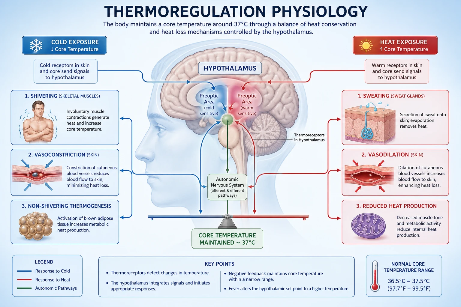
Thermoregulation — the integrated hypothalamic control of body temperature by balancing heat production against heat loss. HEAT PRODUCTION: basal metabolic rate (70-80% of resting heat), shivering thermogenesis (skeletal muscle — up to 5x BMR increase), non-shivering thermogenesis (brown adipose tissue / uncoupling protein 1 — dominant in neonates, reactivated in adult humans), voluntary exercise, and hormonal calorigenesis (thyroid hormone T3/T4 — slow genomic up-regulation of Na+/K+ ATPase; catecholamines — rapid beta-adrenergic stimulation of metabolism). HEAT LOSS: radiation 60% (infrared transfer to cooler surroundings — dominant at rest in a temperate room), convection 12% (air currents carry heat away — basis of forced-air cooling and wind chill), conduction 3% (direct contact — basis of cooling blankets and ice/water immersion), evaporation 25% (sweat 0.58 kcal/g water vaporised + respiratory water loss), and respiration (latent heat of vaporisation + warming of inspired gas). HYPOTHALAMIC CONTROL: the preoptic area of the anterior hypothalamus (PO/AH) houses the central thermoregulatory integrator — it is itself warm-sensitive, contains the defended set point (~37 degrees C core), and integrates central (blood) temperature with peripheral (skin/cold) afferent input to generate autonomic outputs (sweating, shivering, cutaneous vasodilation/vasoconstriction) and behavioural outputs (clothing, seeking shade/shelter). NORMAL RANGE: 36-37.5 degrees C core. RESPONSE TO COLD: peripheral vasoconstriction (sympathetic — reduces shell conductance) → shivering (skeletal muscle) → non-shivering thermogenesis (brown fat) → piloerection (trapped air layer) → TSH/catecholamine release (raise metabolic rate). RESPONSE TO HEAT: cutaneous vasodilation (active vasodilation via cholinergic/nitric oxide — opens arteriovenous anastomoses) → sweating (eccrine glands, sympathetic cholinergic) → behavioural cooling. TARGETED TEMPERATURE MANAGEMENT (TTM): controlled hypothermia 32-36 degrees C after cardiac arrest — reduces cerebral metabolic rate by 6-7% per degree C fall → reduces cerebral O2 demand, attenuates excitotoxicity, reperfusion injury, seizure activity and free-radical generation → neuroprotection. FEVER: prostaglandin E2 (PGE2) synthesised in the PO/AH (via COX-induced conversion of arachidonic acid, driven by endogenous pyrogens IL-1, IL-6, TNF-alpha acting on the organum vasculosum of the lamina terminalis) RAISES the hypothalamic set point → the patient feels cold → shivering + vasoconstriction raise core temperature to the new set point. Antipyretics (paracetamol, NSAIDs) work by BLOCKING PGE2 synthesis (COX inhibition). HYPERTHERMIA: the set point is NORMAL — body temperature exceeds the set point due to excessive external heat, exercise, or uncontrolled heat production (malignant hyperthermia, neuroleptic malignant syndrome, serotonin syndrome, heat stroke) — antipyretics are INEFFECTIVE because the set point is not elevated; treatment is active cooling. POIKILOTHERMIA: loss of hypothalamic thermoregulation — high spinal cord injury (loss of sympathetic outflow below the lesion), brain death — body temperature drifts passively towards ambient temperature.
On this page & tools
Your progress
Saved locally on this device.
7 MCQs with explanations
Target exams
Red flags
Overview
Normal body temperature — definitions and the defended range
The body is conceptualised as a core (deep tissues — brain, heart, viscera — held tightly at ~37 degrees C) and a shell (skin and superficial tissue, which varies widely and acts as the heat-exchange buffer). The defended normal core range is 36-37.5 degrees C, with a characteristic circadian rhythm of ~0.5-1.0 degrees C (nadir in early morning ~04:00-06:00, peak in late afternoon/evening ~16:00-18:00). This rhythm is the reason perioperative temperature thresholds and "fever" definitions must be interpreted against the time of day, and why night-shift ICU fevers may reflect the circadian nadir being exceeded rather than a new infective process. [1]
Core temperature is what the hypothalamus defends and what matters clinically; peripheral/shell temperature is a heat-exchange variable. Measurement sites approximate core to varying fidelity: [1]
Temperature measurement sites — fidelity to core temperature
| Site | Approximates core? | Pros | Cons / caveats |
|---|---|---|---|
| Pulmonary artery (thermistor) | Gold standard — true core (central blood) | Most accurate; reference for research | Invasive; requires a PA catheter |
| Distal oesophagus / nasopharynx | Excellent (close to great vessels and hypothalamic blood) | Tracks rapid change (CPB, TTM) | Oesophageal probe placement; affected by inspired gas if proximal |
| Tympanic membrane (infrared) | Good (shares blood supply — internal carotid → hypothalamus) | Fast, non-invasive | Cerumen, otitis, poor technique reduce accuracy; under-reads in shock |
| Bladder (thermistor catheter) | Good (urine is core-temperature) | Continuous; useful in ICU/TTM | Affected by urine flow rate; cold irrigant distorts |
| Rectal | Moderate (lags core by 30-60 min) | Accessible | Slow to change; faeces, peritoneal blood affect; NOT for rapid TTM titration |
| Axilla / oral / forehead | Poor | Cheap | Oral affected by hot/cold drinks, tachypnoea; axilla very variable — unsuitable for ICU accuracy |
Key point for exams: during TTM, use a fast-responding core surrogate (oesophageal, nasopharyngeal, bladder, or PA) — rectal lags too much to titrate cooling safely. During anaesthesia, the redistribution phase drops core temperature ~1-1.5 degrees C in the first 30-60 minutes as warm core blood is shunted to the periphery by vasodilation — the basis of inadvertent perioperative hypothermia.[4]
Heat production — the five mechanisms
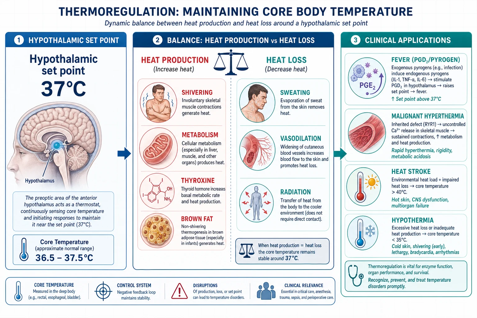
Resting heat production (~80 W at rest) comes overwhelmingly from basal metabolic rate (BMR): the viscera (liver, brain, heart, kidneys) and the obligatory activity of ion pumps (Na+/K+ ATPase consumes ~20-30% of resting ATP). When heat is lost faster than BMR replaces it, the hypothalamus recruits four additional calorigenic mechanisms: [1]
- Basal metabolic rate (BMR) — 70-80% of resting heat. Determined by thyroid hormone set-point, lean body mass, age and sex. This is the baseline over which the other mechanisms operate.
- Shivering thermogenesis — up to ~5x BMR. Involuntary, asynchronous, high-frequency (8-12 Hz) contractions of skeletal muscle (no net limb movement — agonists and antagonists co-contract). Driven by shivering-command neurons in the dorsomedial hypothalamus descending via the reticulospinal tract to anterior horn cells. It is metabolically expensive and limited by muscle glycogen and cardiovascular reserve — which is why uncontrolled shivering during TTM is harmful.
- Non-shivering thermogenesis (NST) — brown adipose tissue (BAT). BAT mitochondria express uncoupling protein 1 (UCP1 / thermogenin) in the inner membrane, which short-circuits the proton gradient so that oxidative phosphorylation dissipates energy as heat rather than driving ATP synthase. NST is the DOMINANT cold-defence in neonates (who cannot shiver effectively and have a large surface-area-to-mass ratio), and was historically thought to be absent in adult humans — modern 18-FDG PET confirms metabolically active BAT depots in the supraclavicular and paraspinal regions of adults, recruited by cold and sympathetic (beta-3 adrenergic) stimulation.
- Voluntary exercise. Skeletal muscle contraction can raise heat production 10-15x briefly; behaviourally recruited (stamping, moving) as a conscious cold response.
- Hormonal calorigenesis. Thyroid hormone (T3/T4) up-regulates Na+/K+ ATPase expression and mitochondrial uncoupling over hours-days (slow, genomic, sets the BMR baseline — hypothyroidism causes cold intolerance and hypothermia; hyperthyroidism causes heat intolerance). Catecholamines (noradrenaline/adrenaline) raise metabolic rate within minutes via beta-adrenergic stimulation of glycogenolysis, lipolysis and BAT. The thyroid-catecholamine synergy (thyroid hormone up-regulates beta-receptor density) explains the explosive hypermetabolism and hyperthermia of thyroid storm. [1]
Heat production mechanisms — relative magnitude and speed
| Mechanism | Magnitude (vs BMR) | Onset | Key physiology | Clinical relevance |
|---|---|---|---|---|
| BMR | 1.0x (baseline) | Constant | Viscera + Na+/K+ ATPase; set by thyroid | Hypothyroid → hypothermia; hyperthyroid → heat intolerance |
| Shivering | up to ~5x | Seconds-minutes | Skeletal muscle, reticulospinal-driven | Dangerous during TTM — must be suppressed |
| Non-shivering (BAT/UCP1) | ~up to 2-2.5x in neonates | Minutes | UCP1 uncouples oxidative phosphorylation | Neonatal cold stress; re-emergent in adults |
| Exercise | up to 10-15x (brief) | Seconds | Voluntary muscle contraction | Exertional hyperthermia / heat stroke |
| Hormonal (T3/T4) | raises BMR set-point | Hours-days (genomic) | Na+/K+ ATPase, beta-receptor density | Thyroid storm hypermetabolism; myxoedema coma hypothermia |
| Hormonal (catecholamines) | ~1.3-1.5x rapid | Minutes | Beta-adrenergic glycogenolysis/lipolysis | Phaeochromocytoma, NMS, serotonin syndrome heat |
Heat loss — the four physical mechanisms
Heat is lost from the body-shell interface to the environment by four physical processes. The proportions are for a resting, clothed person in a temperate room (~22 degrees C): [1]
Heat loss mechanisms — proportion and physiology
| Mechanism | Proportion | Physics | Amplified by | Clinical exploitation |
|---|---|---|---|---|
| Radiation | ~60% | Infrared electromagnetic transfer from warmer body to cooler surfaces — does NOT require contact or air movement; depends on the 4th-power temperature gradient | Cold surrounding surfaces (windows, walls) | Reflective blankets (space blanket) reduce radiative loss; radiant warmers add heat |
| Evaporation | ~25% | Latent heat of vaporisation — 0.58 kcal (2.4 kJ) lost per gram of water vaporised from skin (sweat) or respiratory tract | High ambient temperature (when radiation/convection reverse), low humidity, air movement | Wet sheet + fan cooling for heat stroke; alcohol sponging (faster evaporation) |
| Convection | ~12% | Heat transferred to air at the skin surface, then carried away by air currents; depends on temperature gradient and air velocity | Wind, forced air (Bair Hugger adds heat; fan removes it) | Forced-air cooling; the "wind chill" principle; intraoperative forced-air warming |
| Conduction | ~3% | Direct molecular transfer to a cooler object in contact; proportional to temperature gradient and thermal conductivity | Water (conducts ~25x better than air); metal | Cooling blankets, ice-water immersion for heat stroke, water mattress warming |
| Respiration | (part of evaporation + conduction) | Warming + humidifying inspired gas; vaporisation of water from airways | Cold/dry inspired gas, high minute volume | HME (heat-moisture exchanger) filters conserve this loss in ventilated patients |
Critical exam numerics: radiation ~60%, evaporation ~25%, convection ~12%, conduction ~3%. Note that as ambient temperature rises towards body temperature, radiation/convection/conduction all fall toward zero and then reverse (the environment heats the body), so evaporation (sweating) becomes the ONLY effective heat-loss mechanism when ambient temperature exceeds skin temperature — this is why high ambient humidity (which blocks evaporation) is so dangerous, and why heat stroke is an environmental failure of evaporative loss.[1]
Hypothalamic control — the integrator and the set point
The thermoregulatory system is a classical negative-feedback control loop with a defended set point. The central integrator is the preoptic area of the anterior hypothalamus (PO/AH). [1]
Afferent (input) limb:
- Central (core) thermoreceptors: warm-sensitive neurons in the PO/AH itself fire faster when local brain/blood temperature rises. This is the primary feedback signal — the hypothalamus senses its own temperature.
- Peripheral thermoreceptors: cold and warm receptors in the skin (and some in viscals/spinal cord) provide feed-forward information about the external thermal environment, allowing the hypothalamus to begin defending core temperature BEFORE it changes — this is why stepping into a cold room triggers vasoconstriction almost instantly. [1]
Integrator (the PO/AH): warm-sensitive PO/AH neurons compare the integrated thermal signal with the defended set point. The set point is itself dynamic — it is raised by PGE2 (fever) and by the circadian rhythm, and is influenced by ovarian hormones (luteal-phase ~0.3-0.5 degrees C rise). Additional hypothalamic nodes now recognised include the dorsomedial hypothalamus (DMH), which relays the shivering command, and the raphe pallidus in the medulla, the final common sympathetic output pathway to BAT and skin vasculature.[3]
Efferent (output) limb — two channels: [1]
Thermoregulatory control loop — step by step
- SENSE: warm-sensitive neurons in the preoptic/anterior hypothalamus detect core (blood/brain) temperature; skin cold/warm receptors detect the ambient challenge. Signals travel via the spinothalamic tract and trigeminal afferents to the thalamus and PO/AH.
- INTEGRATE: the PO/AH fuses the central and peripheral signals against the defended set point (~37 degrees C). The magnitude of the error signal (set point minus actual) determines response intensity.
- DECIDE: if core temperature is BELOW set point (cold challenge), heat-conservation and heat-production pathways are recruited in order of metabolic cost; if ABOVE set point (heat challenge), heat-loss pathways are recruited.
- EFFERENT — autonomic (involuntary): sympathetic outflow → (a) skin blood flow via arteriovenous anastomoses of the hands/feet/ears (constriction to conserve heat, dilation to lose heat); (b) sweat glands (eccrine, sympathetic cholinergic); (c) brown adipose tissue (sympathetic beta-3); (d) shivering via reticulospinal tract to anterior horn cells.
- EFFERENT — behavioural (voluntary): the cortex is engaged (clothing, seeking shade/shelter/warmth, altering activity) — in conscious humans this is the MOST powerful defence and is impaired by sedation, anaesthesia, dementia and brain injury, which is why these patients rely entirely on autonomic (and external) control.
- EFFECT: core temperature returns towards set point; the error signal shrinks; the response is gated off (negative feedback).
Cold response vs heat response — the autonomic recruitment ladder
| Response to COLD (recruited in order, cheapest first) | Response to HEAT (recruited in order) |
|---|---|
| 1. Behavioural (clothing, shelter) | 1. Behavioural (shade, rest, fanning) |
| 2. Cutaneous vasoconstriction (sympathetic — closes AV anastomoses; reduces shell conductance; can cut heat loss ~50%) | 2. Cutaneous vasodilation (active vasodilation — cholinergic co-transmission + nitric oxide; opens AV anastomoses; skin blood flow can rise from ~0.5 to ~7 L/min) |
| 3. Non-shivering thermogenesis (BAT/UCP1) | 3. Sweating (eccrine glands, sympathetic cholinergic; up to ~1-2 L/h acutely; heat loss up to ~10x BMR) |
| 4. Shivering (skeletal muscle, up to 5x BMR — costly) | 4. (No further autonomic reserve — decompensation = heat stroke) |
| 5. Hormonal (TSH/thyroid up-regulation, catecholamines — chronic) | |
| 6. Piloerection (vestigial in humans — trapped air layer in fur) |
Fever vs hyperthermia — the pivotal exam distinction
This single distinction is examined relentlessly because it changes management. Both present with a high body temperature, but the underlying pathophysiology is opposite. [1]
Fever — the set point is RAISED
In fever, exogenous pyrogens (bacterial endotoxin/LPS, viral particles) and endogenous pyrogens (IL-1, IL-6, TNF-alpha) act on the organum vasculosum of the lamina terminalis (OVLT) — a circumventricular organ with a permeable blood-brain barrier. This activates phospholipase A2 and cyclo-oxygenase (COX) to convert arachidonic acid to prostaglandin E2 (PGE2) in the PO/AH. PGE2 raises the defended set point (e.g., from 37 to 39 degrees C). The hypothalamus now perceives the body as too COLD relative to the new set point and mounts a cold response: the patient feels cold and shivers ("rigors"), and cuts heat loss by vasoconstriction (pale, cold peripheries), until core temperature climbs to the new set point. Once reached, the patient's temperature plateaus and the peripheries vasodilate ("the fever breaks"). When the pyrogen signal resolves (or an antipyretic is given), PGE2 falls, the set point drops back to 37, and the patient now feels hot and sweats/vasodilates to cool to the new (normal) set point. [1]
Antipyretics work by blocking PGE2 synthesis. Paracetamol (central COX inhibition / putative COX-3), aspirin and NSAIDs (ibuprofen, diclofenac) all reduce PGE2 → lower the set point → the hypothalamus drives heat-loss responses. Antipyretics therefore ONLY work when the set point is elevated (fever) — they have NO effect when the set point is normal (hyperthermia). [1]
Hyperthermia — the set point is NORMAL
In hyperthermia, the set point remains at ~37 degrees C but heat load or heat production exceeds the maximum capacity for heat loss. The hypothalamus is already driving maximal heat-loss responses (sweating, vasodilation) but is overwhelmed. Causes include:
- Environmental: heat stroke (classic or exertional) — failed evaporative loss at high ambient temperature/humidity.
- Uncontrolled endogenous heat production: malignant hyperthermia (ryanodine receptor → uncontrolled SR Ca2+ release → sustained muscle contraction), neuroleptic malignant syndrome (D2 blockade), serotonin syndrome, thyroid storm, status epilepticus, salicylate overdose (uncoupling of oxidative phosphorylation).
- Impaired heat loss: anticholinergic toxicity (blocks sweating), antihistamines, severe skin disease (burns). [1]
Antipyretics are INEFFECTIVE in hyperthermia because the set point is not elevated. Treatment is active cooling (evaporative, ice-water immersion, intravascular cooling, cold IV fluids) plus, where relevant, specific antidotes (dantrolene for malignant hyperthermia; bromocriptine/dantrolene and cooling for NMS; cyproheptadine for serotonin syndrome). [1]
Fever vs hyperthermia — the decision table
| Feature | FEVER | HYPERTHERMIA |
|---|---|---|
| Hypothalamic set point | RAISED (by PGE2) | NORMAL |
| Mediator | PGE2 (via COX, driven by IL-1/IL-6/TNF-alpha on OVLT) | None (set point intact) |
| Patient feels | Cold (during temperature rise) — rigors, pale peripheries | Hot, flushed, sweating (maximal heat-loss attempt) |
| Skin | Cold, pale, constricted during rise | Hot, flushed, vasodilated |
| Response to antipyretics | YES (block PGE2 → lower set point) | NO (set point not elevated) |
| Rate of rise | Gradual, plateau at new set point | Can be explosive (MH: 1 degrees C/5 min) |
| Treatment | Treat cause + antipyretics | ACTIVE COOLING + specific antidote + treat cause |
| Examples | Sepsis, infection, inflammation, malignancy, drug fever (most) | Heat stroke, MH, NMS, serotonin syndrome, thyroid storm, salicylate, NMS, exertional |
Targeted temperature management (TTM) — applied hypothermia
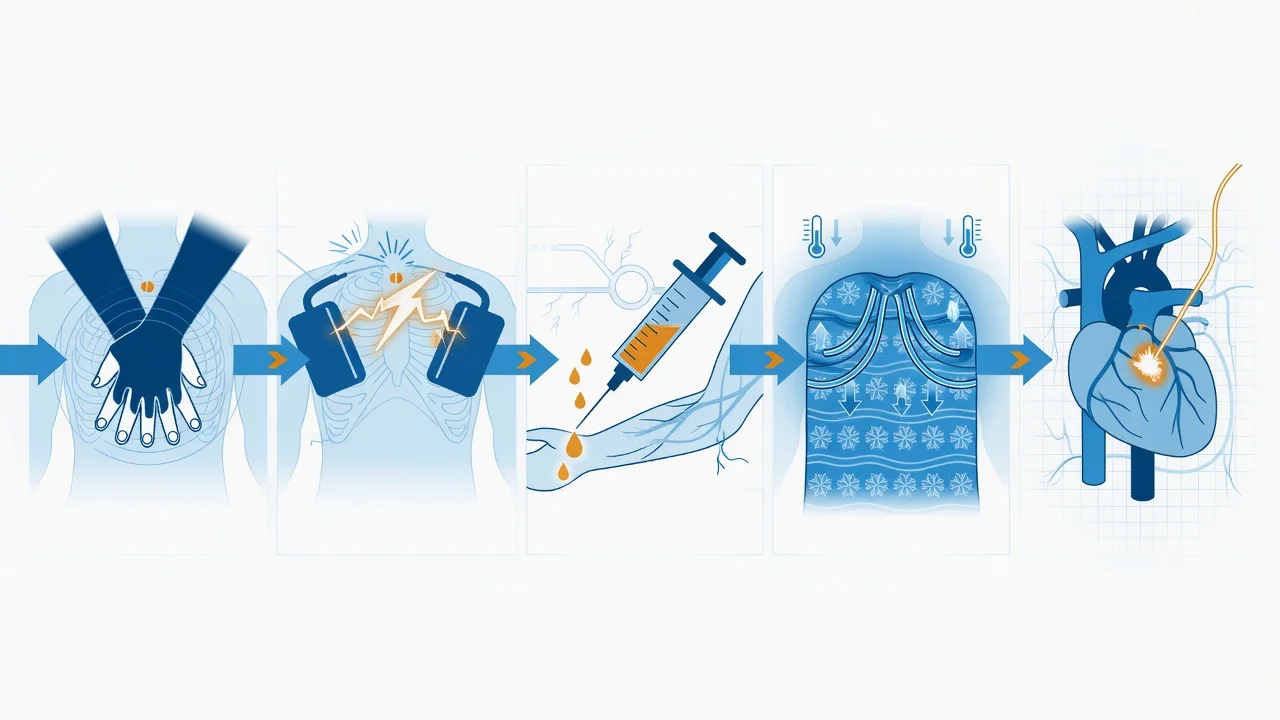
After cardiac arrest, controlled mild hypothermia (now termed targeted temperature management) improves neurological outcome. Current practice (ILCOR) targets 32-36 degrees C for at least 24 hours in comatose adults after witnessed out-of-hospital cardiac arrest with a shockable rhythm, and is considered for other arrests. The physiological rationale rests on the van 't Hoff / Q10 effect on metabolism. [1]
Mechanism of neuroprotection
Lowering temperature reduces the rate of all temperature-dependent biochemical reactions. The clinically taught figure is that cerebral metabolic rate falls by approximately 6-7% for every 1 degree C reduction in temperature — so cooling from 37 to 33 degrees C (a 4 degrees C drop) reduces cerebral oxygen consumption by roughly 25-30%, buying time for the injured, energy-depleted brain during the vulnerable early reperfusion window. Beyond simple metabolic suppression, hypothermia also: [1]
Mechanisms of TTM neuroprotection — step by step
- Reduced cerebral metabolic rate / O2 demand — 6-7% per degree C; the injured brain with impaired mitochondrial ATP production is protected against demand ischaemia.
- Reduced reperfusion injury — suppression of the injurious cascade triggered by reperfusion (free-radical / reactive oxygen species generation, lipid peroxidation, blood-brain barrier disruption, vasogenic oedema).
- Reduced excitotoxicity — lower glutamate release and NMDA-mediated calcium influx, reducing neuronal apoptosis and necrosis.
- Reduced seizure activity — hypothermia raises seizure threshold; post-arrest seizures independently worsen outcome and consume cerebral O2.
- Reduced intracranial pressure — reduced cerebral blood volume and oedema; hypothermia is an ICP-lowering therapy in its own right.
- Suppressed inflammation / apoptosis — modulation of the post-arrest inflammatory (SIRS-like) response and caspase-mediated programmed cell death.
The hazard of shivering during TTM
Because the PO/AH set point is normal (~37 degrees C) in a post-arrest patient, cooling to 33 degrees C creates a 4-degree error signal that the hypothalamus fights with its most powerful weapon: shivering, which can raise metabolic rate up to 5x and cerebral O2 consumption dramatically — directly opposing the neuroprotective goal. Shivering must therefore be suppressed: sedation, opiates (especially meperidine via central shivering-receptor action), buspirone, skin-surface warming (counter-warming of face/limbs reduces the peripheral cold afferent), magnesium, and — if refractory — neuromuscular blockade with continuous EEG monitoring (since the paralysed, cooled patient cannot be clinically assessed for seizures). The Bedside Shivering Assessment Scale (BSAS) is used to titrate this.[5][6]
Complications of TTM
Hypothermia is not benign — every degree of cooling carries predictable physiological effects that must be anticipated and managed: [1]
Predictable physiological effects of TTM (hypothermia 32-36 degrees C)
| System | Effect of hypothermia | Management implication |
|---|---|---|
| Cardiovascular | Initial hypertension/tachycardia (shivering, sympathetic surge); then bradycardia (slowed SA node depolarisation), prolonged QT → torsades risk, arrhythmia (VF below ~30 degrees C) | Continuous cardiac monitoring; correct K+/Mg2+; rewarm if unstable arrhythmia |
| Coagulation | Reversible platelet dysfunction + impaired clotting factor activity → bleeding tendency | Monitor for occult bleeding; avoid routine deep procedures if possible |
| Metabolic/enzyme | Reduced insulin secretion + sensitivity → hyperglycaemia; cold-induced diuresis → hypokalaemia, hypomagnesaemia, hypophosphataemia on rewarming (intracellular shift reverses → rebound hyperkalaemia) | Tight glucose control; check electrolytes q4-6h; anticipate K+ shifts during rewarming |
| Immune | Leucocyte dysfunction → infection risk (especially pneumonia, line sepsis) | Surveillance cultures; strict line care; early de-escalation |
| Pharmacokinetics | Reduced hepatic and renal clearance → drug accumulation (sedatives, analgesics, neuromuscular blockers, vasopressors) prolong their effect | Dose reduction; monitor depth of sedation; prolonged recovery |
| Rewarming | Must be SLOW and controlled (~0.25-0.5 degrees C/h) to avoid rebound cerebral oedema, hypotension (vasodilation), electrolyte shift and seizures | Controlled rewarming protocol; continue sedation through rewarming |
Poikilothermia — loss of the hypothalamic loop
Poikilothermia is the loss of active thermoregulation such that body temperature drifts passively towards ambient (like a reptile). It occurs when the hypothalamic control loop is interrupted at any point: [1]
- High (cervical) spinal cord injury: the hypothalamus (PO/AH) is intact and senses temperature, but the descending sympathetic and somatic efferents cannot reach the heart, vasculature, brown fat or skeletal muscle below the lesion. Cutaneous vasomotor control and shivering (below the lesion) are lost; the patient becomes poikilothermic, drifting towards room temperature — hypothermic in a cold ICU, hyperthermic in a warm room. Limited residual control above the lesion (facial/suprapubic sweating, arm shivering) persists.
- Brain death: destruction of the PO/AH integrator itself removes the central controller; the body becomes poikilothermic. This is one of the clinical observations supporting the diagnosis and a reason organ donors require active warming to maintain a temperature compatible with organ viability.
- Severe hypothermia itself: below ~30 degrees C, the hypothalamic integrator ceases to function effectively and the patient becomes poikilothermic — a key reason not to declare death in severe hypothermia ("not dead until warm and dead").
- Anaesthesia and deep sedation: abolish the BEHAVIOURAL response and blunt the autonomic response, producing functional poikilothermia — the physiological basis of inadvertent perioperative hypothermia and the reason anaesthetised ICU patients need active warming.[4]
Exam practice — SAQs
SAQ — Thermoregulation in sepsis: fever, antipyresis and the hypothermic patient
10 minutes · 10 marks
A 72-year-old woman is admitted to ICU with urosepsis and Escherichia coli bacteraemia in septic shock: T 39.4 degrees C, HR 128, BP 82/48 on noradrenaline 0.3 mcg/kg/min (MAP 62 after 30 mL/kg crystalloid), RR 30, SpO2 94 per cent on FiO2 0.5, lactate 4.2. Six hours in, despite 1 g IV paracetamol, her temperature is 39.1 degrees C and the nurse asks whether she should be actively cooled. On the same round, a second patient — a 58-year-old man with pneumococcal pneumonia of comparable severity — is hypothermic at 34.8 degrees C.
Clinical pearls
[1]Red flags
Key trials and evidence
Hypothermia after Cardiac Arrest Study Group, 2002 — NEJM (PMID 11856793)
Design
Multicentre RCT; 273 comatose survivors of witnessed out-of-hospital VF cardiac arrest. Mild therapeutic hypothermia (32-34 degrees C for 24 h) vs standard normothermia.
Primary outcome
Favourable neurological outcome (CPC 1-2) at 6 months.
Key result
Favourable outcome 55% (hypothermia) vs 39% (normothermia) — number needed to treat ~6. Mortality 41% vs 55% (NNT ~7 for survival).
Significance
Landmark RCT (with Bernard 2002) establishing therapeutic hypothermia as standard after cardiac arrest — the clinical proof of the metabolic-suppression / neuroprotection concept (6-7% per degree C).
Clinical bottom line
Mild hypothermia after VF cardiac arrest roughly doubles the chance of good neurological recovery — the evidence base for modern TTM (32-36 degrees C).
Arrich et al, 2016 — Cochrane (PMID 26878327)
Design
Systematic review and meta-analysis of randomised controlled trials of hypothermia (any method) for neuroprotection in adults after cardiopulmonary resuscitation.
Key finding
Hypothermia improved survival to hospital discharge and favourable neurological outcome vs no hypothermia; the magnitude of benefit narrowed as trial quality and standardisation of post-arrest care improved. No clear difference between 33 and 36 degrees C targets (consistent with the TTM trial).
Significance
The definitive Cochrane synthesis — supports TTM as beneficial while demonstrating that the precise target (33 vs 36 degrees C) is less important than fever prevention and protocolised care.
Clinical bottom line
TTM is beneficial after cardiac arrest; both 33 and 36 degrees C are defensible targets, provided fever/hyperthermia is actively prevented and shivering suppressed.
Bouchama & Knochel, 2002 — NEJM (PMID 12075060)
Type
Definitive clinical review of heat stroke — the pathophysiology and management reference.
Key concepts
Heat stroke = core temperature >40 degrees C with CNS dysfunction, from failed thermoregulation (set point normal — antipyretics ineffective). Classic (passive environmental) vs exertional (exercise-driven heat production). Cooling must be rapid: ice-water immersion (exertional) or evaporative (classic); delay is the main determinant of mortality.
Clinical bottom line
The archetype of hyperthermia with a normal set point — distinguished from fever, treated by active cooling, and the basis for understanding all the antipyretic-resistant hyperthermic syndromes in ICU.
Prognosis
Prognosis in thermoregulatory disorders is dictated by the underlying cause, the magnitude and DURATION of temperature deviation, and the speed of correction: [1]
- Heat stroke: mortality correlates directly with the duration and peak of hyperthermia — cooling delayed beyond ~30 minutes carries mortality up to 50-80%; rapid ice-water immersion (exertional) drops mortality to <5%. End-organ injury (AKI, liver failure, DIC, rhabdomyolysis, ARDS) drives outcome.[1]
- Malignant hyperthermia: untreated mortality ~70-90%; with prompt dantrolene and trigger cessation, mortality is now <5%. The earlier dantrolene is given, the better the outcome.[1]
- Targeted temperature management after cardiac arrest: TTM roughly doubles the odds of good neurological outcome; the HACA and Bernard RCTs gave an NNT of ~6 for favourable outcome. Benefit is greatest in shockable-rhythm out-of-hospital arrest; the precise target (33 vs 36 degrees C) is less important than fever prevention and protocolised post-arrest care.[5][6]
- Poikilothermia (spinal cord injury / brain death): temperature itself is a manage-and-control problem rather than a prognostic marker — outcome is determined by the underlying neurological injury. In organ donors, active warming to maintain normothermia preserves graft viability.
- Severe accidental hypothermia: governed by the Swiss staging (I — conscious, shivering; II — impaired consciousness, no shivering; III — unconscious, vital signs present; IV — no vital signs, apparent death) and the principle of "not dead until warm and dead" — survival with good neurological outcome is possible after prolonged cardiac arrest if the patient is rewarmed with extracorporeal life support before pronouncement.
Key thermoregulation outcomes
References
- [1]Bouchama A, Knochel JP Heat stroke N Engl J Med, 2002.PMID 12075060
- [2]Romanovsky AA Thermoregulation: some concepts have changed. Functional architecture of the thermoregulatory system Am J Physiol Regul Integr Comp Physiol, 2007.PMID 17008453
- [3]DiMicco JA, Zaretsky DV The dorsomedial hypothalamus: a new player in thermoregulation Am J Physiol Regul Integr Comp Physiol, 2007.PMID 16959861
- [4]Lenhardt R The effect of anesthesia on body temperature control Front Biosci (Schol Ed), 2010.PMID 20515846
- [5]Hypothermia after Cardiac Arrest Study Group Mild therapeutic hypothermia to improve the neurologic outcome after cardiac arrest N Engl J Med, 2002.PMID 11856793
- [6]Arrich J, Holzer M, Havel C, Mullner M, Herkner H Hypothermia for neuroprotection in adults after cardiopulmonary resuscitation Cochrane Database Syst Rev, 2016.PMID 26878327