ICU · Gastroenterology
Acute diverticulitis in ICU: complicated presentations
Also known as Diverticulitis · Complicated diverticulitis · Diverticular perforation · Diverticular abscess · Hinchey classification
Diverticular disease: diverticula (mucosal herniations through colonic wall) ± inflammation. Acute diverticulitis: inflammation of diverticula. ICU-relevant complicated diverticulitis (Hinchey III-IV): peritonitis, perforation, abscess, fistula, obstruction. Hinchey classification: I — pericolic abscess, II — distant abscess, III — purulent peritonitis, IV — faeculent peritonitis. Management: Hinchey I-II (abscess) — antibiotics + percutaneous drainage. Hinchey III (purulent peritonitis) — laparoscopic lavage + drainage (controversial) or Hartmann's. Hinchey IV (faeculent peritonitis) — Hartmann's procedure (sigmoid colectomy + end colostomy).
On this page & tools
Your progress
Saved locally on this device.
Target exams
Red flags
Pathophysiology
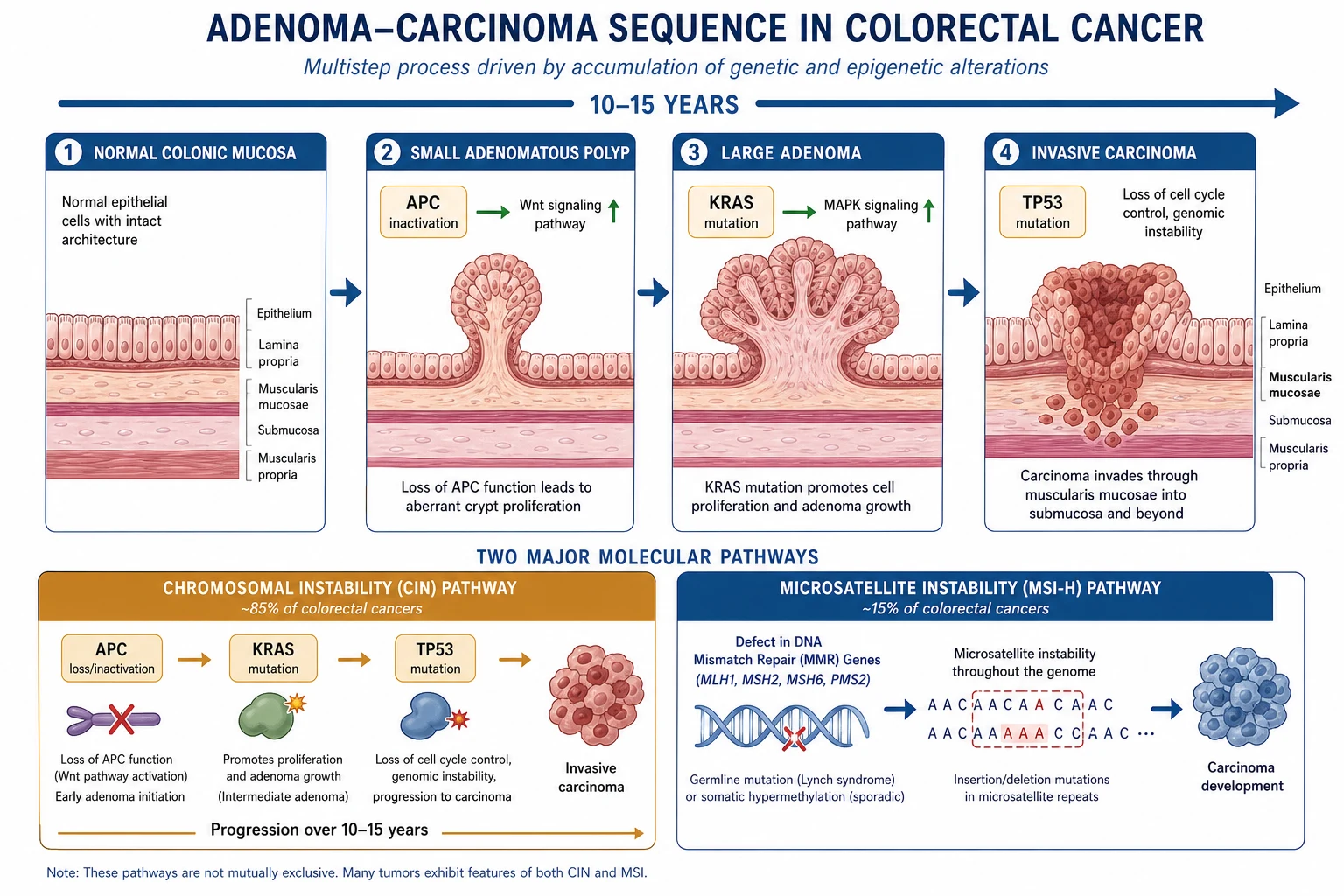
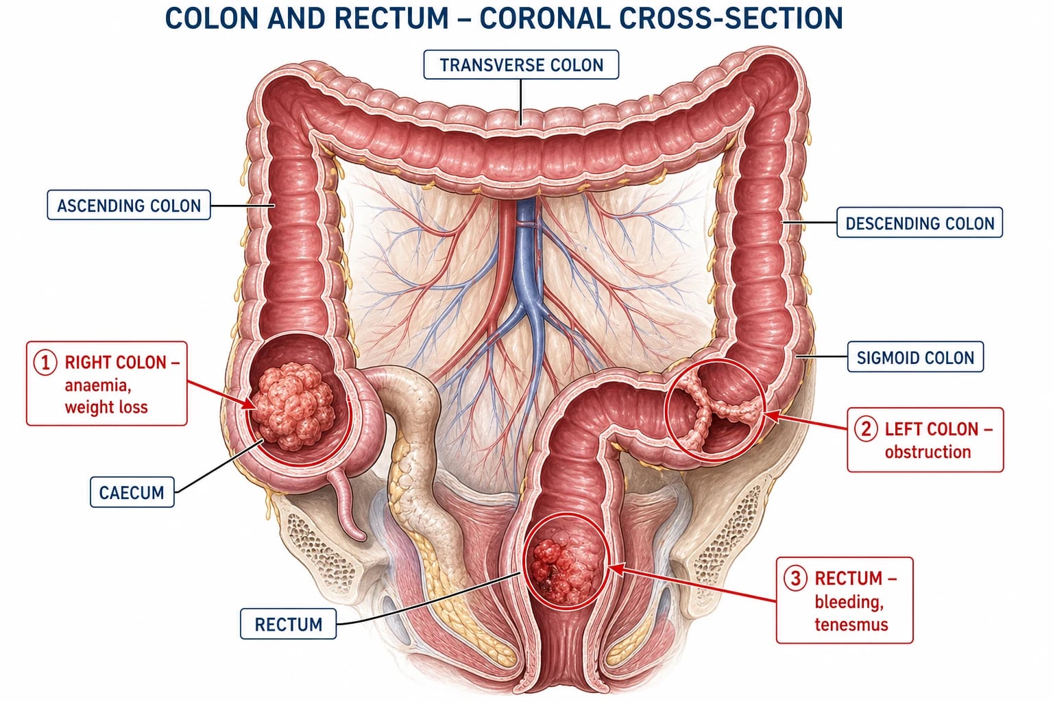
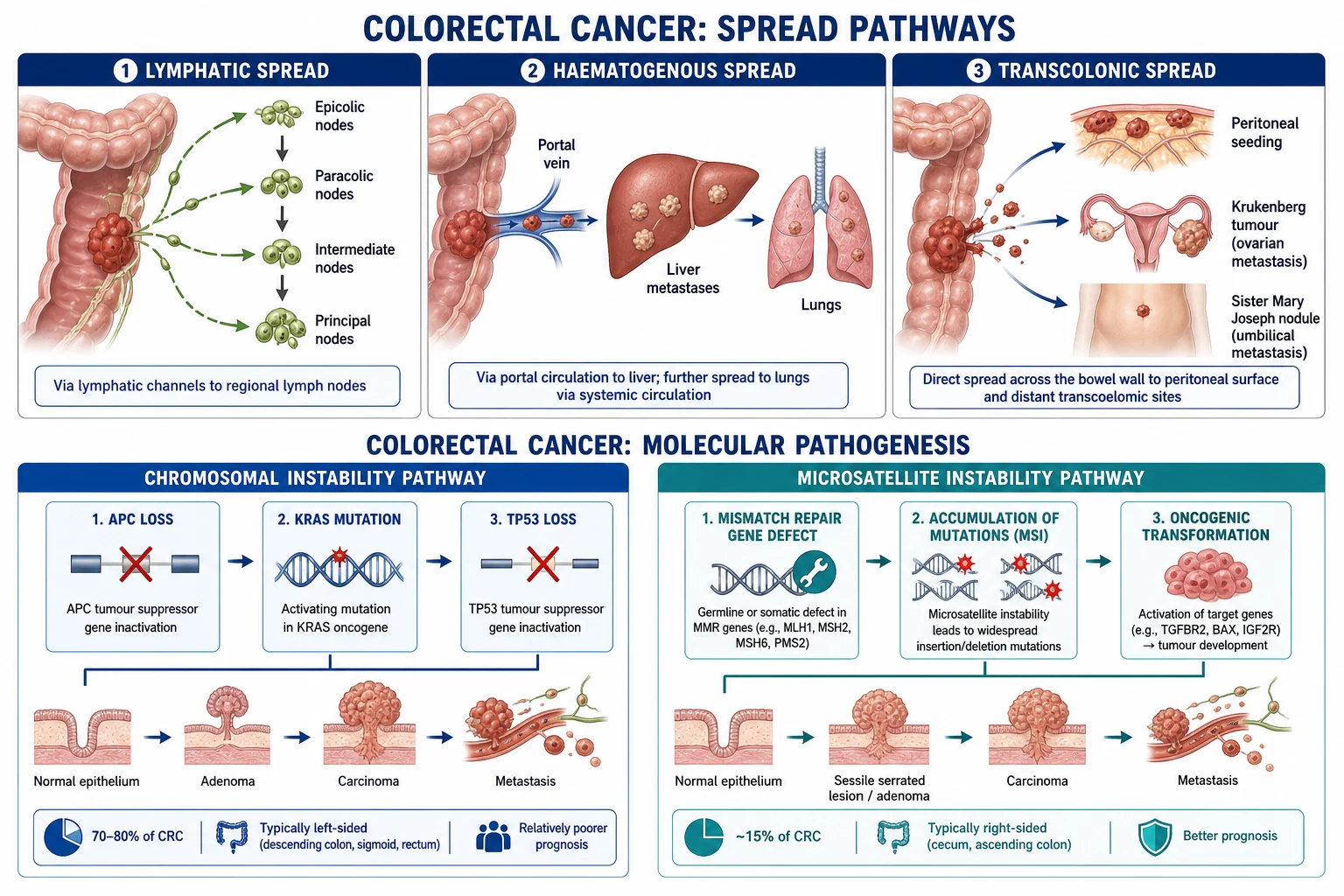
Diverticular anatomy and pathophysiology — the colonic diverticulum
| Structure | Detail |
|---|---|
| Definition | A pseudo-diverticulum — mucosa + submucosa herniate through the muscularis propria at points of vessel penetration (where vasa recta pierce the circular muscle → weak point in the wall). Unlike true diverticula (e.g., Meckel's), the muscular layer is NOT part of the out-pouching. |
| Site | Sigmoid colon (95%) — highest luminal pressure (Laplace's law: P ∝ T/r, narrowest radius → highest pressure). Right-sided disease more common in Asians (~70%); left-sided dominant in Western populations. |
| Risk factors | Low dietary fibre, age >50 (50% by age 60, 70% by age 80), obesity (raises intra-abdominal pressure), sedentary lifestyle, smoking, NSAIDs/opioids, family history, immunosuppression. |
| Trigger for inflammation | Inspissated faecalith or food particle obstructs the narrow diverticular neck → stasis → bacterial overgrowth → micro-perforation of the thin-walled diverticulum (mucosa + submucosa only) → pericolic fat inflammation / phlegmon → abscess formation. |
| Macro-perforation | If inflammation progresses, the diverticulum perforates freely into the peritoneum: purulent peritonitis (pus only — Hinchey III) or faeculent peritonitis (faeces — Hinchey IV, the catastrophic form). |
| Distinguishing bleeding vs inflammation | Diverticular bleeding arises from the vasa recta at the diverticular neck (an arterial source — painless, brisk, maroon). Diverticulitis arises from obstruction/micro-perforation (painful, fever, leukocytosis). Both CAN coexist, but rarely. |
Clinical presentation
Clinical features — what to look for at the bedside
| Symptom / sign | Detail | Pearls |
|---|---|---|
| Abdominal pain | Left lower quadrant (LLQ) in 70-93% (sigmoid dominant). May radiate to back. Diffuse in advanced/generalised peritonitis. | Right-sided diverticulitis (Asian populations, ~5% Western) → RUQ/RLQ pain — often mistaken for appendicitis. |
| Fever | 57-100%. Low-grade in uncomplicated, high-grade rigors in complicated/septicaemia. | Afebrile + unwell immunosuppressed patient → do NOT be reassured; image aggressively. |
| Leucocytosis | 69-83%. Left shift on differential common. | Normal WCC does NOT exclude diverticulitis; CRP/procalcitonin more sensitive. |
| Bowel habit change | Constipation (~50%) or diarrhoea (~25-35%). May mimic gastroenteritis or obstruction. | Obstruction features (distension, absolute constipation, vomiting) → suspect stricture or volvulus. |
| Nausea / vomiting | 20-60% — usually mild; prominent vomiting suggests obstruction or generalised peritonitis. | Ileus in Hinchey III-IV → ER/NGT decompression. |
| Dysuria / urinary frequency | Bladder irritation from adjacent pelvic abscess or colovesical fistula. | Recurrent UTIs + pneumaturia/faecaluria → colovesical fistula until proven otherwise. |
| Peritonism on exam | Localised guarding/rebound in LLQ (Hinchey I-II). Generalised rigidity (Hinchey III-IV) — surgical emergency. | P.R. exam may reveal tender mass, blistering, or boggy fullness in pouch of Douglas (pelvic abscess). |
| Vaginal discharge | Colovaginal fistula — faecal/purulent vaginal discharge. | Suspect in women with recurrent vaginitis and known diverticular disease. |
Differential diagnosis of suspected acute diverticulitis
| Mimic | Key distinguishing features |
|---|---|
| Acute appendicitis | Migration of pain (periumbilical → RIF); McBurney's point tenderness. Caecal diverticulitis or rarely situs inversus can mimic. |
| Colorectal cancer | Insidious, weight loss, anaemia, altered bowel habit, mass. Always exclude cancer in chronic/recurrent disease — colonoscopy 6 weeks after acute episode. |
| Ischaemic colitis | Bloody diarrhoea, "pain out of proportion," vascular disease, AF; left-sided with thumbprinting on imaging. |
| Inflammatory bowel disease | Chronic diarrhoea, blood/mucus, weight loss, extra-intestinal manifestations; younger patients. |
| Ovarian pathology / PID / ectopic | RLQ/LLQ pain in women — pregnancy test mandatory; pelvic ultrasound. |
| Urinary colic / pyelonephritis | Loin-to-groin pain, haematuria; sterile pyuria from adjacent inflammation. |
| Aortic aneurysm leak / aortic dissection | Tearing back pain, hypotension, palpable pulsatile mass. |
| Mesenteric adenitis / GORD / epiploic appendagitis | Self-limiting; CT differentiates (epiploic appendagitis = fat-density inflamed appendage with central vessel). |
Hinchey classification of perforated diverticulitis
| Stage | Finding | Management |
|---|---|---|
| I | Pericolic abscess / phlegmon | Antibiotics ± percutaneous drainage |
| II | Distant (pelvic/abdominal) abscess | Antibiotics + percutaneous drainage; surgery if fails |
| III | Generalised purulent peritonitis (pus) | Laparoscopic lavage (controversial) OR Hartmann's/OR primary anastomosis |
| IV | Generalised faeculent peritonitis (faeces) | Hartmann's procedure (sigmoid colectomy + end colostomy) |
Mortality: I-II ~5% · III ~10-15% · IV ~20-30%
Diagnosis and imaging
CT findings of acute diverticulitis (modality of choice)
| Finding | Detail | Significance |
|---|---|---|
| Bowel wall thickening | ≥4 mm (often ≥5 mm), segmental | Most sensitive sign (~95%). Localises disease to a colonic segment. |
| Fat stranding | Pericolic hazy inflammatory change | Earliest/most common finding. Indicates peri-diverticular inflammation. |
| Diverticula visible | Air-filled outpouchings ± thickened wall | Confirms underlying diverticular disease. Often multiple. |
| Pericolic fluid / phlegmon | Inflammatory mass without drainable pus | Hinchey I (early). Treat with antibiotics, no drainage. |
| Abscess | Rim-enhancing fluid collection with gas/fluid level | Hinchey I (pericolic) or II (distant). Drain if >4-5 cm. |
| Free intra-peritoneal gas | Extra-luminal air — often distant (e.g., falciform ligament sign) | Indicates perforation. Localised → sealed; generalized → free perforation (Hinchey III-IV). |
| Free fluid | Generalised intra-peritoneal fluid | Suggests peritonitis. Non-specific. |
| Extraluminal contrast / faeces | Contrast extravasation or faecal material outside bowel | Free perforation with ongoing leak — emergency. |
Sensitivity/specificity of CT: ~95% / ~95% for acute diverticulitis. IV + oral/rectal contrast improves sensitivity (rectal contrast can demonstrate active leak). In unstable patients proceed straight to laparotomy without CT.[4]
Modified Hinchey classification (Wasvary / Ambrosetti CT-based staging)
| Stage | CT finding | Management |
|---|---|---|
| 0 | Mild clinical diverticulitis, no CT abnormality (or only diverticula) | Antibiotics, often outpatient |
| Ia | Pericolic inflammation / phlegmon — confined to bowel wall | Antibiotics ± observation |
| Ib | Pericolic abscess | Antibiotics ± percutaneous drainage |
| II | Pelvic, distant intra-abdominal, or retroperitoneal abscess | Antibiotics + percutaneous drainage; surgery if fails |
| III | Generalised purulent peritonitis | Laparotomy: Hartmann's or primary anastomosis ± ileostomy |
| IV | Generalised faeculent peritonitis | Emergency laparotomy: Hartmann's (rarely primary anastomosis) |
Antibiotic strategy
Empiric antibiotic regimens in acute diverticulitis by severity
| Severity | Organism cover | Regimen (typical) |
|---|---|---|
| Uncomplicated (Hinchey 0-Ia) | Gram-negative rods + anaerobes | Oral options: co-amoxiclav 625 mg PO TDS alone, OR ciprofloxacin 500 mg PO BD + metronidazole 400 mg PO TDS. 4 days (some evidence observational management without antibiotics in selected low-risk patients). |
| Uncomplicated, admitted (nausea/sepsis) | Same | IV: co-amoxiclav 1.2 g IV TDS, OR ceftriaxone 2 g IV OD + metronidazole 500 mg IV TDS, OR ciprofloxacin + metronidazole. |
| Complicated (Hinchey Ib-II, abscess) | Gram-neg + anaerobes + streptococci | IV piperacillin/tazobactam 4.5 g IV TDS-QDS, OR ceftriaxone + metronidazole. Add drainage if abscess >4-5 cm. |
| Severe / septic shock / Hinchey III-IV | Gram-neg + anaerobes + Enterococcus ± ESBL | IV piperacillin/tazobactam OR meropenem 1 g IV TDS ± metronidazole. Add vancomycin if MRSA/healthcare-associated. Add echinocandin (caspofungin) if Candida risk (prolonged ICU, TPN, post-op, immunosuppression). |
| Duration | — | 4 days after adequate source control (STOP-IT-equivalent). 7-14 days if no source control, bacteraemia, or immunosuppression. |
Surgical approach: Hartmann's vs primary anastomosis
Emergency surgery for Hinchey III-IV — Hartmann's vs primary anastomosis
| Feature | Hartmann's procedure | Primary anastomosis ± defunctioning ileostomy |
|---|---|---|
| Operation | Sigmoid colectomy + end colostomy + rectal stump closure | Sigmoid colectomy + colorectal/colocolic anastomosis ± loop ileostomy |
| Indication | Hinchey IV (faeculent), haemodynamically unstable, unfavourable bowel (oedema, ischaemia, gross contamination), high anaesthetic risk | Hinchey III (purulent), haemodynamically stable, favourable bowel, experienced surgeon, selected Hinchey IV |
| Anastomotic leak risk | None (no anastomosis) | 4-10% (higher in emergency + peritonitis) |
| Stoma at discharge | 100% have colostomy | Variable — temporary ileostomy often reversed within months |
| Reversal rate | Only ~50% ever reversed (high operative risk) | Ileostomy reversal typically ~6 months, >80% reversed |
| Reoperation for reversal | Major laparotomy, high morbidity | Smaller procedure |
| Mortality | 5-15% (Hinchey IV ~20-30%) | Comparable to Hartmann's in selected patients |
| Evidence | Traditional gold standard for perforation | Oberkofler 2012 RCT + meta-analyses: non-inferior to Hartmann's, more reversals, no excess morbidity. Becoming standard for stable Hinchey III. |
Diverticular abscess — management by size
| Abscess size | First-line | Failure / deterioration |
|---|---|---|
| <2 cm | Antibiotics alone (oral) | Re-image if not improved in 48-72h |
| 2-4 cm | IV antibiotics ± percutaneous drainage (drain if accessible) | Drainage if persistent fever / rising CRP / rising WCC |
| >4 cm (Hinchey Ib) | IV antibiotics + percutaneous drainage (PCD) — ~80% success | Surgery (Hartmann's or primary anastomosis) if PCD fails, multi-loculated, or deterioration |
| >5 cm or distant (Hinchey II) | IV antibiotics + PCD + surgical review (likely surgery) | Surgery if source control inadequate |
Management of complicated diverticulitis with sepsis
- Resuscitate — ABC. IV fluids (crystalloid 30 mL/kg bolus for sepsis). Broad-spectrum antibiotics within 1h (piperacillin/tazobactam or meropenem + metronidazole)
- CT abdomen/pelvis with contrast — confirm perforation/abscess, Hinchey stage, guide drainage
- Source control: (a) Hinchey I-II (abscess >4cm): percutaneous drainage + antibiotics. (b) Hinchey III: laparoscopy (lavage) or laparotomy (Hartmann's or primary anastomosis). (c) Hinchey IV: laparotomy + Hartmann's (or primary anastomosis with defunctioning ileostomy in selected patients)
- ICU support — vasopressors, ventilation, renal support as needed
- Post-operative: HDU/ICU. Monitor for anastomotic leak, stoma complications, wound infection, intra-abdominal collection
- Elective reversal of Hartmann's (3-6 months later) — if patient fit
Management of UNCOMPLICATED acute diverticulitis (Hinchey 0-Ia, ambulatory)
- Assess haemodynamic stability — vitals, abdomen exam, septic screen. Most are stable; admit if fever, tachycardia, unable to tolerate oral intake, significant comorbidity, immunosuppression, age >70, pregnancy, or failed outpatient therapy.
- CT abdomen/pelvis (IV contrast) if diagnostic uncertainty, severe features, immunosuppressed, or failure to improve at 48-72h. Avoids the clinical pitfall of treating "diverticulitis" that is actually cancer, appendicitis, ectopic, or ischaemia.
- Antibiotic choice — co-amoxiclav 500/125 mg PO TDS for 4-5 days, OR (if penicillin-allergic) ciprofloxacin 500 mg PO BD + metronidazole 400 mg PO TDS. Selected mild cases may be managed observationally without antibiotics (AVOD trial — see pearls) in low-risk patients.
- Bowel rest — clear fluids for 24-48h, then advance diet as tolerated. No evidence for prolonged "low-residue" diet (legacy practice).
- Analgesia — paracetamol first-line; avoid opioids where possible (constipation, ileus, masking peritonism). NSAIDs controversial (bleeding, perforation risk in some studies).
- Review at 48-72h — clinical improvement expected. If worsening fever/pain/tachycardia → re-image, admit, broaden antibiotics, look for abscess/perforation.
- Elective colonoscopy 6 weeks after acute episode — to exclude underlying colorectal cancer (delayed to allow inflammation to settle; immediate colonoscopy risks perforation).
- Lifestyle — high-fibre diet (20-30 g/day), adequate hydration, weight loss, smoking cessation, regular exercise. Reduce risk of recurrence.[9]
Percutaneous catheter drainage (PCD) of diverticular abscess
- Indication — radiologically accessible abscess >4 cm (Hinchey Ib-II) that is not resolving on antibiotics, or any size with clinical deterioration. Bridges to surgery in unstable patients.
- Pre-procedure — coagulation screen (INR ≤1.5, platelets ≥50); correct coagulopathy. Broad-spectrum antibiotics covering gut flora already running. Consent for bleeding, infection, bowel injury, failure.
- Image guidance — CT (preferred — safer, better visualisation) or ultrasound. Choose a safe trans-abdominal or trans-gluteal route avoiding bowel and vessels.
- Catheter placement — Seldinger technique, 8-12 Fr pigtail catheter. Aspirate and send pus for Gram stain, culture, and sensitivity. Ampicillin-resistant Enterococcus, E. coli, Bacteroides most common.
- Post-procedure — flush catheter 1-3×/day with 5-10 mL saline to maintain patency. Daily output monitoring. Repeat CT at 48-72h to assess cavity size.
- Removal criteria — drainage <10 mL/day for 2-3 consecutive days, no residual cavity, clinical improvement (afebrile, falling CRP/WCC). Average dwell time 7-14 days.
- Failure — persistent fever/rising inflammatory markers after 4-7 days → re-image for undrained locule, catheter migration, fistula formation. Consider surgery.
- Outcome — ~80% success rate. Avoids emergency surgery in majority; allows elective resection later if needed (or none, with shared decision-making).[1]
Hartmann's procedure — operative steps and post-op ICU care
- Indications — Hinchey IV (faeculent peritonitis), unstable Hinchey III, unfavourable bowel (gross contamination, ischaemia, perforated cancer).
- Position — Lloyd-Davies, general anaesthesia, urinary catheter, NGT. Midline laparotomy (can be laparoscopic-assisted in selected).
- Source control — culture peritoneal fluid. Suction pus/faeces. Copious lavage with warm saline.
- Mobilise left colon — divide sigmoid branches of IMA, preserve superior rectal artery. Resect diseased segment (perforated diverticulum-bearing colon) up to proximal healthy bowel.
- End colostomy — mature proximal descending colon as end colostomy in LIF. Close rectal stump with stapler or hand-sewn; place pelvic drain.
- Post-op ICU — ventilation if needed, vasopressors for shock, fluid resuscitation (capillary leak → large positive balance in first 24h), correct acidosis, transfuse for haemorrhage (anastomotic staple line / splenic injury during mobilisation). VTE prophylaxis (LMWH 6 h post-op unless bleeding). Stress-ulcer prophylaxis (PPI) if ventilated/coagulopathic. Glycaemic control 7.8-10 mmol/L.
- Monitor for complications — anastomotic leak (if any), stoma ischaemia/retraction/parastomal hernia, wound dehiscence/infection (~25-40%), pelvic abscess, prolonged ileus, AKI, pneumonia.
- Stoma education — stoma therapy nurse early. Plan reversal (colostomy take-down + colorectal anastomosis) at 3-6 months — but only ~50% ever reversed (operative mortality of reversal ~4%, morbidity ~20%).[6]
Clinical pearls
Additional pearls — complications
Red flags
Prognosis
LOLA trial (Vennix 2015, Lancet)
RCT: 90 patients with perforated diverticulitis (Hinchey III). Laparoscopic lavage vs sigmoidectomy (Hartmann's).
- Primary outcome (major re-intervention within 12 months): lavage 42% vs sigmoidectomy 21% (p=0.03) — lavage WORSE
- Mortality (90 days): lavage 4% vs sigmoidectomy 7% (NS)
- Stoma-free at 6 months: lavage 95% vs sigmoidectomy 53% (benefit of lavage)
- CONCLUSION: Laparoscopic lavage did NOT reduce morbidity and had HIGHER re-intervention rate. NOT recommended for Hinchey III/IV. Hartmann's or primary anastomosis preferred.
LADIES trial (Vennix 2015, Lancet) — overall
Multicentre RCT, the parent trial of LOLA. Two arms: LOLA (lavage vs Hartmann's for Hinchey III) and DIVA (Hartmann's vs sigmoidectomy with primary anastomosis for Hinchey III-IV). LOLA stopped early after interim analysis.
- Stoma-free survival at 12 months: lavage 47% vs Hartmann's 31% (NS)
- Reoperation rate at 12 months: lavage higher (driven by ongoing sepsis, abscess, recurrent diverticulitis)
- Severe adverse events at 90 days: lavage 30% vs Hartmann's 31% (NS)
- CONCLUSION: Laparoscopic lavage is not recommended as definitive treatment for Hinchey III/IV.[14]
SCANDIV trial (Schultz 2015, JAMA)
Multicentre, randomised, open-label Scandinavian trial. 199 patients with Hinchey I-IV (purulent peritonitis on diagnostic laparoscopy). Laparoscopic lavage vs primary resection.
- Primary outcome (major morbidity/mortality at 90 days): lavage 31.0% vs resection 25.6% (NS, p=0.43)
- Reoperation within 90 days: lavage higher (20.3% vs 5.7%)
- Readmission: lavage 20.3% vs resection 12.7%
- CONCLUSION: Lavage did NOT reduce severe complications vs resection. Higher reoperation rate. Lavage not recommended as definitive therapy.[8]
DILALA trial (Angenete 2016, Ann Surg)
Scandinavian multicentre RCT: 83 patients with Hinchey III (purulent peritonitis). Laparoscopic lavage vs Hartmann's (open sigmoidectomy).
- Positive result: lavage patients had shorter hospital stay (difference ~1 day) and fewer reoperations at 90 days (lower than Hartmann's group)
- Conversion/damage control: lavage did NOT require colostomy in most patients
- CONCLUSION: Authors deemed lavage "feasible" — but trial underpowered, and combined with LOLA/SCANDIV evidence, current consensus is lavage has no role as definitive treatment for generalised peritonitis.[7]
AVOD trial (Chabok 2012, Br J Surg) + long-term follow-up (Isacson 2019)
Multicentre RCT, Sweden: 623 patients with CT-confirmed UNCOMPLICATED acute diverticulitis. Antibiotics (IV + oral) vs observational (no antibiotics).
- Complications (abscess, perforation, recurrence): similar in both groups
- Hospital stay: shorter in observational group
- Time to recovery: similar
- Long-term (24-month follow-up): recurrence 22% vs 21% (NS); no difference in complications, surgery, or quality of life
- CONCLUSION: Antibiotic treatment is NOT mandatory for uncomplicated acute diverticulitis in selected patients. Reflects modern selective-use paradigm. Reserve antibiotics for complicated disease, immunosuppression, sepsis, or significant comorbidity.[9]
Oberkofler trial (2012, Ann Surg) — primary anastomosis vs Hartmann's
Multicentre RCT, Switzerland: 62 patients with perforated left colonic diverticulitis (Hinchey III-IV). Primary anastomosis ± defunctioning ileostomy vs Hartmann's procedure.
- Mortality (in-hospital): PA 4% vs Hartmann's 15% (NS, underpowered)
- Morbidity (Dindo-Clavien ≥3): similar (PA 22%, Hartmann's 24%)
- Stoma reversal rate at 12 months: PA 90% vs Hartmann's 57% (p<0.05) — significant benefit
- CONCLUSION: Primary anastomosis is a safe alternative to Hartmann's in stable Hinchey III-IV patients with viable bowel; far superior rate of stoma reversal. Forms the evidence base for the modern shift towards PA in selected patients.[11]
van Dijk IPD meta-analysis (2020, Br J Surg)
Individual-patient-data meta-analysis: 4 RCTs of observational vs antibiotic treatment in uncomplicated acute diverticulitis (n=1394 patients).
- Primary outcome (complicated diverticulitis within 24 months): observational 3.3% vs antibiotic 3.1% (NS)
- Persistent symptoms, recurrence, re-hospitalisation, surgery: no significant differences
- CONCLUSION: Antibiotics can be safely withheld in most patients with uncomplicated acute diverticulitis. Endorse selective antibiotic use.[12]
Kafka-Ritsch damage control surgery RCT (2020, World J Surg)
Prospective randomised trial: perforated diverticulitis with generalised peritonitis. Damage control surgery (resection only, abdominal VAC, second-look) vs primary anastomosis.
- Outcome: damage control group had fewer severe complications and lower reoperation rate vs primary anastomosis in physiologically deranged patients
- CONCLUSION: In the patient in extremis (acidosis, coagulopathy, hypothermia), damage control laparotomy with VAC is superior to definitive surgery — physiology restored before anastomosis.[13]
Prognosis summary
Prognosis and outcomes by Hinchey stage
| Hinchey stage | Mortality | Recurrence (after recovery) | Notes |
|---|---|---|---|
| I | <5% | ~10-30% at 5 years | Excellent with antibiotics ± drainage. Most avoid surgery. |
| II | ~5% | ~20-30% | PCD successful in ~80%; surgery if fails. Elective resection individualised. |
| III | ~10-15% | ~30-50% without elective resection | Significant morbidity. Many undergo elective sigmoid colectomy after recovery. |
| IV | ~20-30% | Variable (often definitive colostomy) | Highest mortality. Most have Hartmann's, only ~50% ever reversed. |
Long-term quality of life and recurrence after complicated diverticulitis
| Outcome | Statistic |
|---|---|
| Recurrence after recovery (any Hinchey) | 10-30% over 5 years |
| Recurrence requiring surgery | 5-15% |
| Stoma reversal after Hartmann's | 40-60% (highly variable; lower in elderly/comorbid) |
| Anastomotic leak (emergency PA) | 4-12% |
| Wound infection post-surgery | 25-40% |
| Long-term quality of life (SF-36, GIQLI) | Lower in stoma patients; similar between Hartmann's (reversed) and primary anastomosis |
| Risk of CRC in resected diverticular specimens | Up to 6-8% incidental — hence mandatory colonoscopy if not resected |
SAQ — Practice scenarios
SAQ — Perforated diverticulitis with septic shock
10 minutes · 10 marks
A 72-year-old man presents with 48 hours of worsening left lower quadrant pain, fever 39.4°C, and obtundation. He is hypotensive BP 78/45 mmHg, HR 128, RR 28, SpO₂ 93% on 15L NRB. Abdomen is distended with generalised rigidity and rebound. Bloods: WCC 24 × 10⁹/L, lactate 6.8 mmol/L, creatinine 245 µmol/L. CT abdomen/pelvis with IV contrast shows sigmoid diverticulosis, free intraperitoneal gas, and free fluid with faecal streaking in the paracolic gutters. Past history: AF (warfarin), CKD, previous MI.
SAQ — Hinchey classification and management algorithm
10 minutes · 10 marks
A 58-year-old woman presents with 5 days of LLQ pain, intermittent fevers, and new-onset dysuria. She is haemodynamically stable (BP 118/72, HR 96, afebrile). Examination reveals a tender LLQ mass with mild localised peritonism. Bloods: WCC 16 × 10⁹/L, CRP 220 mg/L, creatinine 92 µmol/L. CT abdomen/pelvis with IV contrast shows sigmoid diverticulosis with a 6 × 5 × 5 cm rim-enhancing pelvic collection adjacent to the sigmoid, with surrounding fat stranding but no free gas. She is on long-term prednisolone for rheumatoid arthritis.
References
- [1]Feingold D, et al. The Incidence, Risk Factors, and Outcomes of Hyperlactatemia after Heart Transplantation Int Heart J, 2018.PMID 29279533
- [2]Vennix S, et al. Disproportionate subarachnoid space hydrocephalus-outcome and perivascular space Ann Clin Transl Neurol, 2014.PMID 25356428
- [3]Hall JF, et al. Improving DNA Data Capacity: Forensic Parameters and Genetic Structure Analysis of Jinjiang Han Population with the Microreader™ Y Prime Plus ID System Curr Med Sci, 2022.PMID 35403953
- [4]Scheinfeld MH, et al. Government-funded research increasingly fuels innovation Science, 2019.PMID 31221848
- [5]Strate LL, et al. Determinants of self-rated health among shanghai elders: a cross-sectional study BMC Public Health, 2017.PMID 29029627
- [6]Morris AM, Regenbogen SE, Hardiman KM, Hendren S. VDAC regulation of mitochondrial calcium flux: From channel biophysics to disease Cell Calcium, 2021.PMID 33529977
- [7]Angenete E, Thornell A, Burcharth J, et al. Laparoscopic Lavage Is Feasible and Safe for the Treatment of Perforated Diverticulitis With Purulent Peritonitis: The First Results From the Randomized Controlled Trial DILALA Ann Surg, 2016.PMID 25489672
- [8]Schultz JK, Yaqub S, Wallon C, et al. Laparoscopic Lavage vs Primary Resection for Acute Perforated Diverticulitis: The SCANDIV Randomized Clinical Trial JAMA, 2015.PMID 26441181
- [9]Chabok A, Pahlman L, Hjern F, Haapaniemi S, Smedh K, AVOD Study Group. Randomized clinical trial of antibiotics in acute uncomplicated diverticulitis Br J Surg, 2012.PMID 22290281
- [10]Isacson D, Smedh K, Nikberg M, et al. Long-term follow-up of the AVOD randomized trial of antibiotic avoidance in uncomplicated diverticulitis Br J Surg, 2019.PMID 31386199
- [11]Oberkofler CE, Rickenbacher A, Raptis DA, et al. A multicenter randomized clinical trial of primary anastomosis or Hartmann's procedure for perforated left colonic diverticulitis with purulent or fecal peritonitis Ann Surg, 2012.PMID 23095627
- [12]van Dijk ST, Chabok A, Dijkgraaf MG, et al. Observational versus antibiotic treatment for uncomplicated diverticulitis: an individual-patient data meta-analysis Br J Surg, 2020.PMID 32073652
- [13]Kafka-Ritsch R, Zitt M, Perathoner A, et al. Prospectively Randomized Controlled Trial on Damage Control Surgery for Perforated Diverticulitis with Generalized Peritonitis World J Surg, 2020.PMID 32901323
- [14]Vennix S, Musters GD, Mulder IM, et al. Laparoscopic peritoneal lavage or sigmoidectomy for perforated diverticulitis with purulent peritonitis: a multicentre, parallel-group, randomised, open-label trial Lancet, 2015.PMID 26209030
- [15]Hawkins AT, Wise PE, Chan T, et al. Diverticulitis: An Update From the Age Old Paradigm Curr Probl Surg, 2020.PMID 33077029
- [16]Vennix S, Lips DJ, Di Saverio S, et al. Acute laparoscopic and open sigmoidectomy for perforated diverticulitis: a propensity score-matched cohort Surg Endosc, 2016.PMID 26679173