ICU · Gastroenterology
Intestinal obstruction in ICU: mechanical and adynamic
Also known as Bowel obstruction · Small bowel obstruction · Large bowel obstruction · Pseudo-obstruction · Ogilvie syndrome · Volvulus · Strangulated bowel
Intestinal obstruction: blockage of bowel lumen (mechanical) or failure of peristalsis (adynamic/paralytic ileus). Small bowel obstruction (SBO): adhesions (60%), hernias (15%), malignancy (15%), Crohn's, intussusception. Large bowel obstruction (LBO): colorectal cancer (60%), volvulus (10-15%), diverticulitis (10%), faecal impaction. Pseudo-obstruction (Ogilvie): massive colonic dilation without mechanical obstruction, in critically ill patients. Strangulation: blood supply compromised → ischaemia → necrosis → perforation → peritonitis → death. ICU management: resuscitation, NGT decompression, 'drip and suck', surgery for strangulation/peritonitis/failed conservative. Pseudo-obstruction: neostigmine, colonoscopic decompression, surgery if fails.
On this page & tools
Your progress
Saved locally on this device.
Target exams
Red flags
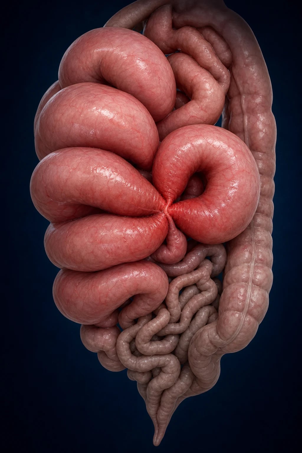
Small bowel vs large bowel obstruction vs pseudo-obstruction
| Feature | Small bowel obstruction | Large bowel obstruction | Ogilvie (pseudo-obstruction) |
|---|---|---|---|
| Common causes | Adhesions (60%), hernia, malignancy, Crohn's | Cancer (60%), volvulus, diverticulitis, faecal impaction | Critically ill ICU patient (sepsis, trauma, opioids, electrolytes) |
| Vomiting | Early, frequent, bilious | Late, may be faeculent | Variable |
| Pain | Colicky, periumbilical | Colicky, lower abdomen | Usually mild or absent |
| Distension | Central, less marked | Marked, peripheral | Marked (especially if delayed) |
| Bowel sounds | High-pitched, tinkling | High-pitched | Decreased or absent |
| Imaging | Dilated SB loops, air-fluid levels, paucity of colonic gas | Dilated colon, 'coffee bean' (volvulus) | Dilated colon throughout, no mechanical obstruction |
| Conservative | NGT + fluids (80% resolve if adhesions) | Often needs surgery (cancer, volvulus) | Neostigmine, colonoscopic decompression |
| Surgical emergency | Strangulation, peritonitis, failed conservative | Perforation, strangulation, volvulus (if endoscopic fails) | Caecum >12cm, perforation, failed neostigmine |
Management of suspected bowel obstruction
- Assess & resuscitate — ABC. IV access. Fluid resuscitation (crystalloid — massive third-space losses). Correct electrolytes (K+, Mg2+). Catheterise (monitor output)
- NGT decompression — 'Drip and suck'. NGT to decompress stomach, reduce vomiting, monitor aspirate
- Imaging — Erect CXR (free gas — perforation), supine/erect abdominal X-ray (dilated loops, air-fluid levels), CT abdomen with contrast (DEFINITIVE — site, cause, strangulation signs)
- Identify strangulation/peritonitis — peritonitis (rigid, guarding), fever, tachycardia, raised lactate, free gas → EMERGENCY SURGERY
- Conservative management (if uncomplicated) — NGT, IV fluids, serial exams, analgesia, avoid early surgery (80% of adhesive SBO resolve). Trial for 48-72h
- Surgery if: failed conservative (no resolution in 48-72h), strangulation, peritonitis, closed-loop obstruction, malignancy
- Specific treatments: Ogilvie — neostigmine. Sigmoid volvulus — endoscopic detorsion + flatus tube. Caecal volvulus — surgery
Clinical pearls
Red flags
Prognosis
Adhesive SBO resolution rates (Bologna guidelines)
Meta-analysis of adhesive SBO managed conservatively:
- Overall resolution with conservative management: 65-80%
- Resolution within 48h: 50-60%
- Resolution within 72h: 70-80%
- Risk of strangulation at presentation: 10-15%
- Mortality: 2-5% (uncomplicated), 10-30% (strangulated)
- Recurrence after conservative management: 30-40% at 5 years [1]
Gastrografin challenge: if contrast reaches colon within 24h → 90% will resolve without surgery. Neostigmine for Ogilvie: 80-90% decompression within minutes.
Pathophysiology
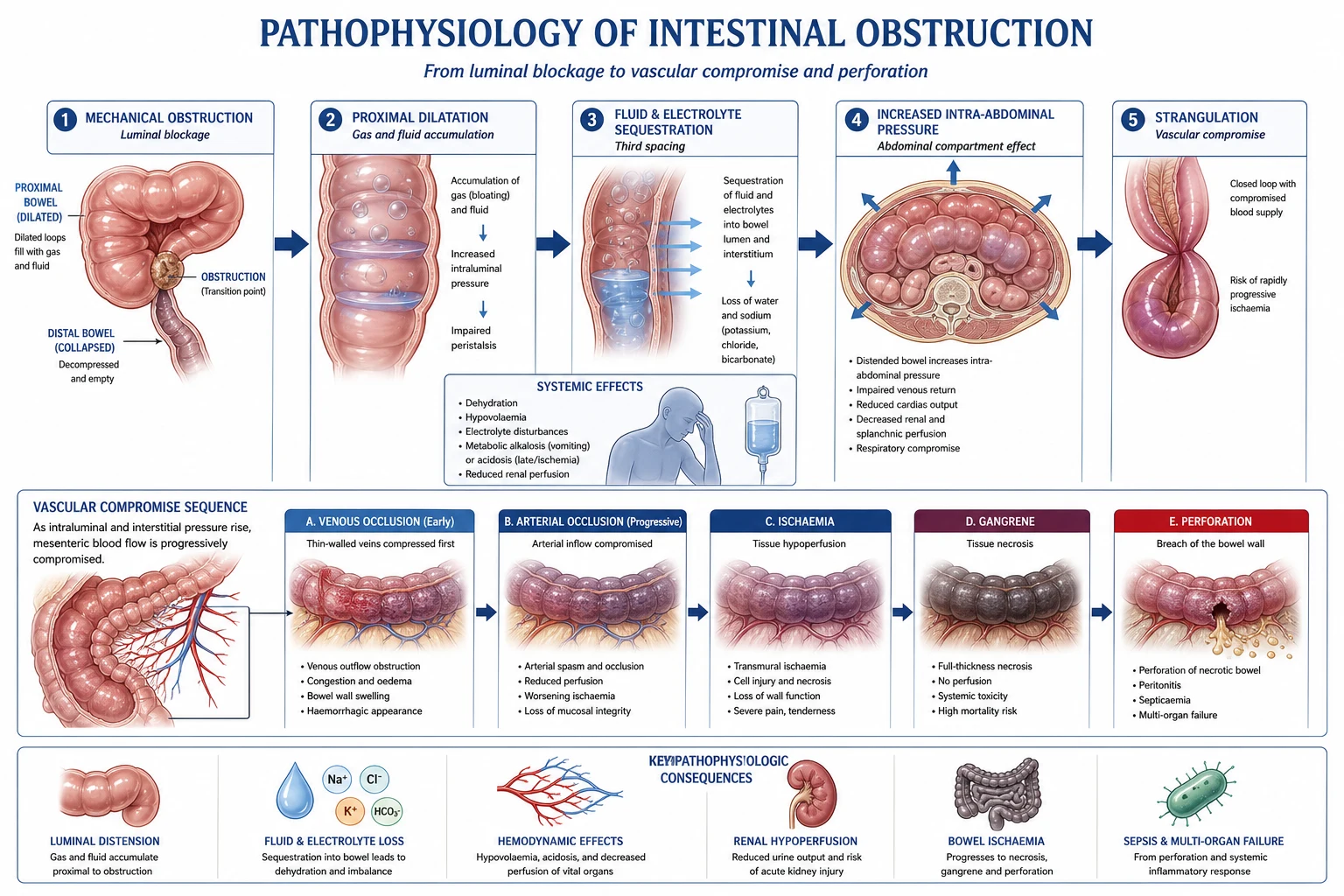
Mechanical obstruction — pathophysiology and progression
| Stage | Pathophysiology | Clinical correlate |
|---|---|---|
| 1. Luminal blockage | Adhesion band, hernia, tumour, volvulus, stricture, intussusception obstructs bowel lumen. Proximal bowel dilates; distal bowel collapses. | Cramping/colicky pain (proximal bowel hyperperistalsis trying to overcome obstruction) |
| 2. Fluid and gas accumulation | Swallowed air + bacterial fermentation gas accumulate proximal to obstruction. Fluid sequesters in bowel lumen (impaired absorption + increased secretion). | Distension, tympany. Massive third-space fluid losses → hypovolaemia, dehydration, AKI |
| 3. Bacterial overgrowth | Stasis proximal to obstruction → bacterial proliferation (especially in closed-loop). Bacteria translocate to mesenteric lymph nodes when mucosal barrier compromised. | Faeculent vomiting (late SBO/LBO). Risk of bacterial translocation → sepsis |
| 4. Venous congestion → arterial compromise | Rising intraluminal pressure compresses venous return first (venous congestion → oedema, wall thickening) → then arterial inflow ceases → ischaemia → infarction → necrosis → perforation | Strangulation. Constant (non-colicky) pain, peritonitis, raised lactate, leucocytosis, fever |
| 5. Perforation → peritonitis | Necrotic bowel wall perforates → spillage of enteric contents → faecal/bilious peritonitis → septic shock → multi-organ failure → death | Rigid abdomen, free gas on imaging, hypotension, high mortality (10-30%) |
Closed-loop vs open-loop obstruction — a critical distinction
| Feature | Open-loop (simple) obstruction | Closed-loop obstruction |
|---|---|---|
| Definition | Single point of obstruction — proximal bowel can decompress (vomit, NGT). Gas/fluid accumulates proximal to obstruction only. | Two points of obstruction along a bowel segment — the segment between both points is trapped ("closed loop") and cannot decompress. |
| Examples | Adhesive SBO (single band), tumour stricture, Crohn's stricture | Volvulus (sigmoid, caecal), incarcerated hernia, adhesive band occluding both afferent and efferent limbs |
| Intraluminal pressure | Moderate — proximal bowel vents via vomitus | Rapidly rising — the closed loop distends enormously, venous then arterial supply compromised |
| Strangulation risk | Low-moderate (develops slowly if at all) | Very high — strangulation can develop within hours |
| Time to ischaemia | Hours to days | Hours |
| CT signs | Transition point, dilated proximal bowel, collapsed distal bowel | "Whirl" sign (volvulus — mesenteric vessels twisting around a fulcrum), "beak" sign (tapering of bowel limbs at obstruction), "coffee bean" (sigmoid volvulus), C-shaped or U-shaped dilated loop, mesenteric stranding |
| Management | Trial of conservative management (drip and suck) safe | Emergency surgery — do not delay for prolonged conservative trial |
| Mortality | 2-5% (uncomplicated) | 10-30% (strangulated) |
Causes of small bowel obstruction — frequency and key features
| Cause | Frequency | Key features |
|---|---|---|
| Adhesions | 60% | Previous abdominal/pelvic surgery (any operation — appendicectomy, gynaecological, laparotomy). Bands cause kinking/compression. Highest yield cause for exams. 80% resolve with conservative management.[1] |
| Hernias | 15% | Inguinal, femoral (high strangulation risk — narrow neck), incisional, umbilical, para-stomal. Always examine all hernial orifices — a strangulated femoral hernia is easily missed. Richter's hernia (only part of wall incarcerated) can strangulate without obstruction. |
| Malignancy | 15% | Primary small bowel tumours (adenocarcinoma, carcinoid, lymphoma, GIST) or metastatic (melanoma, ovarian, colorectal). Usually present with chronic symptoms, weight loss, anaemia. |
| Crohn's disease | 5% | Strictures (fibrotic, post-inflammatory), stricturoplasty sites, adhesions from previous surgery. Often recurrent. |
| Intussusception | Rare in adults | Children (lead point: Meckel's diverticulum, lymphoid hyperplasia, rotavirus). Adults (lead point: tumour, polyp — pathological lead point). "Target" sign on ultrasound/CT. |
| Gallstone ileus | Rare | Elderly women. Cholecystoduodenal fistula → gallstone migrates → impacts at terminal ileum (narrowest point). Rigler's triad: pneumobilia + SBO + ectopic gallstone. |
| Inflammatory | Rare | Radiation enteritis, post-ischaemic stricture, intra-abdominal abscess (extrinsic compression) |
| Meckel's diverticulum | Rare | "Rule of 2s": 2% population, 2 feet from ileocaecal valve, 2 inches long, presents by age 2. Can cause intussusception, volvulus, inflammation (mimics appendicitis). |
| Foreign body | Rare | Bezoar (trichobezoar, phytobezoar), ingested objects, capsule endoscopy retention |
| Drugs | Rare | Enteric-coated potassium, NSAIDs (diaphragm disease), anticoagulants (intramural haematoma) |
Causes of large bowel obstruction — frequency and key features
| Cause | Frequency | Key features |
|---|---|---|
| Colorectal cancer | 60% | Left-sided (circumferential — "apple-core" on imaging, obstruction more common). Right-sided (exophytic, anaemia more common than obstruction). Always suspect in elderly patient with changed bowel habit + weight loss. |
| Volvulus | 10-15% | Sigmoid (75% of volvulus — elderly, constipation, "coffee bean" sign, endoscopic detorsion). Caecal (25% — younger, mobile caecum, surgery required). |
| Diverticular stricture | 10% | Chronic recurrent diverticulitis → fibrotic stricture. Differentiate from cancer (colonoscopy + biopsy). |
| Faecal impaction | 5% | Elderly, institutionalised, immobile, opioid users. Overflow "diarrhoea." Digital rectal exam is diagnostic and therapeutic. |
| Other | 5% | Ischaemic stricture, Crohn's stricture, anastomotic stricture, extrinsic compression (pelvic mass, endometriosis) |
Clinical presentation
The four cardinal features of bowel obstruction — and their nuances
| Feature | Mechanism | SBO pattern | LBO pattern |
|---|---|---|---|
| Colicky pain | Hyperperistalsis proximal to obstruction trying to overcome block. Waves every 3-5 min (SB), every 10-15 min (LB). | Periumbilical, moderate severity, intermittent | Lower abdomen, less frequent cramps, more constant dull ache between cramps |
| Vomiting | Proximal obstruction → early vomiting; distal obstruction → late vomiting | Early, frequent, bilious (proximal) or faeculent (distal/late) | Late, may be absent initially, eventually faeculent |
| Distension | Gas + fluid accumulation proximal to obstruction | Central, less marked (unless distal SBO) | Marked, visible peristalsis, tympanic drum-like abdomen |
| Constipation / obstipation | Obstruction prevents passage of stool and flatus. Obstipation = absolute constipation (no faeces AND no flatus) | May pass some stool/flatus initially (evacuation of distal bowel) → then obstipation | Earlier onset of obstipation (more complete obstruction) |
Key: High obstruction (proximal SBO) = severe vomiting, minimal distension, early presentation. Low obstruction (distal SBO/LBO) = marked distension, late vomiting, colicky pain.
Differential diagnosis of suspected bowel obstruction
| Mimic | Key distinguishing features |
|---|---|
| Paralytic ileus | No mechanical cause. No colicky pain (continuous discomfort or none). Absent bowel sounds (vs tinkling in obstruction). History of recent surgery, opioids, electrolyte disturbance (K+, Mg2+), peritonitis. Resolves with correction of underlying cause. |
| Mesenteric ischaemia | "Pain out of proportion" to exam. AF, vascular disease, recent MI. Bloody diarrhoea. Raised lactate, metabolic acidosis. CT: bowel wall thickening, pneumatosis, portal venous gas, mesenteric arterial/venous occlusion. |
| Gastroenteritis / enteritis | Cramping + diarrhoea (not constipation/obstipation). Vomiting prominent but no distension. Self-limiting. |
| Acute pancreatitis | Epigastric pain radiating to back. Raised amylase/lipase. CT shows pancreatic inflammation. Can cause paralytic ileus. |
| Intra-abdominal sepsis / peritonitis | Generalised peritonitis (rigid, guarding). May have ileus secondary to peritonitis. CT identifies abscess/perforation. |
| Pseudo-obstruction (Ogilvie) | Massive colonic dilation without mechanical obstruction. Critically ill patient. Exclude mechanical obstruction with CT/water-soluble contrast enema BEFORE neostigmine. |
| Toxic megacolon | Severe colitis (C. difficile, IBD, ischaemic). Systemically unwell. Markedly dilated colon with mucosal oedema/thumbprinting on imaging. |
| Ruptured abdominal aortic aneurysm | Tearing back/flank pain, collapse, palpable pulsatile mass. Not truly a mimic but can present with abdominal pain + distension. |
Imaging
Imaging modalities in suspected bowel obstruction
| Modality | Role | Findings | Limitations |
|---|---|---|---|
| Erect CXR | First-line: detect free gas (perforation) | Free air under diaphragm (R hemidiaphragm on erect CXR). Sensitive for perforation. | Does not diagnose obstruction. May miss small amounts of free gas (sensitivity ~60%). |
| Supine AXR | First-line: assess bowel gas pattern | Dilated small bowel loops (>3 cm, central, valvulae conniventes — "stepladder" air-fluid levels), dilated colon (>6 cm, peripheral, haustra), "coffee bean" sign (sigmoid volvulus), absent rectal gas | Low sensitivity for early/partial SBO. Cannot determine cause or strangulation. |
| Erect AXR | Confirm air-fluid levels | Multiple air-fluid levels in dilated small bowel loops. "String of beads" sign (small amounts of gas trapped between valvulae conniventes in fluid-filled loops). | Largely superseded by CT. Less useful in LBO. |
| CT abdomen/pelvis with IV contrast | GOLD STANDARD — diagnostic test of choice | Transition point (abrupt change from dilated to collapsed bowel). Cause: adhesions (no mass, abrupt transition), hernia, tumour, volvulus. Strangulation: reduced/absent wall enhancement, bowel wall thickening, mesenteric fluid/stranding, ascites, pneumatosis, portal venous gas. Perforation: free gas, free fluid. Closed-loop: "whirl" sign, U/C-shaped loop. | Radiation, IV contrast nephropathy (caution in AKI). Requires haemodynamic stability. |
| Water-soluble contrast study (Gastrografin) | Diagnostic + therapeutic for adhesive SBO | 100 mL Gastrografin via NGT. If contrast reaches colon within 4-24 h → obstruction will resolve (high sensitivity ~90-97%). Therapeutic effect (osmotic). | Only for adhesive SBO without strangulation. Contraindicated if peritonitis. |
| Ultrasound (POCUS) | Bedside in unstable patients | Dilated bowel loops (>3 cm SB, >6 cm LB), antiperistalsis, "keyboard" sign (valvulae conniventes), fluid-filled loops, "to-and-fro" peristalsis. Can identify free fluid and the "target" sign (intussusception). | Operator-dependent. Bowel gas limits views. Cannot reliably identify strangulation. |
CT signs of strangulation / bowel ischaemia — surgical urgency markers
| CT sign | Pathophysiology | Significance |
|---|---|---|
| Reduced/absent bowel wall enhancement | Arterial inflow compromised → ischaemia. IV contrast does not perfuse the ischaemic segment. | Most specific sign of strangulation (>95%). Indication for emergency surgery. |
| Bowel wall thickening (>3 mm SB, >6 mm LB) | Venous congestion → mural oedema, intramural haemorrhage | Non-specific (also seen in inflammation). But combined with other signs, supports ischaemia. |
| Mesenteric stranding / haziness | Mesenteric venous congestion, oedema, fat infiltration from inflammation | Non-specific. More specific when localised to the affected segment's mesentery. |
| Mesenteric fluid / ascites | Venous congestion → transudation of fluid into mesentery and peritoneum | Non-specific but concerning when combined with other signs. Dark haemorrhagic fluid at surgery = infarction. |
| "Whirl" sign | Mesentery twists around a fulcrum (vascular pedicle) → volvulus / closed-loop | Pathognomonic for volvulus (sigmoid or midgut). Indicates closed-loop obstruction. |
| "Beak" sign | Tapering of bowel limbs at the site of obstruction | Seen in volvulus and closed-loop obstruction. |
| Pneumatosis intestinalis | Gas in bowel wall from ischaemia → mucosal breakdown, gas-forming bacteria invade wall | Late sign — indicates transmural infarction. Emergency. |
| Portal venous gas | Gas from necrotic bowel enters mesenteric veins → portal venous system | Very late sign — indicates extensive infarction. Very high mortality. |
| Free gas | Perforation of necrotic/ischaemic bowel | Emergency — surgical exploration mandatory. |
| "Target" sign | Bowel wall showing alternating layers of high/low attenuation (submucosal oedema + haemorrhage) | Indicates ischaemia or inflammation. |
Management — detailed approach
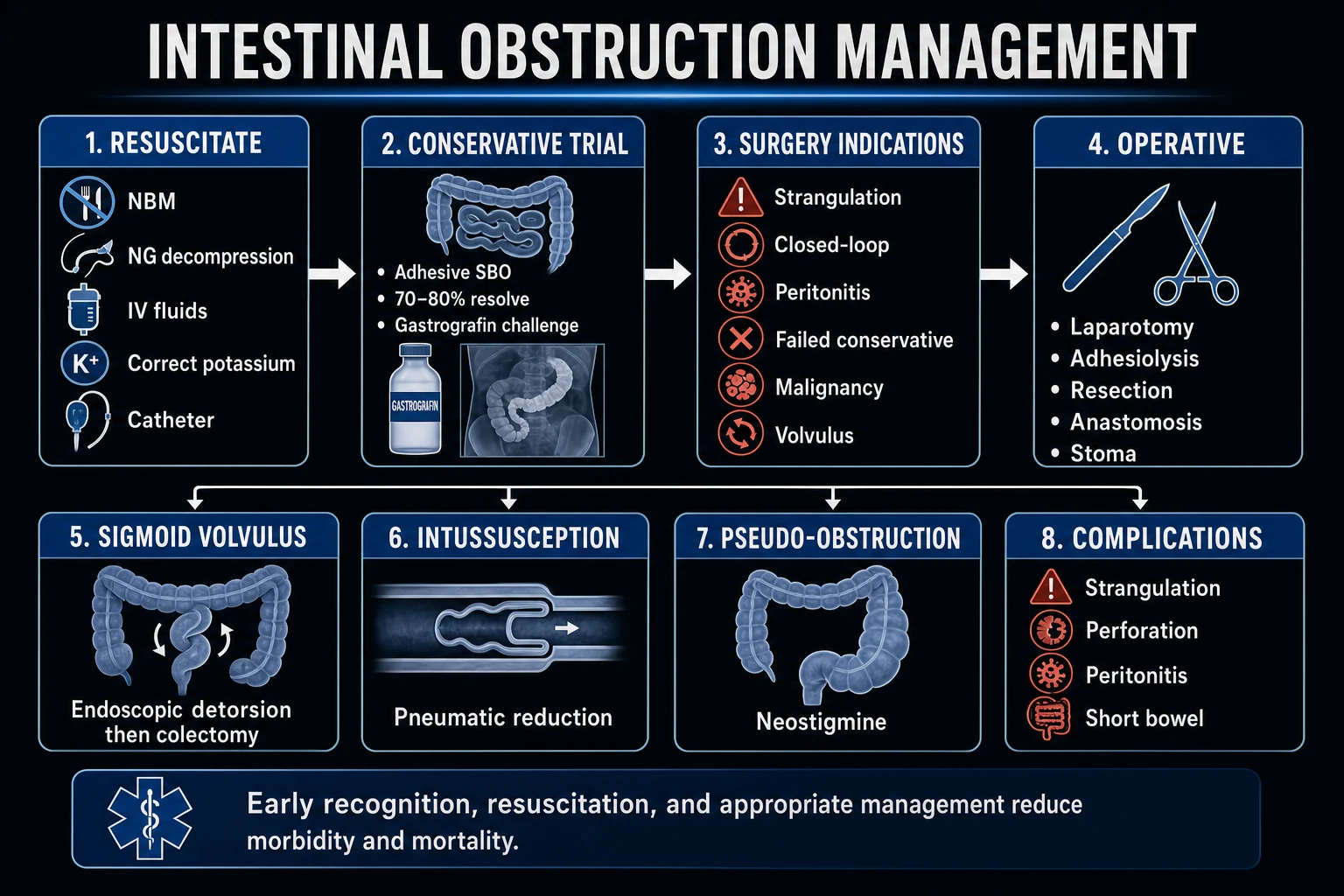
Resuscitation and initial stabilisation
Initial resuscitation — 'Drip and Suck' for all obstructions
- ABC + IV access — two large-bore cannulae. Oxygen if hypoxic. Cardiac monitor (elderly, electrolyte disturbance). Assess for sepsis (peritonitis → septic shock).
- Fluid resuscitation — balanced crystalloid (Hartmann's / Plasma-Lyte). Massive third-space losses into obstructed bowel: 1-3 L deficit common. Bolus 500 mL aliquots, reassess. Correct hypokalaemia (K+ lost in vomitus + third-space sequestration), hypomagnesaemia, hyponatraemia. Avoid aggressive 0.9% saline (hyperchloraemic metabolic acidosis, AKI risk — SMART trial).
- Nasogastric tube (NGT) — "Suck." Decompresses stomach, reduces vomiting and aspiration risk, monitors aspirate volume (falling aspirate = improving obstruction), relieves proximal distension. Large-bore NGT (16-18 Fr) preferred over fine-bore for viscous secretions. Document aspirate character (bilious, faeculent).
- Urinary catheter — monitor urine output (target >0.5 mL/kg/h). AKI from hypovolaemia. Fluid balance chart.
- Analgesia — morphine 2.5-5 mg IV titrated (avoid NSAIDs — renal impairment). Antiemetics (ondansetron 4 mg IV). Do not give prokinetics (metoclopramide, erythromycin) in mechanical obstruction — may worsen strangulation risk or perforation.
- Broad-spectrum antibiotics — if strangulation, peritonitis, or sepsis suspected (e.g., co-amoxiclav or ceftriaxone + metronidazole). Not routinely required for uncomplicated adhesive SBO.
- Surgical referral — involve surgical team early. Serial abdominal examinations (q4-6h). Set clear thresholds for surgical intervention.
Fluid management in bowel obstruction — principles
| Principle | Rationale | Practical approach |
|---|---|---|
| Crystalloid first-line | Replace isotonic third-space losses | Hartmann's or Plasma-Lyte (balanced — avoids hyperchloraemic acidosis of 0.9% saline) |
| Volume | Sequestration into obstructed bowel lumen + vomiting → significant deficit (1-3 L) | 1-2 L bolus, then maintenance + ongoing losses. Rate guided by clinical response (HR, BP, urine output, lactate, capillary refill) |
| Potassium | Hypokalaemia universal (vomiting + third-space loss). Hypokalaemia worsens ileus and cardiac arrhythmias | 20-40 mmol/L in maintenance fluids. Check U&E q12-24h. Target K+ 4.0-4.5 mmol/L. |
| Magnesium | Hypomagnesaemia common (vomiting, poor intake). Refractory hypokalaemia if Mg2+ not corrected | 10-20 mmol MgSO4 IV. Check Mg2+ level. |
| Avoid hypotonic fluids | SIADH risk from stress/nausea/pain + possible hyponatraemia from GI losses | Use isotonic crystalloid for resuscitation |
| Monitor | AKI risk from hypovolaemia; fluid overload risk in elderly/cardiac disease | Urine output >0.5 mL/kg/h. Daily weights. Lactate trend (clearing = resuscitation adequate). Reassess q4-6h. |
Gastrografin (water-soluble contrast) challenge
Water-soluble contrast challenge (Gastrografin) for adhesive SBO
- Indication — uncomplicated adhesive SBO without signs of strangulation/peritonitis. CT shows single transition point, no ischaemia. Aids both diagnosis (predicts need for surgery) and therapy (osmotic effect).[13]
- Patient selection — no peritonitis, no free gas, no signs of strangulation, partial or complete SBO with adhesive aetiology (previous surgery, CT showing abrupt transition with no mass). NOT for hernia, volvulus, tumour, or closed-loop obstruction.
- Procedure — 100 mL Gastrografin (diatrizoate meglumine — hyperosmolar water-soluble contrast) administered via NGT. Clamp NGT for 2 h. Patient mobilises/changes position to aid transit.
- Assessment — serial abdominal X-rays at 4, 8, and 24 h. If contrast reaches colon (caecum) within 24 h → obstruction will resolve (sensitivity 92-97%, specificity 60-80%). If contrast does NOT reach colon within 24 h → high likelihood of needing surgery (proceed to laparotomy).
- Therapeutic mechanism — Gastrografin is hyperosmolar (1900 mOsm/L) → draws fluid into bowel lumen → oedema reduction at obstruction site + stimulates peristalsis. May hasten resolution by 24-48 h compared to conservative management alone.
- Outcome — Meta-analyses show Gastrografin reduces need for surgery (odds ratio ~0.6) and shortens hospital stay. Does NOT reduce recurrence rate.[13]
- Contraindications — suspected perforation (use barium for fistula — water-soluble extravasates but is resorbed; barium causes peritonitis), peritonitis, complete obstruction from non-adhesive cause.
Surgical management
Indications for surgery in small bowel obstruction
| Indication | Rationale | Timing |
|---|---|---|
| Strangulation (ischaemia) | Blood supply compromised → necrosis → perforation → death. CT signs: reduced wall enhancement, mesenteric fluid, pneumatosis. Clinical: peritonitis, raised lactate, fever, constant pain. | Emergency — immediate laparotomy |
| Peritonitis (perforation) | Free gas on imaging. Rigid abdomen. Faecal/bilious peritonitis → septic shock. | Emergency — immediate laparotomy |
| Closed-loop obstruction | Volvulus (whirl sign), incarcerated hernia. Rapid strangulation risk. | Urgent — surgery within hours (caecal volvulus = surgery; sigmoid volvulus = endoscopic detorsion first) |
| Failed conservative management | No resolution within 48-72 h of drip-and-suck + Gastrografin challenge. Worsening distension/pain/aspirate. | Urgent — surgery within 24 h of declaring failure |
| Non-adhesive cause | Hernia (unless easily reducible and reducible on presentation), malignancy, intussusception, gallstone ileus (may require surgery), Crohn's stricture. | Urgent-elective — timing depends on cause |
| Recurrent obstruction | Multiple admissions for adhesive SBO → consider elective adhesiolysis or bowel resection. | Elective — after resolution of current episode |
Surgical approach to SBO — laparotomy vs laparoscopy
- Pre-operative optimisation — resuscitate first (fluids, electrolytes, antibiotics if strangulation/peritonitis). Cross-match blood. NGT in situ (decompressed stomach reduces aspiration on induction). Urinary catheter. DVT prophylaxis (LMNH — hold if emergency surgery, mechanical prophylaxis intraoperatively).
- Approach selection — Open laparotomy (midline incision): standard for strangulation, peritonitis, multiple previous surgeries, haemodynamic instability. Laparoscopic adhesiolysis: selected patients (≤2 previous surgeries, localised adhesions, no peritonitis, stable). Experienced surgeon only — conversion to open if dense adhesions, bowel injury, or unable to identify obstruction. Conversion rate 20-50%.[10][14]
- Operative steps (laparotomy) — systematic "run-through" of entire small bowel from ligament of Treitz to ileocaecal valve. Identify transition point (dilated → collapsed bowel). Identify and treat cause: adhesiolysis (divide adhesions), hernia repair, tumour resection. Assess bowel viability — colour, peristalsis, mesenteric pulsation. If viability uncertain → warm packs 15-20 min + 100% O2 → reassess. If still non-viable → resect (strangulated/ischaemic segment) with primary anastomosis or stoma. Washout if peritonitis/perforation.
- Bowel preservation principle — minimise resection (short bowel syndrome risk). Resect only frankly non-viable bowel. For adhesive SBO: adhesiolysis alone (no resection needed if bowel viable). For strangulated bowel: resect from viable proximal to viable distal margin.
- Laparoscopic adhesiolysis outcomes — shorter hospital stay (2-3 days less), fewer wound infections, earlier return of bowel function. BUT higher risk of unrecognised enterotomy (bowel injury during dissection — always check for enterotomy before closing).[10]
- Post-operative ICU care — fluid resuscitation (capillary leak → third-space losses), monitor for anastomotic leak (days 5-7: fever, tachycardia, abdominal pain, raised inflammatory markers → CT with water-soluble contrast), wound infection, ileus (expected 3-5 days — NGT, fluids, mobilise), AKI, pneumonia. VTE prophylaxis (LMNH 6 h post-op). Stress-ulcer prophylaxis (PPI) if ventilated/coagulopathic/steroid. Glycaemic control 7.8-10 mmol/L.
Post-operative complications after bowel obstruction surgery
| Complication | Incidence | Features | Management |
|---|---|---|---|
| Prolonged ileus | 10-20% | No flatus/stool >5 days post-op. Distension, no bowel sounds. Nausea, NGT aspirate persists. | Conservative: NGT, IV fluids, correct electrolytes, minimise opioids, mobilise. Exclude mechanical obstruction with CT if >7 days. |
| Anastomotic leak | 4-10% (emergency, higher) | Days 5-7: fever, tachycardia, abdominal pain, pelvic/abdominal collection, raised CRP/WCC. Septic shock. | CT with water-soluble contrast. Drain collection (radiological or surgical). Defunctioning stoma. Re-laparotomy if unstable. |
| Surgical site infection | 10-25% (contaminated/dirty wounds) | Erythema, purulent discharge, dehiscence. | Wound swab, antibiotics (flucloxacillin or based on culture), open/incise/drain. |
| Intra-abdominal collection/abscess | 5-10% | Fever, raised WCC/CRP, persistent ileus. CT confirms. | Percutaneous drainage (if accessible) or surgical drainage. Antibiotics. |
| Enterocutaneous fistula | 1-5% | Enteric contents draining from wound/drain site. Days 7-14. | Conservative first (TPN, somatostatin analogue, wound care). Surgery if fails to close after months. |
| Adhesive SBO recurrence | 30-40% at 5 years | Readmission with SBO. | Initially conservative. Elective adhesiolysis/barrier adhesion prevention (Seprafilm) at subsequent surgery. |
| Short bowel syndrome | Variable | Extensive resection (>200 cm SB). Diarrhoea, malabsorption, weight loss, electrolyte disturbance. | TPN, anti-diarrhoeals, intestinal rehabilitation. Rare in obstruction surgery. |
Ogilvie syndrome (acute colonic pseudo-obstruction)
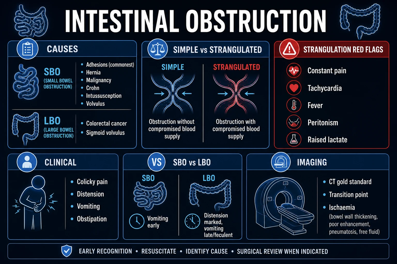
Ogilvie syndrome — risk factors in ICU patients
| Category | Risk factors |
|---|---|
| Critical illness | Sepsis, severe pneumonia, ARDS, multi-organ failure, severe burns, hypoxia |
| Surgical | Recent abdominal/pelvic surgery, caesarean section, hip/knee replacement, cardiac surgery, transplant surgery |
| Trauma | Pelvic/lumbar fractures, retroperitoneal haematoma, spinal cord injury |
| Metabolic/electrolyte | Hypokalaemia, hypomagnesaemia, hypocalcaemia, hypothyroidism, uraemia |
| Drugs | Opioids (major), anticholinergics, phenothiazines, calcium channel blockers, clonidine, SSRIs |
| Cardiovascular | Heart failure, myocardial infarction, cardiac arrest |
| Neurological | Stroke, Parkinson's disease, multiple sclerosis, spinal cord injury, dementia |
| Infectious | Herpes zoster, cytomegalovirus, systemic infection |
Ogilvie syndrome — pathophysiology and the parasympathetic theory
| Mechanism | Detail |
|---|---|
| Core concept | Massive colonic dilation (especially caecum and right colon) WITHOUT mechanical obstruction. The colon is atonic — no peristalsis. |
| Sympathetic overactivity | Critical illness, pain, stress → excessive sympathetic output → inhibitory effect on colonic motility |
| Parasympathetic suppression | Pelvic splanchnic nerves (S2-S4) supply parasympathetic innervation to distal colon/rectum. Vagus supplies proximal colon. Imbalance between sacral parasympathetic (loss) and vagal (excess) → segmental atony → functional obstruction → gas accumulation → massive dilation. |
| Why the caecum? | Laplace's law: T = P × r. The caecum has the largest radius (r) → greatest wall tension (T) at any given intraluminal pressure (P). This makes the caecum the most vulnerable to perforation. Caecal diameter >12 cm = high perforation risk. Perforation risk also depends on duration of dilation (>6 days = higher risk) and rate of dilation (rapid = higher risk). |
| Outcome | Spontaneous resolution in ~70-80% with conservative measures. Pharmacological decompression (neostigmine) successful in 80-90%. Colonoscopic decompression in ~70-80% of non-responders. Surgery required in 1-5%. Perforation risk 3-15%. Mortality 10-30% (driven by underlying critical illness + perforation).[12] |
Management of Ogilvie syndrome (acute colonic pseudo-obstruction)
- Diagnose and exclude mechanical obstruction — CT abdomen/pelvis is essential. Shows massively dilated colon (especially caecum/right colon) throughout, no mechanical obstruction / no transition point. Also excludes caecal/sigmoid volvulus, tumour, and assesses for perforation/ischaemia. Water-soluble contrast enema can also exclude distal mechanical obstruction.[12]
- Conservative measures (first-line) — Correct ALL electrolyte abnormalities (K+ >4.0, Mg2+ >0.8, Ca2+ normal). Stop all exacerbating drugs (opioids → use multimodal analgesia, anticholinergics, calcium channel blockers). Mobilise / frequent position changes. Nasogastric decompression (reduce swallowed air). Rectal tube (decompress rectum/sigmoid). Restrict oral intake. Serial abdominal X-rays q12-24h to monitor caecal diameter. Trial for 48-72 h. ~70-80% resolve with conservative measures.[12]
- Neostigmine (second-line) — if no improvement after 48-72 h of conservative measures AND caecal diameter >10-12 cm (or rapidly increasing). MUST exclude mechanical obstruction first (CT) — neostigmine in mechanical obstruction is dangerous (increases pressure against obstruction → perforation).[8]
- Neostigmine administration protocol — 2 mg IV over 3-5 min (slow push). Patient supine on bedpan/commode. Continuous ECG, BP, HR monitoring. Have atropine 0.5-1 mg drawn up (for bradycardia). Onset of effect: within minutes (typically passes flatus/stool within 30 min). Efficacy: 80-90% decompression. May repeat once after 2-4 h if partial response. Contraindications: mechanical obstruction (unexcluded), perforation, severe bradycardia (<60 bpm), active bronchospasm, recent MI.[8]
- Colonoscopic decompression (third-line) — if neostigmine fails, contraindicated, or recurrent after initial response. Unprepared colonoscopy (no bowel prep — difficult and potentially dangerous in dilated, thin-walled colon). Gentle air insufflation (minimal). Advance to caecum (success 40-80% — may not reach caecum). Aspirate gas throughout withdrawal. Leave decompression tube in caecum (14-18 Fr) for 48-72 h. Success: 70-80%. Recurrence: 20-40% (may need repeat). Perforation risk: 2-3% (higher than diagnostic colonoscopy).[16]
- Surgery (last resort) — if neostigmine + colonoscopy fail, OR if perforation/ischaemia develops. Caecostomy (tube caecostomy — minimally invasive, can be percutaneous or open). Subtotal colectomy (if ischaemia/perforation — resect non-viable colon, primary anastomosis or ileostomy). High mortality (15-40% — reflects critical illness + emergency surgery).[12]
- Monitoring during treatment — serial abdominal X-rays q12-24h. Target: caecal diameter <9 cm. Clinical exam: watch for peritonitis (perforation). If caecum >12 cm for >6 days → escalating intervention. Recurrence after neostigmine: 20-30% → consider repeat dose or colonoscopy.[12]
Ogilvie syndrome — caecal diameter and perforation risk
| Caecal diameter | Perforation risk | Recommended action |
|---|---|---|
| <10 cm | Low | Conservative management (correct electrolytes, stop drugs, mobilise). Monitor with serial AXR. |
| 10-12 cm | Moderate | Conservative measures intensified. Consider neostigmine if no resolution in 48 h or diameter increasing. |
| >12 cm | High | Neostigmine (if no contraindication and mechanical obstruction excluded). If neostigmine fails → colonoscopic decompression. Surgery if colonoscopy fails. |
| >12 cm for >6 days | Very high | Urgent intervention (neostigmine → colonoscopy → surgery). Perforation risk increases significantly with prolonged dilation. |
| >14 cm OR signs of perforation | Critical | Emergency surgery (caecostomy or colectomy). Free gas on imaging = perforation. |
Note: Caecal diameter is the primary determinant, but perforation can occur at smaller diameters with rapid onset or ischaemia. Clinical context (peritonitis, sepsis, underlying critical illness) guides urgency.
Volvulus
Sigmoid vs caecal volvulus
| Feature | Sigmoid volvulus | Caecal volvulus |
|---|---|---|
| Frequency | 75% of colonic volvulus | 25% |
| Demographics | Elderly, nursing-home residents, chronic constipation, institutionalised, psychiatric medications, high-fibre diet in some populations (Africa, "volvulus belt") | Younger (30-60 years), mobile caecum (incomplete fixation — 10-25% population) |
| Mechanism | Sigmoid colon twists (180-540°) around its mesentery. Elongated redundant sigmoid + narrow mesenteric base = predisposition. Closed-loop obstruction. | Caecum twists (clockwise) around its mesentery ± axial volvulus. Mobile caecum allows torsion. Can also be caecal bascule (anterior fold, not true torsion). |
| Imaging | "Coffee bean" sign — massively dilated sigmoid loop arising from pelvis, projecting toward RUQ. The mesenteric crease creates the "bean's" central crease. Plain AXR diagnostic in 60-80%. CT confirms ("whirl" sign of mesenteric twist). | Dilated caecum in LUQ or epigastrium (flipped superiorly). "Coffee bean" may be seen but oriented differently. CT: "whirl" sign, caecal "beak." May mimic SBO (dilated SB loops if ileocaecal valve incompetent). |
| Presentation | Massive abdominal distension, cramping lower abdominal pain, constipation/obstipation. May be chronically recurrent (intermittent torsion/detorsion). | SBO-like picture: distension, vomiting, colicky pain. Rapid strangulation risk. |
| First-line management | Endoscopic detorsion — flexible sigmoidoscopy, gently advance past twist, deflate/decompress. Insert flatus tube (rectal tube) left in situ 48-72 h. Success ~70-80%. Recurrence 40-60%. | Surgery — endoscopic detorsion is NOT recommended (unsafe, caecum not reachable via colonoscopy reliably, high perforation risk in closed loop). Right hemicolectomy ± primary anastomosis. |
| Surgery indications | Endoscopic failure, ischaemia/perforation, recurrent (elective sigmoid colectomy after successful detorsion). | All caecal volvulus → surgery (right hemicolectomy). |
| Recurrence | 40-60% after successful endoscopic detorsion alone. Elective sigmoid colectomy after successful detorsion reduces recurrence to <5%. | Right hemicolectomy is definitive (recurrence ~1%). |
| Mortality | 5-10% (elective detorsion); 20-50% if strangulated/perforated | 10-20% (caecal volvulus has higher emergency surgery rate) |
Endoscopic detorsion of sigmoid volvulus
- Indication — sigmoid volvulus without signs of strangulation/perforation (no peritonitis, no free gas, clinically stable, viable bowel on imaging). First-line therapy.[9][15]
- Preparation — consent (perforation risk ~1-2%, failure). Broad-spectrum antibiotics (contaminated field). Decompression: gentle rectal examination, some centres gently pass flatus tube to decompress gas before scope.
- Procedure — flexible sigmoidoscopy (unprepared bowel — usually little stool as bowel proximal is obstructed). Gently advance scope. At the twist: spiral "whirl" of mucosa, congested mucosa (assess viability — pale/dusky/black = ischaemia → ABORT, go to surgery). Gently insufflate and manipulate scope past twist. Sudden rush of gas/liquid stool = successful detorsion. Advance to proximal sigmoid/decompress.
- Assess mucosal viability — if mucosa is viable (pink, normal), proceed. If ischaemic (dusky, purple, sloughing, black) → ABORT endoscopy → emergency surgery (sigmoid colectomy — strangulated bowel).
- Flatus tube placement — insert large-bore rectal tube (28-32 Fr) past detorsion point, leave in situ 48-72 h to maintain detorsion and allow continued decompression. Confirm position on AXR.
- Post-procedure — NBM, IV fluids, monitor for recurrence (serial abdominal exams, AXR). Consider elective sigmoid colectomy during same admission (3-5 days after detorsion) to prevent recurrence (40-60% recurrence if no surgery).[9]
- Outcome — Success 70-80%. Recurrence 40-60% (without elective surgery). Mortality of detorsion 1-5%. Mortality of strangulated sigmoid volvulus requiring emergency surgery 20-50%.[15]
Additional clinical pearls
Additional red flags
Prognosis and outcomes
Outcomes by obstruction type and complication
| Scenario | Mortality | Key outcome data |
|---|---|---|
| Uncomplicated adhesive SBO (conservative) | 2-5% | 65-80% resolve with drip-and-suck. Recurrence 30-40% at 5 years. Gastrografin reduces surgery need by ~36%.[3][13] |
| Strangulated SBO (surgery) | 10-30% | Mortality driven by delay to surgery, comorbidity, septic shock. Early laparotomy (within 24 h of strangulation signs) improves survival.[2] |
| LBO from colorectal cancer | 10-20% (emergency) | Emergency surgery (resection ± stoma) higher mortality than elective. Stenting (self-expanding metal stent) as bridge to elective surgery in selected unfit patients.[5] |
| Sigmoid volvulus (endoscopic detorsion) | 1-5% | Success 70-80%. Recurrence 40-60% without elective colectomy.[9] |
| Sigmoid volvulus (emergency surgery) | 20-50% | High mortality reflects elderly, comorbid population + emergency setting.[15] |
| Caecal volvulus (surgery) | 10-20% | Right hemicolectomy definitive. Recurrence ~1%.[15] |
| Ogilvie syndrome (conservative) | 5-15% | 70-80% resolve with conservative measures. Mortality driven by underlying critical illness.[12] |
| Ogilvie (neostigmine) | 5-15% | 80-90% decompression. Recurrence 20-30%.[8] |
| Ogilvie (colonoscopic decompression) | 10-20% | Success 70-80% of neostigmine failures. Perforation risk 2-3%.[16] |
| Ogilvie with perforation | 40-50% | Emergency surgery (caecostomy/colectomy). High mortality reflects critical illness + abdominal sepsis.[12] |
| Gallstone ileus | 5-15% | Enterolithotomy definitive for acute obstruction. Recurrence of gallstone ileus rare (<5%). Cholecystectomy may be deferred.[6] |
Ponec et al. 1999 — Neostigmine for Ogilvie syndrome (landmark RCT)
Randomised, double-blind, placebo-controlled trial. 21 patients with Ogilvie (caecum >10 cm, no mechanical obstruction, failed conservative management for 48 h).
- Intervention: Neostigmine 2 mg IV vs placebo
- Primary outcome: Clinical decompression (passing flatus/stool + reduction in abdominal distension) within 3 h
- Results: Decompression in 10/11 (91%) neostigmine vs 0/10 (0%) placebo (p<0.001). Median time to response 4 min (neostigmine).
- Adverse events: Bradycardia 2/11 (required atropine), mild abdominal cramping, vomiting, salivation, lacrimation. No perforation.
- Recurrent dilation: 3/11 (27%) recurred after initial response → 2 responded to second dose.
- Significance: Established neostigmine as first-line pharmacological therapy for Ogilvie syndrome. Subsequent studies (Paran et al. 2000, van der Snock 2008, Valle et al. 2015) confirmed efficacy with 80-90% decompression rates and acceptable safety.[8]
Abbas et al. (Cochrane, 2007/2015) — Water-soluble contrast for adhesive SBO
Systematic review and meta-analysis of RCTs evaluating oral water-soluble contrast in adhesive SBO.
- Trials included: 8 RCTs, 710 patients
- Adhesive SBO resolution: Pooled OR 2.30 (95% CI 1.43-3.71) favouring water-soluble contrast over conservative management alone
- Need for surgery: Pooled OR 0.62 (95% CI 0.43-0.89) — 36% relative reduction in need for surgery
- Time to resolution: Mean difference -1.46 days (95% CI -2.17 to -0.75) — faster resolution
- Hospital stay: Mean difference -1.85 days (95% CI -2.86 to -0.83) — shorter stay
- Adverse events: No significant difference (water-soluble contrast well tolerated)
- Conclusion: Water-soluble contrast (Gastrografin) is both diagnostic (predicts need for surgery) and therapeutic (hastens resolution, reduces need for surgery). Recommended for uncomplicated adhesive SBO without strangulation signs.[13]
Halabi et al. (2014) — ACS-NSQIP: Laparoscopic vs open surgery for SBO
Retrospective analysis of 6,722 patients from the American College of Surgeons National Surgical Quality Improvement Program (ACS-NSQIP) database.
- Laparoscopic group (n=989) vs Open group (n=5,733)
- Mortality: 1.0% (laparoscopic) vs 3.1% (open) — OR 0.45 (p<0.001)
- Overall morbidity: 12.3% vs 25.0% — OR 0.45 (p<0.001)
- Surgical site infection: 3.4% vs 8.5% — OR 0.44 (p<0.001)
- Pneumonia: 1.3% vs 3.7% — OR 0.41 (p<0.001)
- Hospital stay: 4 days (laparoscopic) vs 8 days (open) (p<0.001)
- Conversion rate: 20.2% (higher with dense adhesions, previous open surgery)
- Conclusion: Laparoscopic adhesiolysis associated with lower morbidity, mortality, and shorter stay — but selection bias (healthier, less complex patients selected for laparoscopy). Demonstrates feasibility and safety of laparoscopic approach in selected SBO patients.[14]
ICU-specific considerations for intestinal obstruction
| Consideration | Detail |
|---|---|
| Sepsis from strangulated bowel | Bacterial translocation from ischaemic bowel → gram-negative sepsis, septic shock. Resuscitate (30 mL/kg crystalloid), vasopressors (noradrenaline), broad-spectrum antibiotics (piperacillin-tazobactam ± aminoglycoside), source control (emergency laparotomy + resection). Lactate clearance guides resuscitation. Mortality 30-50%. |
| AKI from hypovolaemia | Third-space losses → pre-renal AKI. Resuscitate early. Monitor urine output. Avoid nephrotoxins (NSAIDs, aminoglycosides unless indicated). CT contrast may worsen AKI — weigh risk/benefit. |
| Abdominal compartment syndrome | Dilated bowel → raised intra-abdominal pressure. Bladder pressure >20 mmHg + organ dysfunction (AKI, respiratory failure, hypotension). Decompress (NGT, rectal tube, neostigmine, surgery). Mortality 40-70%. |
| Nutrition | Prolonged obstruction/ileus → malnutrition. Early enteral nutrition if conservative management (post-pyloric NGT if gastric stasis). TPN if prolonged (>7 days) and enteral not possible. Refeeding syndrome risk if malnourished. |
| VTE prophylaxis | Bowel obstruction = high VTE risk (immobility, dehydration, inflammation, cancer). LMWH unless contraindicated (active bleeding, recent surgery — mechanical prophylaxis). Hold LMNH 12 h pre-surgery. |
| Stress ulcer prophylaxis | PPI (pantoprazole 40 mg IV OD) if ventilated >48 h, coagulopathy, shock, previous GI bleed. Not routinely for all patients (COOK trial — excess pneumonia with routine PPI). |
| Delirium | Common in elderly ICU patients with obstruction. Manage pain (paracetamol first, opioid-sparing), treat sepsis, correct electrolytes, mobilise, sleep hygiene. Avoid benzodiazepines (worsen delirium) unless alcohol/benzo withdrawal. |
| Goals of care / ethics | Malignant bowel obstruction in advanced cancer → discuss goals of care. Surgery may be futile. Palliative approach (decompressive gastrostomy, octreotide, steroids, analgesia) may be appropriate. Involve palliative care team early. |
ICU management checklist for intestinal obstruction
- Resuscitate — IV access, crystalloid bolus (correct hypovolaemia from third-space losses), correct K+/Mg2+. Catheterise (monitor UO). Broad-spectrum antibiotics if sepsis/peritonitis.
- Decompress — NGT (large-bore, 16-18 Fr). Monitor aspirate. Consider rectal tube for distal obstruction.
- Image — Erect CXR (free gas), supine AXR (dilated loops, coffee bean), CT abdomen/pelvis with IV contrast (gold standard — site, cause, strangulation, perforation, closed-loop).
- Triage — Strangulation/peritonitis/closed-loop → emergency surgery. Uncomplicated adhesive SBO → drip-and-suck + Gastrografin challenge. Ogilvie → exclude mechanical obstruction → conservative → neostigmine → colonoscopy → surgery. Volvulus → sigmoid (endoscopic detorsion) vs caecal (surgery).
- Monitor — Serial abdominal exams (q4-6h). Vital signs, urine output, lactate. Serial AXR (Ogilvie caecal diameter). Falling NGT aspirate + passing flatus = improving.
- Surgical liaison — Involve surgeons early. Set clear triggers for surgery: peritonitis, free gas, CT ischaemia signs, failed conservative at 72 h, closed-loop.
- Optimise comorbidities — Cardiac, respiratory, renal, diabetes. VTE prophylaxis (LMNH). Glycaemic control (7.8-10 mmol/L). Stress ulcer prophylaxis if indicated. Nutrition plan (enteral preferred).
- Post-operative (if surgery) — HDU/ICU. Monitor for ileus, anastomotic leak, wound infection, intra-abdominal collection, AKI, pneumonia. Early mobilisation. ERAS protocol (early feeding, opioid-sparing analgesia).
Exam practice
SAQ — Small bowel obstruction (SBO): initial ICU management
10 minutes · 10 marks
A 68-year-old man with a background of open colectomy for diverticular disease 4 years ago and an incisional hernia presents with 48 hours of colicky central abdominal pain, distension, bilious vomiting and absolute constipation (no flatus, no stool). He is febrile 37.9 °C, HR 108, BP 102/68, RR 22, SpO2 95% on 2 L NP. Abdomen is distended and tympanitic with active tinkling bowel sounds and a midline hernia that is soft and reducible. Labs: WCC 14.2 x10^9/L, lactate 1.4 mmol/L, K+ 3.2 mmol/L, creatinine 142 umol/L, CRP 88 mg/L. AXR shows multiple dilated small-bowel loops with valvulae conniventes and a stepladder pattern.
SAQ — Strangulation and ischaemia in bowel obstruction
10 minutes · 10 marks
A 74-year-old woman with atrial fibrillation (not anticoagulated — refused warfarin) presents with 36 hours of severe constant periumbilical pain (now out of proportion to examination), three episodes of dark-red rectal bleeding and a single vomit. She is febrile 38.6 °C, HR 128 (irregularly irregular), BP 88/54, RR 26, SpO2 92% on 4 L NP, GCS 14. Abdomen: soft, mildly distended, no guarding, but with guarding on deep palpation in the right lower quadrant and a mass. Digital rectal exam: dark-red blood on glove. Labs: Hb 168 g/L, WCC 21 x10^9/L, lactate 5.8 mmol/L, K+ 5.6 mmol/L, creatinine 188 umol/L, INR 1.6. CT angiogram (performed before review): small-bowel wall thickening, pneumatosis intestinalis in the distal ileum, mesenteric venous gas, and a filling defect in the superior mesenteric artery with cutoff at a distal branch.
References
- [1]Ten Broek RPG, et al. Self-reported history of seizure and baseline neurocognitive test performance in student-athletes: an initial investigation Phys Sportsmed, 2017.PMID 28838286
- [2]Maung AA, et al. Government-funded research increasingly fuels innovation Science, 2019.PMID 31221848
- [3]Di Saverio S, et al. Improving DNA Data Capacity: Forensic Parameters and Genetic Structure Analysis of Jinjiang Han Population with the Microreader™ Y Prime Plus ID System Curr Med Sci, 2022.PMID 35403953
- [4]Saunders MD, Kimmey MB. Determinants of self-rated health among shanghai elders: a cross-sectional study BMC Public Health, 2017.PMID 29029627
- [5]Vogel JD, et al. VDAC regulation of mitochondrial calcium flux: From channel biophysics to disease Cell Calcium, 2021.PMID 33529977
- [6]Townsend CM, et al. Can sand nourishment material affect dune vegetation through nutrient addition? Sci Total Environ, 2020.PMID 32278174
- [7]Branicka O, et al. Taking Procrustes' Axe to Professor Fisher's response J Eval Clin Pract, 2011.PMID 21951938
- [8]Ponec RJ, Saunders MD, Kimmey MB. Hospital admission policy for tuberculosis in pulmonary centres in Italy: a national survey. AIPO Tuberculosis Study Group. Italian Association of Hospital Pulmonologists Int J Tuberc Lung Dis, 1999.PMID 10587320
- [9]Triantafyllidis I, et al. The Diesel Exhaust in Miners Study provides no evidence for an increase in risk for lung cancer in miners exposed to diesel engine emissions Eur J Epidemiol, 2018.PMID 30382498
- [10]Cirocchi R, et al. Development and validation of a novel panel of 16 STR markers for simultaneous diagnosis of β-thalassemia, aneuploidy screening, maternal cell contamination detection and fetal sample authenticity in PND and PGD/PGS cases Sci Rep, 2019.PMID 31092881
- [11]Foster NM, et al. Accelerometer measured levels of moderate-to-vigorous intensity physical activity and sedentary time in children and adolescents with chronic disease: A systematic review and meta-analysis PLoS One, 2017.PMID 28640907
- [12]Saunders MD. Amphetamine- and scopolamine-induced locomotor activity following treatment with chlorphenvinphos or chlorphyriphos in rats Int J Occup Med Environ Health, 2005.PMID 16201202
- [13]Abbas S, Bissett IP, Parry BR. Expression of Das-1, Ki67 and sulfuric proteins in gastric cardia adenocarcinoma and intestinal metaplasia lesions Exp Ther Med, 2013.PMID 23837030
- [14]Halabi WJ, et al. [ALK-positive large B-cell lymphoma: report of a case] Zhonghua Bing Li Xue Za Zhi, 2011.PMID 22169650
- [15]Gouge F, et al. Intrathyroidal multiloculated proliferating thymic cyst Endocr Pathol, 2015.PMID 25600589
- [16]ASGE Standards of Practice Committee. Nitrite oxidizing bacteria (NOB) dominating in nitrifying community in full-scale biological nutrient removal wastewater treatment plants AMB Express, 2017.PMID 28116698