ICU · Gastroenterology
Peritonitis in ICU: spontaneous bacterial, secondary, and tertiary
Also known as Spontaneous bacterial peritonitis · SBP · Secondary peritonitis · Tertiary peritonitis · Catheter-related peritonitis
Peritonitis: inflammation of peritoneum. THREE types in ICU: (1) SPONTANEOUS BACTERIAL PERITONITIS (SBP): infection of ascites WITHOUT obvious intra-abdominal source, in cirrhotic patients. Diagnosis: ascitic PMN 250 cells/mm³. Treatment: cefotaxime/ceftriaxone, albumin (prevents HRS). (2) SECONDARY PERITONitis: infection from intra-abdominal source (perforation, abscess, post-surgical leak). Polymicrobial. Treatment: surgery (source control) + broad-spectrum antibiotics. (3) TERTIARY PERITONITIS: persistent or recurrent peritonitis in critically ill, often with resistant organisms (Enterococcus, Candida, Pseudomonas, MRSA). High mortality. Also: CAPD peritonitis (peritoneal dialysis patients).
On this page & tools
Your progress
Saved locally on this device.
Target exams
Red flags
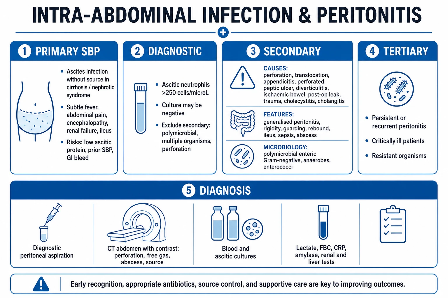
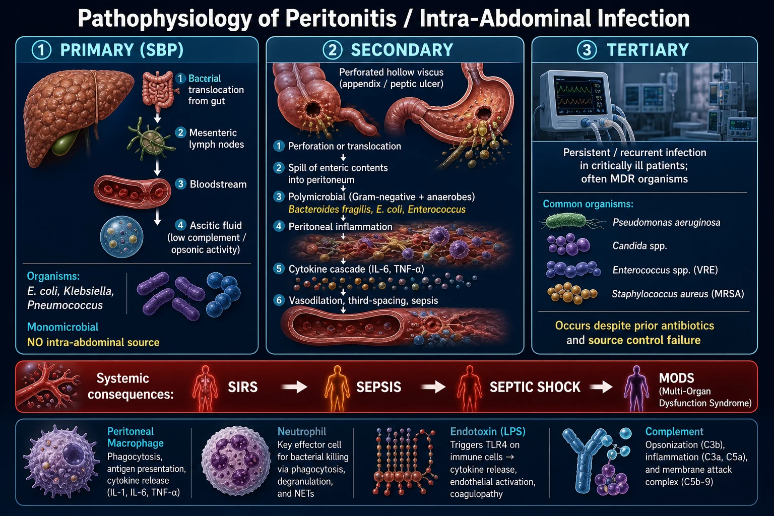
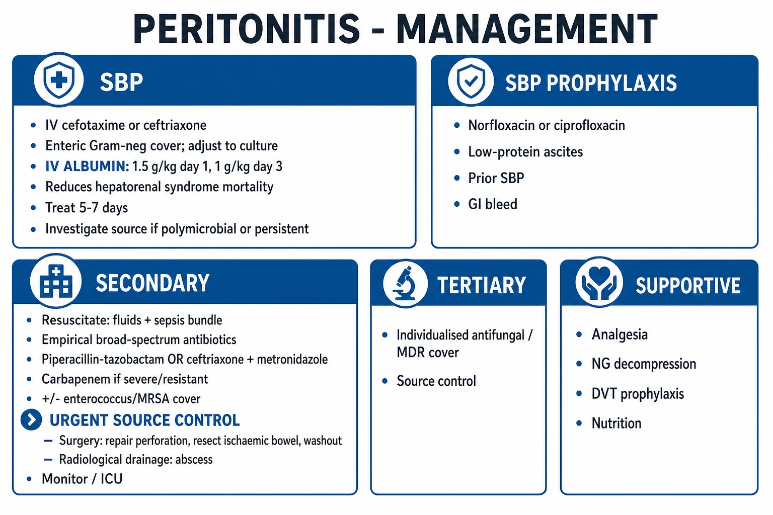
Types of peritonitis
| Feature | SBP | Secondary | Tertiary | CAPD |
|---|---|---|---|---|
| Source | None (spontaneous) | Perforation, leak, abscess | Persistent/recurrent after secondary | Peritoneal dialysis catheter |
| Population | Cirrhosis with ascites | Anyone with abdominal pathology | Critically ill ICU patients | PD patients |
| Organisms | Monomicrobial (E. coli, Klebsiella) | Polymicrobial (gram-negative, anaerobes) | Resistant (Enterococcus, Candida, Pseudomonas, MRSA) | Staphylococcus (epidermidis, aureus) |
| Diagnosis | Ascitic PMN >250/mm³ | CT, laparotomy | Persistent sepsis, cultures positive | Cloudy dialysate, PMN >100/mm³ |
| Treatment | Cefotaxime + ALBUMIN | SURGERY (source control) + broad antibiotics | Anti-fungal, anti-Enterococcus, anti-Pseudomonas | Intraperitoneal antibiotics ± catheter removal |
| Mortality | 10-30% | 5-20% | 30-50% | <5% (per episode) |
Management of suspected secondary peritonitis
- Resuscitate — ABC. IV fluids (septic patients often hypovolaemic). Vasopressors (noradrenaline) for shock. Broad-spectrum antibiotics within 1h
- CT abdomen with contrast — identify source (perforation, abscess, leak, ischaemia)
- Source control — SURGERY is MANDATORY: (a) Perforation — repair/resect. (b) Abscess — drain (percutaneous or surgical). (c) Leak — repair/re-anastomose/stoma. (d) Ischaemia — resect necrotic bowel. (e) Lavage (wash out peritoneum)
- Antibiotics — broad-spectrum, polymicrobial cover: piperacillin/tazobactam OR meropenem + metronidazole. Cover gram-negative, anaerobes, Enterococcus. Consider antifungal (candida) if prolonged, immunosuppressed
- ICU support — ventilation, renal support, vasopressors
- Duration: 4 days after source control (STOP-IT trial showed no benefit of longer)
- Monitor for tertiary peritonitis — persistent sepsis despite source control → resistant organisms → broaden antibiotics
SAQ — Spontaneous bacterial peritonitis in a cirrhotic with decompensation
10 minutes · 10 marks
A 54-year-old man with known alcohol-related cirrhosis (Child-Pugh C, MELD-Na 22) presents with 36 hours of abdominal pain, fever 38.6 degrees C, and increasing confusion. He is on no SBP prophylaxis. On arrival he is drowsy (GCS 13), BP 88/54 (MAP 65), HR 112, RR 22, SpO2 96 per cent on room air, T 38.4 degrees C. The abdomen is distended with a positive fluid thrill and generalised mild tenderness without guarding. Blood tests: Hb 96, WCC 14.2, neutrophils 11.8, platelets 78, creatinine 138 micromol/L (baseline 92), bilirubin 92 micromol/L, INR 1.9, albumin 22 g/L, lactate 1.6. Diagnostic paracentesis returns turbid fluid; the bedside dipstick is leukocyte esterase 3+.
SAQ — Secondary peritonitis from a perforated viscus with septic shock
10 minutes · 10 marks
A 67-year-old man presents with 14 hours of severe generalised abdominal pain and vomiting. He is on apixaban for non-valvular atrial fibrillation. On arrival he is clammy and drowsy (GCS 14), temperature 38.9 degrees C, HR 132 in AF, BP 78/42 (MAP 54) on 5 L/min nasal spec, RR 28, SpO2 94 per cent. The abdomen is rigid and distended with generalised peritonism and absent bowel sounds. Bloods: Hb 121, WCC 22.4, lactate 5.8, creatinine 188 micromol/L, venous pH 7.21, base excess -10, INR 1.4. An erect chest x-ray shows free gas under the diaphragm. He has had no recent surgery; he takes apixaban 5 mg BD with last dose 8 hours ago.
Clinical pearls
Red flags
Prognosis
Albumin in SBP (Sort 1999, NEJM)
RCT: 126 cirrhotic patients with SBP. Cefotaxime + albumin vs cefotaxime alone.
- Hepatorenal syndrome: 10% (albumin) vs 33% (control) — p=0.002, 67% relative reduction
- In-hospital mortality: 10% (albumin) vs 29% (control) — p=0.01, 65% relative reduction
- 3-month mortality: 22% vs 41% — p=0.03 [1]
Albumin dose: 1.5 g/kg day 1, 1 g/kg day 3. CONCLUSION: Albumin is STANDARD OF CARE for SBP (especially with bilirubin >68 μmol/L or creatinine >88 μmol/L).
SBP — spontaneous bacterial peritonitis: deep dive [1]
Pathophysiology and portal of entry — why the four peritonitides differ
| Type | Source / portal of entry | Microbiology | Key host factor |
|---|---|---|---|
| SBP | Haematogenous / lymphatic seeding of ascites; intestinal bacterial translocation across oedematous, more-permeable cirrhotic gut wall. NO visible intra-abdominal source on imaging. | Monomicrobial — single organism in 90%+ of culture-positive cases | Cirrhosis with ascites: low ascitic opsonic/complement activity (protein <15 g/L, C3 low) impairs phagocytosis. Diseased Kupffer cells fail to clear portal bacteraemia. |
| Secondary | Spillage from a breached viscus — perforation, ischaemic necrosis, anastomotic leak, abscess, trauma. Source is anatomical and IMAGING-DETECTABLE. | Polymicrobial — gut flora: gram-negative bacilli, anaerobes (Bacteroides, Clostridium), Enterococcus, streptococci; ± Candida | Any patient with abdominal pathology; severity depends on load, virulence, host, and source-control TIMING. |
| Tertiary | Persistent or recurrent infection after apparent control of secondary peritonitis — failure of source control or host immunity. Often NO clear new source. | MDR/fermenters/fungi — Enterococcus (incl. VRE), Candida, Pseudomonas, Stenotrophomonas, MRSA, Acinetobacter | Critically ill ICU patient: immune paralysis, prolonged antibiotics, foreign material (mesh, drains), open abdomen, TPN. Reflects immunoparesis more than new contamination. |
| CAPD / catheter-related | Inoculation along the Tenckhoff catheter (touch contamination, exit-site/tunnel infection) or trans-visceral migration. Peritoneum is the dialyser. | Staphylococci (CoNS 30-45%, S. aureus 10-20%), gram-negatives (15-25%), Streptococcus (10%), fungal (2-5%) | Peritoneal dialysis patients; defence diluted by ~2 L dialysate, low opsonin, catheter as foreign body. |
Ascitic fluid analysis — interpreting the diagnostic paracentesis
| Result | Definition | Significance | Action |
|---|---|---|---|
| SBP | Ascitic PMN ≥ 250 cells/mm³ (with or without positive culture) | Diagnostic of SBP — treat empirically NOW. Culture positive in only ~50%. | Cefotaxime 2 g IV BD-TDS × 5-7 days + albumin 1.5 g/kg day 1, 1 g/kg day 3.[1] |
| Culture-negative neutrocytic ascites (CNNA) | PMN ≥ 250/mm³ AND negative culture (and no antibiotics in prior 7 days) | A variant of SBP — identical prognosis and management. The 50% culture-negative group. | Treat as SBP. Not a false positive — usually due to culture technique; bedside inoculation into blood-culture bottles ↑ yield to 70-80%.[1] |
| Monomicrobial non-neutrocytic bacterascites (MNB) | Single organism on culture AND PMN < 250/mm³ | Often transient/colonisation that self-clears; BUT treat if symptomatic (fever, pain) or cirrhotic — risk of progression to SBP. | Treat if symptomatic; repeat tap in 24-48 h if asymptomatic. |
| Polymicrobial bacterascites | Multiple organisms AND PMN < 250/mm³ | Suggests PERFORATION — bowel leak inoculating ascites. Diagnostic clue that this is NOT SBP. | Image (CT) for perforation → surgical source control. |
| Secondary peritonitis (in cirrhotic with ascites) | PMN ≥ 250 AND polymicrobial culture (multiple organisms, including anaerobes/yeast) AND ascitic: total protein >10 g/L, glucose <2.8 mmol/L, LDH >225 U/L | Differentiate from SBP: SBP is monomicrobial, no anaerobes, normal glucose/LDH. Secondary needs surgery. | CT + surgery; broaden to cover anaerobes + Enterococcus. |
Diagnostic workup of a cirrhotic patient with ascites and fever/altered mental state
- Diagnostic paracentesis is MANDATORY — every cirrhotic with ascites + any of: fever/abdominal pain/altered mental state/renal impairment/GI bleed/leucocytosis. Also tap on admission and at first sign of decompensation. Up to 30% of asymptomatic cirrhotic inpatients have SBP on admission.[1]
- Bedside technique — supine, LLQ or RLQ (midline 2 cm below umbilicus in obese). Clean skin. 20-30 mL aspirated. Inoculate blood-culture bottles directly at the bedside (aerobic + anaerobic) — doubles culture yield vs delayed lab inoculation. Send: cell count + differential, protein, albumin, glucose, LDH, Gram stain, culture.
- Reagent strip (dipstick) PMN — a leukocyte esterase dipstick (≥2+) gives a rapid bedside PMN surrogate when the lab count is delayed. Sensitivity for SBP ~90%.
- Diagnosis = PMN ≥ 250 cells/mm³ — start antibiotics immediately; do NOT wait for culture. If PMN borderline (e.g., 200-250), repeat in 24-48 h.
- Coagulopathy is NOT a contraindication — cirrhotic coagulopathy/ascites do not require fresh frozen plasma/platelets before a therapeutic or diagnostic tap (mild bleeding risk <1%, life-threatening <0.1%).[1]
- If PMN ≥ 250 → CT abdomen/pelvis if any feature of secondary peritonitis (polymicrobial Gram stain, anaerobes, ascitic glucose <2.8 or LDH >225, free gas on CXR/CT) to exclude a perforated viscus.
- Reassess at 48 h — repeat tap. PMN should fall by ≥ 25% at 48 h with appropriate antibiotic. If not, suspect resistant organism or secondary peritonitis → broaden + image.
Organisms in SBP — predicted microbiology
| Group | Frequency | Organisms | Antibiotic implication |
|---|---|---|---|
| Gram-negative bacilli | 70-75% | Escherichia coli (most common, ~50%), Klebsiella pneumoniae, other Enterobacterales | Third-generation cephalosporin (cefotaxime/ceftriaxone) — covers ~95% of community-acquired SBP. |
| Gram-positive cocci | 20-25% | Streptococcus pneumoniae (pneumococcus), other streptococci, Enterococcus (more common nosocomial) | Cefotaxime covers streptococci/pneumococcus; Enterococcus needs ampicillin/vancomycin (cover if nosocomial). |
| Anaerobes | <5% (RARE) | Bacteroides, Clostridium | The low ascitic oxygen tension + presence of complement selects against anaerobes — do NOT add metronidazole routinely for classic SBP. |
| Nosocomial / healthcare-associated SBP | rising | MDR gram-negatives (ESBL, KPC), Enterococcus (incl. VRE), Pseudomonas | Switch to piperacillin/tazobactam, meropenem, or ceftolozane/tazobactam based on local antibiogram. Cefotaxime fails in ~20% of nosocomial SBP.[3] |
| Fungal | <2% | Candida | Only in heavily antibiotic-exposed/immunosuppressed — add echinocandin. |
SBP antibiotic therapy — empiric regimens by clinical context
| Clinical context | First-line | Alternative / allergy | Duration |
|---|---|---|---|
| Community-acquired SBP, uncomplicated | Cefotaxime 2 g IV BD-TDS OR ceftriaxone 2 g IV OD | Levofloxacin 500 mg IV/PO OD or moxifloxacin (if no prior quinolone prophylaxis, no vomiting/shock) | 5 days (as effective as 10 days) |
| SBP with sepsis/septic shock | Cefotaxime/ceftriaxone ± piperacillin/tazobactam (broaden for healthcare acquisition) | Meropenem 1 g IV TDS | 7 days; reassess at 48 h by PMN fall |
| Healthcare-associated / nosocomial SBP | Piperacillin/tazobactam 4.5 g IV TDS-QDS OR meropenem | Add vancomycin if VRE/Enterococcus risk; consider ceftolozane/tazobactam for MDR Pseudomonas | 7 days, de-escalate on culture |
| Quinolone prophylaxis failure | NEVER use ofloxacin/ciprofloxacin empirically — high quinolone resistance | Cefotaxime/ceftriaxone (1st) | 5-7 days |
| Add ALBUMIN to ALL SBP | 1.5 g/kg IV day 1, 1 g/kg day 3 | (Saline is NOT a substitute — must be albumin) | Days 1 and 3 only |
SBP management algorithm — bedside sequence
- Confirm diagnosis — ascitic PMN ≥ 250 cells/mm³.
- Cefotaxime 2 g IV BD-TDS (or ceftriaxone 2 g IV OD) — start within 1 h of tap. Send blood cultures (bacteraemia in 30-50%).
- Albumin 1.5 g/kg IV on day 1, 1 g/kg IV on day 3 — give to ALL SBP patients; MANDATORY if any of: creatinine >88 μmol/L, BUN >30 mg/dL, bilirubin >68 μmol/L.[2]
- Re-tap at 48 h — PMN must fall ≥ 25%. If not: suspect resistant organism or secondary peritonitis → broaden (piperacillin/tazobactam or meropenem), CT to exclude perforation.
- Search for and treat precipitant — GI bleed (give SBP prophylaxis), urinary/biliary infection, prior paracentesis without albumin, dehydration, large-volume paracentesis without albumin replacement.
- Continue therapy 5 days total for uncomplicated SBP; 7 days for sepsis/bacteraemia.
- Start SECONDARY PROPHYLAXIS at discharge — norfloxacin 400 mg OD or ciprofloxacin 500 mg OD (lifelong, until transplant). Recurrence 70% at 1 year without prophylaxis vs 20% with.[1]
- Refer for liver transplant evaluation — SBP is a decompensating event that doubles 1-year mortality (30-50%). MELD-Na drives listing priority.
Albumin in cirrhosis — three landmark trials, three different conclusions
| Trial | Population | Intervention | Key result | Interpretation |
|---|---|---|---|---|
| Sort 1999 (NEJM) | 126 SBP patients | Cefotaxime + albumin (1.5 g/kg D1, 1 g/kg D3) vs cefotaxime alone | HRS 10% vs 33%; hospital mortality 10% vs 29% | Albumin is STANDARD OF CARE for SBP.[2] |
| ANSWER 2018 (Lancet) | 440 decompensated cirrhosis (no SBP) | Long-term weekly albumin × 18 months vs standard care | 50% reduction in SBP, fewer decompensation events, 18-month survival improved (74% vs 62%) | Long-term albumin reduces SBP incidence and improves survival in selected outpatients.[8] |
| ATTIRE 2021 (NEJM) | 777 hospitalised cirrhosis patients | Targeted 20% albumin infusions to keep serum albumin >30 g/L vs standard care | No reduction in new infection, renal dysfunction, or mortality | Albumin infusions to a target level in unselected inpatients do NOT prevent infection. Be selective — benefit proven in SBP and ANSWER-selected outpatients only.[9] |
ANSWER trial — Caraceni 2018, Lancet (long-term albumin in decompensated cirrhosis)
Open-label RCT, Italy. 440 patients with decompensated cirrhosis, no SBP at entry.
- Intervention: human albumin 1.5 g/kg/week for 2 weeks, then 40 g/week for 18 months, vs standard care.
- Primary endpoint — 18-month mortality: 77/220 (albumin) vs 99/220 (standard) — HR 0.66, borderline.
- Incident SBP: 10% vs 24% — hazard ratio 0.40 (60% reduction).
- Grade 3-4 ascites, renal dysfunction, bacterial infection: all significantly reduced.
- CONCLUSION: In selected outpatients with decompensated cirrhosis, long-term albumin reduces SBP incidence and may improve survival. Selective use, not blanket ATTIRE-style infusions.[8]
ATTIRE — China 2021, NEJM (albumin in hospitalised cirrhosis)
Multicentre RCT, UK. 777 hospitalised patients with cirrhosis and serum albumin <30 g/L.
- Intervention: 20% human albumin to maintain serum albumin ≥30 g/L (target-led) up to 14 days vs standard care.
- Primary composite (new infection, renal dysfunction, death) at 15 days: 29.7% vs 30.2% — no difference.
- Adverse events: pulmonary oedema more common with albumin.
- CONCLUSION: Routine albumin infusions in unselected hospitalised cirrhosis do NOT improve outcomes. Albumin should be reserved for SBP, large-volume paracentesis (≥5 L), HRS, and selected ANSWER-style outpatients.[9]
Secondary peritonitis: deep dive — sources, organisms, source control
Sources of secondary peritonitis — predicted by anatomical site of perforation
| Source | Classic organism profile | Clinical clues | Imaging |
|---|---|---|---|
| Perforated appendicitis | E. coli, Bacteroides fragilis, Streptococcus anginosus group | Migration of periumbilical→RLQ pain; McBurney's point tenderness; young male; perforation → diffuse peritonitis/sepsis | CT: peri-appendiceal fat stranding, free fluid ± free gas, appendicolith |
| Perforated diverticulitis (Hinchey III-IV) | E. coli, Klebsiella, Bacteroides, Clostridium; ± Enterococcus | LLQ pain in older patient; acute severe abdominal pain + rigid abdomen in Hinchey IV; faeculent peritonitis | CT: sigmoid diverticula, free gas ± faeces; Hinchey stage |
| Perforated peptic ulcer (PUD) | Streptococcus, E. coli, anaerobes (after several hours) | Sudden "tearing" epigastric pain → rigid abdomen; "Valentino's sign" (RIF pain from gastric fluid tracking down right paracolic gutter); standing CXR: free subdiaphragmatic gas | Erect CXR; CT abdomen; "Valsalva/computed tomography" shows anterior free gas |
| Ischaemic bowel / mesenteric infarction | E. coli, Klebsiella, Clostridium perfringens, Enterococcus; high risk of bacteraemia | AF, heart failure, vasopressors; "pain out of proportion"; metabolic acidosis, rising lactate; bloody diarrhoea late | CT: pneumatosis intestinalis, portal venous gas, mesenteric vessel occlusion, thickened oedematous loops |
| Anastomotic leak (postoperative, day 3-7) | Enterococcus (ampicillin-resistant), Candida, Pseudomonas, MDR GNR — reflects hospital flora | Fever, ileus, drain output (faeculent/bilious/purulent), sepsis after GI surgery | CT with water-soluble oral/rectal contrast (NOT barium); contrast extravasation |
| Biliary source (cholecystitis, cholangitis, bile leak) | E. coli, Klebsiella, Enterococcus, Enterobacter; less often anaerobes (Clostridium perfringens — emphysematous) | RUQ pain, jaundice, Charcot's/Reynold's pentad; post-cholecystectomy/ERCP; bile-stained peritoneal fluid | MRCP/ERCP; CT: wall thickening, pericholecystic fluid, ductal dilatation |
| Post-traumatic (blunt or penetrating bowel injury) | E. coli, Bacteroides, Streptococcus | Mechanism (seatbelt sign, handlebar, GSW); delayed presentation; persistent tachycardia/lactataemia after trauma | CT: free gas, free fluid, bowel wall thickening, mesenteric stranding |
Predicted organisms and antibiotic cover by source (community vs healthcare-associated)
| Severity / context | Predicted cover required | Empiric regimen |
|---|---|---|
| Community-acquired, mild-moderate | Gram-negative rods + anaerobes | Ceftriaxone 2 g IV OD + metronidazole 500 mg IV TDS, OR co-amoxiclav 1.2 g IV TDS + metronidazole, OR moxifloxacin 400 mg IV OD (monotherapy) |
| Community-acquired, high-severity / sepsis | Gram-negative rods + anaerobes + Enterococcus | Piperacillin/tazobactam 4.5 g IV TDS-QDS, OR meropenem 1 g IV TDS + metronidazole (if ESBL risk) |
| Healthcare-associated / postoperative | Gram-negatives (incl. ESBL/Pseudomonas) + Enterococcus + Candida | Piperacillin/tazobactam OR meropenem + vancomycin (Enterococcus/MRSA) + echinocandin (caspofungin 70 mg then 50 mg OD) if recent fluoroquinolone/cephalosporin exposure, TPN, prolonged ICU stay |
| Biliary source | Gram-neg + Enterococcus (anaerobes RARE in pure biliary disease unless emphysematous) | Ceftriaxone + metronidazole; piperacillin/tazobactam monotherapy if severe |
| Candida peritonitis (post-op, multiple abdominal re-explorations, necrotising pancreatitis, immunosuppressed) | C. albicans, C. glabrata, C. krusei | Echinocandin (caspofungin/micafungin/anidulafungin) if critically ill; step down to fluconazole if stable and C. albicans susceptible. Remove infected foreign material; source control.[10] |
Principles of SOURCE CONTROL — the four D's
- DRAINAGE — percutaneous (image-guided) or surgical drainage of collections/abscess. Aim: evacuating pus and contaminated fluid.
- DEBRIDEMENT — excision of necrotic, ischaemic or grossly contaminated tissue (non-viable bowel, omental slough). Necrotic tissue is a culture medium that antibiotics cannot penetrate.
- DECOMPRESSION / DIVERSION — relieve obstruction (NGT, rectal tube, defunctioning stoma); prevent ongoing contamination.
- DEFINITIVE REPAIR — close or resect the breach: primary repair, resection + anastomosis, resection + stoma (Hartmann's), or controlled fistula formation (open abdomen with VAC, "planned re-laparotomy"). [1]
Timing: Source control within 6 h of septic shock onset is associated with reduced mortality (Surviving Sepsis). Each additional hour of delay in the hypotensive patient increases mortality ~7-8%.[11]
Management of suspected perforated viscus with septic shock
- ABCDE resuscitation — oxygen, two large-bore IVs. Crystalloid 30 mL/kg bolus for sepsis-induced hypoperfusion. Noradrenaline first-line vasopressor if MAP <65 mmHg after fluids. Add vasopressin / adrenaline if refractory.
- Broad-spectrum antibiotics within 1 h (sepsis hour-1 bundle) — piperacillin/tazobactam 4.5 g IV ± vancomycin (Enterococcus/MRSA) ± echinocandin if Candida risk. Add metronidazole if using ceftriaxone/cefepime alone.
- Stat surgical review + anaesthetics — declare a surgical emergency; consent for laparotomy.
- Cross-match 4-6 units, group-and-save, check coagulation, platelets, lactate, venous gas, lactate trend.
- CT abdomen/pelvis with IV + water-soluble oral/rectal contrast — only if the patient can be stabilised (CT is inadvisable in extremis — go straight to theatre). Confirm free gas, perforation site, abscess.
- Urinary catheter + arterial line + CVC — guide fluid responsiveness and vasopressors; target MAP ≥65, lactate clearance ≥10%/h, ScvO₂ ≥70%.
- Theatre — emergency laparotomy: peritoneal lavage, identify and repair/resect source, drain. Damage-control surgery in unstable patients (leave abdomen open, VAC, planned second-look).
- Post-op ICU — vasopressor wean, lung-protective ventilation, renal support, glycaemic control 6-10 mmol/L, deep vein thrombosis prophylaxis once bleeding controlled, stress ulcer prophylaxis, enteral nutrition within 24-48 h.
- Antibiotic duration: 4 days after adequate source control (STOP-IT).[4] Prolong if persistent sepsis, bacteraemia, fungaemia, source control incomplete.
STOP-IT trial — Sawyer 2015, NEJM (duration of antibiotics after source control)
Multicentre RCT, USA. 518 patients with complicated intra-abdominal infection and adequate source control.
- Intervention: antibiotics stopped after a fixed ~4-day course (approx. 96 h) vs continued until physician-satisfied (~8 days).
- Primary composite (surgical-site infection, recurrent IAI, death) at 30 days: 21.8% (short) vs 22.3% (long) — non-inferior.
- Median duration: 4 days (short) vs 8 days (long).
- CONCLUSION: Once adequate source control is achieved, ~4 days of antibiotics is sufficient. Prolonged courses do not improve outcomes and select for resistance/C. difficile.[4]
Tertiary peritonitis: deep dive — the failing host
Secondary vs tertiary peritonitis — distinguishing the ICU phenotype
| Feature | Secondary peritonitis | Tertiary peritonitis |
|---|---|---|
| Timing | Acute — first presentation of perforation/leak | Persistent or recurrent after ≥48 h of treatment of secondary, OR no source identifiable |
| Host | Often previously well; new intra-abdominal catastrophe | Critically ill; prolonged ICU stay, multi-organ failure, immunoparesis |
| Source | Identifiable perforation/leak/abscess | Often NO source found at re-operation; widespread serosal inflammation, "sticky" fibrinous peritoneum |
| Microbiology | Predictable gut flora | MDR/low-virulence/opportunistic — Enterococcus (incl. VRE), Candida, Pseudomonas aeruginosa, Stenotrophomonas maltophilia, coag-neg staph, MRSA, Acinetobacter |
| Cultures | Often polymicrobial with predictable gut organisms | Often multiple isolates of hospital-acquired MDR organisms; may be culture-negative despite florid sepsis |
| Fever pattern | High-grade, swinging | Afebrile or low-grade — host too sick to mount fever; hypothermia a poor sign |
| WCC | Leucocytosis with left shift | Often leucopenia or normal WCC in immunoparalysed host |
| Treatment | Surgery (source control) + narrow antibiotics | Broad-spectrum anti-MDR + antifungal + immune-supportive care; surgery often unhelpful (no source to fix) |
| Mortality | 5-20% | 30-50% — driven by multi-organ failure rather than uncontrolled infection per se |
| Conceptual model | A surgical disease | A marker of critical illness / immune dysfunction — "the peritoneum as the organ that fails"[5] |
Organisms in tertiary peritonitis and rationale for cover
| Organism | Why it dominates in tertiary peritonitis | Antibiotic of choice |
|---|---|---|
| Enterococcus (E. faecium → VRE, E. faecalis) | Selected by cephalosporin use (inherently resistant); intrinsically resistant gut coloniser | Ampicillin (E. faecalis); linezolid or daptomycin for VRE (E. faecium) |
| Candida (albicans, glabrata, krusei) | Broad-spectrum antibiotics deplete bacteria; TPN; central lines; multiple laparotomies | Echinocandin (caspofungin) initially; fluconazole step-down |
| Pseudomonas aeruginosa | Biofilm on catheters/drains; prior antibiotic exposure selects MDR strains | Piperacillin/tazobactam, ceftolozane/tazobactam, ceftazidime-avibactam, or meropenem (depending on sensitivities); add aminoglycoside in severe |
| Stenotrophomonas maltophilia | Selected by carbapenems; environmental water organism in ICU sinks | TMP-sulfamethoxazole (only reliable agent) |
| MRSA | Lines, wounds, prolonged ICU stay | Vancomycin or linezolid |
| Acinetobacter baumannii | survives on ICU surfaces; extreme MDR | Sulbactam combinations, polymyxin/colistin, tigecycline (organism-dependent) |
| Coagulase-negative staphylococci | Foreign material (mesh, drains), low-virulence | Often contaminants; treat only if repeatedly isolated + clinical infection |
Approach to tertiary peritonitis — the patient who isn't getting better
- Confirm the diagnosis — persistent or recurrent intra-abdominal sepsis ≥48 h after adequate source control of secondary peritonitis. Low-grade fever, persistent leucocytosis/thrombocytopenia, rising lactate, ongoing vasopressor requirement, persistent ileus.
- Re-image — CT abdomen/pelvis with contrast looking for undrained collection, leak, ischaemia, abscess. False negatives are common in tertiary peritonitis — small loculated collections may not be drainable.
- Re-evaluate source control — is there a drainable collection? A missed enterocutaneous fistula? Anastomotic leak? Consider diagnostic laparoscopy/laparotomy if imaging equivocal — BUT surgery in tertiary peritonitis is often unrewarding ("sticky" peritoneum, no clear source, high surgical risk).
- Re-culture aggressively — peritoneal fluid (from drains), blood cultures, line tips. Send for fungal and atypical cultures. Antimicrobial stewardship review.
- Broaden empirically — anti-MDR cover: meropenem + vancomycin + echinocandin (caspofungin). Adjust to local antibiogram and isolate sensitivities. Add aminoglycoside or colistin for extreme MDR GNR.
- Address the failing host — this is a disease of immune paresis. Enteral nutrition (gut mucosal trophism, reduces translocation), tight glycaemic control (6-10 mmol/L), lung-protective ventilation, renal support, avoid iatrogenic immunosuppression where possible. Consider IVIG/G-CSF only in selected cases (no routine evidence).
- Reassess daily — serial CRP/procalcitonin trends, lactate clearance, vasopressor weaning, organ support weaning. De-escalate antibiotics as soon as sensitivities allow.
- Mortality is 30-50% — discuss goals of care early with family and MDT. Persistent multi-organ failure beyond day 7-10 of tertiary peritonitis carries >80% mortality.[5]
Catheter-related / peritoneal dialysis (CAPD) peritonitis
PD peritonitis — organisms, source and expected course
| Organism | % of cases | Source | Typical course |
|---|---|---|---|
| Coagulase-negative staphylococci (S. epidermidis) | 30-45% | Touch contamination at exchange — most common, usually mild | Responds to intraperitoneal (IP) vancomycin/cefazolin; catheter usually retained |
| Staphylococcus aureus | 10-20% | Exit-site / tunnel infection; biofilm on catheter | More severe; may need catheter removal if tunnel infection or refractory |
| Streptococcus / Enterococcus | 10-15% | Oropharyngeal/gut source; trans-visceral migration | Treat with IP vancomycin/cefazolin + IP aminoglycoside; Enterococcus — ampicillin if susceptible |
| Gram-negative bacilli (E. coli, Klebsiella, Pseudomonas) | 15-25% | Gut translocation, urinary source; Pseudomonas from water/biofilm | Pseudomonas → catheter removal usually required; cover with IP ceftazidime/gentamicin, then per sensitivities |
| Fungal (Candida) | 2-5% | Heavily antibiotic-exposed, immunosuppressed, recent bacterial peritonitis | Catheter removal MANDATORY; systemic antifungal (echinocandin/fluconazole); mortality 10-25% |
| Culture-negative | 10-20% | — | Usually responds to IP antibiotics; rule out fastidious organisms (mycobacteria, fungi) if persistent |
Indications for Tenckhoff catheter removal in PD peritonitis
| Indication | Reason |
|---|---|
| Fungal peritonitis (any Candida) | Catheter universally infected/biofilm-coated; cure impossible with catheter in situ. Remove immediately on diagnosis. |
| Refractory peritonitis (no clinical/culture response at 5 days of appropriate IP antibiotics) | Biofilm; needs catheter removal + 2-3 weeks of systemic antibiotics before reinsertion |
| Recurrent peritonitis (same organism <30 days after completion of therapy) | Suggests biofilm or tunnel focus; remove catheter |
| Pseudomonas / Stenotrophomonas | Often refractory; catheter removal usually required to cure |
| Repeating peritonitis (different organism <30 days after therapy) | Often dialysate/tubing contamination; review exchange technique |
| Tunnel infection not responding to antibiotics | Suggests catheter focus; removal prevents recurrent |
| Severe intra-abdominal pathology (appendicitis, ischaemia, perforation) | Differentiate from PD peritonitis (PD = monomicrobial usually); treat as secondary peritonitis + remove catheter |
Empiric management of suspected PD peritonitis — intraperitoneal antibiotics
- Diagnosis — cloudy dialysate + abdominal pain ± fever. The cardinal sign is cloudy effluent. Send: dialysate cell count + differential (after ≥2 h dwell), Gram stain, culture. PD peritonitis = dialysate WCC >100/mm³ with PMN >50% (after a dwell).[6]
- Empiric IP antibiotics within 2-3 h of presentation — IP vancomycin or cefazolin (gram-positive cover) + IP ceftazidime or gentamicin (gram-negative cover). IP dosing delivers the antibiotic directly to the peritoneum — higher local levels than IV. Loading dose added to one exchange; dwell ≥6 h.
- Add heparin 500-1000 U/L to dialysate — prevents fibrin clots obstructing the catheter.
- Once culture results (24-72 h) — narrow to organism. CoNS → IP cefazolin or vancomycin alone; S. aureus → IP vancomycin + review exit site; gram-negative → IP ceftazidime (or gentamicin if not aminoglycoside-sparing); fungal → remove catheter + systemic antifungal.
- Duration: 2 weeks (coNS, strep); 3 weeks (S. aureus, gram-negative, Enterococcus); until catheter removed + 2-3 weeks systemic (fungal).
- Catheter removal if: fungal, refractory at 5 days, recurrent (same organism), Pseudomonas/Stenotrophomonas, or severe intra-abdominal pathology.
- Prevention — meticulous exit-site care (mupirocin daily to nares + exit site), training/re-training on aseptic technique (disconnect systems reduce touch contamination), prophylactic mupirocin/rifampicin for S. aureus nasal carriage.
Routes of antibiotic administration in PD peritonitis — IP vs IV
| Route | Pros | Cons | When |
|---|---|---|---|
| Intraperitoneal (IP) | Preferred — achieves high peritoneal levels; outperforms IV for PD peritonitis; avoids systemic exposure | Adds to dialysate; requires dwell ≥6 h; compatibility (vancomycin + ceftazidime may precipitate — give in separate exchanges) | First-line for all PD peritonitis; load in the long (overnight) dwell.[6] |
| Intravenous (IV) | Easy, well tolerated | Lower peritoneal penetration than IP; less effective | Reserve for severe sepsis, systemic infection, or when PD temporarily held; combine with IP |
Additional clinical pearls
Additional red flags
Summary — what the CICM/FFICM/EDIC candidate must know
[1]Peritonitis at a glance — the one-table revision
| Feature | SBP | Secondary | Tertiary | CAPD |
|---|---|---|---|---|
| Population | Cirrhosis + ascites | Any abdominal catastrophe | Critically ill ICU patient | PD patient |
| Source | None (haematogenous) | Perforation/leak/abscess | Persistent/recurrent; often no source | Catheter (touch contamination) |
| Microbiology | Monomicrobial GNR (E. coli, Klebsiella, S. pneumo) | Polymicrobial gut flora + anaerobes | MDR (Enterococcus, Candida, Pseudomonas, MRSA) | Staph (CoNS, S. aureus), GNR, fungal |
| Diagnosis | Ascitic PMN ≥ 250/mm³ | CT + surgery | Persistent sepsis, cultures positive | Dialysate WCC >100/mm³, PMN >50% |
| First-line treatment | Cefotaxime + ALBUMIN | Surgery + piperacillin/tazobactam | Meropenem + vancomycin + echinocandin | Intraperitoneal vancomycin/cefazolin + ceftazidime |
| Source control | NOT required (no source) | MANDATORY | Often unrewarding | Catheter removal for specific indications |
| Antibiotic duration | 5-7 days | 4 days after source control | Often prolonged (until clinically resolving) | 2-3 weeks |
| Mortality | 10-30% (albumin halves high-risk) | 5-20% (driven by source-control timing) | 30-50% | <5% per episode |
| Key trial | Sort 1999 (albumin) | STOP-IT 2015 (4 days) | Evans 2001 (tertiary phenotype) | ISPD 2016 (IP antibiotics) |
References
- [1]Runyon BA, AASLD. Introduction to the revised American Association for the Study of Liver Diseases Practice Guideline management of adult patients with ascites due to cirrhosis 2012 Hepatology, 2013.PMID 23463403
- [2]Sort P, Navasa M, Arroyo V, et al. Effect of intravenous albumin on renal impairment and mortality in patients with cirrhosis and spontaneous bacterial peritonitis N Engl J Med, 1999.PMID 10432325
- [3]Solomkin JS, Mazuski JE, Bradley JS, et al. Diagnosis and management of complicated intra-abdominal infection in adults and children: guidelines by the Surgical Infection Society and the Infectious Diseases Society of America Clin Infect Dis, 2010.PMID 20034345
- [4]Sawyer RG, Claridge JA, Nathens AB, et al. Trial of short-course antimicrobial therapy for intraabdominal infection N Engl J Med, 2015.PMID 25992746
- [5]Evans HL, Raymond DP, Pelletier SJ, et al. Tertiary peritonitis (recurrent diffuse or localized disease) is not an independent predictor of mortality in surgical patients with intraabdominal infection Surg Infect (Larchmt), 2001.PMID 12593701
- [6]Li PK-T, Szeto CC, Piraino B, et al. ISPD Peritonitis Recommendations: 2016 Update on Prevention and Treatment Perit Dial Int, 2016.PMID 27282851
- [7]Mazuski JE, Tessier JM, May AK, et al. The Surgical Infection Society Revised Guidelines on the Management of Intra-Abdominal Infection Surg Infect (Larchmt), 2017.PMID 28085573
- [8]Caraceni P, Riggio O, Angeli P, et al.; ANSWER Study Investigators. Long-term albumin administration in decompensated cirrhosis (ANSWER): an open-label randomised trial Lancet, 2018.PMID 29861076
- [9]China L, Freemantle N, Forrest E, et al.; ATTIRE trial investigators. A Randomized Trial of Albumin Infusions in Hospitalized Patients with Cirrhosis N Engl J Med, 2021.PMID 33657293
- [10]Pappas PG, Kauffman CA, Andes D, et al. Clinical practice guidelines for the management of candidiasis: 2009 update by the Infectious Diseases Society of America Clin Infect Dis, 2009.PMID 19191635
- [11]Sartelli M, Mazuski JE, Coccolini F, et al. Intra-abdominal infections survival guide: a position statement by the Global Alliance For Infections In Surgery World J Emerg Surg, 2024.PMID 38851700
- [12]Coccolini F, Sartelli M, Podda M, et al. Acute abdomen in the immunocompromised patient: WSES, SIS-E, WSIS, AAST, and GAIS guidelines World J Emerg Surg, 2021.PMID 34372902