ICU · GI/Nutrition
Intra-abdominal hypertension and abdominal compartment syndrome
Also known as Intra-abdominal hypertension (IAH) · Abdominal compartment syndrome (ACS) · Intra-abdominal pressure (IAP) · Abdominal perfusion pressure (APP) · Surgical decompression
Intra-abdominal hypertension (IAH) is sustained intra-abdominal pressure 12 mmHg. Abdominal compartment syndrome (ACS) is sustained IAP 20 mmHg with NEW ONSET organ failure (kidney, respiratory, cardiovascular). Causes: abdominal trauma/surgery, massive fluid resuscitation, pancreatitis, ileus, peritonitis, burns. Measurement: bladder pressure (gold standard). Effects of raised IAP: renal failure (venous congestion, reduced perfusion), respiratory failure (elevated diaphragm, reduced compliance, high ventilator pressures), cardiovascular (reduced venous return, reduced cardiac output), intestinal ischaemia, raised intracranial pressure. Treatment: decompressive laparotomy for ACS. Prevent: avoid excessive fluid resuscitation, nasogastric decompression, prokinetics.
On this page & tools
Your progress
Saved locally on this device.
Target exams
Red flags
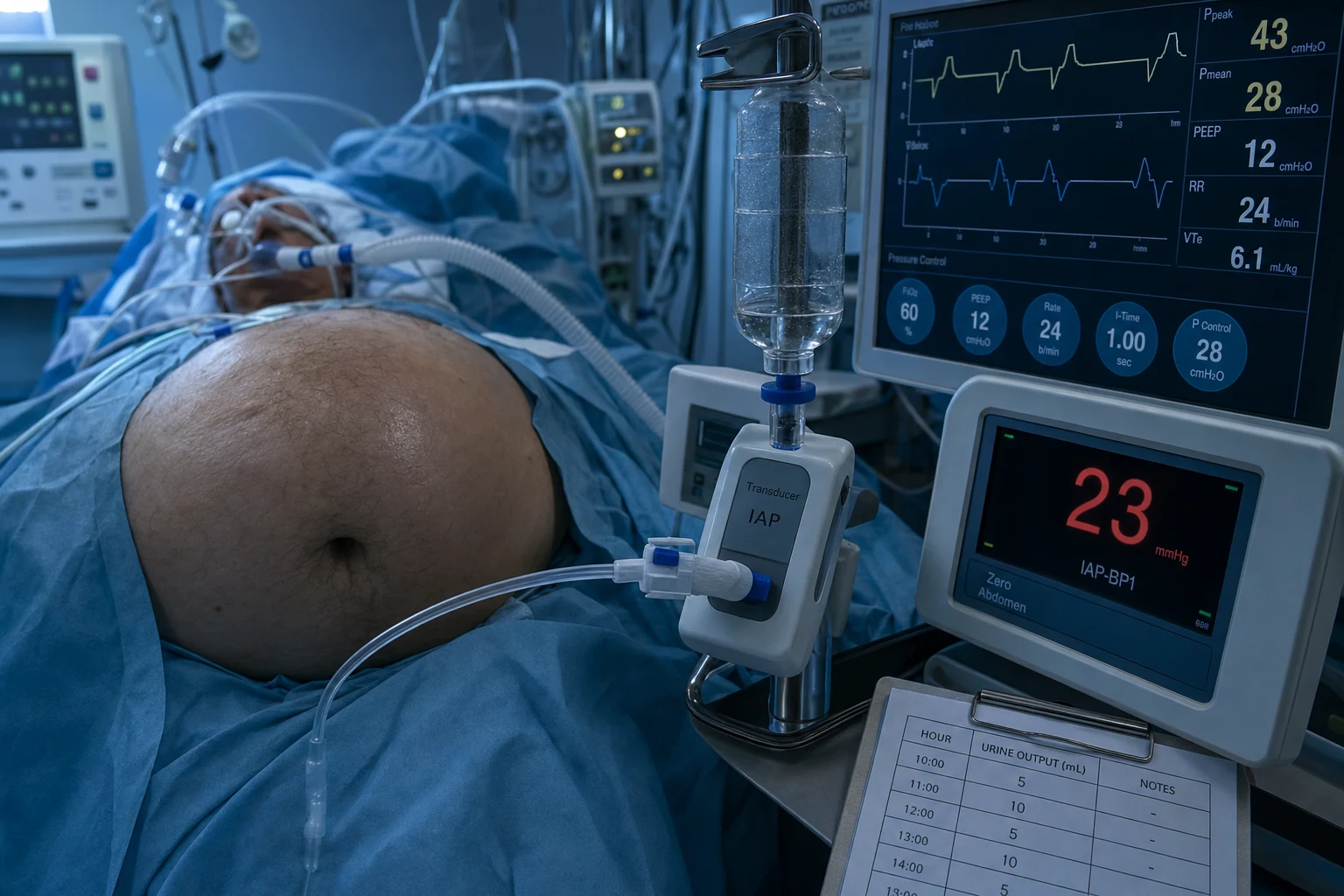
Definitions (WSACS)
WSACS grading of intra-abdominal pressure (click each)
IAP 16-20 mmHg
Moderate IAH. May begin to affect organ function. Monitor closely. Consider percutaneous catheter drainage if ascites/fluid collection.
Organ effects of raised IAP
Renal
#1 affected organ
- Oliguria → anuria (pressure on renal veins → venous congestion)
- Reduced renal plasma flow and GFR
- Renovascular compression → RAAS activation
- Mechanism: increased renal venous pressure > reduced arterial perfusion
- Often the FIRST sign of ACS — unexplained oliguria in ICU patient
Respiratory
Ventilation compromise
- Elevated diaphragm → reduced functional residual capacity
- Increased intrathoracic pressure → high peak and plateau pressures
- Reduced chest wall compliance
- Hypoxia, hypercarbia, ventilator dyssynchrony
- May mimic ARDS but responds to decompression
Cardiovascular
Reduced cardiac output
- Reduced venous return (IVC compression)
- Increased afterload (intrathoracic pressure)
- Reduced cardiac output → hypotension
- Elevated CVP and PCWP (falsely high — do not use for fluid decisions)
Other
Multi-system
- Intestinal ischaemia (mesenteric venous congestion → bacterial translocation)
- Raised intracranial pressure (important in trauma with TBI + abdominal injury)
- Hepatic dysfunction (reduced portal flow)
- Metabolic acidosis (reduced tissue perfusion)
Measurement
[1]Management
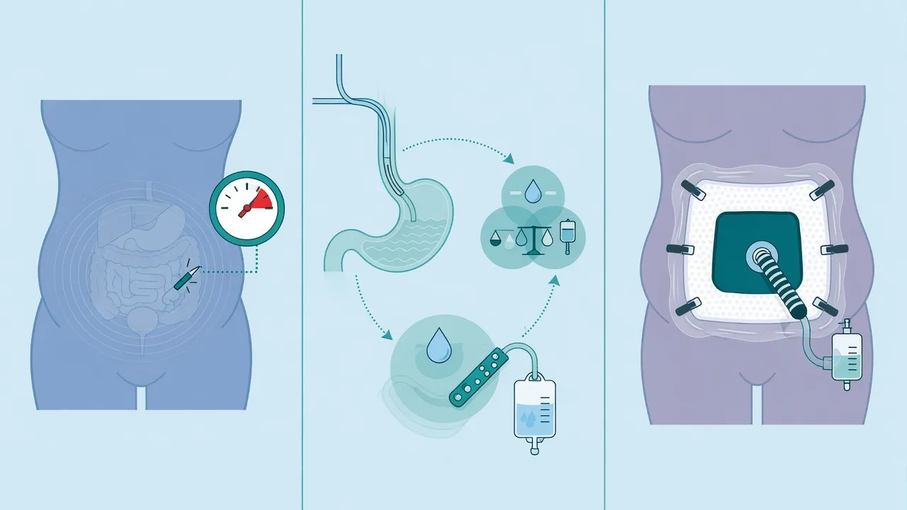
IAH/ACS management — escalating interventions
Grade I-II (IAP 12-20): medical management
(1) Ensure adequate sedation/analgesia (reduce abdominal muscle tone). (2) NG tube for gastric decompression. (3) Rectal tube/enema for colonic decompression. (4) Prokinetics (metoclopramide, erythromycin). (5) Avoid excessive fluid resuscitation (#1 cause of secondary ACS). (6) Body positioning: avoid head elevation >30 degrees (increases IAP). (7) Treat ileus.
Grade III (IAP 21-25): percutaneous drainage
If ascites, haemorrhage, or fluid collection present: percutaneous catheter drainage (pigtail catheter under ultrasound guidance). Can rapidly reduce IAP if fluid is the cause. Send fluid for culture if infection suspected.
ACS (IAP >20 + organ failure): decompressive laparotomy
SURGICAL EMERGENCY. Decompressive laparotomy: open the abdomen, leave open with temporary closure (vacuum dressing, Bogota bag). Mortality without decompression: 40-100%. After decompression: IAP drops immediately, organ function often improves within hours. Delayed closure (days-weeks later) once swelling resolves.<Cite id="3" />}
Optimise abdominal perfusion pressure (APP)
APP = MAP - IAP. Target APP >60 mmHg. May need vasopressors (noradrenaline) to increase MAP if IAP cannot be lowered immediately. Do NOT give more fluids to raise MAP — worsens IAP (vicious cycle of fluid → oedema → higher IAP → lower perfusion → more fluids).
Prevention
[1]SAQs
SAQ — ACS with bladder pressure 30 mmHg after ruptured AAA repair
10 minutes · 10 marks
A 62-year-old man is in ICU on day 2 after an emergency open repair of a ruptured abdominal aortic aneurysm. He received 8 units of packed red cells, 6 units of FFP and 6 L of crystalloid intra-operatively. He is now oliguric (8 mL/h despite two furosemide boluses), the abdomen is tense and distended, the ventilator peak airway pressure has risen from 28 to 46 cmH₂O with plateau 42 cmH₂O, and his MAP is 66 mmHg on noradrenaline 0.35 mcg/kg/min. Bladder pressure, measured with 25 mL saline instilled supine at end-expiration zeroed at the iliac crest, is 30 mmHg.
SAQ — Decompressive laparotomy decision in severe acute pancreatitis
10 minutes · 10 marks
A 48-year-old woman with severe acute necrotising pancreatitis is ICU day 6. Over the last 12 hours she has become oliguric (15 mL/h) with creatinine rising from 180 to 340 µmol/L, plateau pressure has increased from 30 to 39 cmH₂O at the same tidal volume, lactate has risen to 3.4 mmol/L, and she requires noradrenaline 0.25 mcg/kg/min for a MAP of 68 mmHg. Serial bladder pressures over 24 h are 22, 24 and 26 mmHg. The surgical team is reluctant to operate given the inflamed retroperitoneum and are asking whether continued medical management is reasonable.
Clinical pearls
Red flags
WSACS definitions — primary, secondary and recurrent ACS
The World Society of the Abdominal Compartment Syndrome (WSACS) standardised terminology in 2004, revised in 2006, 2013 and again in 2024. The definitions rest on three pillars: a pressure threshold (IAH), a graded severity (I–IV), and a pressure-plus-organ-failure diagnosis (ACS).[1][2][10]
[1]Primary ACS
Intra-abdominal cause
- Arises from injury or disease within the abdomino-pelvic cavity.
- Trauma (blunt or penetrating), ruptured AAA, abdominal surgery.
- Peritonitis / intra-abdominal sepsis, perforated viscus.
- Severe acute pancreatitis with necrosis and fluid collections.
- Post-operative haemorrhage or visceral oedema after laparotomy.
Secondary ACS
Extra-abdominal cause
- A condition that is NOT of abdomino-pelvic origin.
- The hallmark driver: massive crystalloid resuscitation.
- Major burns (> 40% TBSA) with capillary leak and aggressive fluid.
- Sepsis, shock and septic shock requiring high-volume resuscitation.
- Coagulopathy and massive transfusion with resultant bowel-wall oedema.
Recurrent ACS
Re-emerges after treatment
- Re-development of ACS after surgical or medical treatment.
- Seen in the open abdomen that has been prematurely closed.
- Re-accumulation of fluid or haematoma behind a temporary closure.
- Carries the worst prognosis of the three categories.
- Demands a low threshold to re-explore the open abdomen.
WSACS grading of intra-abdominal hypertension (Grades I–IV)
WSACS grades IAH by the IAP value, independently of organ function. ACS is not Grade V — it is a separate diagnosis superimposed on any grade when organ failure appears.[2]
Grade I
IAP 12–15 mmHg
- Mild IAH. Conservative measures usually suffice.
- Sedation/analgesia, NG decompression, prokinetics, fluid discipline.
- Monitor IAP every 4–6 h; reassess fluid balance hourly.
Grade II
IAP 16–20 mmHg
- Moderate IAH. Some organ cross-talk begins (subtle oliguria, rising plateau pressure).
- Add body-positioning changes; consider percutaneous drainage if fluid is present.
- Escalate to a negative fluid balance; rule out ongoing third-space losses.
Grade III
IAP 21–25 mmHg
- Severe IAH. Significant organ dysfunction is common.
- Aggressive medical decompression: neuromuscular blockade, fluid removal (diuretics/CRRT).
- Percutaneous catheter drainage of any drainable collection.
- Surgical decompression if APP cannot be kept > 60 mmHg or organs fail.
Grade IV
IAP > 25 mmHg
- Critical IAH — usually already ACS if organs are failing.
- Decompressive laparotomy is the default; medical therapy is temporising only.
- Open-abdomen management with staged closure after swelling resolves.
Pathophysiology — the compartment pressure cascade
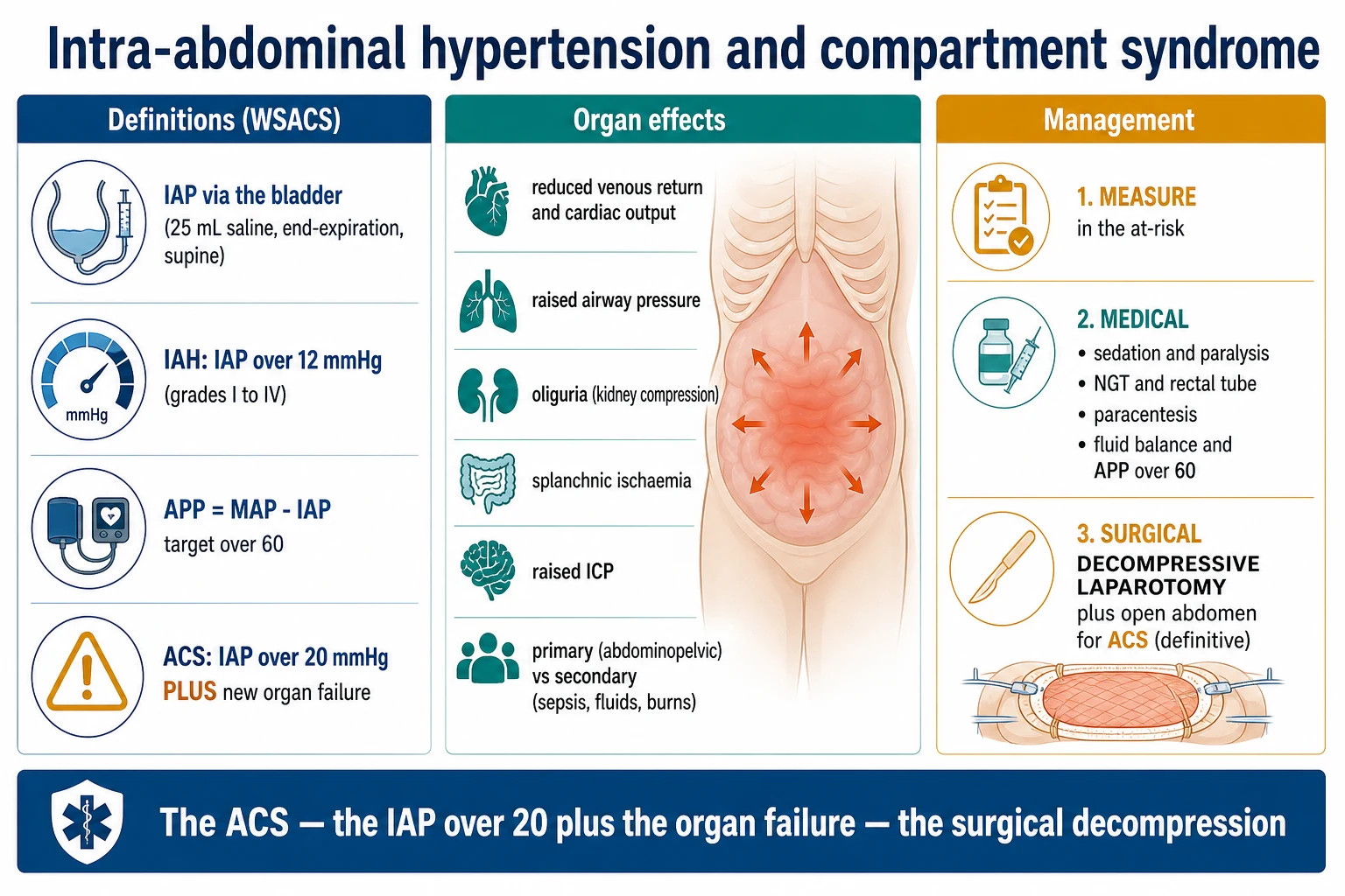
Raised IAP injures organs through a small number of mechanical and humoral mechanisms that converge on reduced perfusion, increased afterload, and end-organ congestion.[2]
How intra-abdominal hypertension becomes multi-organ failure
1. Pressure rise
IAP rises as intra-abdominal volume increases (oedematous bowel, ascites, blood, gas) within the relatively non-compliant abdominal compartment. Above ~20 mmHg the diaphragm is pushed cranially and the inferior vena cava and renal veins are compressed.
2. Reduced venous return
IVC and pelvic venous compression reduce preload. Compensatory increase in intrathoracic pressure further reduces the venous gradient. Cardiac output falls. CVP and wedge pressure read artificially high — the classic trap.
3. Increased afterload
Intrathoracic pressure and direct vascular compression raise systemic vascular resistance. Combined with reduced preload, stroke volume and MAP fall. Reflex tachycardia is blunted by sedation.
4. Renal failure
Renal venous congestion (not just reduced arterial flow) drops the filtration gradient. The renin–angiotensin–aldosterone axis is activated; renocortical blood flow falls. Oliguria progressing to anuria is frequently the first clinical sign.
5. Respiratory failure
Cephalad diaphragm displacement reduces functional residual capacity and lung compliance; atelectasis and shunt cause hypoxaemia. Peak and plateau pressures climb, risking barotrauma and mimicking ARDS.
6. Splanchnic and cerebral effects
Mesenteric venous congestion causes bowel-wall oedema, bacterial translocation and possible ischaemia. Reduced venous drainage from the head plus raised intrathoracic pressure elevates intracranial pressure — critical in the multiply-injured patient.
Measuring intra-abdominal pressure — technique and pitfalls
The intravesical (bladder) pressure method is the WSACS reference standard: cheap, repeatable, and minimally invasive.[1]
Standardised bladder-pressure technique (WSACS 2013)
1. Prepare the patient
Supine, fully relaxed, end-expiration, no active abdominal muscle contraction. Zero the transducer at the iliac crest in the mid-axillary line (the phlebostatic zero for the abdomen).
2. Empty the bladder
Drain the bladder completely through the Foley catheter. Clamp or disconnect the drainage bag.
3. Instil 25 mL warm sterile saline
Inject a standard 25 mL (maximum 50 mL in obese or large-bladder patients) of warmed 0.9% saline through the sampling port. Larger volumes falsely elevate the reading.
4. Connect a pressure transducer
Attach a pressure transducer via a three-way tap to the Foley catheter. Allow 30–60 s for bladder detrusor relaxation before reading.
5. Read at end-expiration
Record the value in mmHg at end-expiration after a stable trace. Take the mean of three readings.
6. Standardise the frequency
In at-risk patients measure every 4 h (minimum). Trend rather than act on a single value. Switch to continuous monitoring once IAH is established.
Intravesical (bladder)
Reference standard
- WSACS gold standard; cheapest and most widely available.
- Via Foley + 25 mL saline + transducer.
- Inaccurate with intravesical Botox, neurogenic bladder, pelvic haematoma, or bladder rupture.
- Cannot be used when there is no bladder access (post-cystectomy).
Intragastric
Alternative route
- Via naso/ogastric Foley connected to a water column or transducer.
- Useful when bladder access is lost.
- Affected by gastric tone, NG feed and peristalsis — more noise.
Inferior vena cava (IVC)
Ultrasound surrogate
- IVC diameter and collapsibility correlate loosely with IAP.
- No direct reading; a screening surrogate only.
Direct intraperitoneal
Invasive / continuous
- Catheter placed at laparoscopy, laparotomy or percutaneously.
- Reserved for the open abdomen or research; allows continuous IAP tracing.
- Emerging continuous systems (e.g. TraumaGuard) are under validation.
Risk stratification — who to monitor
In a multicentre cohort, approximately 50% of critically ill patients developed IAH and around 8% developed ACS; IAH independently predicted mortality.[4]
Diminished wall compliance
Mechanical restriction
- Major burns with abdominal eschar (limit wall expansion).
- Prone ventilation, tight surgical closure, rectus sheath haematoma.
- Obesity (BMI > 30) and high PEEP raising intrathoracic pressure.
Increased luminal volume
Hollow viscus
- Gastroparesis, ileus, pseudo-obstruction (Ogilvie).
- Volvulus, colonic obstruction, NG/PEG feed intolerance.
Increased abdominal contents
Solid / mass effect
- Ascites, haemoperitoneum, intra-abdominal haematoma.
- Severe acute pancreatitis with fluid collections or necrosis.
- Retroperitoneal bleed (ruptured AAA, pelvic fracture).
Capillary leak / fluid overload
The dominant driver
- Massive crystalloid resuscitation, sepsis, septic shock.
- Major trauma with damage-control resuscitation.
- Massive transfusion and coagulopathy.
Miscellaneous
Other
- Laparoscopy (pneumoperitoneum), peritoneal dialysis.
- Liver transplantation (large graft + reperfusion oedema).
- Pregnancy (gravid uterus) — see special populations.
Mechanical ventilation in intra-abdominal hypertension
IAH transforms the lung into a stiff, heavy organ that is easily injured by the ventilator, and ventilator settings in turn feed back onto IAP.[7][8]
Lung mechanics
What changes
- Reduced chest-wall compliance; lung compliance preserved early then falls.
- Plateau rises more than peak pressure; raised transpulmonary pressure is needed to inflate.
- Basal atelectasis, intrapulmonary shunt, hypoxaemia.
- Reduced functional residual capacity; propensity to cyclic collapse.
Set the ventilator
Practical rules
- Set PEEP by **transpulmonary pressure** (oesophageal) — abdominal load may require higher applied PEEP.
- Use a **lower tidal volume (4–6 mL/kg PBW)**; accept permissive hypercapnia.
- Aim for transalveolar-safe pressures: plateau < 30 cmH2O where the transpulmonary pressure allows.
- Consider **neuromuscular blockade** to abolish expiratory muscle activity that raises IAP.
Reciprocal danger
Ventilator to abdomen
- High intrathoracic pressure is transmitted across the diaphragm, further raising IAP.
- High PEEP reduces renal venous return and splanchnic perfusion.
- Decompression can cause reperfusion and a transient spike in airway pressures followed by improvement.
Medical management ladder (non-surgical decompression)
For Grade I–III IAH and as temporising therapy before surgery, a graded medical decompression ladder is applied. Most of these measures work by reducing intra-abdominal volume (lumen, fluid, wall tone) rather than changing the container.[1]
Five-step medical decompression ladder
Step 1 — Evacuate intraluminal contents
NG tube and/or rectal tube; enemas; prokinetics (metoclopramide 10 mg IV q6h plus or minus erythromycin 200 mg IV q12h); consider colonoscopic decompression for pseudo-obstruction.
Step 2 — Evacuate intra-abdominal fluid collections
Ultrasound-guided percutaneous drainage of ascites, haematoma or loculated fluid — the fastest effective single manoeuvre when fluid is the cause. (See percutaneous drainage section.)<Cite id="6" />
Step 3 — Improve abdominal wall compliance
Adequate sedation and analgesia (opioid plus benzodiazepine, plus or minus dexmedetomidine). Avoid head-of-bed > 30°. Remove tight eschar (escharotomy in burns). Reverse Trendelenburg can worsen IAP — keep the patient flat.
Step 4 — Correct positive fluid balance
Avoid further crystalloid. Aim for a **negative fluid balance**. Use diuretics (furosemide), **CRRT with net ultrafiltration**, or albumin plus diuretic for hypoalbuminaemia. Do NOT bolus fluid to "treat" the falling MAP — this is the cardinal error.
Step 5 — Optimise APP with vasoactives and neuromuscular blockade
Noradrenaline to raise MAP and APP > 60 mmHg (vasopressin if refractory). **Neuromuscular blockade** (cisatracurium or rocuronium infusion) reduces abdominal muscle tone and can drop IAP by 5–10 mmHg, buying time and sometimes avoiding surgery.<Cite id="5" />
Percutaneous catheter drainage
When imaging shows a drainable intra-abdominal collection, percutaneous drainage can drop IAP within minutes and may obviate surgery, particularly in secondary ACS driven by fluid/ascites or in severe pancreatitis.[6]
Percutaneous drainage workflow
1. Image first
Bedside ultrasound or CT to localise ascites, haematoma, biloma or walled-off collection and to plan a safe route that avoids bowel and vessels.
2. Choose the catheter
8–14 Fr pigtail for free fluid; a larger catheter or catheter-over-guidewire for viscous haematoma or necrosis. Multiple catheters may be needed.
3. Drain and re-measure
Aspirate, leave to free drainage, then re-measure IAP within 1 h. A meaningful drop confirms the collection was the dominant contributor.
4. Send and reassess
Send fluid for culture, amylase (pancreatic) and cytology. Repeat imaging if IAP fails to fall — look for undrained locules or ongoing haemorrhage.
Cheatham & Safcsak (Chest 2011)
Chest 2011
Retrospective case series — 81 patients with IAH/ACS (125 catheter placements); percutaneous catheter drainage of intra-abdominal fluid under ultrasound guidance
Key finding
Mean IAP fell from 26 mmHg to 16 mmHg after drainage; catheter drainage alone definitively treated IAH/ACS in roughly 70% of patients, avoiding laparotomy in the majority
Practice change
Established percutaneous catheter drainage as first-line intervention for IAH/ACS with a drainable collection, particularly secondary ACS and pancreatitis
Decompressive laparotomy and the open abdomen
Decompressive laparotomy is the definitive treatment for ACS. IAP falls immediately, and organ function typically improves within hours, though mortality remains high (40–60%) because of late presentation and the underlying insult.[3]
Decompressive laparotomy — operative and post-operative pathway
1. Indications
Sustained IAP > 20 mmHg with **new organ dysfunction or failure** (especially anuria, rising ventilator pressures, haemodynamic collapse) that has not responded to the medical ladder. Grade IV IAH usually warrants surgery.
2. The operation
Full midline laparotomy. Release the abdominal wall; evacuate fluid or haematoma. IAP drops immediately. The abdomen is left **open** (laparostomy) — primary fascial closure at this stage causes recurrent ACS.
3. Temporary abdominal closure (TAC)
Cover the viscera with a temporary dressing — negative-pressure wound therapy (NPWT/ABThera), Wittmann patch, Bogota bag (sterile IV bag sutured to skin) or a sandwich vacuum dressing. NPWT is preferred: it protects viscera, manages fluid and aids fascial traction.
4. Reperfusion precautions
Release of the clamp causes reperfusion of ischaemic bowel and a surge of potassium, lactate and acid — expect transient haemodynamic instability; have vasopressors, calcium and bicarbonate ready.
5. Staged re-look and closure
Return to theatre every 24–48 h for washout, debridement and attempted gradual fascial closure. Definitive closure when IAP is normal, oedema resolved and no further source — usually days to weeks. Delayed closure reduces recurrent ACS but risks entero-atmospheric fistula.
Negative-pressure therapy (NPWT)
Preferred TAC
- The modern default (e.g. ABThera).
- Manages fluid losses, splints fascia laterally, protects viscera.
- Higher rates of primary fascial closure than other methods.
Wittmann patch
Velcro-like
- Two prosthetic sheets sutured to fascia; gradually tightened.
- Useful for planned, staged closure.
- Does not manage fluid losses.
Bogota bag
Simple cover
- A sterile 3 L irrigation bag sutured to skin.
- Cheap and rapid where NPWT is unavailable.
- No fluid management; risks adhesions and fistula.
Mesh / mesh-mediated traction
Bridging
- Absorbable or biological mesh sewn to fascia.
- Used when fascia cannot be approximated.
- High planned ventral hernia rate.
PANCEV (BMC Surg 2010 — design)
BMC Surgery 2010
Multicentre randomised controlled trial — decompressive laparotomy with temporary closure vs percutaneous puncture plus abdominal catheter in ACS complicating severe acute pancreatitis
Key finding
Designed to compare surgical decompression with percutaneous drainage in pancreatitis-induced ACS; primary endpoints death and major complications. Established the only RCT framework for ACS management in pancreatitis
Practice change
Set the randomised evidence base for choosing percutaneous vs surgical decompression in pancreatitis-related ACS
Malbrain multicentre cohort (CCM 2005)
Crit Care Med 2005
Prospective multicentre epidemiological study — 265 critically ill patients across 14 ICUs with daily IAP measurement
Key finding
IAH in ~50% and ACS in ~8% of ICU patients; IAH an independent predictor of mortality; incidence under-recognised in routine practice
Practice change
Established the epidemiological importance of IAH/ACS in the general ICU and the case for routine IAP monitoring in at-risk patients
Special populations
ACS behaves differently in specific contexts; the threshold to decompress and the operative approach vary.[9]
Severe acute pancreatitis
Common medical-ICU cause
- Capillary leak + retroperitoneal fluid + ileus drive an early high IAP.
- Infected necrosis and fluid collections drive IAH — drain percutaneously first.
- Surgical decompression is reserved for refractory ACS; often combined with necrosectomy.
- The PANCEV trial informs percutaneous vs surgical approach.
Major burns
> 40% TBSA
- Capillary leak + eschar + large-volume resuscitation.
- Abdominal escharotomy restores wall compliance; may avert ACS.
- Strict fluid stewardship and early enteral nutrition reduce incidence.
Trauma / damage-control
Primary ACS
- Damage-control laparotomy already leaves the abdomen open — recurrence indicates ongoing bleeding or third-space loss.
- Rewarm, correct coagulopathy and acidosis; avoid a crystalloid chase.
Ruptured AAA / post-EVAR
Vascular
- Retroperitoneal haematoma and reperfusion oedema drive IAH.
- The abdominal compartment is often left open after emergency repair.
- High mortality; vigilance for mesenteric and renal ischaemia.
Pregnancy and peripartum
Obstetric ACS
- Gravid uterus raises baseline IAP; pre-eclampsia, sepsis or haemorrhage precipitate ACS.
- Left lateral tilt decompresses the IVC; deliver the baby if undelivered.
- Multidisciplinary (obstetrics, ICU, surgery) decision-making; percutaneous drainage preferred before laparotomy where possible.
Liver transplantation
Post-reperfusion
- Large graft + reperfusion oedema + ascites raise IAP.
- Risk to graft and renal function; low threshold to leave open or re-explore.
- IAH post-OLT predicts graft dysfunction and AKI.
Paediatric
Children
- Same definitions, with weight-based fluid targets.
- Common in necrotising enterocolitis, post-cardiac surgery, sepsis.
- Bladder pressure is validated; smaller instillation volume (1 mL/kg).
Prevention bundle — the WSACS approach
Most ACS is preventable; prevention is overwhelmingly about fluid discipline and early source control.[1]
WSACS prevention bundle
1. Fluid stewardship
Use goal-directed, balanced resuscitation; prefer blood products in trauma; monitor net fluid balance hourly; aim for neutral or negative balance after the resuscitation phase.
2. Monitor at-risk patients
Measure bladder pressure every 4–6 h in any patient meeting a risk criterion (massive fluid, pancreatitis, burns, sepsis, major trauma, post-EVAR).
3. Evacuate early
NG/rectal tubes, prokinetics, and percutaneous drainage of collections before IAP climbs.
4. Maintain wall compliance
Adequate analgesia and sedation; avoid unnecessary prone positioning or head elevation; escharotomy for circumferential burns.
5. Early definitive surgery
Operate for the intra-abdominal source (perforation, ischaemia, bleeding) without delay; do not temporise with fluids.
Distinguishing IAH from ACS — and from mimics
IAH
Pressure-only
- Sustained IAP > 12 mmHg.
- No new organ failure.
- Treat medically along the decompression ladder.
ACS
Pressure + failure
- Sustained IAP > 20 mmHg **plus** new organ failure.
- APP typically < 60 mmHg.
- Surgical emergency: decompressive laparotomy.
Sepsis / ARDS mimic
Looks similar
- High ventilator pressures, oliguria, hypotension, high lactate.
- Differentiate by measuring IAP — the only way to know.
- Both may coexist; IAH worsens sepsis and vice-versa.
Key trials and evidence summary
Kirkpatrick — WSACS 2013 guidelines
Intensive Care Med 2013
International multidisciplinary consensus — systematic review plus Delphi; updated definitions and 17 clinical practice guidelines for IAH/ACS
Key finding
Standardised definitions (IAH > 12 mmHg, ACS > 20 mmHg + organ failure, APP > 60 mmHg) and a graded management algorithm from medical decompression to laparotomy
Practice change
The current worldwide reference standard for IAH/ACS definitions, measurement and management
Cheatham — APP as resuscitation endpoint
J Trauma 2000
Prospective observational study — 159 trauma/surgical ICU patients; IAP, APP, MAP and survival
Key finding
APP (MAP - IAP) > 60 mmHg was the only resuscitation endpoint independently associated with survival; superior to MAP or IAP alone
Practice change
Established APP > 60 mmHg as the target resuscitation endpoint in IAH/ACS
Red flags — extended
Quick-revision summary
References
- [1]Kirkpatrick AW, Roberts DJ, De Waele J, et al. Intra-abdominal hypertension and the abdominal compartment syndrome: updated consensus definitions and clinical practice guidelines from the World Society of the Abdominal Compartment Syndrome Intensive Care Med, 2013.PMID 23673399
- [2]Malbrain ML, Cheatham ML, Kirkpatrick A, et al. Results from the International Conference of Experts on Intra-abdominal Hypertension and Abdominal Compartment Syndrome. I. Definitions Intensive Care Med, 2006.PMID 16967294
- [3]Nasa P, Chanchalani G, Juneja D, et al. Surgical decompression for the management of abdominal compartment syndrome with severe acute pancreatitis: A narrative review World J Gastrointest Surg, 2023.PMID 37901738
- [4]Malbrain ML, Chiumello D, Pelosi P, et al. Incidence and prognosis of intraabdominal hypertension in a mixed population of critically ill patients: a multiple-center epidemiological study Crit Care Med, 2005.PMID 15699833
- [5]Cheatham ML, White MW, Sagraves SG, et al. Abdominal perfusion pressure: a superior parameter in the assessment of intra-abdominal hypertension J Trauma, 2000.PMID 11038078
- [6]Cheatham ML, Safcsak K Percutaneous catheter decompression in the treatment of elevated intraabdominal pressure Chest, 2011.PMID 21903735
- [7]Regli A, Pelosi P, Malbrain MLNG Ventilation in patients with intra-abdominal hypertension: what every critical care physician needs to know Ann Intensive Care, 2019.PMID 31025221
- [8]Regli A, Reintam Blaser A, De Keulenaer B, et al. Intra-abdominal hypertension and hypoxic respiratory failure together predict adverse outcome - A sub-analysis of a prospective cohort J Crit Care, 2021.PMID 33906106
- [9]Lozada MJ, Goyal V, Levin D, et al. Management of peripartum intra-abdominal hypertension and abdominal compartment syndrome Acta Obstet Gynecol Scand, 2019.PMID 31070780
- [10]Nasa P, Wise RD, Smit M, et al. International cross-sectional survey on current and updated definitions of intra-abdominal hypertension and abdominal compartment syndrome World J Emerg Surg, 2024.PMID 39609850
- [11]Radenkovic DV, Bajec D, Ivancevic N, et al. Decompressive laparotomy with temporary abdominal closure versus percutaneous puncture with placement of abdominal catheter in patients with abdominal compartment syndrome during acute pancreatitis: background and design of multicenter, randomised, controlled study BMC Surg, 2010.PMID 20624281