ICU · GI & nutrition / surgical
Abdominal Sepsis & Peritonitis — Source Control, SBP & the Polymicrobial
Also known as Peritonitis · Abdominal sepsis · Intra-abdominal sepsis · Spontaneous bacterial peritonitis · SBP · Secondary peritonitis · Tertiary peritonitis · Source control · Peritonism · Piperacillin-tazobactam · Metronidazole · Complicated intra-abdominal infection · cIAI · Damage control laparotomy · Mannheim Peritonitis Index · Hepatorenal syndrome
The peritonitis is the inflammation of the peritoneum, usually from the intra-abdominal infection; the abdominal sepsis is the sepsis from the intra-abdominal source. The three types: the primary (the spontaneous bacterial peritonitis — the SBP in the cirrhosis and the ascites, the monomicrobial, the ascitic PMN over 250 cells per cubic mm, the cefotaxime plus the albumin), the secondary (the GI perforation or the ischaemia, the polymicrobial — the Gram-negative and the anaerobes, the source control plus the antibiotics), and the tertiary (the persistent or the recurrent after 48 hours of the failed initial treatment, the resistant — the Enterococcus, the Candida, the MDR organisms). The diagnosis: the peritonism (the rigid abdomen, the guarding, the rebound), the CT (the gold standard — the free gas, the fluid, the source), the lactate, the cultures. The management: the Sepsis-6 bundle, the broad antibiotics (the piperacillin-tazobactam ± the vancomycin ± the antifungal, or the ceftriaxone plus the metronidazole or the meropenem), the duration 4 to 7 days after the source control, and the SOURCE CONTROL (the surgery or the IR — the drainage, the debridement, the repair) — the definitive; the delayed source control worsens the mortality.
On this page & tools
Your progress
Saved locally on this device.
8 MCQs with explanations
Target exams
Red flags
Overview & definition
The peritonitis is the inflammation of the peritoneum, usually from the intra-abdominal infection. The abdominal sepsis is the sepsis from the intra-abdominal source. The two pillars of the management: (1) the antibiotics (the broad cover) and (2) the source control (the drainage, the debridement, the repair — the definitive). The delayed source control worsens the mortality. The three types — the primary (the SBP), the secondary (the GI perforation), the tertiary (the persistent) — have the different management.[1]
A modern and exam-important refinement is the intra-abdominal infection (IAI) terminology used by the WSES 2017 guidelines, which classifies IAI by acquisition (community-acquired, CA-IAI vs healthcare-associated, HA-IAI) and by severity (complicated, cIAI = infection beyond the source organ into the peritoneal space, vs uncomplicated). The practical point: an HA-IAI or a high-severity cIAI demands broader empiric cover (carbapenem, anti-Enterococcus, antifungal) and a lower threshold for source control than a community-acquired appendicitis.[1]

Pathophysiology & the peritoneal response
The peritoneum is a serous membrane (~1.7 m², comparable to the skin) with a mesothelial layer and an underlying rich capillary and lymphatic network. The normal peritoneal cavity contains <100 mL of ultrafiltrate; the omentum, mesentery, and peritoneal macrophages provide innate immunity. When bacteria or irritants (bile, blood, gastric acid, pancreatic enzymes, faeces) breach the cavity, the response is stereotyped:[1]
- Inflammation — mesothelial and mast cells release cytokines (IL-1, IL-6, TNF-α), increasing vascular permeability. Protein-rich fluid exudes; this is the early peritoneal exudate.
- Fluid sequestration — large volumes (litres) of fluid can third-space into the peritoneal cavity within hours, producing hypovolaemia and haemoconcentration.
- Bacterial clearance vs overgrowth — diaphragmatic lymphatics clear bacteria into the systemic circulation (bacteraemia), but if the inoculum overwhelms clearance, organisms multiply. Within 6 hours a single E. coli can become 10&sup8; organisms; anaerobes (Bacteroides) establish synergistically with facultative aerobes.
- Abscess formation vs diffuse peritonitis — the omentum and bowel "wall off" a localised source into an abscess; if containment fails (large inoculum, virulent organisms, impaired immunity, free perforation), the result is diffuse peritonitis with systemic sepsis.
- Systemic inflammation → organ dysfunction — cytokine spillover drives vasodilatory shock, capillary leak, ARDS, AKI, and the septic coagulopathy that defines septic shock. [1]
Bacterial translocation from the ischaemic or obstructed gut (mesenteric lymph nodes to systemic circulation) is the proposed mechanism linking gut dysfunction to multiple organ failure in severe peritonitis, and is the rationale for early enteral nutrition to maintain mucosal integrity.[1]
The clinical consequence: the patient with diffuse peritonitis loses fluid at a rate comparable to a major burn — aggressive crystalloid resuscitation in the first hours is mandatory, but is a bridge to source control, not a substitute for it. [1]
Classification of peritonitis
Peritonitis is classified by the mechanism of peritoneal contamination into three types — primary, secondary, and tertiary. The classification is not academic: it dictates the microbiology, the need for source control, and the empiric antibiotic spectrum.[1][1]
Primary (SBP)
No GI source — cirrhosis/ascites
- Monomicrobial — E. coli (~50%), Klebsiella, pneumococcus; one organism only
- Ascitic PMN >250 cells/mm³ OR positive culture in a patient with cirrhosis and ascites
- NO source control needed — no perforation to drain or repair
- Cefotaxime 2 g q8h PLUS albumin 1.5 g/kg day 1, 1 g/kg day 3
- Albumin prevents hepatorenal syndrome (HRS) — the sort of detail examiners love
- Secondary prophylaxis with daily norfloxacin after the first episode
Secondary
GI perforation / ischaemia — polymicrobial
- Polymicrobial — Gram-negatives + anaerobes (Bacteroides) ± Enterococcus ± Candida
- Causes: appendicitis, diverticulitis, perforated peptic ulcer, ischaemic bowel, post-op leak, trauma
- SOURCE CONTROL is the definitive treatment — antibiotics alone fail
- Piperacillin-tazobactam OR ceftriaxone + metronidazole; meropenem if severe/healthcare-associated
- Antifungal (echinocandin) if Candida risk; consider anti-Enterococcus cover in HA-IAI
- Duration 4–7 days AFTER adequate source control (STOP-IT)
Tertiary
Persistent/recurrent after 48 h — MDR
- Persistent or recurrent infection ≥48 h after apparently adequate initial management
- MDR organisms — Enterococcus (incl. VRE), Candida, Pseudomonas, Stenotrophomonas, Acinetobacter
- Driven by inadequate initial source control + immunoparalysis + prior antibiotic pressure
- First EXCLUDE an undrained source / ongoing leak (CT) before attributing to tertiary
- Carbapenem ± antifungal ± anti-MDR cover guided by local antibiogram; planned relaparotomy
- Poor outcomes — mortality 30–60% vs ~10–20% for secondary
The WSES 2017 acquisition framework
The WSES 2017 guidelines layer a second, orthogonal classification on top of the primary/secondary/tertiary scheme — by acquisition, which is the strongest single predictor of the likely microbiology and thus the empiric regimen:[1][1]
- Community-acquired IAI (CA-IAI) — infection present on or within 48 h of admission. Typical organisms: E. coli and other Enterobacterales, Bacteroides, Streptococcus. Empiric cover with a single broad agent (piperacillin-tazobactam) or ceftriaxone + metronidazole is adequate for most.
- Healthcare-associated IAI (HA-IAI) — infection developing >48 h after admission, or within 28 days of hospital discharge, or after a procedure. Higher likelihood of resistant Enterobacterales (ESBL), Enterococcus, Pseudomonas, and Candida. Requires broader empiric cover (carbapenem ± anti-Enterococcus ± antifungal) and de-escalation from cultures. [1]
The two classifications are used together: a patient with secondary (perforated viscus) AND healthcare-associated IAI needs the broadest empiric cover; a patient with primary SBP needs neither source control nor anaerobic cover. [1]
The three types of the peritonitis
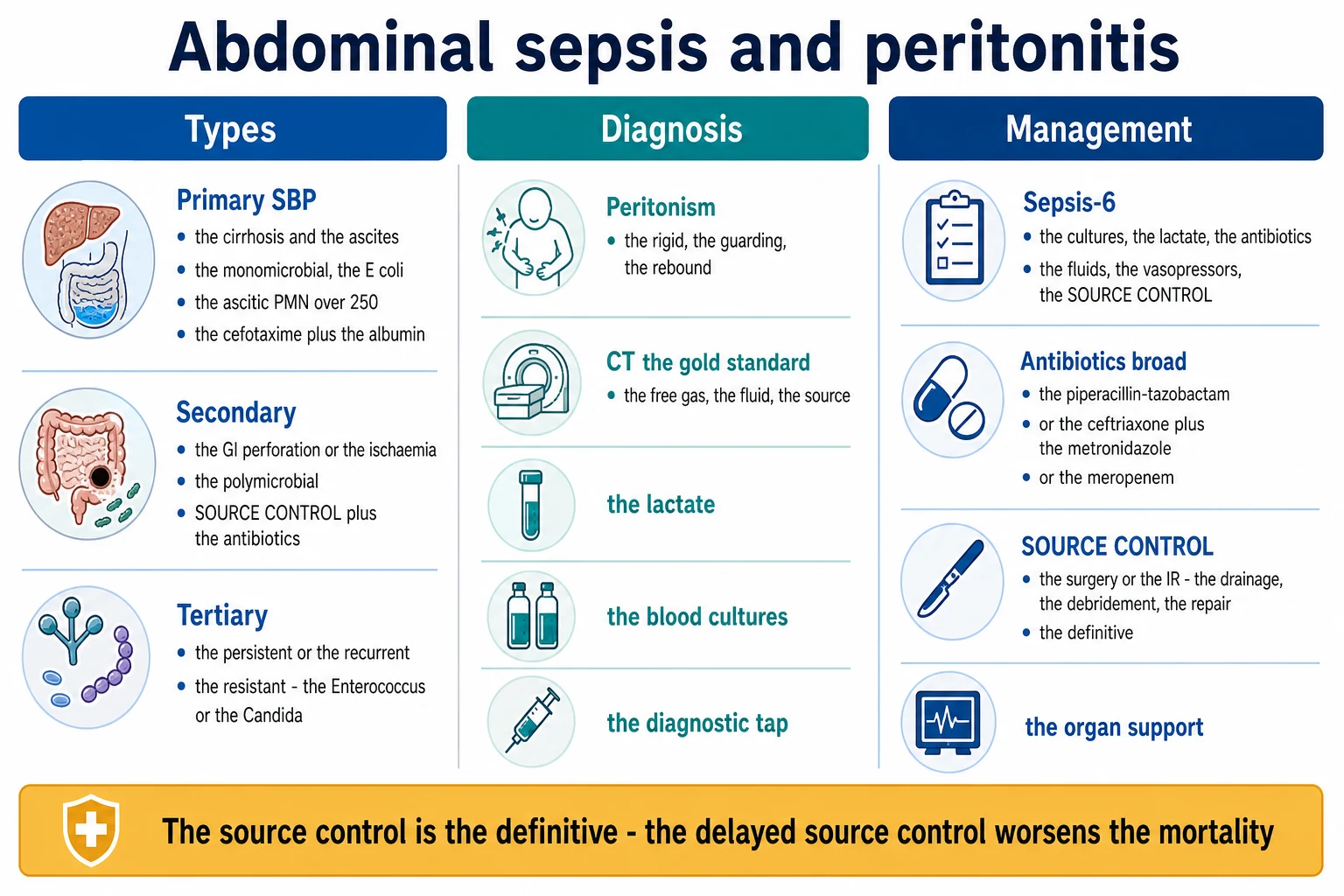
The primary (the spontaneous bacterial peritonitis, the SBP)
- In the cirrhosis and the ascites, no obvious GI source. The monomicrobial (the E. coli, the Klebsiella).[1]
- The diagnosis — the ascitic fluid PMN over 250 cells per cubic mm (or the positive culture). The diagnostic tap.[1]
- The management — the cefotaxime (or the ceftriaxone) PLUS the albumin (the 1.5 g per kg on the day 1, the 1 g per kg on the day 3) to prevent the hepatorenal syndrome. The secondary prophylaxis (the norfloxacin) after the first episode.[1]
The secondary peritonitis
- From the GI source — the perforation (the appendix, the diverticular, the peptic ulcer, the colon), the ischaemia, the post-operative leak.[1]
- The polymicrobial — the Gram-negative, the anaerobes, the enterococci.[1]
- The management — the source control (the surgery) PLUS the broad antibiotics. The definitive. The delayed worsens the mortality.[1]
The tertiary peritonitis
- The persistent or the recurrent peritonitis after the failed initial treatment. The often the resistant organisms — the Enterococcus, the Candida, the Stenotrophomonas, the Pseudomonas.[1]
- The poor outcomes. The broad antibiotics (the carbapenem, the antifungal), the repeated source control, the review of the resistance.[1]
Causes by type — the surgical and microbiological map
Primary SBP — risk factors and microbiology
Spontaneous bacterial peritonitis occurs almost exclusively in cirrhotic ascites (and rarely in nephrotic syndrome, heart failure ascites, or peritoneal dialysis — where it is "culture-negative neutrocytic peritonitis" or PD-peritonitis). The pathogenesis is bacterial translocation from the gut (impaired mucosal barrier, reticuloendothelial dysfunction, low ascitic protein <1 g/dL, low complement opsonic activity).[1]
- Organisms: Enterobacterales dominate — E. coli (~50%), Klebsiella pneumoniae, then Streptococcus pneumoniae, other streptococci, and enterococci. Strictly monomicrobial by definition; polymicrobial ascitic culture suggests a perforated viscus (think secondary, image the patient).
- Risk factors: low ascitic fluid protein (<1 g/dL — the single best predictor), severe liver disease (high MELD, bilirubin >2.5), variceal haemorrhage, prior SBP (1-year recurrence ~70% without prophylaxis).
- Variants: bacterascites (positive culture, PMN <250 — treat if symptomatic), culture-negative neutrocytic ascites (PMN >250, negative culture — treat as SBP), poly-microbial bacterascites (polymicrobial culture without PMN rise — raises perforation, image the patient). [1]
Secondary peritonitis — the organ-specific sources
Upper GI
Foregut — acid/bile
- Perforated peptic ulcer — free gas under the right hemidiaphragm; chemical then bacterial peritonitis
- Perforated gastric/small-bowel tumour, iatrogenic perforation (post-ERCP, post-endoscopy)
- Microbiology initially sparse (acid sterilises) → becomes Gram-negatives + streptococci + Candida after ~6–12 h
Lower GI
Colonic — faecal, polymicrobial
- Appendicitis, diverticulitis (sigmoid), perforated colonic malignancy, ischaemic colitis, volvulus
- Polymicrobial faecal load — E. coli, Bacteroides fragilis (essential anaerobic cover), Enterococcus, Klebsiella
- Highest inoculum and highest mortality of the secondary causes; lowest threshold for source control
Ischaemia
Bowel necrosis
- Mesenteric arterial embolus/thrombus, venous thrombosis, strangulated hernia/volvulus, non-occlusive (low-flow)
- Transmural infarction releases the full faecal microbiome into the peritoneum — catastrophic, polymicrobial
- Surgical emergency — resect non-viable bowel; damage-control laparotomy if haemodynamically unstable
Post-operative
Anastomotic leak
- Leak from a gastro-intestinal, biliary, or pancreatic anastomosis, typically days 5–10 post-op
- Healthcare-associated microbiology — resistant Enterobacterales (ESBL), Enterococcus, Pseudomonas, Candida
- Low threshold to re-image and re-operate; the post-op patient with new sepsis has a leak until proven otherwise
Tertiary peritonitis — the MDR landscape
Tertiary peritonitis is defined as persistent or recurrent infection ≥48 hours after adequate initial management of a secondary peritonitis. It reflects inadequate source control, immunoparalysis (persistent inflammation-immunosuppression catabolism, PICS), and antibiotic selection pressure, and is characterised by organisms that are opportunistic and resistant:[1][1]
- Enterococcus (including VRE) — the signature organism; cover with ampicillin/vancomycin ± daptomycin/linezolid per sensitivity.
- Candida spp. (C. albicans and increasingly C. glabrata, C. parapsilosis) — intra-abdominal candidiasis doubles mortality; treat with an echinocandin, de-escalate to fluconazole if susceptible.
- Pseudomonas aeruginosa, Acinetobacter, Stenotrophomonas maltophilia — driven by prior broad-spectrum exposure.
- Coagulase-negative staphylococci — occasionally in the long-stay, line-bearing, immunoparalysed patient. [1]
The crucial exam point: always re-image to exclude an undrained collection or ongoing leak before attributing failure to "tertiary peritonitis" — most apparent tertiary cases are actually persistent secondary infection from inadequate source control. [1]
Diagnosis
- The peritonism — the rigid abdomen, the guarding, the rebound. The clinical signs of the diffuse peritoneal inflammation.[1]
- The CT — the gold standard (the free gas, the intra-abdominal fluid, the source — the perforation, the abscess, the ischaemia). Do the CT with the contrast.[1]
- The lactate, the blood cultures, the diagnostic tap (the SBP), the diagnostic laparoscopy if the uncertain.[1]
The diagnostic workup — a structured approach
Diagnostic workup in suspected abdominal sepsis
1. Clinical assessment — the peritonism screen
Examine for rigidity, voluntary/involuntary guarding, rebound tenderness, and absent bowel sounds (paralytic ileus). Remember: the rigid, board-like abdomen is a LATE sign — the elderly, immunosuppressed, and spinal-injured patient may have minimal signs. A normal abdomen does NOT exclude perforation.
2. Bloods — lactate, FBE, CRP, blood cultures (×2), VBG/ABG
Lactate >2 mmol/L suggests sepsis; >4 with hypotension = septic shock (Surviving Sepsis). Send blood cultures BEFORE antibiotics when feasible without delaying the 1-hour bundle. Check amylase/lipase (pancreatitis masquerades), β-hCG in women, and group-and-save.
3. Imaging — CT abdomen/pelvis with IV contrast is the gold standard
Look for FREE INTRAPERITONEAL GAS (perforation), free fluid, fat stranding, bowel wall thickening / pneumatosis (ischaemia), abscess collection, and the source (appendix, diverticulum, ulcer). CT is more sensitive than erect CXR for free gas. Bedside ultrasound (POCUS) is a rapid triage tool for free fluid and gallbladder/appendiceal pathology in the unstable patient who cannot travel to CT.
4. Erect CXR or lateral decubitus film
Free subdiaphragmatic gas (Riedel's lobe area) on an erect CXR detects ~70% of perforations — a useful bedside adjunct when CT is delayed, but a negative film does not exclude perforation.
5. Diagnostic tap in cirrhosis with ascites
In EVERY cirrhotic with ascites and sepsis (or new abdominal pain, encephalopathy, renal dysfunction), perform a diagnostic paracentesis: cell count (PMN >250 cells/mm³ = SBP), culture (in blood-culture bottles), albumin (for serum-ascites albumin gradient), and total protein.
6. Diagnostic laparoscopy / laparotomy when imaging is non-diagnostic
In the unstable or imaging-negative patient with peritonism, diagnostic laparoscopy is both diagnostic and therapeutic — it can confirm and treat a perforated ulcer, appendicitis, or ischaemic segment in one operation.
Severity scoring — the Mannheim Peritonitis Index (MPI)
The Mannheim Peritonitis Index is the most widely used intra-operative severity score for peritonitis, validated to predict mortality. It is calculated at laparotomy:[1]
- Age >50 years (5 points) · Female (5) · Organ failure (7) · Malignancy (4) · Pre-operative duration >24 h (4)
- Origin of sepsis not colonic (4) · Diffuse generalized peritonitis (6) · Exudate clear (0) / purulent (6) / faecal (12) [1]
| MPI score | Mortality |
|---|---|
| <21 | ~0% |
| 21–29 | ~15% |
| >29 | ~50–60% |
The MPI is less used in the modern era of APACHE II / SOFA, but examiners (especially the CICM First Part) still expect recognition of it as the operative severity tool for peritonitis, and it remains useful for risk-stratifying the need for ICU admission and planned relaparotomy. [1]
Management
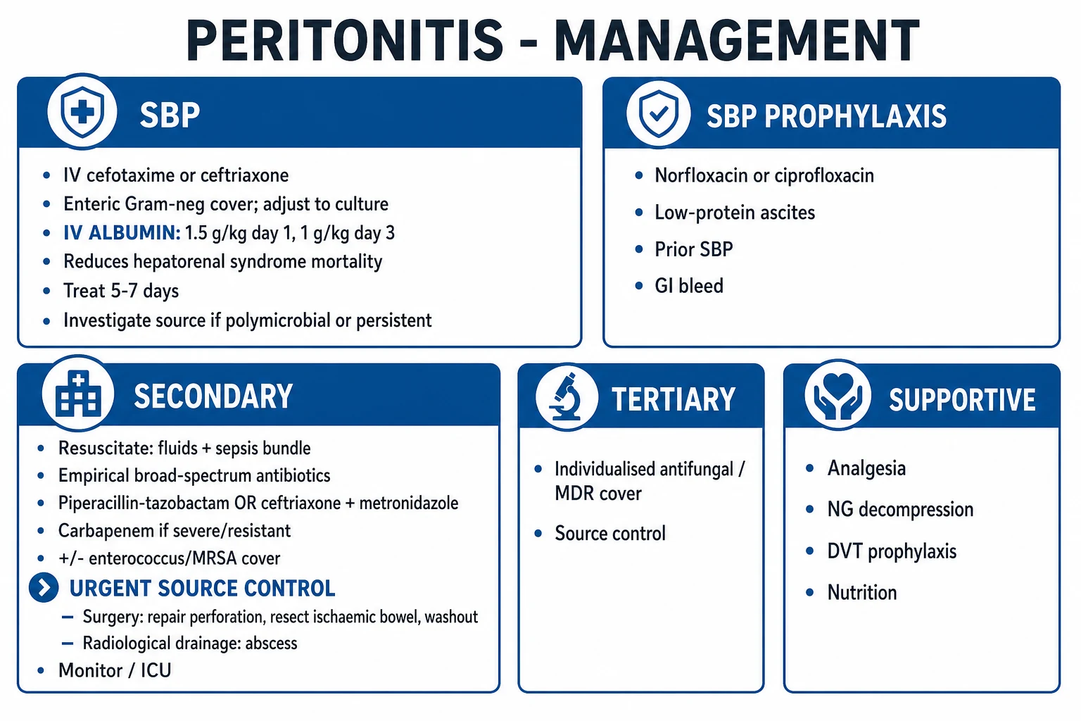
1. The Sepsis-6 bundle (the first hour)
The cultures (the blood, the peritoneal), the lactate, the broad antibiotics, the fluids, the vasopressors, and the source control. The Surviving Sepsis 1-hour bundle.[1][1]
The Surviving Sepsis Campaign 2021 guidelines make the priorities explicit: (1) measure lactate and resample to guide clearance; (2) obtain blood cultures before antibiotics without delaying therapy; (3) administer broad-spectrum antibiotics within 1 hour for sepsis/septic shock; (4) begin rapid 30 mL/kg crystalloid for sepsis-induced hypoperfusion or shock; (5) apply vasopressors to maintain MAP ≥65 mmHg (norepinephrine first-line); and (6) achieve source control as soon as medically/logistically practical — ideally within 6–12 hours for the severely ill.[1]
2. The broad antibiotics
- The community-acquired — the piperacillin-tazobactam OR the ceftriaxone plus the metronidazole (the Gram-negative plus the anaerobic cover).[1]
- The healthcare-associated or the severe — the meropenem (the carbapenem) plus the antifungal (the Candida risk).[1]
- The antifungal (the echinocandin, the fluconazole) for the Candida risk (the immunocompromise, the prolonged antibiotics, the post-operative, the recurrent).[1]
- The de-escalate once the cultures and the sensitivities return (the 24 to 48 hours).[1]
The empiric antibiotic choice is governed by acquisition and severity. The WSES 2017 and SIS-IDSA 2010/2017 guidelines converge on the following framework:[1][1][1]
CA-IAI, mild-moderate
Community appendicitis etc.
- Ceftriaxone 2 g IV daily + metronidazole 500 mg q8h, OR
- Cefoxitin 2 g q8h, OR moxifloxacin 400 mg daily, OR ertapenem 1 g daily
- Piperacillin-tazobactam 4.5 g q8h is a convenient single agent and is a reasonable empiric choice in the ED/ICU
- NO empiric anti-Enterococcus or antifungal needed for the community-acquired case
CA-IAI, high severity
Shock / diffuse peritonitis
- Piperacillin-tazobactam 4.5 g q6–8h (or q8h extended infusion), OR
- Cefepime 2 g q8h + metronidazole, OR meropenem 1 g q8h
- ADD vancomycin if MRSA risk or line infection, or to broaden Gram-positive cover
- Consider empiric echinocandin (caspofungin/micafungin) if multiple Candida risk factors
HA-IAI
Post-op / healthcare-associated
- Meropenem 1 g q8h ± vancomycin (covers ESBL, Pseudomonas, Enterococcus)
- Empiric echinocandin for Candida (post-op upper-GI/biliary, prolonged antibiotics, immunocompromise)
- De-escalate aggressively once cultures and sensitivities return (usually 48–72 h)
- Engage infectious diseases / antimicrobial stewardship early — local antibiogram is king
When to add vancomycin: empiric anti-Enterococcus / MRSA cover (vancomycin) is justified in healthcare-associated infection, post-operative infection, immunocompromise, prior broad-spectrum antibiotics, prosthetic heart valves, and severe sepsis with a high suspicion of resistant organisms. Routine empiric vancomycin for community-acquired peritonitis is not recommended.[1][1]
When to add an antifungal: empiric antifungal therapy (an echinocandin such as caspofungin 70 mg then 50 mg daily, or micafungin 100 mg daily; de-escalate to fluconazole if susceptible) for intra-abdominal candidiasis is indicated in: post-operative intra-abdominal infection (especially upper-GI/biliary/pancreatic), immunocompromise, necrotising pancreatitis, prolonged broad-spectrum antibiotics, recurrent intra-abdominal infection, and any critically-ill patient with Candida isolated from a normally sterile intra-abdominal site. Intra-abdominal Candida doubles mortality.[1]
Antibiotic duration: the STOP-IT randomised trial established that a fixed ~4-day course (median 4 days) after adequate source control is non-inferior to an ~8-day course for complicated intra-abdominal infection. Current guidance: 4–7 days of antibiotics after adequate source control; stop earlier if source control is excellent and the patient is clinically well; extend only for persistent infection, inadequate source control, or undrained collections. Prolonged courses select for resistance and for C. difficile.[1]
3. The source control (the definitive)
- The physical measures to control the source — the drainage of the infected fluid, the debridement of the necrotic tissue, the repair or the resection of the perforation.[1]
- The surgery (the laparotomy, the laparoscopy) or the interventional radiology (the percutaneous drainage of the abscess).[1]
- The early, the adequate, the least physiological insult — the "perfect source control". The delayed source control (over the 6 to 12 hours in the severe) worsens the mortality.[1]
- The "source control before the antibiotics fade" — the source matters more than the duration of the antibiotics.[1]
Source control strategies — the surgical decision
Source control is the definitive treatment for secondary and tertiary peritonitis. The specific intervention depends on the source, the stability of the patient, and the localisation of the infection:[1][1]
Source control strategies in intra-abdominal infection
1. Definitive surgery (single operation)
For the stable patient with a localised, correctable source — e.g. perforated appendicitis (appendicectomy), perforated peptic ulcer (primary closure + omental patch / Graham patch), perforated diverticulitis (Hartmann's procedure or primary anastomosis). Aim: control the source, wash out the contamination, and close in one operation.
2. Percutaneous drainage (interventional radiology)
For a localised, accessible abscess or collection (e.g. post-operative pelvic abscess, diverticular abscess) without diffuse peritonitis. CT- or ultrasound-guided drain placement + antibiotics; converts surgery to a delayed, elective, single-stage procedure. The stable patient with a walled-off collection is the ideal candidate.
3. Damage control laparotomy (DCL)
For the severely unstable, acidotic, coagulopathic, hypothermic patient ("the lethal triad"). The operation is abbreviated to ONLY (a) control contamination (close the perforation rapidly, staple off non-viable bowel), (b) wash out, and (c) leave the abdomen OPEN with a temporary closure (Bogota bag / negative-pressure wound therapy). The patient returns to ICU for resuscitation and to theatre for re-look at 24–48 h. Definitive reconstruction and anastomosis are deferred.
4. Planned relaparotomy (staged re-operation)
A scheduled return to theatre at 24–48 h to re-explore, wash out, and assess bowel viability — used when the source cannot be controlled in one operation (massive contamination, doubtful viability) or when ongoing contamination is expected.
5. On-demand relaparotomy (the modern default)
Re-operation ONLY if clinically indicated (worsening sepsis, signs of ongoing leak, rising lactate, failure to improve) rather than routinely. The STOP-IT-era and meta-analysis data (Lamme 2002) favour ON-DEMAND over planned relaparotomy — equivalent outcomes, fewer operations, less abdominal-wall damage. Imaging (CT) guides the decision to re-operate.
6. Open abdomen with negative-pressure therapy
For the patient with severe diffuse peritonitis requiring multiple re-looks, or abdominal compartment syndrome. Maintains access for relaparotomy, prevents compartment syndrome, and manages the open wound; definitive closure is achieved once sepsis resolves and the abdomen is clean.
On-demand vs planned relaparotomy: the meta-analysis by Lamme et al (2002) and subsequent studies found that on-demand relaparotomy (driven by clinical deterioration or imaging) achieves equivalent or better outcomes than planned/elective relaparotomy, with fewer re-operations, shorter ICU stay, and fewer abdominal-wall complications (incisional hernia, fistula). The exceptions where planned relaparotomy remains justified: inability to achieve adequate source control at the first operation, gross peritoneal soiling with doubtful bowel viability, and necrotising pancreatic infection.[1]
4. The organ support
The ventilation (the ARDS), the vasopressors (the shock), the renal replacement therapy (the AKI), the DVT prophylaxis, the glycaemic control, the nutrition (the early enteral).[1]
Key ICU organ-support points specific to abdominal sepsis: norepinephrine is the first-line vasopressor; add vasopressin if escalating. Lung-protective ventilation (6 mL/kg tidal volume) if ARDS develops. Early enteral nutrition within 24–72 h is preferred over parenteral in the patient with a functioning gut — it preserves mucosal integrity and reduces bacterial translocation; parenteral nutrition is reserved for the prolonged nil-by-mouth patient or those with prolonged ileus/upper-GI leak. Monitor for intra-abdominal hypertension (IAH) — measure intravesical pressure in the tight, oliguric, ventilator-disagreeing patient and treat abdominal compartment syndrome (pressure >20 mmHg with new organ failure) with surgical decompression.[1]
Spontaneous bacterial peritonitis (SBP) — the comprehensive protocol
SBP deserves its own section because its diagnosis, antibiotic regimen, and the albumin protocol (which prevents hepatorenal syndrome) are high-yield exam material and frequently mis-managed.[1][1]
Diagnostic criteria
Diagnose SBP in a cirrhotic patient with ascites when the ascitic fluid polymorphonuclear (PMN) count is >250 cells/mm³ (the diagnostic threshold, regardless of culture result), OR a positive ascitic culture with PMN <250 (bacterascites, treat if symptomatic). Send ascites in blood-culture bottles to maximise yield (~50% culture-negative otherwise). Always also send cell count, total protein, albumin (for SAAG).[1]
Treatment
- Cefotaxime 2 g IV q8h for 5 days (or ceftriaxone 2 g IV daily) — covers >95% of causative Enterobacterales. Oral ofloxacin 400 mg q12h is an alternative in the stable, culture-sensitive patient.
- PLUS albumin 1.5 g/kg on day 1, then 1 g/kg on day 3 — the Sort et al (NEJM 1999) randomised trial showed this reduces the incidence of hepatorenal syndrome (HRS) from 33% to 16% and reduces 3-month mortality from 29% to 10%. Albumin is most beneficial in patients with bilirubin >4 mg/dL, creatinine >1 mg/dL, or blood urea nitrogen >30 mg/dL.[1]
- No source control is required — SBP has no perforation to drain. If polymicrobial organisms grow, suspect a perforated viscus and re-image.
Secondary prophylaxis
After the first episode of SBP, lifelong secondary prophylaxis with daily oral norfloxacin 400 mg (or ciprofloxacin, or cotrimoxazole) reduces 1-year recurrence from ~70% to ~20%. Primary prophylaxis (norfloxacin) is indicated in cirrhotics with ascitic protein <1.5 g/dL plus impaired renal function (creatinine ≥1.2, BUN ≥25, Na <130) or liver failure (Child-Pugh ≥9 + bilirubin ≥3) — but prophylaxis increases the risk of Gram-positive and quinolone-resistant Gram-negative infections, so it should be targeted, not universal.[1][1]
The WSES 2017 clinical pathways
The World Society of Emergency Surgery (WSES) 2017 guidelines translate the classification into three practical, severity-stratified clinical pathways for the empiric management of intra-abdominal infection. The examiner expects you to know that the empiric antibiotic regimen is driven by acquisition (CA vs HA) and severity (the WSES sepsis severity score, WSESS):[1]
Pathway A — Community-acquired, lower severity (WSESS <3)
- Source control: early surgical or radiological intervention, definitive if feasible.
- Antibiotics: ceftriaxone + metronidazole, OR piperacillin-tazobactam, OR ertapenem. No empiric anti-Enterococcus or antifungal.
- Duration: stop within 24 h after adequate source control if source removed and patient recovering; otherwise 4–7 days.
Pathway B — Community-acquired, higher severity (WSESS ≥3) or any critically ill
- Source control: damage control if unstable; lower threshold for surgery.
- Antibiotics: piperacillin-tazobactam (extended infusion) OR cefepime + metronidazole OR meropenem. Add vancomycin if MRSA/Enterococcus risk.
- Antifungal: empiric echinocandin if multiple Candida risk factors (upper-GI source, immunocompromise, prolonged antibiotics).
- Duration: 4–7 days after source control; reassess daily.
Pathway C — Healthcare-associated (HA-IAI)
- Source control: surgical or radiological; assume resistant organisms.
- Antibiotics: meropenem (or imipenem-cilastatin) ± vancomycin ± echinocandin, tailored to the local antibiogram and prior cultures.
- De-escalation: central — engage antimicrobial stewardship; narrow to the narrowest effective agent once sensitivities return.
- Duration: 4–7 days after adequate source control.
The exam-ready summary: CA + mild = cephalosporin + metronidazole; CA + severe = pip-tazo or carbapenem; HA = carbapenem ± vancomycin ± antifungal; always de-escalate; always 4–7 days after source control.[1][1]
Key trials and guidelines
STOP-IT (Sawyer, NEJM 2015)
N Engl J Med 2015
Multicentre RCT, 518 pts with complicated IAI after adequate source control — ~4 days vs ~8 days of antibiotics
Key finding
No difference in surgical-site infection, recurrent IAI, or death (~22% each). Short course non-inferior to long course.
Practice change
Established ~4-day fixed course after adequate source control as standard; ended "treat to clinical wellness" prolonged courses
Sort (NEJM 1999) — albumin in SBP
N Engl J Med 1999
RCT, 126 cirrhotics with SBP — cefotaxime ± IV albumin (1.5 g/kg day 1, 1 g/kg day 3)
Key finding
Renal impairment 33%→10%; HRS 21%→10%; in-hospital mortality 21%→10%; 3-month mortality 41%→22%
Practice change
Made albumin (1.5 g/kg day 1, 1 g/kg day 3) standard of care in SBP to prevent hepatorenal syndrome
WSES 2017 guidelines
World J Emerg Surg 2017
International consensus guidelines on intra-abdominal infections — classification by acquisition (CA vs HA) and WSES severity score
Key finding
Stratified empiric antibiotic regimens and source-control strategies by acquisition and severity
Practice change
Codified the CA-vs-HA framework and severity-stratified pathways now used worldwide
Lamme meta-analysis (BJS 2002)
Br J Surg 2002
Meta-analysis of RCTs — planned (elective) relaparotomy vs on-demand relaparotomy in secondary peritonitis
Key finding
On-demand relaparotomy equivalent outcomes with fewer operations, shorter ICU stay, fewer abdominal-wall complications
Practice change
Shifted practice from routine planned re-operation to on-demand re-operation guided by clinical course and imaging
SIS Revised Guidelines (Mazuski, 2017)
Surg Infect 2017
Surgical Infection Society revised evidence-based guidelines for intra-abdominal infection management
Key finding
Reinforced empiric regimens by acquisition/severity; defined anti-Enterococcus and antifungal indications; 4-day post-source-control course
Practice change
Aligned SIS/IDSA practice with STOP-IT short-course data and the WSES framework
Surviving Sepsis 2021
Crit Care Med 2021
International guidelines (SCCM/ESICM) for sepsis and septic shock management
Key finding
Source control "as soon as medically and logistically practical" (weak, low quality, ideally within 6–12 h); 1-hour antibiotic bundle for sepsis/septic shock
Practice change
Embedded source control and rapid antibiotics as bundle elements in all sepsis pathways including abdominal sepsis
Mnemonic — the four "S"s of abdominal sepsis
The four S's of abdominal sepsis management
- Sepsis-6 — the first-hour bundle (cultures, lactate, antibiotics, fluids, vasopressors, source control)
- Source control — the definitive treatment; surgery or IR; delay >6–12 h in the severe patient worsens mortality
- Spectrum — empiric cover driven by acquisition: CA = pip-tazo or cephalosporin + metronidazole; HA = carbapenem ± vancomycin ± antifungal
- Short course — 4–7 days AFTER adequate source control (STOP-IT); always de-escalate from cultures
Complications
- Intra-abdominal hypertension (IAH) and abdominal compartment syndrome (ACS) — severe peritonitis with capillary leak and aggressive fluid resuscitation can raise intra-abdominal pressure. Measure intravesical pressure in the tight, oliguric, ventilator-disagreeing patient. ACS (pressure >20 mmHg with new organ failure) requires surgical decompression.[1]
- Intra-abdominal abscess — a walled-off infected collection; managed by percutaneous drainage + antibiotics (IR first-line) or operative drainage if complex/multiple.[1]
- Enterocutaneous fistula — a late complication of open abdomen, anastomotic leak, or bowel injury; managed by nutrition (preferably enteral), sepsis control, and staged surgical repair once the patient is well-nourished and sepsis-free.
- C. difficile colitis — broad-spectrum antibiotics predispose; check GDH + toxin in any post-operative deterioration with leucocytosis or ileus; treat with oral vancomycin (fidaxomicin second-line).[1]
- Tertiary peritonitis — failure of source control or immunoparalysis; MDR organisms; poor outcomes.[1]
- Multiple organ dysfunction syndrome (MODS) — the final common pathway of uncontrolled abdominal sepsis; mortality rises steeply with the number of failing organs.
Prognosis
Mortality in abdominal sepsis is determined by (1) the source (colonic/faecal > upper-GI > biliary), (2) the adequacy and timing of source control, (3) the host (age, comorbidity, immunosuppression, malignancy), and (4) the severity of organ dysfunction at presentation (SOFA, APACHE II, lactate).[1][1]
- Secondary peritonitis, adequately managed: mortality ~10–20%.
- Tertiary peritonitis: mortality 30–60%, driven by MDR organisms and immunoparalysis.
- SBP: mortality ~10–20% per episode, 60–70% 1-year mortality after the first episode (reflecting advanced liver disease); the albumin protocol halves the early renal/mortality risk.[1]
- Predictors of poor outcome: delay to source control >24 h, lactate >4 mmol/L, age >70, malignancy, high MPI (>29) or APACHE II, immunosuppression, and Gram-negative bacteraemia.
Exam practice — SAQs
SAQ — Tertiary peritonitis with multidrug-resistant organisms after perforated diverticulitis
10 minutes · 10 marks
A 64-year-old man is day 14 in ICU after an emergency Hartmann procedure for perforated sigmoid diverticulitis (Hinchey IV). He initially improved on piperacillin-tazobactam 4.5 g q8h but on day 10 developed recurrent fever (38.9 degrees C), rising lactate (3.8 mmol/L), oliguria (0.3 mL/kg/h), and purulent output from his pelvic drain. A CT abdomen showed a residual 6 x 5 cm pelvic collection; he underwent relaparotomy with washout and drain placement on day 11. Peritoneal cultures from theatre grow vancomycin-resistant Enterococcus faecium (VRE), Candida glabrata, and carbapenem-resistant Pseudomonas aeruginosa susceptible only to colistin. He remains in septic shock on noradrenaline 0.4 mcg/kg/min (MAP 62), norfloxacin prophylaxis for a prior SBP episode, TPN-dependent, with an open abdomen under negative-pressure wound therapy.
SAQ — Source control principles and antibiotic duration after perforated appendicitis (STOP-IT trial)
10 minutes · 10 marks
A 67-year-old woman is admitted to ICU after emergency laparotomy for perforated appendicitis with diffuse faeculent peritonitis. At operation she underwent appendicectomy, peritoneal lavage with 6 litres of warm saline, and primary fascial closure. She was given piperacillin-tazobactam 4.5 g IV q8h within one hour of recognition of septic shock and received 30 mL/kg crystalloid. Postoperatively she is noradrenaline-independent, afebrile by day 2, with a falling lactate (4.2 to 1.1 mmol/L), resolving ileus, and is tolerating a diet on day 4. The surgical team asks when antibiotics can be stopped. Blood cultures are negative.
Clinical pearls
Exam pitfalls
Red flags
One-paragraph exam answer
Abdominal sepsis & peritonitis is inflammation of the peritoneum from an intra-abdominal source, classified into three types. Primary (SBP) — in cirrhosis with ascites, monomicrobial (E. coli, Klebsiella), diagnosed by ascitic PMN >250 cells/mm³, treated with cefotaxime 2 g q8h for 5 days PLUS albumin 1.5 g/kg day 1 and 1 g/kg day 3 (prevents hepatorenal syndrome), no source control needed. Secondary — from a GI source (appendicitis, diverticulitis, perforated peptic ulcer, ischaemia, post-op leak), polymicrobial (Gram-negatives + anaerobes ± Enterococcus ± Candida), treated with source control (surgery or IR — drainage, debridement, repair) PLUS empiric antibiotics (piperacillin-tazobactam or ceftriaxone + metronidazole; meropenem ± vancomycin ± antifungal for severe/healthcare-associated), for 4–7 days after adequate source control (STOP-IT). Tertiary — persistent/recurrent after 48 h, MDR organisms (Enterococcus, Candida, Pseudomonas), poor outcomes — re-image to exclude an undrained source first, then broad cover (carbapenem + antifungal) guided by the antibiogram. Diagnosis: peritonism (rigid abdomen, guarding, rebound), CT with contrast is the gold standard (free gas, fluid, source), lactate, blood cultures, diagnostic tap in cirrhosis. Management: the Surviving Sepsis 1-hour bundle (cultures, lactate, antibiotics, fluids, vasopressors, source control within 6–12 h), source control as soon as practical, de-escalate antibiotics from cultures, and organ support (ventilation, RRT, early enteral nutrition, VTE prophylaxis, monitor for abdominal compartment syndrome). The WSES 2017 pathways stratify empiric therapy by acquisition (CA vs HA) and severity. Source control is the definitive treatment — the delayed source control worsens the mortality. [1]
References
- [1]Pisanty-Alarid J, Marra AR, Camargo LFA, et al. Source Control and Antibiotics in Intra-Abdominal Infections. Antibiotics (Basel), 2024.PMID 39200076
- [2]Sartelli M, Chichom-Mefire A, Labricciosa FM, et al The management of intra-abdominal infections from a global perspective: 2017 WSES guidelines for management of intra-abdominal infections. World Journal of Emergency Surgery, 2017.PMID 28702076
- [3]Sawyer RG, Claridge JA, Nathens AB, et al Trial of short-course antimicrobial therapy for intraabdominal infection. New England Journal of Medicine, 2015.PMID 25992746
- [4]Sort P, Navasa M, Arroyo V, et al Effect of intravenous albumin on renal impairment and mortality in patients with cirrhosis and spontaneous bacterial peritonitis. New England Journal of Medicine, 1999.PMID 10432325
- [5]Runyon BA (AASLD Practice Guidelines Committee) Introduction to the revised American Association for the Study of Liver Diseases Practice Guideline management of adult patients with ascites due to cirrhosis 2012. Hepatology, 2013.PMID 23463403
- [6]Solomkin JS, Mazuski JE, Bradley JS, et al Diagnosis and management of complicated intra-abdominal infection in adults and children: guidelines by the Surgical Infection Society and the Infectious Diseases Society of America. Clinical Infectious Diseases, 2010.PMID 20034345
- [7]Evans L, Rhodes A, Alhazzani W, et al Surviving Sepsis Campaign: International Guidelines for Management of Sepsis and Septic Shock 2021. Critical Care Medicine, 2021.PMID 34605781
- [8]Linder MM, Wacha H, Feldmann U, et al [The Mannheim peritonitis index. An instrument for the intraoperative prognosis of peritonitis]. Chirurg, 1987.PMID 3568820
- [9]Mazuski JE, Tessier JM, May AK, et al The Surgical Infection Society Revised Guidelines on the Management of Intra-Abdominal Infection. Surgical Infections, 2017.PMID 28085573
- [10]Lamme B, Boermeester MA, Reitsma JB, Mahler CW, Obertop H, Gouma DJ Meta-analysis of relaparotomy for secondary peritonitis. British Journal of Surgery, 2002.PMID 12445059
- [11]Bassetti M, Marchetti M, Chakrabarti A, et al A research agenda on the management of intra-abdominal candidiasis: results from a consensus of multinational experts. Intensive Care Medicine, 2013.PMID 24105327
- [12]Fernández J, Tandon P, Mensa J, Garcia-Tsao G Antibiotic prophylaxis in cirrhosis: Good and bad. Hepatology, 2016.PMID 26528864