ICU · GI & nutrition / surgical
Acute cholangitis and biliary sepsis
Also known as Acute cholangitis · Ascending cholangitis · Biliary sepsis · Biliary obstruction · Charcot triad · Reynolds pentad · Tokyo Guidelines (TG18) · Choledocholithiasis · ERCP biliary drainage · Suppurative cholangitis
Acute cholangitis is bacterial infection of an obstructed biliary tree — a life-threatening emergency. The pathophysiology is a pressure-driven cascade: biliary obstruction raises intraductal pressure, bacteria proliferate in stagnant bile, and infected bile refluxes across disrupted bile-duct canaliculi (cholangio-venous reflux) into the systemic circulation producing bacteraemia and septic shock. Charcot triad (fever + jaundice + RUQ pain) is present in 50-70% and is now only one limb of the Tokyo Guidelines 2018 diagnostic criteria. Reynolds pentad (Charcot + hypotension + altered mental status) indicates severe (Grade III) cholangitis with septic shock. Management: (1) antibiotics within 1 hour (piperacillin-tazobactam covers Enterobacteriaceae — E. coli, Klebsiella, Enterobacter — plus Enterococcus and anaerobes), (2) biliary DRAINAGE via ERCP (sphincterotomy + stone extraction ± stent), the definitive treatment — within 24h for severe, 24-48h for moderate, elective for mild. Without drainage, mortality approaches 100%. Distinguish from acute cholecystitis (gallbladder, not duct): cholangitis is duct obstruction with systemic sepsis and needs ERCP; cholecystitis is gallbladder inflammation managed with cholecystectomy.
On this page & tools
Your progress
Saved locally on this device.
Target exams
Red flags
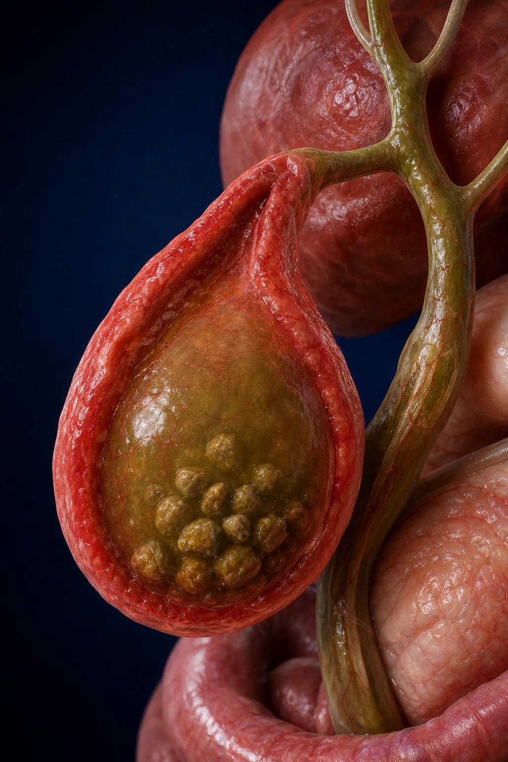
Pathophysiology — the pressure-driven cascade
Acute cholangitis is fundamentally a disease of stagnation under pressure. Normal biliary physiology depends on unidirectional flow from the canaliculi through the intra- and extra-hepatic ducts into the duodenum. Bile is normally sterile because continuous flow flushes any ascending bacteria, and the sphincter of Oddi acts as a mechanical barrier. Three events conspire to produce cholangitis:[4]
- Biliary obstruction — any lesion that blocks bile flow (stone, tumour, stricture, stent, parasite) raises intraductal pressure. Normal bile-duct pressure is 7-14 cmH₂O; with obstruction it can exceed 20-30 cmH₂O.
- Bacterial proliferation — stagnant bile becomes a culture medium. Bacteria enter the duct either by ascending from the duodenum (when the sphincter barrier is breached by stones, stents or previous sphincterotomy) or, less commonly, via the portal vein from the gut. Bacterial counts in bile rise logarithmically once flow ceases.
- Cholangio-venous reflux — this is the pivotal event. At high intraductal pressure, the hepatic tight junctions between hepatocytes and the canalicular-biliary interface break down. Bacteria, endotoxin and infected bile reflux directly across the disrupted barrier into the hepatic venous and lymphatic systems, producing bacteraemia, endotoxaemia and the systemic inflammatory response. This is why blood cultures are positive in 30-50% of cholangitis (far higher than cholecystitis) and why the disease can progress to septic shock so rapidly. [1]
The bacteria most commonly cultured reflect gut origin — predominantly gram-negative Enterobacteriaceae (E. coli, Klebsiella, Enterobacter, Citrobacter) and gram-positive Enterococcus, with anaerobes (Bacteroides, Clostridium) in mixed or healthcare-associated infection. Bacteraemia is documented in roughly half of cases, and polymicrobial infection is common with biliary stents or prior instrumentation. Endotoxaemia drives the fever, rigors and vasodilatory shock; bilirubin rises from the mechanical obstruction combined with sepsis-induced hepatic dysfunction. [1]
Definition and diagnosis
Acute cholangitis is a bacterial infection of the bile ducts associated with biliary obstruction. The Tokyo Guidelines 2018 (TG18) introduced a structured diagnostic criterion — suspected diagnosis requires one systemic (A), one cholestatic (B) and one imaging (C) finding, with the "definite" diagnosis additionally confirmed by imaging of the cause:[1][6]
A. Systemic signs of inflammation (need at least 1)
- Fever over 38°C and/or rigors
- Laboratory evidence of inflammation: abnormal WBC (under 4 or over 12 × 10⁹/L), or CRP over 10 mg/L [1]
B. Cholestatic findings (need at least 1)
- Jaundice (clinically apparent)
- Abnormal liver function tests: alkaline phosphatase, GGT or ALT/AST elevated to over 1.5× upper limit, or bilirubin elevated
C. Imaging findings (need at least 1)
- Biliary dilatation on ultrasound, CT or MRCP
- Evidence of the cause: stone, stricture, stent, tumour
A suspected diagnosis = (one A + one B + one C); a definite diagnosis additionally confirms the cause on imaging. Charcot triad alone is now a historical clinical clue — its sensitivity is only 50-70% and its absence does NOT exclude cholangitis, particularly in the elderly and immunocompromised. [1]
Clinical presentation
The classical clinical picture is dominated by the triad Jean-Martin Charcot described in 1877, and its more malignant extension described by Benedict Reynolds in 1959:[1]
Charcot triad
Present in only 50-70%
- FEVER (often with rigors — reflects bacteraemia)
- JAUNDICE (obstructive — elevated conjugated bilirubin, dark urine, pale stools)
- RIGHT UPPER QUADRANT PAIN (constant, may radiate to back/shoulder)
- Presence of all three = high probability of cholangitis
- BUT absence does NOT exclude — especially in elderly/immunocompromised
- Now only one limb of the structured TG18 diagnostic criteria
Reynolds pentad
Severe cholangitis + septic shock
- Charcot triad PLUS:
- HYPOTENSION (septic shock, often vasoplegic)
- ALTERED MENTAL STATUS (confusion, agitation, drowsiness)
- Indicates severe / Grade III cholangitis
- Requires URGENT ERCP within 24h + ICU admission
- Mortality 25-50% without prompt drainage
Causes of biliary obstruction
Any cause of mechanical bile-duct blockage can precipitate cholangitis. The aetiology is broadly split into community-acquired (typically stones) and healthcare-associated (stents, strictures, instrumentation — with a more resistant microbiology):[2]
[1]Community-acquired
Typically stones
- Choledocholithiasis — the dominant cause
- Organisms: E. coli, Klebsiella, Enterococcus
- Lower rates of resistant organisms
- Piperacillin-tazobactam adequate empirically
- Cholecystectomy after resolution to prevent recurrence
Healthcare-associated
Stents, strictures, instrumentation
- Occluded biliary stent — classic
- Post-surgical / transplant stricture
- Prior ERCP or biliary instrumentation
- Polymicrobial, more resistant (VRE, ESBL, Pseudomonas, Candida)
- Add vancomycin ± antifungal; broaden per local antibiogram
Cholangitis vs cholecystitis — the critical distinction
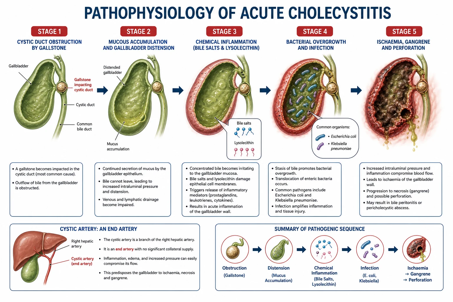
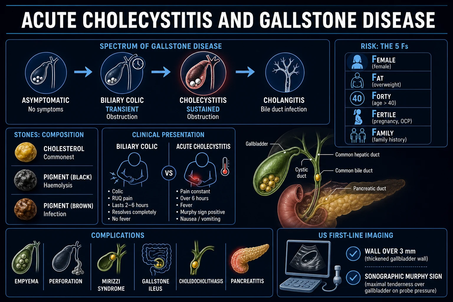
This is the single most frequently tested distinction in biliary sepsis and the one most often confused at the bedside. Acute cholangitis is infection of an obstructed bile duct with systemic sepsis; acute cholecystitis is inflammation of the gallbladder (usually from cystic duct obstruction by a stone), which is typically more localised. The management diverges sharply — cholangitis needs ERCP for duct decompression, cholecystitis needs cholecystectomy.[4]
Acute cholangitis
Duct obstruction + sepsis
- Obstruction of the COMMON BILE DUCT
- JAUNDICE prominent (obstructive, high bilirubin)
- Fever with rigors (bacteraemia in 30-50%)
- RUQ pain often diffuse, less focal than cholecystitis
- Dilated CBD on imaging; ALP/GGT elevated
- Definitive treatment: ERCP (sphincterotomy + stone extraction ± stent)
- Higher systemic illness — can progress to septic shock (Reynolds pentad)
- Higher blood-culture positivity
Acute cholecystitis
Gallbladder inflammation
- Obstruction of the CYSTIC DUCT (gallbladder neck)
- JAUNDICE usually ABSENT (CBD not obstructed)
- Fever may be present but rigors less common
- RUQ pain localised, with positive MURPHY SIGN
- Gallbladder wall thickening, pericholecystic fluid on US
- Definitive treatment: laparoscopic CHOLECYSTECTOMY (early, within 72h)
- Localised peritoneal signs; septic shock less common
- Lower blood-culture positivity
Differential diagnosis
The differential for fever + jaundice + RUQ pain / sepsis is broad. The structured TG18 criteria and imaging resolve most uncertainty:[1]
Biliary
Same anatomic region
- Acute cholecystitis (gallbladder, no jaundice)
- Choledocholithiasis without infection (obstructive jaundice, no sepsis)
- Biliary leak / biloma (post-cholecystectomy)
- Mirizzi syndrome (stone impacted in cystic duct compressing CBD)
- Cholangiocarcinoma with superimposed infection
- Acalculous cholecystitis (critically ill, ICU patient)
Non-biliary
Mimics to exclude
- Acute pancreatitis (epigastric pain, high lipase)
- Right basal pneumonia / empyema (referred pain)
- Hepatic abscess (often solitary, post-portal bacteraemia)
- Acute viral hepatitis (high ALT/AST, viral serology)
- Perforated peptic ulcer (free air on imaging)
- Appendicitis (retrocaecal) or mesenteric ischaemia
- Drug-induced liver injury / sepsis-induced cholestasis
Tokyo Guidelines severity grading
Severity drives timing of drainage and is the single most important triage decision after the diagnosis is made. The Tokyo Guidelines 2018 three-tier grading is applied at presentation, based on response to initial antibiotics and the presence of organ dysfunction:[1][6]
Tokyo Guidelines (TG18) severity grading (click each)
Organ dysfunction = ICU
At least ONE organ dysfunction at onset: cardiovascular (noradrenaline/dopamine needed for hypotension), neurological (altered mental status / decreased consciousness), respiratory (PaO2/FiO2 under 300), renal (creatinine over 177 µmol/L / 2 mg/dL), liver (PT-INR over 1.5), haematological (platelets under 100 × 10⁹/L). Requires URGENT biliary drainage — ERCP within 24 hours — plus ICU organ support.
Investigations
Diagnosis combines blood tests (systemic inflammation + cholestasis), imaging (to demonstrate obstruction and its cause), and blood cultures (before antibiotics). [1]
Blood tests
- FBC — leukocytosis (or leucopenia under 4); thrombocytopenia may herald severe disease/DIC.
- CRP — elevated; CRP over 10 mg/L is part of TG18 criterion A.
- LFTs — the cholestatic pattern dominates: ALP and GGT elevated, bilirubin elevated (conjugated/direct). ALT/AST may also be raised. The bilirubin level often correlates with the degree of obstruction.
- Coagulation — PT-INR may be elevated (hepatic dysfunction + vitamin K malabsorption from obstruction); correct before ERCP if possible.
- Lactate — elevated in severe disease / septic shock; a key resuscitation target.
- Amylase/lipase — to exclude concomitant pancreatitis (a CBD stone can cause both gallstone pancreatitis and cholangitis).
- Blood cultures — draw BEFORE antibiotics if possible; positive in 30-50%, polymicrobial common. Repeat after drainage if persistent fever.
- Bile cultures (obtained at ERCP) — guide de-escalation; always send. [1]
Imaging
Ultrasound
First-line, bedside
- Identifies gallstones and biliary dilatation (CBD over 6-8 mm)
- Cheap, portable, no radiation, repeatable
- Limited by bowel gas and body habitus
- Often cannot see the obstructing stone itself (CBD stones missed in up to 70%)
- Confirms or excludes gallbladder pathology (cholecystitis)
CT abdomen/pelvis
Second-line, especially if septic
- Shows biliary dilatation, the level of obstruction, and often the cause
- Identifies complications (abscess, portal vein thrombosis, perforation)
- Better for malignant obstruction and pancreatitis
- Excludes alternative diagnoses (perforation, mesenteric ischaemia)
- Contrast — caution in AKI; use low threshold in the septic patient
MRCP
Gold standard non-invasive duct imaging
- Best sensitivity for CBD stones and strictures (over 90%)
- No radiation; no contrast needed
- Does NOT allow therapeutic intervention
- Use when US/CT suggest obstruction and ERCP is not immediately planned
- Helps plan ERCP strategy (anatomy, stone size/number)
EUS
Problem-solving
- Highest sensitivity for small CBD stones and ampullary lesions
- Can exclude stones before ERCP (avoiding unnecessary ERCP)
- Allows FNA of suspicious masses (cholangiocarcinoma)
- Reserved for indeterminate cases after MRCP
Management — the ICU bundle
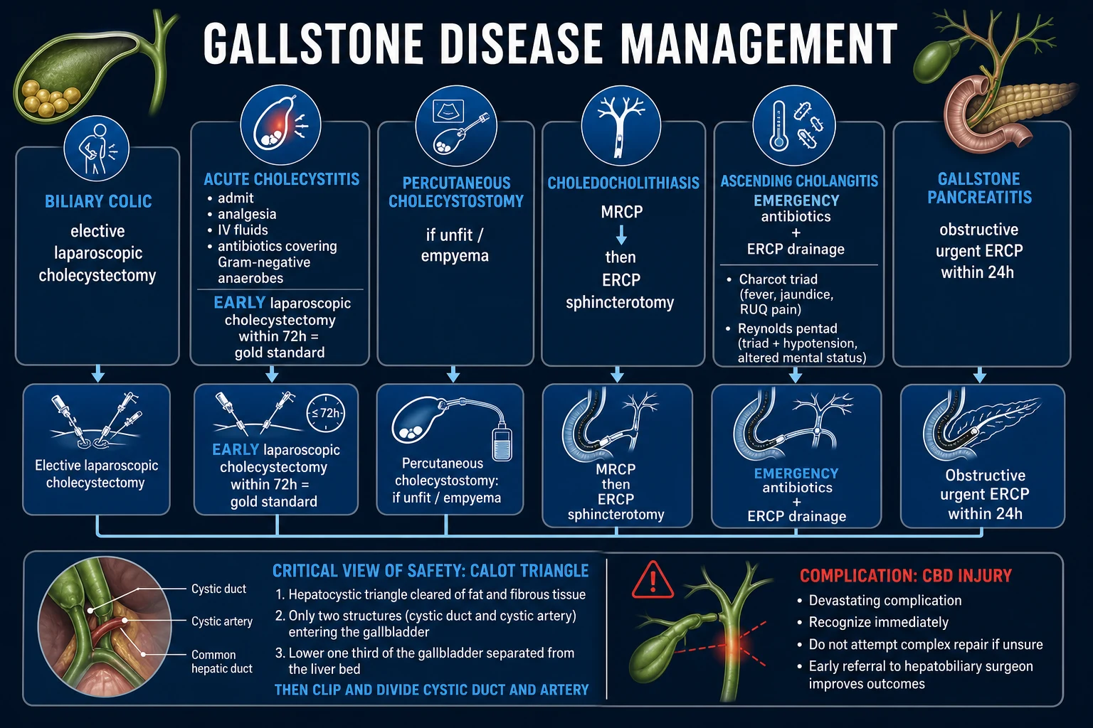
Management has three concurrent streams: antibiotics, biliary drainage (definitive source control), and resuscitation / organ support. The Tokyo Guidelines 2018 "management bundle" emphasises that these run in parallel, not in series.[6][7]
Acute cholangitis management protocol
1. Antibiotics within 1 hour (after blood cultures)
Empiric broad-spectrum: **piperacillin-tazobactam 4.5 g IV TDS** — covers Enterobacteriaceae (E. coli, Klebsiella, Enterobacter), Enterococcus and anaerobes. Alternatives: ceftriaxone 2 g + metronidazole, or cefepime + metronidazole. Add **vancomycin** if healthcare-associated, prior antibiotics, known colonisation, or severe (Grade III). **Severe penicillin allergy**: aztreonam + vancomycin + metronidazole, or a fluoroquinolone-based regimen. De-escalate on culture results from blood AND bile. Duration: 4-7 days after adequate drainage (shorter with good source control).<Cite id="3" /><Cite id="5" />
2. Fluid resuscitation + sepsis bundle
Crystalloid 30 mL/kg balanced-solution bolus for hypotension or lactate over 2 mmol/L. Vasopressors (noradrenaline first-line) for septic shock targeting MAP over 65. Monitor lactate clearance and urine output (over 0.5 mL/kg/h). Follow the Surviving Sepsis Campaign bundle — antibiotics within 1 hour, cultures first, measure lactate. Correct coagulopathy (vitamin K, FFP) before ERCP where feasible — but **do not let coagulopathy delay drainage in severe disease**.
3. Biliary DRAINAGE via ERCP — the definitive treatment
ERCP = endoscopic retrograde cholangiopancreatography. **Timing by severity**: URGENT (under 24h) for severe (Grade III); EARLY (under 48h) for moderate (Grade II); ELECTIVE for mild (Grade I). Technique: **cannulation of the CBD, sphincterotomy (incision of the sphincter of Oddi), stone extraction (balloon/basket), and stent placement** if a stricture or incomplete clearance. If ERCP fails or is unavailable: **percutaneous transhepatic biliary drainage (PTBD)** or surgical drainage.<Cite id="2" /><Cite id="6" />
4. Address the underlying cause
Once the patient stabilises after drainage, treat the cause: **cholecystectomy** during the same admission or within 2-4 weeks for choledocholithiasis (to prevent recurrence); management of malignant obstruction (definitive metal stent, oncology referral); dilatation or stenting of benign strictures; removal of parasites.
5. Organ support and monitoring
ICU admission for Grade III and unstable Grade II. Lung-protective ventilation if ARDS, noradrenaline ± vasopressin for vasodilatory shock, renal replacement therapy for severe AKI. Monitor for post-ERCP complications (pancreatitis, bleeding, perforation, worsening cholangitis). VTE prophylaxis, stress-ulcer prophylaxis if indicated, glycaemic control 6-10 mmol/L.
6. Cholecystectomy after resolution (if gallstone cause)
If cholangitis was caused by gallstones: perform **laparoscopic cholecystectomy during the SAME admission or within 2-4 weeks** to prevent recurrence (~10-20% recurrence within weeks if the gallbladder is left in situ). If unfit for surgery: consider **percutaneous cholecystostomy** or expectant management with a retained CBD stent.
Antibiotics — empiric and tailored
Empiric antibiotics must cover the Enterobacteriaceae (especially E. coli and Klebsiella), Enterococcus and anaerobes, with broadening for healthcare-associated or severe disease. The TG18 antimicrobial guideline stratifies by both severity and clinical setting.[5]
Community-acquired, mild-moderate
Piperacillin-tazobactam first-line
- Piperacillin-tazobactam 4.5 g IV TDS — covers all targets, first-line
- Alternative: ceftriaxone 2 g IV OD + metronidazole 500 mg IV TDS
- Alternative: cefepime + metronidazole
- Ciprofloxacin + metronidazole (if penicillin-allergic, per antibiogram)
- Duration 4-7 days after adequate drainage
Severe (Grade III) / healthcare-associated
Broaden + add MRSA cover
- Piperacillin-tazobactam PLUS vancomycin (MRSA / resistant Enterococcus)
- Consider carbapenem (meropenem) if ESBL risk, prior resistant organisms, or critical illness
- Add antifungal (echinocandin) if prolonged broad-spectrum, TPN, or known Candida colonisation
- Send blood AND bile cultures; de-escalate aggressively
- Reassess at 48-72 hours — fever should resolve within 2-3 days of drainage
ERCP timing and technique
ERCP is the cornerstone of definitive management. The timing is dictated by severity, and the technique by the underlying cause.[6]
Timing by severity
Severe (Grade III)
Urgent — under 24 hours
- Any one organ dysfunction = Grade III
- ERCP as soon as resuscitation begun — within 24 hours
- Do NOT wait for response to antibiotics
- Resuscitate in parallel; noradrenaline infusions can run during ERCP
- Highest mortality if delayed
Moderate (Grade II)
Early — under 24-48 hours
- No response to antibiotics within 24h, or meets two TG18 moderate criteria
- ERCP within 24-48 hours of presentation
- Often possible to stabilise first, then semi-elective ERCP
- Monitor closely for progression to Grade III
Mild (Grade I)
Elective
- Responds to initial antibiotics, no organ dysfunction
- ERCP can be performed electively during the admission
- Address cause — cholecystectomy for stones
- Reassess regularly — mild can progress to severe if obstruction persists
ERCP technique
ERCP combines endoscopy (side-viewing duodenoscope) with fluoroscopy to access and treat the biliary tree via the ampulla of Vater. The standard therapeutic sequence for choledocholithiasis:[2]
- Cannulation — the CBD is selectively cannulated with a catheter or sphincterotome; contrast is injected to outline the duct and confirm the obstruction/stone.
- Sphincterotomy — the sphincter of Oddi is incised with electrocautery, enlarging the ampullary opening to allow stone extraction and future stone passage; this is the mainstay that prevents recurrence.
- Stone extraction — stones are removed with a balloon catheter or Dormia basket, swept from the CBD into the duodenum.
- Stent placement — a plastic (or self-expanding metal) biliary stent is placed if: stones cannot be fully cleared (large/impacted stones, intrahepatic stones), a stricture is present, or the patient is too unstable for prolonged instrumentation. A stent re-establishes drainage and bridges to definitive therapy.
- Nasobiliary drain — an alternative to a stent when ongoing external drainage or repeat cholangiography is desired. [1]
For large or difficult stones, adjuncts include mechanical lithotripsy, laser/electrohydraulic lithotripsy, and balloon dilation of the biliary sphincter. For malignant obstruction, a self-expanding metal stent (SEMS) provides durable palliative drainage. [1]
Alternative biliary drainage — when ERCP fails
ERCP is successful in over 90% of cases, but fails or is impossible in some settings (prior gastric surgery/Roux-en-Y, periampullary diverticulum, tumour infiltration, failed cannulation, unavailable expertise). The alternatives:[2]
PTBD
Percutaneous transhepatic
- Radiologist places a drain through the liver into a dilated intrahepatic duct
- Used when ERCP fails, is unavailable, or anatomy is unfavourable
- Requires dilated intrahepatic ducts (easier in malignant obstruction)
- Risks: bleeding, bile leak, pneumothorax, catheter dislodgement
- Can be bridged to internal stent or definitive surgery
EUS-BD
Endoscopic ultrasound-guided
- EUS-guided rendezvous or direct transluminal drainage (hepaticogastrostomy, choledochoduodenostomy)
- Performed at specialist centres by advanced endoscopists
- Increasingly preferred over PTBD after failed ERCP — internal drainage
- Useful in altered anatomy (Roux-en-Y gastric bypass)
- Risks: bile leak, bleeding, stent migration
Surgical
Open / laparoscopic CBD exploration
- Common bile duct exploration (CBDE) with T-tube placement
- Reserved for failure of all endoscopic/percutaneous approaches
- Higher morbidity in the acutely septic patient
- Can be combined with cholecystectomy at index operation
Post-ERCP complications
ERCP is therapeutic but carries a defined complication burden — the ICU team must monitor for these after the procedure:[2]
Pancreatitis
3-10% — commonest
- Most common ERCP complication (3-10%)
- Mechanism: mechanical/thermal injury to the pancreatic duct orifice
- Presents with new epigastric pain, rising amylase/lipase within 24h
- Risk reduced by prophylactic rectal NSAID (diclofenac/indomethacin) and wire-guided cannulation
- Higher risk with difficult cannulation, sphincter of Oddi dysfunction, repeated injection
Bleeding
2-5% — after sphincterotomy
- Usually from the sphincterotomy site (sphincterotomy is the main risk)
- Immediate or delayed (up to 1-2 weeks)
- Higher risk with coagulopathy, anticoagulants, larger sphincterotomy
- Most cases settle with endoscopic haemostasis (clips, adrenaline, cautery)
- Prevent by correcting INR/platelets before ERCP where possible
Perforation
~1%
- Duodenal wall (lateral), periampullary, or bile-duct perforation
- Presents with abdominal pain, free air, sepsis
- Most duodenal perforations managed conservatively (NPO, antibiotics, drainage)
- Large perforations or bile-duct injuries may need surgery
Cholangitis / sepsis
1-3%
- Failed clearance, retained stones, or stent occlusion
- Incomplete drainage = ongoing source of sepsis
- Risk if contrast injected under pressure into an obstructed, infected duct
- Treat with antibiotics and repeat ERCP / further drainage
Post-ERCP complication rates
Microbiology
Bile is normally sterile; the organisms in cholangitis reflect gut origin and ascent. E. coli is consistently the #1 pathogen, with Klebsiella and Enterococcus close behind. Anaerobes are increasingly important in mixed or healthcare-associated infection.[3]
Biliary sepsis organisms
Special situations
Post-cholecystectomy
Stricture or retained stone
- Benign post-surgical stricture (clip injury, ischaemia) — commonest late cause
- Retained CBD stone (missed at cholecystectomy) — presents weeks later
- Manage with ERCP stenting / extraction; balloon dilatation for strictures
- Surgical reconstruction (hepaticojejunostomy) for recurrent or complex strictures
Malignant obstruction
Pancreatic, cholangio, ampullary
- Pancreatic head cancer — commonest malignant cause; painless jaundice
- Cholangiocarcinoma, ampullary cancer, gallbladder cancer
- Cholangitis often heralds or complicates the obstruction
- Definitive SEMS (self-expanding metal stent) for palliation; oncology referral
- Higher rate of multidrug-resistant organisms if stented long-term
Post-liver transplant
Anastomotic stricture
- Anastomotic (duct-to-duct) stricture at the bile-duct junction
- High immunosuppression — atypical presentation, opportunistic organisms (CMV)
- ERCP with balloon dilatation and sequential stenting first-line
- Coordinate with transplant team; risk of hepatic artery thrombosis (causes ischaemic strictures)
Pregnancy
Choledocholithiasis in pregnancy
- Increased gallstone formation from hormonal biliary stasis
- ERCP is safe in pregnancy with lead shielding and left lateral positioning
- Preferably in second trimester; sphincterotomy + stone extraction
- Defer cholecystectomy to postpartum unless recurrent
Recurrent pyogenic cholangitis
Oriental cholangiohepatitis
- Endemic in East Asia; intrahepatic pigment stones and strictures
- Recurrent episodes of cholangitis; biliary parasites (Clonorchis) contribute
- Difficult to clear; often needs repeated ERCP, percutaneous and surgical drainage
- Hepatic resection for segmental disease; high recurrence rate
Immunocompromised / neutropenic
Atypical, low-grade
- May not mount fever or leukocytosis — atypical presentation
- Opportunistic organisms (fungal, CMV, atypical bacteria)
- Low threshold for imaging and cultures; broaden empiric cover
- Acalculous cholecystitis and biliary candidiasis more common
Evidence and landmark trials
Tokyo Guidelines 2018
JHBPS 2018 / 2021
International consensus guideline series — diagnostic criteria, severity grading, antimicrobial therapy, drainage timing, and management bundles for acute cholangitis and cholecystitis
Key finding
Structured diagnostic criteria (systemic + cholestasis + imaging), three-tier severity grading driving drainage timing, standardised antibiotic stratification by severity and setting
Practice change
Became the global standard for diagnosis, severity grading and management; replaced earlier Tokyo Guidelines versions
Urgent vs Early ERCP
Gut 2026
RCT — urgent ERCP (under 24h) vs early ERCP (24-48h) in mild-to-moderate acute cholangitis
Key finding
No difference in 30-day mortality, in-hospital mortality or organ failure; urgent ERCP had higher procedure-related adverse events
Practice change
In stable mild-to-moderate cholangitis, completing resuscitation before ERCP within 48h is safe; severe disease still needs urgent drainage
TG18 antimicrobial
JHBPS 2018
Consensus guideline — stratified empiric antibiotic regimens by severity (Grade I-III) and clinical setting (community vs healthcare-associated)
Key finding
Piperacillin-tazobactam appropriate for most community-acquired; broaden with vancomycin/carbapenem for healthcare-associated or severe; de-escalate on culture
Practice change
Standardised empiric antibiotic selection in acute biliary infection
Prophylactic rectal NSAID
NEJM 2012
Meta-analysis and subsequent RCTs — rectal diclofenac/indomethacin to prevent post-ERCP pancreatitis
Key finding
Rectal NSAID immediately before/after ERCP reduces post-ERCP pancreatitis by roughly half in average-risk patients
Practice change
Routine prophylactic rectal NSAID now standard before ERCP
Complications
Local
Biliary and hepatic
- Hepatic abscess — from ascending infection; needs drainage
- Biliary sepsis with multi-organ failure if drainage delayed
- Pancreatitis — if the obstructing stone also blocks the pancreatic duct
- Sepsis-induced hepatic dysfunction / jaundice worsening
- Recurrence — if the underlying cause (gallbladder stones) is not addressed
Systemic
Septic and organ failure
- Septic shock — distributive, often high output; noradrenaline first-line
- Acute kidney injury — ATN from hypoperfusion and sepsis; may need RRT
- ARDS — lung-protective ventilation
- Disseminated intravascular coagulation — from gram-negative endotoxaemia
- Metabolic: hyperglycaemia, electrolyte disturbance from sepsis
Prognosis
Mortality is driven by severity at presentation and the timeliness of biliary drainage:[1][2]
Outcomes by severity (TG18)
Predictors of poor outcome include advanced age, high Charlson comorbidity index, malignant obstruction, delayed drainage, high bilirubin, high lactate, bacteraemia with resistant organisms, and the presence of multiple organ dysfunctions at presentation. Reynolds pentad at presentation is itself a marker of high mortality. Prompt drainage within 24-48 hours is the single most modifiable determinant of survival in severe disease. [1]
Exam practice
SAQ — Severe (Grade III) acute cholangitis
20 minutes · 10 marks
A 74-year-old man presents to the emergency department with a 24-hour history of rigors, right upper quadrant pain and progressive confusion. On examination: temperature 39.4°C, HR 128, BP 82/48 (MAP 59), RR 28, SpO2 94% on room air, GCS 13 (E3 V4 M6), icteric with RUQ tenderness. Bloods: WBC 24.6, bilirubin 138 µmol/L (direct 102), ALP 410, ALT 180, INR 1.7, creatinine 210 µmol/L, lactate 4.2 mmol/L, platelets 92. Bedside ultrasound shows a dilated common bile duct of 12 mm with a stone in the distal CBD and intrahepatic duct dilatation. Blood cultures have been taken.
SAQ — Cholangitis vs cholecystitis distinction
10 minutes · 8 marks
A 52-year-old woman presents with 12 hours of right upper quadrant pain, fever 38.6°C and nausea. She is tender in the RUQ with a positive Murphy sign. Bloods: WBC 15.2, bilirubin 22 µmol/L, ALP 180, ALT 90. Ultrasound shows gallstones, a thickened gallbladder wall with pericholecystic fluid, and a common bile duct of 5 mm.
Clinical pearls
Red flags
References
- [1]Yokoe M, Hata J, Takada T, et al. Ginkgolide A alleviates cardiac remodeling in mice with myocardial infarction via binding to matrix metalloproteinase-9 to attenuate inflammation Eur J Pharmacol, 2022.PMID 35367419
- [2]Lau WY, et al. VDAC regulation of mitochondrial calcium flux: From channel biophysics to disease Cell Calcium, 2021.PMID 33529977
- [3]Solomkin JS, et al. Know thy neighbor: Modeling spatiotemporal cell-fate patterns in a neural stem cell niche Cell Stem Cell, 2021.PMID 34358437
- [4]Takada T. Tokyo Guidelines 2018: updated Tokyo Guidelines for the management of acute cholangitis/acute cholecystitis J Hepatobiliary Pancreat Sci, 2018.PMID 29334699
- [5]Gomi H, Solomkin JS, Takada T, et al. Tokyo Guidelines 2018: antimicrobial therapy for acute cholangitis and cholecystitis J Hepatobiliary Pancreat Sci, 2018.PMID 29090866
- [6]Miura F, Okamoto K, Takada T, et al. Tokyo Guidelines 2018: initial management of acute biliary infection and flowchart for acute cholangitis J Hepatobiliary Pancreat Sci, 2018.PMID 28941329
- [7]Mayumi T, Okamoto K, Takada T, et al. Tokyo Guidelines 2018: management bundles for acute cholangitis and cholecystitis J Hepatobiliary Pancreat Sci, 2018.PMID 29090868