ICU · GI/Nutrition
Acute liver failure (hepatic failure)
Also known as Acute liver failure (ALF) · Fulminant hepatic failure · King's College Criteria · N-acetylcysteine (NAC) · Liver transplant for ALF · Cerebral oedema in ALF · O'Grady classification · Hyperacute / acute / subacute liver failure
Acute liver failure (ALF) is severe acute liver injury with encephalopathy and coagulopathy (INR >1.5) within 26 weeks, in a patient without pre-existing liver disease. Classification by tempo (O'Grady): hyperacute (<7 days), acute (8-28 days), subacute (29 days-12 weeks) — prognosis varies by interval. Causes: paracetamol (1 in UK/Aus), drug-induced (idiosyncratic), viral hepatitis (HBV 1 viral), Wilson disease, pregnancy-related (AFLP, HELLP), mushroom (Amanita phalloides), ischaemic, autoimmune, indeterminate. Pathophysiology of cerebral oedema: ammonia crosses BBB - astrocytic glutamine synthetase - glutamine accumulation - astrocyte swelling (cytotoxic oedema) - raised ICP - tonsillar herniation (1 cause of death). Management: N-acetylcysteine (NAC) for ALL causes (Lee 2009 showed transplant-free survival benefit beyond paracetamol), supportive care for complications, and urgent liver transplant assessment using King's College Criteria. Lactate is a key prognostic marker (early >3.5 mmol/L or persistent elevation predicts poor outcome). Do NOT routinely correct INR with FFP — it is a liver function marker, not a bleeding-risk indicator (rebalanced haemostasis).
On this page & tools
Your progress
Saved locally on this device.
18 MCQs with explanations
Target exams
Red flags

Definition
Acute liver failure (ALF) is the onset of coagulopathy (INR >1.5) and any degree of hepatic encephalopathy within 26 weeks of the first symptoms of liver injury, in a patient without pre-existing cirrhosis or liver disease. The 26-week ceiling separates ALF from acute-on-chronic liver failure (ACLF), which has a distinct management pathway.[1]
The older term "fulminant hepatic failure" (Trey & Davidson, 1970) required encephalopathy within 8 weeks of the first symptom; "late-onset" or "subfulminant" referred to cases developing encephalopathy later. This binary has been superseded by the O'Grady tempo-based classification (below), which carries prognostic meaning. [1]
[1]Classification by tempo — the O'Grady system
The interval between jaundice and encephalopathy defines three syndromes with distinct aetiologies and outcomes.[3]
O'Grady tempo classification — interval from jaundice to encephalopathy
29 days - 12 weeks
Encephalopathy 5-12 weeks after jaundice. Typical causes: seronegative ("non-A non-B") hepatitis, idiosyncratic drug-induced liver injury (DILI), some Wilson presentations. Carries the WORST transplant-free survival (often <20%) — cerebral oedema is less common but multi-organ failure and portal hypertension dominate. Urgent transplant is frequently the only option.
Hyperacute (<7 d)
Best prognosis
- Paracetamol overdose — the archetype
- Ischaemic hepatitis / "shock liver"
- Amanita phalloides mushroom poisoning
- Cerebral oedema: COMMON and early (the dominant threat)
- Transplant-free survival ~50-60% (highest of the three)
Acute (8-28 d)
Intermediate
- Viral hepatitis — HBV (most common viral cause), HAV, HEV
- Some idiosyncratic drug reactions
- Autoimmune hepatitis (acute presentation)
- Cerebral oedema: moderate risk
- Transplant-free survival ~30-40%
Subacute (29 d - 12 wk)
Worst prognosis
- Seronegative ("non-A non-B") hepatitis — often indeterminate
- Idiosyncratic drug-induced liver injury (DILI)
- Wilson disease (acute decompensation)
- Cerebral oedema: LESS common; portal hypertension and renal failure dominate
- Transplant-free survival often <20% — transplant is frequently the only exit
Classification at the bedside — apply on admission
Confirm ALF
Acute liver injury (AST/ALT markedly elevated) + coagulopathy INR >1.5 + encephalopathy (any grade), within 26 weeks, AND no known pre-existing liver disease. If chronic liver disease/cirrhosis is present, re-classify as ACLF — different pathway.
Record the tempo
Date of first symptom (often jaundice or nausea) -> date of encephalopathy. <7 d = hyperacute; 8-28 d = acute; 29 d-12 wk = subacute. The tempo refines prognosis and frames how aggressively to push transplant assessment.
Identify aetiology
Paracetamol level, viral serology, ceruloplasmin + slit-lamp, autoimmune panel, pregnancy test, drug/herbal history, Amanita exposure (delayed GI symptoms then liver failure). Aetiology determines specific therapy AND prognosis.
Stratify for transplant
Apply King's College Criteria on admission and reapply serially. Add lactate and arterial ammonia to the prognostic picture. Involve the transplant centre EARLY — the window between meeting criteria and brain death from cerebral oedema can be hours.
Causes
Common causes
West / UK / Aus
- Paracetamol (acetaminophen) overdose — #1 cause in UK, Australia, US (~40-50% of ALF). Both deliberate and therapeutic misadventure (staggered dosing in pain or malnutrition).
- Drug-induced (idiosyncratic) — isoniazid, valproate, halothane, nitrofurantoin, phenytoin, statins, herbal/dietary supplements (e.g. kava, green-tea extract, Polygonum multiflorum), recreational MDMA
- Viral hepatitis — HBV (#1 viral cause worldwide; acute infection or reactivation with immunosuppression/chemotherapy), HAV, HEV (endemic and zoonotic), HSV (immunocompromised/pregnant), EBV, CMV
- Ischaemic hepatitis ("shock liver") — profound hypotension, cardiac arrest, severe heart failure. Often recovers with haemodynamic support.
- Autoimmune hepatitis (acute presentation)
- Indeterminate (~20%) — "seronegative hepatitis"; common in subacute ALF and carries a poor prognosis
Less common / specific
Selected populations
- Mushroom poisoning (Amanita phalloides) — delayed GI symptoms (6-24 h) then liver failure at 48-72 h. Classically after foraging. Treat with silibinin + NAC + aggressive fluid.
- Wilson disease — young patient, ALF + Coombs-negative haemolysis + low ceruloplasmin + Kayser-Fleischer rings + markedly low alkaline phosphatase relative to bilirubin. Always fatal without transplant.
- Pregnancy-related: acute fatty liver of pregnancy (AFLP) and HELLP — third trimester; delivery is the definitive treatment.
- Heat stroke / malignant hyperthermia
- Malignant infiltration (lymphoma, melanoma, breast) — hepatomegaly, imaging
- Budd-Chiari syndrome (hepatic venous outflow obstruction)
- Veno-occlusive disease / sinusoidal obstruction syndrome (post-chemotherapy, haematopoietic stem cell transplant)
Aetiology and prognosis
Transplant-free survival
- Paracetamol — best (~50-60% spontaneous survival)
- Ischaemic hepatitis — good with haemodynamic correction
- Pregnancy-related (delivery) — good once delivered
- HAV — relatively good
- HBV — moderate (~25-35%)
- Drug-induced (idiosyncratic) — poor (~20-30%)
- Autoimmune — moderate with steroids, often needs transplant
- Seronegative / indeterminate — poor (~15-25%)
- Wilson disease — near-100% fatal without transplant; urgent listing
- Amanita — variable; silibinin + NAC improve outcomes
Aetiology-specific therapy
Beyond supportive care
- Paracetamol — NAC (regardless of time since overdose if any hepatotoxicity)
- Amanita — silibinin (milk thistle) + NAC + aggressive crystalloid diuresis
- HBV — nucleos(t)ide analogue (entecavir/tenofovir)
- HSV — IV aciclovir
- Autoimmune — corticosteroids (prednisolone 40-60 mg)
- Wilson — chelation (penicillamine) usually futile in acute crisis; TRANSPLANT
- Pregnancy (AFLP/HELLP) — URGENT DELIVERY of the fetus
- Budd-Chiari — anticoagulation, TIPS, transplant
- Drug-induced — STOP the offending agent immediately
Paracetamol — the leading cause in the West
Paracetamol (acetaminophen) hepatotoxicity is the single largest cause of ALF in the UK, Australia and the US. The toxic metabolite NAPQI (formed by CYP2E1) depletes glutathione and binds hepatocyte proteins causing centrilobular (zone 3) necrosis. Risk is amplified by fasting, malnutrition, alcohol, and concurrent CYP2E1 inducers. [1]
Paracetamol overdose — diagnostic and management cascade
Establish timing
Single ingestion time is essential for the Rumack-Matthew nomogram. If staggered or unknown ingestion time, the nomogram is NOT valid — treat with NAC if any evidence of hepatotoxicity (ALT rising or detectable paracetamol level).
Rumack-Matthew nomogram
Plots serum paracetamol level (y-axis) against time post-ingestion (x-axis, from 4 to 24 h). Level ABOVE the treatment line ("200" line at 4 h in some units, "150"/100 in others) -> give NAC. Use the local nomogram. NOT applicable after 24 h or in staggered overdose.
Give NAC
150 mg/kg IV over 1 h (loading), then 50 mg/kg over 4 h, then 100 mg/kg over 16 h. Newer 12 h and 20 h protocols exist. Continue beyond the standard course if ALT rising, INR >2, or paracetamol still detectable. Anaphylactoid reactions (rash, bronchospasm) are common in the loading dose — slow the infusion and treat with antihistamine; do NOT stop NAC.
Monitor for King's College Criteria
Check pH, INR, creatinine, lactate, and grade of encephalopathy at baseline and serially (4-12 h). Any deterioration toward KCC triggers urgent transplant referral.
Specific aetiologies — high-yield patterns
[1] [1] [1] [1]Pathophysiology of cerebral oedema — the leading cause of death
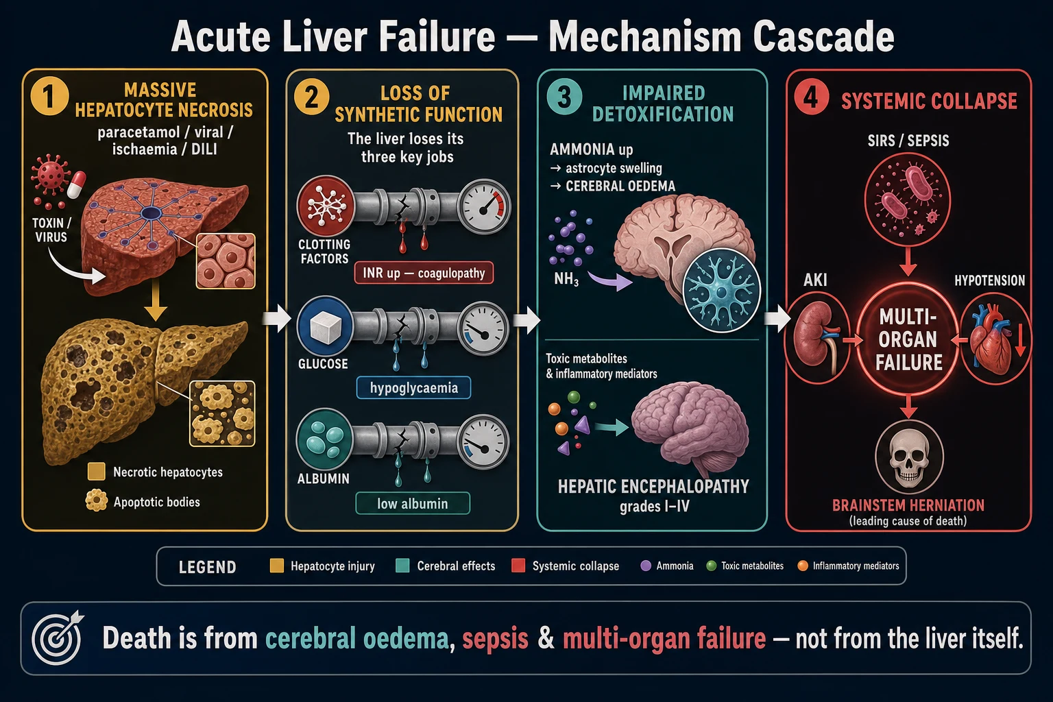
Cerebral oedema with intracranial hypertension is the #1 cause of death in ALF. Its incidence tracks encephalopathy grade: rare in Grade I-II, ~25-35% in Grade III, and >75% in Grade IV encephalopathy. The mechanism is cytotoxic (intracellular) oedema driven by the ammonia-glutamine axis.[2]
The ammonia-glutamine cascade
Ammonia -> glutamine -> astrocyte swelling -> herniation
Ammonia generation and failed clearance
Gut bacteria and enterocytes generate ammonia from dietary protein and glutamine. The failing liver cannot convert ammonia to urea (urea cycle), and portosystemic shunting bypasses residual hepatocytes. Serum ammonia rises.
Ammonia crosses the blood-brain barrier
Ammonia diffuses readily across the BBB (as NH3) and is also actively transported. In ALF the BBB itself becomes more permeable, accelerating influx.
Astrocytic glutamine synthetase
Within astrocytes, glutamine synthetase condenses ammonia with glutamate to form **glutamine**. This is the brain's only significant ammonia-detoxification route. The astrocyte is therefore the cellular target of ammonia toxicity.
Glutamine accumulation -> osmotic stress
Glutamine accumulates intracellularly and acts as an osmolyte, drawing water into the astrocyte. The astrocyte swells (**cytotoxic oedema**). Mitochondrial glutamine also enters and is deaminated back to ammonia by glutaminase, generating intramitochondrial ammonia and reactive oxygen species ("the Trojan horse" hypothesis).
Astrocyte swelling -> cerebral oedema
Swollen astrocytes (the foot processes regulate the blood-brain barrier and capillary diameter) compress microvasculature, reduce cerebral perfusion, and disrupt the extracellular matrix. Cytotoxic oedema generalises. Brain volume increases within a fixed skull.
Raised ICP -> herniation
Intracranial pressure rises; cerebral perfusion pressure (CPP = MAP - ICP) falls. The swollen brain shifts: uncal herniation (CN III palsy — fixed dilated pupil), then tonsillar herniation with brainstem compression and Cushing's triad (hypertension, bradycardia, irregular respiration), then brain death. This is why Grade III-IV encephalopathy mandates intubation and ICP-targeted therapy.
Why cerebral oedema is uniquely dangerous in ALF (vs cirrhosis)
ALF cerebral oedema
Acute, cytotoxic
- Rapid, massive ammonia rise -> acute astrocyte swelling
- True cytotoxic cerebral oedema -> intracranial HYPERTENSION -> herniation
- Common and lethal in Grade III-IV (~75% in Grade IV)
- Astrocytes have no time to compensate (no osmotic adaptation)
- Requires hyperosmolar therapy, head elevation, often ICP monitoring
Cirrhotic HE
Chronic, compensated
- Chronic, lower-grade ammonia exposure -> astrocyte Alzheimer type II change
- Astrocytes adapt osmotically (extrude myo-inositol) -> minimal oedema
- Intracranial hypertension and herniation are RARE
- Lactulose + rifaximin effective; management is precipitant-driven
- Different disease, different management pathway
Management
General principles
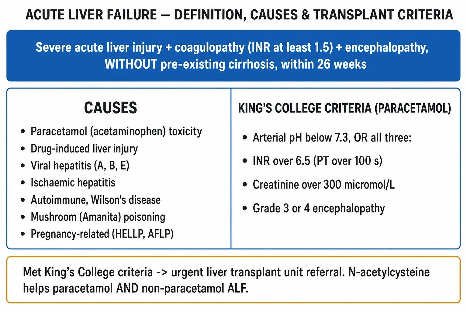
ALF is a time-critical, multi-organ disease managed (ideally) in a transplant-capable centre. The four pillars are: (1) aetiology-directed therapy (NAC, silibinin, antivirals, steroids, delivery); (2) cerebral oedema prevention and control; (3) multi-organ supportive care (glucose, renal, haemodynamics, infection, coagulation); and (4) early transplant assessment against King's College Criteria. Refer to a transplant centre as soon as ALF is diagnosed — do not wait for deterioration.[1][2]
ALF ICU management protocol — the full bundle
NAC for ALL causes
N-acetylcysteine 150 mg/kg IV over 1 h, then 50 mg/kg over 4 h, then 100 mg/kg over 16 h. Lee et al (2009, Gastroenterology) showed improved transplant-free survival in non-paracetamol ALF, especially early-stage (Grade I-II). Give to ALL ALF patients regardless of cause. Low risk, potential benefit, cheap. Continue until INR improving, encephalopathy resolving, or transplant.
Cerebral oedema management
Grade III-IV encephalopathy = high risk of intracranial hypertension. INTUBATE for airway protection and to enable ICP-targeted care. Head of bed elevated 30 degrees, midline. Maintain normocapnia (PaCO2 35-40), normoxia, normothermia (avoid fever), normoglycaemia. Hyperosmolar therapy: hypertonic 3% saline (target Na 145-155 mmol/L) or mannitol 0.5-1 g/kg if not anuric. Consider ICP monitor in Grade IV. Avoid hypotension/hypoxia (each episode worsens secondary brain injury). Induced hypothermia (32-34 C) and hypernatraemia as rescue. Hyperventilation as a TEMPORARY bridge only (causes cerebral vasoconstriction lowering ICP but reduces cerebral perfusion if sustained).
Coagulopathy — do NOT chase the INR
INR is a liver FUNCTION marker (prognostic, central to King's College Criteria), NOT a bleeding-risk indicator. ALF is a state of "rebalanced haemostasis": both procoagulant factors AND anticoagulant factors (protein C/S, antithrombin) are reduced, and platelets are often dysfunctional but thrombopoietin-deficient. Viscoelastic testing (TEG/ROTEM) better reflects true haemostasis than INR. Give vitamin K 10 mg IV. Use FFP ONLY for active bleeding or before invasive procedures. Recombinant activated factor VII for emergencies. Do not transfuse platelets prophylactically unless bleeding.
Hypoglycaemia
Check glucose hourly. Maintain >4 mmol/L with a 10% dextrose infusion (and 50% boluses if needed). Impaired hepatic gluconeogenesis and depleted glycogen make hypoglycaemia both common and dangerous — it worsens the already-injured brain and can mimic or precipitate encephalopathy.
Renal support
AKI is common (~50-70%): hepatorenal syndrome, ATN from hypotension/paracetamol nephrotoxicity, or contrast. Avoid nephrotoxins. CRRT is PREFERRED over intermittent haemodialysis — continuous therapy avoids the rapid solute shifts and blood pressure swings that worsen cerebral oedema and raise ICP. Maintain adequate MAP for cerebral and renal perfusion.
Haemodynamics
ALF is a hyperdynamic, vasodilated state (NO-mediated). Target MAP >75 mmHg and CPP >60 mmHg. Norepinephrine is first-line vasopressor (alpha agonism preserves CPP). Add vasopressin/terlipressin for refractory vasodilation. Avoid pure beta-agonists. Fluid resuscitate with balanced crystalloid; albumin for hypoalbuminaemia. Invasive arterial and central monitoring essential.
Infection surveillance
Marked immune paralysis -> very high infection risk (bacterial ~60-80%, fungal ~30%). Surveillance cultures (blood, urine, sputum, line) daily. Empirical broad-spectrum antibiotics for any clinical deterioration (new fever, rising WCC/w lactate, worsening encephalopathy or haemodynamics) — infection precipitates multi-organ failure and amplifies cerebral oedema. Add antifungal cover for prolonged ICU stay. Prophylactic antibiotics/antifungals are used in many units.
Liver transplant assessment
Urgent referral to a transplant centre at diagnosis — transfer BEFORE deterioration. Apply King's College Criteria on admission AND serially (they are dynamic). Time is critical: brain death from cerebral oedema is the #1 cause of death. List EARLY; do not wait for maximal deterioration, because many die or develop contraindications (sepsis, brain death) while listed.
N-acetylcysteine (NAC) — for ALL causes
NAC is the one therapy in ALF with the broadest evidence base. In paracetamol overdose it restores hepatic glutathione and detoxifies NAPQI. In non-paracetamol ALF it improves transplant-free survival through pleiotropic effects: antioxidant (replenishes glutathione systemically), improved microcirculation and oxygen delivery, anti-inflammatory, and attenuation of cerebral oedema.[4]
Lee 2009 — NAC for non-paracetamol ALF (Gastroenterology; PMID 19524577)
Source
Multicentre double-blind RCT, US Acute Liver Failure Study Group (ALFSG); 173 patients with non-acetaminophen ALF, stratified by grade I-II vs III-IV encephalopathy
Intervention
IV N-acetylcysteine vs placebo, standard regimen
Primary outcome
Overall survival at 3 weeks: 70% NAC vs 66% placebo (1-sided P=0.28) — NOT significant for the whole cohort
Subgroup (Grade I-II HE)
Transplant-free survival SIGNIFICANTLY higher with NAC (52% vs 30%, P=0.02) — the patients who matter, before they deteriorate
Subgroup (Grade III-IV HE)
No survival benefit (most were already too sick / destined for transplant)
Clinical bottom line
Give NAC to ALL ALF patients, especially early-stage (Grade I-II) disease of ANY aetiology — it improves transplant-free survival and is safe, cheap, and easy. The benefit extends well beyond paracetamol.
NAC administration — practical
Indication
ALL patients with ALF of any aetiology, AND any paracetamol patient with hepatotoxicity (rising ALT or detectable level) regardless of time since ingestion.
Dose (standard IV regimen)
Loading 150 mg/kg in 200 mL 5% dextrose over 1 h (60 min); then 50 mg/kg in 500 mL over 4 h; then 100 mg/kg in 1000 mL over 16 h. Total 21 h. Total dose 300 mg/kg. Modify volume for fluid restriction.
Adverse effects
Anaphylactoid (not IgE) reaction in ~10-20% during loading: flush, rash, bronchospasm, rarely hypotension. More common in asthmatics and with rapid infusion. Management: slow/stop infusion temporarily, antihistamine (chlorphenamine), bronchodilator if needed, then RESTART at slower rate. Do NOT abandon NAC for a rash.
When to stop
Continue beyond the 21 h course if INR still rising or >2, paracetamol still detectable, or encephalopathy present. Stop when INR <2 and falling, encephalopathy resolving, paracetamol undetectable — OR at transplant.
King's College Criteria — transplant listing
The King's College Criteria (KCC), derived by O'Grady et al from the King's College London ALF cohort, remain the most widely used and validated criteria for predicting death without transplant and therefore for listing for emergency liver transplantation. They are aetiology-specific (paracetamol vs non-paracetamol) and should be applied on admission and serially.[5]
[5] [1]Paracetamol KCC
Single or combined
- Arterial pH <7.25 after resuscitation (any grade of HE), OR
- All three combined: INR >6.5 AND creatinine >300 umol/L AND Grade III-IV HE
- Lactate adjunct: early arterial lactate >3.5 mmol/L (pre-resuscitation) or >3.0 after resuscitation predicts mortality
- Phosphate adjunct: hypophosphataemia (Phosphate <0.55 mmol/L day 2-3) reflects hepatic regeneration failure
Non-paracetamol KCC
INR or 3 of 5
- INR >6.5 (PT >100 s) alone, OR
- Any 3 of 5: INR >3.5 / age <10 or >40 / unfavourable aetiology (seronegative, halothane, idiosyncratic) / jaundice-to-encephalopathy >7 d / bilirubin >300 umol/L
- Aetiology-specific Wilson and seronegative cases almost always meet criteria
- Less sensitive than paracetamol KCC; combine with lactate and clinical trajectory
Prognostic markers — beyond King's College
ALF prognostication is multi-modal. No single marker is sufficient; the trajectory matters as much as any single value. [1]
Static markers
On / near admission
- King's College Criteria — aetiology-specific (above)
- Arterial lactate — >3.5 mmol/L early (or >3.0 after resuscitation) predicts poor outcome
- Arterial ammonia — >150-200 umol/L predicts intracranial hypertension
- Phosphate — low phosphate (day 2-3) = failed regeneration = poor
- Aetiology — paracetamol/ischaemic/pregnancy good; seronegative/DILI/Wilson poor
- Tempo — hyperacute best; subacute worst
- Grade of encephalopathy at presentation
Dynamic markers
Trajectory over hours
- Rising INR despite therapy = worsening synthetic function
- Rising arterial lactate despite resuscitation = worsening perfusion/clearance
- Progression of encephalopathy grade (II -> III -> IV) = cerebral oedema risk
- Rising ammonia = intracranial hypertension risk
- Development of renal failure / vasopressor dependence / new infection
- Falling CPP on ICP monitoring
Alternative and composite scores
Other prognostic tools (know they exist)
Clichy criteria (France)
Historically used for HBV-related ALF: factor V level <20% of normal in patients <30 years, or <30% in patients >30 years, combined with Grade III-IV encephalopathy. Less used outside France.
MELD
Model for End-stage Liver Disease — validated in cirrhosis; used in some ALF settings but less discriminating than KCC for ALF. Rising MELD reflects worsening.
CLIF-C ACLF / ALF scores
Derived from the CANONIC ACLF cohort and extended to ALF; combine organ-failure score with age and white-cell count. Useful for mortality prediction and goals-of-care discussions.
APACHE II
Generic ICU score; reasonable for overall mortality prediction but not designed for ALF transplant decisions. Not a substitute for KCC.
Complications of ALF
Cerebral
#1 cause of death
- Cerebral oedema / intracranial hypertension -> tonsillar herniation
- Seizures (often non-convulsive — low threshold for cEEG)
- Cerebral hypoperfusion (low CPP) -> secondary injury
Metabolic / haematologic
- Hypoglycaemia (impaired gluconeogenesis) — check hourly
- Coagulopathy (low factors + low anticoagulants = rebalanced) — bleeding LESS common than INR suggests
- Thrombocytopenia (consumption, splenomegaly, sepsis)
- Metabolic acidosis (lactate) — prognostic
- Hypophosphataemia, hypokalaemia, hypomagnesaemia
Renal / circulatory
- AKI (~50-70%) — HRS, ATN, paracetamol nephrotoxicity
- Hyperdynamic vasodilatory shock (NO-mediated) — norepinephrine first-line
- Pancreatitis (paracetamol association)
Infective
- Bacterial infection (~60-80%) — Gram-positive and Gram-negative; immune paralysis
- Fungal infection (~30%) — Candida; prolonged ICU stay
- Sepsis precipitates multi-organ failure and amplifies cerebral oedema
Key trials and evidence
O'Grady 1989 — derivation of the King's College Criteria (Gastroenterology; PMID 2490426)
Source
Retrospective analysis of 588 ALF patients (King's College London, 1973-1985), validated prospectively in 175 patients (1986-1987)
Objective
Identify early, objective indicators of prognosis to select patients for liver transplantation
Paracetamol criteria
Arterial pH <7.30 (revised to <7.25), OR all three of (INR/PT >100 s + creatinine >300 umol/L + grade III-IV encephalopathy)
Non-paracetamol criteria
INR >6.5 alone, OR any 3 of 5 (INR >3.5, age <10/>40, unfavourable aetiology, jaundice-to-encephalopathy >7 d, bilirubin >300 umol/L)
Performance
Sensitivity ~70%, specificity ~90% for death without transplant — high specificity (meeting criteria = very likely to die), modest sensitivity (not meeting criteria does NOT guarantee survival)
Clinical bottom line
Apply KCC on admission AND serially; meeting them triggers urgent transplant referral. They remain the global standard for ALF transplant listing 35 years on.
Lee 2009 — NAC for non-paracetamol ALF (Gastroenterology; PMID 19524577)
Source
Multicentre double-blind RCT, US ALFSG; 173 patients with non-acetaminophen ALF
Intervention
IV N-acetylcysteine vs placebo
Whole-cohort survival
70% NAC vs 66% placebo at 3 weeks — not significant
Grade I-II subgroup
Transplant-free survival 52% NAC vs 30% placebo (P=0.02) — significant
Clinical bottom line
NAC improves transplant-free survival in early-stage non-paracetamol ALF -> give NAC to ALL ALF, especially early-grade disease. Cheap, safe, broad benefit.
O'Grady 1993 — redefining the syndromes of ALF (Lancet; PMID 8491116)
Source
Conceptual reclassification of ALF by the interval between jaundice and encephalopathy, grounded in the King's College cohort
Contribution
Defined hyperacute (<7 d), acute (8-28 d), and subacute (5-12+ weeks) sub-syndromes with distinct aetiologies, complication profiles, and prognoses
Key insight
The faster the encephalopathy, paradoxically the BETTER the transplant-free survival (hyperacute/paracetamol regenerates; subacute/seronegative does not)
Clinical bottom line
The classification that frames every modern ALF discussion — tempo plus aetiology determines prognosis and transplant urgency.
ALF evidence and outcomes — the synthesis
NAC
Mortality benefit in paracetamol ALF even when started >24 h after ingestion; transplant-free survival benefit in non-paracetamol ALF (Lee 2009). Give to ALL ALF.
King's College Criteria
Specificity ~90%, sensitivity ~70% for death without transplant. Apply serially; adjunct lactate/ammonia/phosphate refine.
Transplantation
1-year survival ~80-90% for ALF — the definitive therapy for those who meet criteria.
MARS / albumin dialysis
RELIEF and subsequent trials: no mortality benefit; may improve encephalopathy and bridge to transplant. Not standard of care for survival.
Spontaneous survival
Overall ~40-60% without transplant; aetiology-dependent (paracetamol/pregnancy/ischaemic best; seronegative/DILI/Wilson worst).
Cerebral oedema
#1 cause of death; hypertonic saline + mannitol + ICP-directed care in Grade III-IV. Induced hypothermia and indometacin as rescue.
SAQ — Paracetamol-induced acute liver failure: King's College Criteria and the transplant decision
10 minutes · 10 marks
A 24-year-old woman is brought to the emergency department 48 hours after a staggered paracetamol overdose (estimated 25 g). She is drowsy (GCS 13), jaundiced and has been vomiting. Bloods: ALT 8,500 U/L, INR 6.8, creatinine 312 umol/L, arterial pH 7.18, lactate 5.2 mmol/L, glucose 2.6 mmol/L, paracetamol level 220 mg/L at 48 h. She has received 30 mL/kg crystalloid. The liver transplant unit is on the phone asking whether to accept her for listing.
SAQ — Cerebral oedema and intracranial hypertension in acute liver failure
10 minutes · 10 marks
A 30-year-old man with paracetamol-induced acute liver failure (INR 7.2, ALT 11,000 U/L) has progressed from Grade II to Grade IV encephalopathy over 6 hours. He is intubated and ventilated. He becomes hypertensive (BP 188/96) with bradycardia (HR 44), his pupils become asymmetric (right 6 mm and fixed; left 3 mm) and he develops decerebrate posturing to stimuli. Serum ammonia is 210 umol/L. You are called to manage suspected intracranial hypertension.
Clinical pearls
[1] [1]Red flags
Exam summary — the ten things to take to the exam
ALF at a glance — viva-ready
Define
Acute liver injury + INR >1.5 + encephalopathy, within 26 weeks, no pre-existing liver disease.
Classify (O'Grady)
Hyperacute <7 d (paracetamol, best); acute 8-28 d (viral); subacute 29 d-12 wk (seronegative/DILI, worst). Faster = better.
Causes
Paracetamol #1 (UK/Aus), idiosyncratic drugs, viral (HBV), ischaemic, autoimmune, Wilson, pregnancy (AFLP/HELLP), Amanita, indeterminate.
Mechanism of death
Ammonia -> astrocyte glutamine -> astrocyte swelling -> cytotoxic cerebral oedema -> raised ICP -> herniation (#1 cause of death).
Drug all patients get
N-acetylcysteine (NAC) for ALL causes — Lee 2009 showed transplant-free survival benefit beyond paracetamol. Also vitamin K 10 mg.
Transplant trigger
King's College Criteria (paracetamol: pH <7.25 OR INR >6.5 + Cr >300 + grade III-IV HE; non-paracetamol: INR >6.5 OR >=3 of 5). Apply serially.
Prognostic markers
KCC + arterial lactate (>3.5 early = poor) + ammonia (>150-200 = intracranial HTN risk) + phosphate (low = failed regeneration) + aetiology + tempo.
Do NOT do
Do NOT routinely correct INR with FFP. Do NOT use intermittent dialysis (use CRRT). Do NOT delay transplant referral. Do NOT use the nomogram in staggered paracetamol overdose.
Supportive bundle
Hypoglycaemia (10% dextrose, hourly glucose), cerebral oedema (intubate Grade III-IV, head up, hypertonic saline/mannitol, ICP monitor), AKI (CRRT), infection (surveillance + empirical antibiotics), haemodynamics (norepinephrine, MAP >75, CPP >60).
Outcomes
Spontaneous survival ~40-60% (aetiology-dependent); transplant 1-year survival ~80-90%. Refer early.
References
- [1]Lee WM, Stravitz RT, Larsen RA. Introduction to the revised American Association for the Study of Liver Diseases Position Paper on acute liver failure 2011 Hepatology, 2012.PMID 22213561
- [2]Bernal W, Wendon J. Acute liver failure N Engl J Med, 2013.PMID 24369077
- [3]O'Grady JG, Schalm SW, Williams R. Acute liver failure: redefining the syndromes Lancet, 1993.PMID 8101303
- [4]Lee WM, Hynan LS, Rossaro L, Fontana RJ, Stravitz RT, Schiodt FV, et al. Intravenous N-acetylcysteine improves transplant-free survival in early stage non-acetaminophen acute liver failure Gastroenterology, 2009.PMID 19524577
- [5]O'Grady JG, Alexander GJ, Hayllar KM, Williams R. Early indicators of prognosis in fulminant hepatic failure Gastroenterology, 1989.PMID 2490426