ICU · GI & nutrition / surgical
Acute Pancreatitis in ICU — Atlanta Severity, Goal-Directed Fluids & the Step-Up Approach
Also known as Acute pancreatitis · Severe acute pancreatitis · Necrotising pancreatitis · Infected necrosis · Atlanta classification · BISAP · Ranson criteria · WATERFALL trial · PONI trial · Step-up necrosectomy · Walled-off necrosis
Acute pancreatitis is an acute inflammatory process of the pancreas diagnosed by two of three criteria (characteristic epigastric pain radiating to the back, serum lipase or amylase over 3x the upper limit of normal, and characteristic imaging). The revised Atlanta classification grades severity as mild (no organ failure, no complications — ~80%), moderate (transient organ failure under 48h or local/systemic complications), and severe (persistent organ failure over 48h — mortality up to 30-40%). Gallstones and alcohol account for ~75-80% of cases. ICU management of severe disease centres on goal-directed moderate-rate fluid resuscitation with Ringer’s lactate (the WATERFALL trial showed aggressive fluids harmful), early enteral nutrition within 48 hours (not NPO), analgesia, NO routine prophylactic antibiotics, urgent ERCP for gallstone pancreatitis with cholangitis, and the minimally invasive step-up approach (drainage then necrosectomy) for infected necrosis, delayed ~4 weeks if possible.
On this page & tools
Your progress
Saved locally on this device.
8 MCQs with explanations
Target exams
Red flags
Definition and diagnosis
Acute pancreatitis is an acute inflammatory process of the pancreas with variable involvement of other regional tissues or remote organ systems. The diagnosis requires two of three criteria (revised Atlanta):[1][3]
- Characteristic abdominal pain — acute onset of persistent, severe epigastric pain, often radiating straight through to the back.
- Serum lipase (or amylase) at least 3x the upper limit of normal — lipase is preferred (more specific, remains elevated longer).
- Characteristic imaging findings — contrast-enhanced CT, MRI or transabdominal ultrasound. Imaging is NOT required to make the diagnosis if the pain plus enzyme criteria are met. [1]
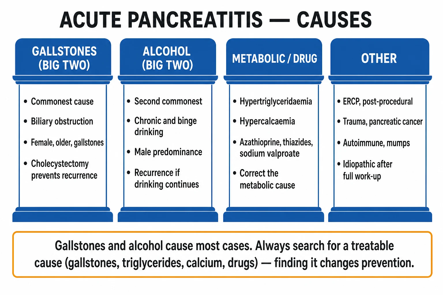
The causes cluster around the I GET SMASHED mnemonic, with gallstones (40-50%) and alcohol (25-30%) together accounting for the majority:[9]
- Gallstones — the commonest cause worldwide; microlithiasis/sludge included.
- Ethanol (alcohol) — the second commonest; chronic + binge pattern.
- Trauma, Steroids, Mumps/malignancy, Autoimmune, Scorpion sting, Hypercalcaemia/Hypertriglyceridaemia, ERCP, Drugs (azathioprine, thiazides, sodium valproate, didanosine, mesalazine, oestrogens). [1]
The revised Atlanta classification of severity
Severity is determined by the presence and duration of organ failure and by local/systemic complications, classified within 48 hours of presentation. The three-tier system replaces older terms (oedematous vs necrotising) and directly guides triage:[1]
Revised Atlanta severity (click each tier)
Persistent organ failure over 48h
Severe acute pancreatitis (SAP). Persistent organ failure (over 48h) using a modified Marshall (or SOFA) score of 2 or more in respiratory, cardiovascular or renal systems. Often coexists with pancreatic necrosis. Mandates ICU admission; infected necrosis is the major late cause of death.
Organ failure is defined by a modified Marshall score of 2 or more in any of the three systems (respiratory: PaO2/FiO2; cardiovascular: systolic BP; renal: creatinine). Persistent means the dysfunction lasts beyond 48 hours; transient means it resolves within that window. [1]
Local complications and the temporal taxonomy
The Atlanta revision also standardises the names of the local collections by content (fluid vs necrotic) and time (under or over 4 weeks):[1][9]
| Collection | Under 4 weeks | Over 4 weeks |
|---|---|---|
| Fluid (no necrosis) | Acute peripancreatic fluid collection | Pancreatic pseudocyst |
| Necrotic (solid + fluid) | Acute necrotic collection (peripancreatic and/or pancreatic) | Walled-off necrosis (WON) |
This matters because the timing dictates the intervention: walled-off necrosis (mature, encapsulated) drains and necrosectomises far better than an immature acute necrotic collection, which is why intervention is delayed ~4 weeks whenever clinically possible. [1]
Severity scoring
No single score predicts severe pancreatitis with high accuracy; scores are used in combination with clinical judgement and the persistent-SIRS rule. The early ICU scores:[3][4]
BISAP
Bedside, 24h, 5 items
- BUN over 25 mg/dL (8.9 mmol/L)
- Impaired mental status (GCS under 15)
- SIRS (2 or more of 4 criteria)
- Age over 60 years
- Pleural effusion on imaging
- Score of 3 or more predicts severe disease and mortality
APACHE II
Continuous, complex, ICU
- Most accurate but complex; recalculated over time
- Score of 8 or more traditionally predicts severe disease
- Acute physiology + age + chronic health
- The default ICU severity score; not pancreatitis-specific
Ranson
Gallstone vs non-gallstone variants
- 5 criteria at admission + 6 (non-gallstone) or 5 (gallstone) at 48h
- Admission: age, WBC, glucose, AST, LDH
- At 48h: haematocrit fall, BUN rise, calcium, PaO2, base deficit, fluid sequestration
- Score of 3 or more = severe; slow and cumbersome
Glasgow (Imrie)
Common in UK/ANZ at 48h
- 8 criteria measured within 48 hours
- Age over 55, WBC over 15, glucose over 10, LDH over 600
- Urea over 16, calcium under 2, PaO2 under 8, albumin under 32
- 3 or more positive = severe pancreatitis
Pathophysiology
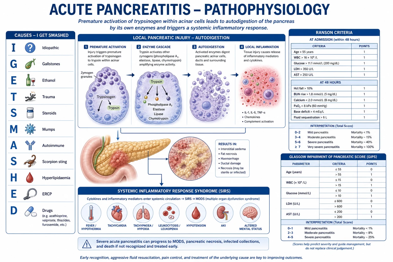
Acute pancreatitis begins with intra-acinar activation of trypsinogen to trypsin — a premature, intracellular activation of digestive proenzymes that should occur only in the duodenal lumen. The consequences unfold as a self-amplifying cascade:[9]
- Acinar cell injury — trypsin activates phospholipase A2, elastase and other enzymes; the acinar cell undergoes autophagy and necrosis.
- Local inflammation — release of cytokines (IL-6, IL-8, TNF-alpha) generates oedema, fat necrosis (the chalky white calcium soap deposits of saponification), and varying degrees of haemorrhage.
- Systemic inflammatory response syndrome (SIRS) — the cytokine storm spreads systemically, producing capillary leak, profound third-space fluid sequestration (retroperitoneal and peripancreatic), hypovolaemia, and vasodilatory shock.
- Distant organ dysfunction — SIRS-driven acute respiratory distress syndrome (ARDS), acute kidney injury (ATN from hypoperfusion and inflammation), and distributive shock define the persistent organ failure of severe disease.
- Necrosis and infection — devitalised pancreatic and peripancreatic tissue becomes a culture medium; secondary infection (typically gut-derived gram-negatives and anaerobes) develops in 20-30% of necrotising cases and converts a survivable illness into the major late cause of death. [1]
Clinical presentation
- Pain — sudden-onset, severe, persistent epigastric pain radiating through to the back, eased partly by sitting forward. Onset over hours (gallstone) or after a binge (alcohol).
- Nausea and vomiting — almost universal; persistent vomiting raises the possibility of ileus or gastric outlet compression by a large fluid collection.
- Signs of severe disease / organ failure — tachycardia, hypotension, tachypnoea/hypoxia (early ARDS or pleural effusion), oliguria, altered mental status, mottled peripheries.
- Abdominal examination — disproportionate epigastric tenderness relative to the (often mild) rigidity; Cullen’s sign (periumbilical bruising) and Grey Turner’s sign (flank bruising) indicate severe haemorrhagic necrotising pancreatitis but are late and uncommon (<3%).
- Fever beyond the first 48h or new fever later in the course — suspect infected necrosis or cholangitis. [1]
Investigations
Blood tests
- Lipase (and amylase) — over 3x upper limit supports the diagnosis; the level does NOT correlate with severity.
- Full blood count — haemoconcentration (haematocrit over 44% at admission or rising) predicts necrosis; thrombocytopenia and rising INR suggest evolving SIRS/coagulopathy.
- Urea and creatinine — rising BUN despite fluids is a marker of severe disease and is built into BISAP.
- Liver enzymes — ALT over 150 U/L has a high positive predictive value for a gallstone aetiology; rising bilirubin and ALP suggest an obstructed biliary tree (cholangitis until proven otherwise).[3]
- Calcium, magnesium, triglycerides — hypocalcaemia reflects severity; triglycerides confirm the metabolic aetiology.
- CRP — over 150 mg/L at 48 hours is a robust severity marker.
- Arterial or venous blood gas — hypoxaemia, metabolic acidosis and a rising lactate flag organ failure.
Imaging
- Abdominal ultrasound — first-line to identify gallstones and biliary dilation; limited by bowel gas and obesity.
- Contrast-enhanced CT — the definitive imaging modality. Perform at 72 hours or later if severity or aetiology is unclear — early CT understates necrosis (necrosis takes 2-3 days to declare). Look for the Balthazar CT severity index: necrosis over 30%, extrapancreatic complications, and gas within collections (infected necrosis). Avoid contrast in early AKI.[3]
- MRCP / EUS — for idiopathic pancreatitis, microlithiasis, bile duct stones, and to characterise walled-off necrosis (solid vs fluid content) before intervention.
- Chest X-ray — pleural effusions (especially left-sided) are a severity marker.
Management — the severe pancreatitis bundle
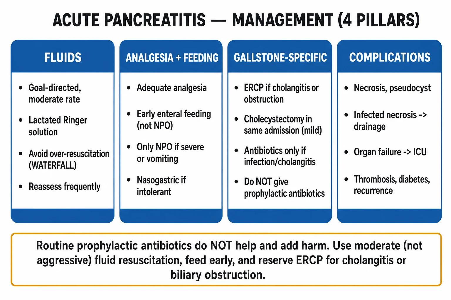
The ICU management protocol for severe acute pancreatitis
1. Goal-directed MODERATE fluid resuscitation
Ringer’s lactate preferred (balanced crystalloid). Goal-directed, NOT fixed bolus: titrate to BUN fall, haematocrit 35-44%, lactate clearance, mean arterial pressure over 65, and urine output over 0.5 mL/kg/h. Reassess every 6h. The WATERFALL trial proved aggressive (15 mL/kg bolus) resuscitation HARMFUL — more fluid overload with no mortality benefit.
2. Early enteral nutrition within 48 hours
Begin oral or nasogastric feeding within 48 hours — the standard of care. NPO (bowel rest) is outdated: early feeding reduces infected necrosis, organ failure and mortality by maintaining gut mucosal integrity. Nasogastric is as effective as nasojejunal. Reserve NPO only for persistent vomiting or ileus, and even then prioritise early nasojejunal feeding.
3. Analgesia and supportive care
Adequate opioid analgesia (no single agent is superior — avoid the outdated claim that morphine causes sphincter of Oddi spasm). DVT prophylaxis (high risk in severe disease), stress-ulcer prophylaxis if mechanically ventilated or coagulopathic, glycaemic control 6-10 mmol/L, and correction of electrolytes (calcium, magnesium, potassium).
4. NO routine prophylactic antibiotics
Do NOT give prophylactic antibiotics — multiple RCTs and a trial sequential analysis confirm no reduction in infected necrosis or mortality, with increased fungal and resistant infections. Reserve antibiotics for PROVEN infected necrosis, cholangitis, or another documented infection. When infected necrosis is confirmed, use a carbapenem (good pancreatic penetration).
5. ERCP for gallstone pancreatitis with cholangitis
Urgent ERCP with sphincterotomy within 24 hours ONLY if there is ascending cholangitis (fever, jaundice, pain — Charcot triad) or strong evidence of ongoing biliary obstruction. ERCP is NOT indicated for mild gallstone pancreatitis without cholangitis. Cholecystectomy during the index admission for mild disease; deferred for severe.
6. Organ support
Escalate to the failing system: lung-protective ventilation for ARDS (Vt 6 mL/kg predicted body weight), noradrenaline for vasodilatory shock, and renal replacement therapy for severe AKI or fluid overload. Target MAP over 65, lactate clearance, and resolution of SIRS.
7. Step-up approach for infected necrosis
For documented infected necrosis, use the minimally invasive step-up: percutaneous or endoscopic (transluminal) DRAINAGE first, with minimally invasive necrosectomy only if drainage fails. Delay intervention ~4 weeks if the patient is stable, allowing walled-off necrosis to mature. Open necrosectomy is the last resort — the PONI trial showed the step-up approach reduces major complications and death.
Fluid resuscitation — what the evidence says
The historical practice of aggressive large-volume crystalloid resuscitation (the "pancreatitis is a burn of the retroperitoneum" doctrine) was overturned by the WATERFALL trial.[2]
Moderate (preferred)
Goal-directed, Ringer’s lactate
- Bolus only with hypovolaemia/hypotension
- Then 1.5 mL/kg/h (3 mL/kg/h if目标 losses)
- Reassess every 6h — titrate to BUN, haematocrit, urine
- Lower rate of fluid overload (WATERFALL)
Aggressive (harmful)
Fixed high-rate bolus
- Up to 15-20 mL/kg/h plus repeated boluses
- More fluid overload (WATERFALL: 20.5% vs 6.3%)
- No mortality or complication benefit
- Risks: abdominal compartment syndrome, pulmonary oedema
The ACG 2024 guideline endorses aggressive resuscitation only for hypovolaemic shock, then a transition to a defined goal-directed rate — the practical message is titrate, do not over-resuscitate.[3]
Nutrition — early enteral is the standard
The dogma of "resting the pancreas" (NPO with total parenteral nutrition) was abandoned after multiple trials and meta-analyses demonstrated that early enteral nutrition within 48 hours reduces infectious complications, organ failure and mortality.[3][9]
- Route: nasogastric is non-inferior to nasojejunal and simpler — start NG unless there is gastric intolerance.
- Timing: within 48 hours of admission.
- Composition: a standard polymeric formula is adequate for most; elemental formulas offer no consistent advantage.
- Parenteral nutrition is reserved for the rare patient who cannot tolerate enteral feeding despite prokinetics and post-pyloric placement — it carries higher infection and cost burdens. [1]
Antibiotics — the negative evidence
Gallstone-specific management
- ERCP with sphincterotomy within 24 hours if there is cholangitis (Charcot’s triad: fever, jaundice, RUQ pain; Reynolds' pentad adds shock and altered mental state) or strong evidence of persistent biliary obstruction (rising bilirubin, dilated CBD on imaging).[8]
- No ERCP for mild gallstone pancreatitis without cholangitis — a Cochrane review found no mortality benefit and a higher risk of complications from routine early ERCP in this group; the stone has usually passed.[8]
- Cholecystectomy — same admission for mild gallstone pancreatitis (to prevent recurrence within weeks); deferred until recovery for severe disease with necrosis or collections.
Infected necrosis — the major late cause of death
Sterile necrosis is the rule early on; secondary infection develops in 20-30% of necrotising pancreatitis, typically in the second to third week, and is the dominant determinant of late mortality.[9]
Diagnosis is clinical-radiological:
- Gas within the necrotic collection on CT — the most specific finding.
- New or worsening fever, rising inflammatory markers, or clinical deterioration after an initial improvement.
- Positive culture — historically from CT-guided fine-needle aspiration; now largely a clinical-radiological diagnosis, with culture obtained at the time of drainage. [1]
Management — the step-up approach (PONI trial, NEJM 2010):[5]
The step-up approach for (suspected) infected necrosis
1. Antibiotics for confirmed/suspected infection
Carbapenem (e.g. meropenem) for broad gram-negative and anaerobic cover with good pancreatic penetration; add an antifungal if prolonged broad-spectrum exposure. De-escalate on culture results.
2. Delay intervention if clinically stable
Postpone drainage/necrosectomy for ~4 weeks if the patient is stable with antibiotics, allowing the collection to mature into walled-off necrosis — mature collections drain and necrosectomise far better and with fewer complications.
3. Percutaneous or endoscopic transluminal DRAINAGE first
Drain the collection via a percutaneous catheter (radiological) or endoscopic transluminal (transgastric) route. Drainage alone resolves infection in many patients by creating a controlled fistula.
4. Minimally invasive NECROSECTOMY if drainage fails
If drainage is insufficient, proceed to minimally invasive necrosectomy — percutaneous (videoscopic-assisted retroperitoneal debridement, VARD) or endoscopic transluminal. Endoscopic and surgical step-up are broadly comparable (TENSION/ExTENSION long-term follow-up).
5. Open necrosectomy as last resort
Reserve open necrosectomy for failure of the step-up, catastrophic deterioration, or when minimally invasive expertise is unavailable. It carries the highest mortality and fistula/bleeding rates — the PONI trial established the step-up as default.
The TENSION trial and its long-term ExTENSION follow-up compared endoscopic versus surgical step-up for infected necrotising pancreatitis and found broadly comparable outcomes, with the endoscopic route offering potential advantages in fistula rates and shorter recovery — supporting a multidisciplinary, individualised choice.[6]
Evidence and landmark trials
WATERFALL
NEJM 2022
249 pts with acute pancreatitis — aggressive (15 mL/kg bolus then 5 mL/kg/h) vs moderate (1.5 mL/kg/h) LR
Key finding
Fluid overload significantly higher with aggressive (20.5% vs 6.3%); no difference in pancreatitis-related complications
Practice change
Goal-directed moderate resuscitation preferred over aggressive bolus therapy
PONI
NEJM 2010
88 pts with necrotising pancreatitis + suspected infection — minimally invasive step-up vs primary open necrosectomy
Key finding
Step-up reduced major complications/death (40% vs 69%) and new-onset diabetes; 35% managed with drainage alone
Practice change
Minimally invasive step-up replaced primary open necrosectomy as the standard
TENSION / ExTENSION
Gastroenterology 2022
Multicentre RCT — endoscopic vs surgical step-up for infected necrotising pancreatitis, with long-term follow-up
Key finding
Broadly comparable outcomes; endoscopic route offers potential advantages in fistula rates and recovery
Practice change
Multidisciplinary, individualised choice between endoscopic and surgical step-up
Prophylactic antibiotics (TSA)
Antibiotics 2022
Systematic review with trial sequential analysis of prophylactic antibiotics in acute pancreatitis
Key finding
No reduction in infected necrosis or mortality; trial sequence confirms no further trials warranted
Practice change
Routine prophylactic antibiotics NOT recommended
Cochrane ERCP
Cochrane 2012
Systematic review — early routine ERCP vs conservative management in acute gallstone pancreatitis
Key finding
No mortality benefit in mild or severe gallstone pancreatitis without cholangitis; ERCP indicated only for cholangitis/obstruction
Practice change
ERCP reserved for cholangitis or biliary obstruction, not routine
Complications
Local
Within and around the pancreas
- Acute necrotic collection / walled-off necrosis
- Peripancreatic fluid collection / pseudocyst
- Infected necrosis — the major late cause of death
- Haemorrhage from pseudoaneurysm (splenic, gastroduodenal artery)
- Splenic/portal vein thrombosis; gastric outlet obstruction
- Intra-abdominal hypertension / abdominal compartment syndrome
Systemic
Distant organ failure
- ARDS and ventilatory failure (lung-protective ventilation)
- Acute kidney injury — ATN from hypoperfusion; RRT if severe
- Distributive / hypovolaemic shock — noradrenaline
- Disseminated intravascular coagulation
- Hypocalcaemia, hyperglycaemia, hypomagnesaemia
Prognosis
Outcomes by severity (revised Atlanta)
Overall mortality across all severities is approximately 2-5%; in severe disease with infected necrosis it climbs to 20-30%. Resolution of SIRS within 48 hours carries a mortality near zero; persistent SIRS beyond 48 hours is the dominant prognostic marker.[3][4]
Exam practice
SAQ — Severe acute pancreatitis with infected necrosis
15 minutes · 10 marks
A 58-year-old man is admitted with severe epigastric pain radiating to his back, vomiting, and a serum lipase of 1800 U/L (3x ULN). Ultrasound shows gallstones. By 72 hours he is in the ICU: HR 122, BP 88/52 (MAP 62) on noradrenaline 0.2 mcg/kg/min, RR 28, SpO2 92% on FiO2 0.6, urine output 20 mL/h, and he is confused. Lactate 3.8 mmol/L, BUN 12 mmol/L, haematocrit 0.46, calcium 1.9 mmol/L, CRP 220. Contrast CT at 72h shows 40% pancreatic necrosis with gas in the collection. He has received 4 L of Ringer’s lactate in the first 24 hours.
Clinical pearls
Red flags
References
- [1]Banks PA, Bollen TL, Dervenis C, et al. Classification of acute pancreatitis--2012: revision of the Atlanta classification and definitions by international consensus Gut, 2013.PMID 23100216
- [2]de-Madaria E, Buxbaum JL, Maisonneuve P, et al. Aggressive or Moderate Fluid Resuscitation in Acute Pancreatitis N Engl J Med, 2022.PMID 36103415
- [3]Tenner S, Vege SS, Sheth SG, et al. American College of Gastroenterology Guidelines: Management of Acute Pancreatitis Am J Gastroenterol, 2024.PMID 38857482
- [4]Wu BU, Johannes RS, Sun X, et al. The early prediction of mortality in acute pancreatitis: a large population-based study Gut, 2008.PMID 18519429
- [5]van Santvoort HC, Besselink MG, Bakker OJ, et al. A step-up approach or open necrosectomy for necrotizing pancreatitis N Engl J Med, 2010.PMID 20410514
- [6]Onnekink AM, Boxhoorn L, Timmerhuis HC, et al. Endoscopic Versus Surgical Step-Up Approach for Infected Necrotizing Pancreatitis (ExTENSION): Long-term Follow-up of a Randomized Trial Gastroenterology, 2022.PMID 35580661
- [7]Poropat G, Gorianec K, Lackovic A, et al. Systematic Review with Trial Sequential Analysis of Prophylactic Antibiotics for Acute Pancreatitis Antibiotics (Basel), 2022.PMID 36139970
- [8]Tse F, Yuan Y. Early routine endoscopic retrograde cholangiopancreatography strategy versus early conservative management strategy in acute gallstone pancreatitis Cochrane Database Syst Rev, 2012.PMID 22592743
- [9]Boxhoorn L, Voermans RP, Bouwense SA, et al. Acute pancreatitis Lancet, 2020.PMID 32891214