ICU · gi-nutrition
Acute Severe Pancreatitis — Comprehensive (Revised Atlanta, WATERFALL, PANTER, Step-Up, Infected Necrosis)
Also known as Severe acute pancreatitis · Necrotising pancreatitis · Infected pancreatic necrosis · WATERFALL trial · PANTER trial · TENSION trial · Step-up approach · Walled-off necrosis · Revised Atlanta classification · Pancreatic pseudocyst · BISAP score · Acute necrotic collection
Acute severe pancreatitis (approximately 15-20% of all acute pancreatitis) is defined by the Revised Atlanta Classification (2012) as acute pancreatitis with PERSISTENT organ failure lasting 48 hours — respiratory (PaO2/FiO2 <300), cardiovascular (SBP <90 despite fluids / need for vasopressors), or renal (creatinine 170 umol/L) — and carries a mortality of 15-30%. The two pathological phases dictate ICU management. EARLY PHASE (week 1): uncontrolled SYSTEMIC INFLAMMATION (SIRS) driven by pancreatic enzyme activation and cytokine storm (IL-6, TNF-alpha, IL-1) → capillary leak, 'third-space' fluid sequestration into the retroperitoneum and bowel wall → hypovolaemia, haemoconcentration, haemodynamic instability, AKI, and the early killers ARDS and shock. Aetiology: gallstones (1, ~40%), alcohol (2, ~30%), ERCP, hypertriglyceridaemia (11 mmol/L), drugs (azathioprine, mesalazine, didanosine, sodium valproate, thiazides), trauma, post-ERCP, autoimmune, idiopathic. EARLY ICU MANAGEMENT is SUPPORTIVE and evidence-based: (1) GOAL-DIRECTED FLUID RESUSCITATION with lactated Ringer's — the WATERFALL trial (NEJM 2022, de-Madaria) proved AGGRESSIVE fluids (20 mL/kg bolus + 3 mL/kg/hr) CAUSE HARM (fluid overload 20.5% vs 6.3%, no outcome benefit) — use moderate, goal-directed resuscitation (1.5 mL/kg/hr + small boluses only if hypovolaemic); (2) EARLY ENTERAL NUTRITION within 48 hours (NOT 'NPO + TPN' — old practice refuted by multiple RCTs and meta-analyses) — maintains gut mucosal barrier, reduces bacterial translocation, reduces infected necrosis and mortality; oral or nasogastric route is as effective as nasojejunal in most patients; (3) NO PROPHYLACTIC ANTIBIOTICS — the PANTER trial, ACG (Tenner 2024), and IAP/APA guidelines all confirm antibiotics do NOT prevent infected necrosis and increase resistance, fungal superinfection, and C. difficile; give antibiotics ONLY for documented infection (infected necrosis, cholangitis, pneumonia); (4) analgesia (opioid — morphine/fentanyl, PCA); (5) ERCP within 24-72h ONLY if concomitant cholangitis or ongoing biliary obstruction. LATE PHASE (week 2+): local complications dominate — pancreatic/peripancreatic NECROSIS (sterile in ~70%, infected in ~30%), walled-off necrosis, pseudocyst. Infected necrosis is the principal late killer (mortality 30-40% historically). Diagnosis: gas within necrosis on CT is pathognomonic; otherwise clinical deterioration + positive fine-needle aspiration (FNA) culture. Management of infected necrosis is the STEP-UP APPROACH (PANTER trial, NEJM 2010, van Santvoort): percutaneous or endoscopic DRAINAGE first, then minimally-invasive necrosectomy only if drainage fails — step-up halved the composite of major complications/death (40% vs 69%) versus open necrosectomy, and 35% needed drainage alone. The TENSION trial (Lancet 2018, van Brunschot) showed ENDO- scopic step-up (EUS-guided transluminal drainage ± endoscopic necrosectomy) is NOT superior to surgical step-up for the primary endpoint but causes FEWER pancreatic fistulas and shorter hospital stay — endoscopic step-up is now the preferred first approach when expertise exists. CRITICAL: DELAY invasive intervention ~4 weeks whenever possible (let necrosis become walled-off/encapsulated → safer drainage). Scoring: APACHE II =8, Ranson =3, BISAP =3, CRP 150 mg/L at 48h, and the Revised Atlanta organ-failure criteria all predict severity. Systemic complications: SIRS → MODS, ARDS (lung-protective ventilation), AKI (CRRT if shocked), abdominal compartment syndrome (monitor bladder pressure), and sepsis from infected necrosis.
On this page & tools
Your progress
Saved locally on this device.
7 MCQs with explanations
Target exams
Red flags
Overview
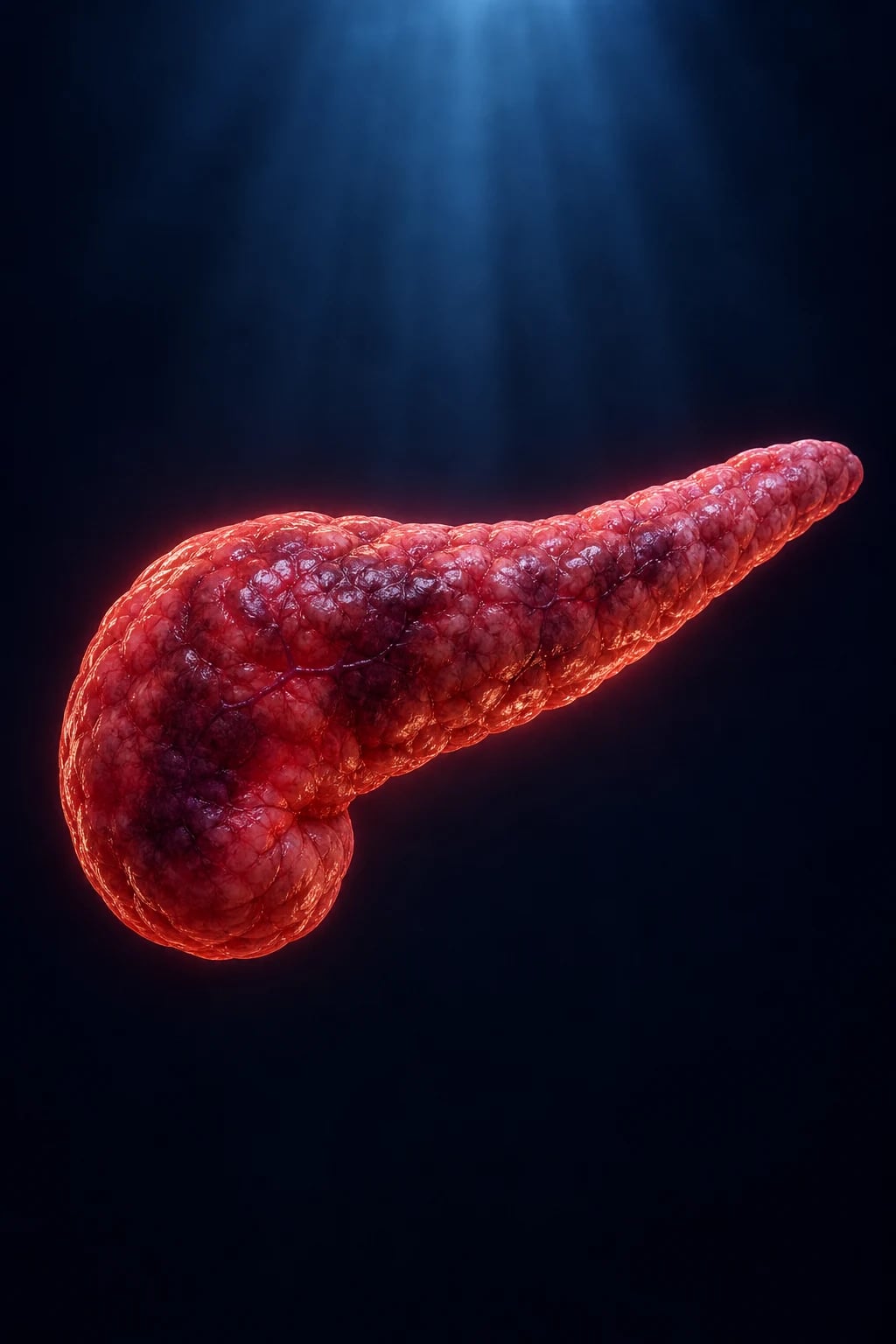
Severe acute pancreatitis is one of the most demanding ICU diagnoses: it is simultaneously a disease of uncontrolled systemic inflammation (the early phase, where the intensivist saves the patient by supporting failing organs and resuscitating judiciously) and a disease of local destructive complications (the late phase, where necrosis and superinfection demand a disciplined, multidisciplinary 'step-up' intervention strategy). The fellowship-level danger is that much of traditional teaching — 'aggressive fluids', 'NPO and TPN', 'prophylactic carbapenems', 'open necrosectomy' — has been directly refuted by landmark randomised trials (WATERFALL, PANTER, TENSION) and modern guidelines (ACG 2024, IAP/APA 2013). The intensivist who still practices the old dogma harms patients. This topic covers the full fellowship-exhaustive syllabus: revised Atlanta classification, aetiology, severity scoring, every local and systemic complication, and the evidence-based, trial-anchored management pathway.[1][2][3]
Revised Atlanta Classification (2012) — the current standard
The Revised Atlanta Classification replaced the ambiguous 1992 original with clear, reproducible definitions based on organ failure and morphology. It is the single most examinable framework in pancreatitis.[1]
Severity classification — Revised Atlanta (2012)
| Severity | Definition | Organ failure | Local complications | Incidence | Mortality |
|---|---|---|---|---|---|
| Mild | No organ failure, no local/systemic complications | None | None | ~80% | <1% |
| Moderately severe | Transient organ failure (<48h) resolving, OR local complications, OR exacerbation of comorbidity | Transient (<48h) | Peripancreatic fluid collection, necrosis, pseudocyst, walled-off necrosis | ~15-20% | ~5% |
| Severe | PERSISTENT organ failure >48h (one or more organs) — single or multiple | Persistent (>48h) | Usually present | ~5% | 15-30% |
Morphological (imaging) classification — two types
| Type | Frequency | Definition | Local collections (early, <4 wk) | Local collections (late, >4 wk) |
|---|---|---|---|---|
| Interstitial oedematous pancreatitis | ~80-90% | No necrosis; diffuse pancreatic enlargement | Acute peripancreatic fluid collection (APFC) | Pancreatic pseudocyst |
| Necrotising pancreatitis | ~10-20% | Pancreatic and/or peripancreatic necrosis (non-enhancing tissue on contrast CT) | Acute necrotic collection (ANC) | Walled-off necrosis (WON) |
Organ failure is graded using a modified Marshall score (or SOFA) across three systems: respiratory (PaO2/FiO2 <300 = score >=2), cardiovascular (SBP <90 mmHg unresponsive to fluids = score >=2), and renal (creatinine >=170 umol/L = score >=2). Persistent = score >=2 in the same system for >48 hours. The distinction between transient (<48h, resolving) and persistent (>48h) organ failure is THE determinant of severe disease — a patient with necrosis but no organ failure is NOT severe, whereas a patient with no necrosis but persistent organ failure IS severe.[1]
Aetiology — find and remove the cause
Causes of acute pancreatitis — frequency and ICU relevance
| Cause | Frequency | Mechanism | Specific ICU action |
|---|---|---|---|
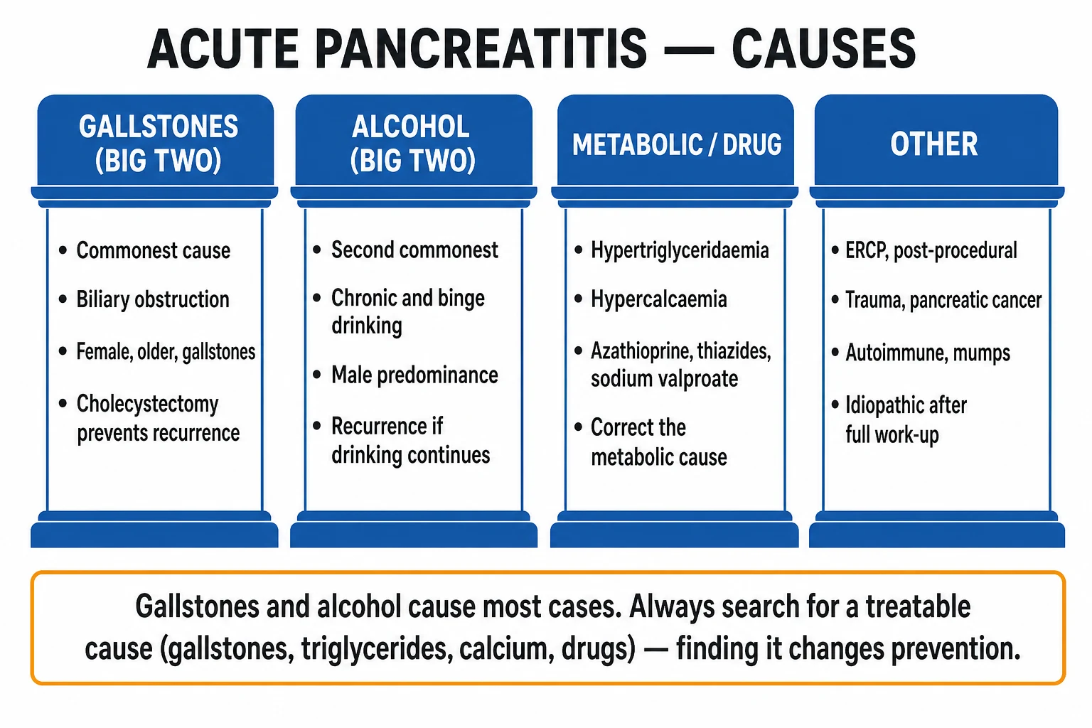
| Gallstones (including microlithiasis/sludge) | ~40% (#1) | Gallstone impacts at ampulla of Vater → obstructs pancreatic duct → ductal hypertension + bile reflux into pancreatic duct → premature trypsinogen activation | Ultrasound for stones/dilated CBD; MRCP/EUS to confirm CBD stone; ERCP within 24-72h if cholangitis/obstruction; cholecystectomy before discharge (mild) or after recovery (severe) |
| Alcohol | ~30% (#2) | Direct toxic effect on acinar cells + pancreatic ductal protein plug formation + oxidative stress | Cessation counselling; thiamine; alcohol withdrawal prophylaxis (CIWA-Ar) |
| Hypertriglyceridaemia | ~5-10% | Triglycerides >11 mmol/L → chylomicrons → lipase hydrolysis → free fatty acids toxic to acinar cells → capillary microthrombi | Insulin/dextrose infusion (activates lipoprotein lipase); plasmapheresis if severe/refractory or pregnancy; avoid lipid emulsion; fibrate + omega-3 long term |
| ERCP / post-procedural | ~3-5% | Mechanical/chemical injury from contrast or instrumentation; papillary oedema | Highest risk with sphincter of Oddi manometry; prophylactic rectal NSAID (diclofenac 100 mg PR) reduces risk |
| Drugs | ~2-5% | Idiosyncratic or toxic: azathioprine/6-MP, mesalazine, didanosine, sodium valproate, thiazides, oestrogens, tetracyclines, sulphonamides, L-asparaginase | Stop offending drug; usually resolves on withdrawal |
| Trauma / post-operative | <2% | Ductal disruption from blunt abdominal injury (handlebar) or surgery | CT for duct injury; ERCP stenting if main duct disrupted |
| Autoimmune (AIP) | <2% | IgG4-mediated lymphoplasmacytic infiltration | Corticosteroids (prednisolone 40 mg/day) — dramatic response |
| Idiopathic | ~10-20% | Unidentified after standard workup | Repeat ultrasound for microlithiasis; EUS; consider autoimmune/genetic |
The mnemonic 'I GET SMASHED' captures aetiology: Idiopathic, Gallstones, Ethanol, Trauma, Steroids/drugs, Mumps/mumps, Autoimmune, Scorpion sting (and snake bite), Hypercalcaemia/hypertriglyceridaemia/hypothermia, ERCP, Drugs. The two intensive-care imperatives are (1) recognise cholangitis in the gallstone patient (fever + jaundice + RUQ pain = Charcot triad → emergency ERCP), and (2) recognise severe hypertriglyceridaemia (lipaemic milky serum, TG >11 mmol/L) because it has a specific, effective treatment (insulin/dextrose ± plasmapheresis).[2][3]
Severity scoring — predict who will deteriorate
Severity scoring guides the intensity of monitoring and the threshold for ICU admission. No single score is perfect; the Revised Atlanta organ-failure criteria remain the gold standard for 'severe', while bedside scores (BISAP) and biomarkers (CRP) add early prediction.[1][2]
Severity scoring systems compared
| Score | Timing | Criteria | 'Severe' threshold | Pros | Cons |
|---|---|---|---|---|---|
| Revised Atlanta (organ failure) | Continuous | Persistent organ failure >48h (Marshall >=2 in resp/CV/renal) | Persistent OF >48h = severe | Current international standard; simple | Needs 48h to declare; doesn't predict prospectively |
| APACHE II | Admission, daily | 12 physiological variables + age + chronic health | >=8 predicts severe | Best single predictor at admission; dynamic | Complex; not bedside; not pancreatitis-specific |
| Ranson | Admission + 48h | 5 criteria at admission + 6 at 48h (11 total) | >=3 severe; >=6 very severe | Classic; well validated | Cumbersome; needs 48h; labour intensive |
| BISAP | First 24h | BUN >35, Impaired mental status, SIRS, Age >60, Pleural effusion (1 point each) | >=3 predicts severe/mortality | Simple; bedside; first 24h | Less sensitive; underused |
| CRP | 48h | Acute-phase protein | >150 mg/L at 48h = severe/necrosis | Cheap; widely available; necrosis surrogate | Late (48h); non-specific |
| CT Severity Index (Balthazar) | After 72h | Inflammation grade (A-E) + necrosis % (0/30/50/100) | >=4 moderate; >=7 severe | Quantifies necrosis (predicts complications) | Contrast load; underestimates before 72h |
| HAPS / Harmless score | Admission | No rebound/peritonitis, normal creatinine, normal haematocrit | All absent = 'harmless' (mild) | Excellent rule-OUT at admission | Only identifies the very mild |
Practical approach: combine BISAP and APACHE II at admission (early triage), CRP at 48h (necrosis proxy), and a contrast CT after 72 hours if severe (define morphology/necrosis). Haemoconcentration (haematocrit rising or failing to fall) at 24-48h is a simple, powerful marker of ongoing fluid sequestration and impending necrosis. [1]
Clinical features and early ICU assessment
The presentation — epigastric pain radiating straight through to the back, relieved by sitting forward, with vomiting and retching — is familiar. The ICU-relevant assessment focuses on severity, organ support requirements, and the search for a treatable cause. [1]
First-hour ICU assessment of suspected severe acute pancreatitis
- CONFIRM THE DIAGNOSIS — two of three: (a) characteristic epigastric pain radiating to back; (b) lipase (or amylase) >3x upper limit of normal; (c) characteristic imaging (CT/usound). Lipase is more specific and stays elevated longer than amylase. NOTE: a normal/low amylase does NOT exclude pancreatitis (alcoholic, hypertriglyceridaemic pancreatitis can have near-normal enzymes; lipase degraded in renal failure can be low). Send lipase, amylase, FBC, U&E, LFTs, glucose, calcium, triglycerides, lactate, CRP, blood gas, blood cultures.
- GRADE SEVERITY / ORGAN FAILURE — calculate BISAP and APACHE II; assess for SIRS (the engine of early deterioration); examine for peritonism (suggests severe/necrotising); check oxygenation, blood pressure, urine output. Determine need for ICU vs HDU vs ward.
- IDENTIFY AND TREAT ORGAN FAILURE — oxygen/ventilation for respiratory failure (lung-protective if ARDS), vasopressors (noradrenaline) for shock, renal replacement therapy for refractory AKI. These are the EARLY killers — death in week 1 is from MODS, not infection.
- SEARCH FOR THE CAUSE — abdominal ultrasound (gallstones, CBD diameter, biliary sludge), LFTs (obstructive picture suggests gallstone aetiology/cholangitis), triglycerides (>11 mmol/L = hypertriglyceridaemic), calcium (hypercalcaemia), alcohol history, drug history, recent ERCP. If aetiology unclear, MRCP/EUS to look for CBD stones/microlithiasis.
- DECIDE ON ERCP — ask two questions: (a) Is there cholangitis (Charcot triad: fever + jaundice + RUQ pain; Reynolds pentad + shock/altered mental state)? (b) Is there strong evidence of persistent biliary obstruction (dilated CBD, rising/undropping bilirubin, confirmed stone)? If YES to either → emergency/urgent ERCP within 24-72h. If NO (stone likely already passed) → manage pancreatitis; cholecystectomy later. Do NOT do 'routine' ERCP for uncomplicated gallstone pancreatitis — it adds risk without benefit.
- INSTITUTE EVIDENCE-BASED EARLY MANAGEMENT — moderate goal-directed lactated Ringer's resuscitation (NOT aggressive — WATERFALL); analgesia; plan early enteral nutrition within 48h; do NOT start prophylactic antibiotics.
- PLAN IMAGING — an early CT (within 72h) is usually unnecessary unless the diagnosis is in doubt or there is diagnostic uncertainty; necrosis is underestimated before 72h. A contrast CT at 72h+ (if severe) defines necrosis extent and collections. Reserve MRI for characterising ductal anatomy or when contrast is contraindicated.
Early-phase management — fluid resuscitation (WATERFALL)
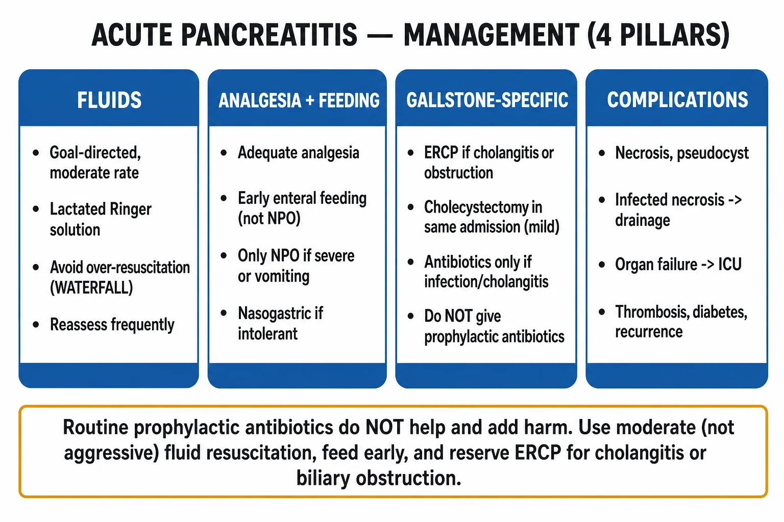
Fluid resuscitation is the cornerstone of early management — but the dogma of 'aggressive hydration' has been overturned. Severe pancreatitis causes massive capillary leak and third-space sequestration (retroperitoneum, bowel wall), producing hypovolaemia and haemoconcentration that compromise pancreatic microcirculation and worsen necrosis. Fluids ARE needed — but the rate matters.[4]
WATERFALL trial (NEJM 2022, de-Madaria) — aggressive vs moderate fluids
| Feature | Aggressive resuscitation | Moderate resuscitation |
|---|---|---|
| Bolus | Lactated Ringer's 20 mL/kg | 10 mL/kg ONLY if hypovolaemic; NO bolus if normovolaemic |
| Maintenance | 3 mL/kg/hr | 1.5 mL/kg/hr |
| Fluid overload | 20.5% | 6.3% (adjusted RR 2.85; p=0.004) |
| Moderate/severe pancreatitis | 22.1% | 17.3% (no significant difference; p=0.32) |
| Hospital stay (median) | 6 days | 5 days |
| Outcome | Trial HALTED EARLY by DSMB for harm (fluid overload) without any clinical benefit | — |
| Conclusion | Aggressive fluids CAUSE HARM (overload) without benefit → use MODERATE, goal-directed resuscitation | — |
Goal-directed fluid resuscitation protocol (post-WATERFALL)
- FLUID TYPE: Lactated Ringer's is preferred over 0.9% saline (reduces SIRS and CRP; less hyperchloroaemic metabolic acidosis which can worsen coagulopathy and renal vasoconstriction).
- INITIAL RATE: 1.5 mL/kg/hr maintenance. Add a 250-500 mL bolus ONLY if the patient is hypovolaemic (hypotension, tachycardia, poor perfusion, raised lactate). Do NOT give a routine 10-20 mL/kg bolus to a normovolaemic patient.
- REASSESS EVERY 4-6 HOURS using goals: urine output >0.5 mL/kg/hr, MAP >65 mmHg, heart rate <120, lactate falling, haematocrit trending toward 35-44% (falling from a haemoconcentrated baseline is the goal — do NOT push it below normal).
- DE-ESCALATE as soon as euvolaemic — switch to a lower maintenance rate. The commonest error is continuing high-rate fluids for days, causing pulmonary oedema, worsening ARDS, and abdominal compartment syndrome.
- AVOID 'panic bolusing' — repeated 500-1000 mL boluses in a haemodynamically stable patient cause cumulative overload. If hypotension persists despite adequate filling, add a vasopressor (noradrenaline) — do not chase the pressure with more fluid.
- MONITOR FOR FLUID OVERLOAD — daily weights, fluid balance, oxygen requirement, lung bases, and (in severe cases) bladder pressure for intra-abdominal hypertension.
The pathophysiological insight: pancreatic perfusion depends on maintaining an adequate intravascular volume to prevent haemoconcentration-driven microcirculatory thrombosis and ischaemic necrosis — but over-resuscitation raises intra-abdominal pressure, worsens pulmonary oedema and ARDS, and may itself promote pancreatic injury. The 'sweet spot' is euvolaemia, achieved by frequent reassessment rather than a fixed aggressive protocol.[4][2]
Early-phase management — nutrition (NOT 'NPO + TPN')
The traditional 'bowel rest (NPO) + total parenteral nutrition' dogma is refuted. Prolonged fasting causes gut mucosal atrophy, increases bacterial translocation across the gut barrier (the mechanism of late infected necrosis), and TPN adds catheter-related bloodstream infection, hyperglycaemia, and cost. Multiple RCTs and meta-analyses confirm that early enteral nutrition (within 48 hours) reduces infected necrosis, multi-organ failure, and mortality.[2][3]
Nutrition in severe acute pancreatitis — the evidence
| Strategy | Recommendation | Evidence / rationale |
|---|---|---|
| Timing | Start enteral nutrition WITHIN 48 HOURS of admission | Meta-analyses: early EN reduces infected necrosis, MOF, and mortality vs delayed/TPN |
| Route — first choice | ORAL (low-fat solids when nausea/ileus resolves) OR NASOGASTRIC (NG) feed | NG is as effective and safe as NJ in most trials; simpler; easier to place |
| Route — if gastric outlet obstruction/vomiting uncontrolled | NASOJEJUNAL (NJ) feed | Bypasses stomach; historically preferred (less 'pancreatic stimulation') but NO proven superiority to NG in most patients |
| Formula | Standard polymeric formula in most; semi-elemental if malabsorption | No clear superiority of 'elemental' or immune-enhancing formulas |
| Total parenteral nutrition (TPN) | ONLY if enteral feeding is IMPOSSIBLE for >5-7 days (ileus, obstruction, prolonged intolerance) or as a supplement if target enteral volume unachievable | TPN increases infection, hyperglycaemia, cost |
| Refeeding | Resume oral intake as soon as nausea/pain improves; do NOT keep NPO awaiting normal enzymes | ACG 2024: early refeeding is safe and reduces complications |
The key exam point: early enteral nutrition is a treatment, not just supportive care — it maintains gut mucosal integrity, prevents bacterial translocation (the source of late infected necrosis), modulates the inflammatory response, and reduces mortality. Withholding it is harmful. The route (oral/NG vs NJ) matters far less than the principle of feeding the gut early.[2]
Early-phase management — antibiotics (NOT prophylactic)
This is among the most examined and most over-applied areas. Prophylactic antibiotics (historically carbapenems, given their penetration into pancreatic tissue) were once standard for all severe pancreatitis, on the theory that they would prevent infected necrosis. They do not, and they cause harm.[2][3]
Prophylactic antibiotics in severe pancreatitis — the verdict
| Question | Answer | Evidence |
|---|---|---|
| Do prophylactic antibiotics prevent infected necrosis? | NO | Multiple RCTs (including the PANTER-trial era data) and meta-analyses: no reduction in infected necrosis |
| Do they reduce mortality? | NO | No mortality benefit in meta-analyses (ACG 2024, IAP/APA 2013) |
| Do they cause harm? | YES | Increased antibiotic resistance, fungal superinfection (Candida), Clostridioides difficile colitis |
| What is the recommendation? | DO NOT give prophylactic antibiotics for predicted-severe or necrotising pancreatitis | ACG 2024 (strong): 'Antibiotics should NOT be routinely administered'. IAP/APA 2013: same |
| When ARE antibiotics indicated? | (1) DOCUMENTED infected necrosis (gas on CT, positive FNA culture, clinical sepsis with necrosis); (2) CHOLANGITIS or documented extrapancreatic infection (pneumonia, line infection, UTI); (3) as empiric therapy while evaluating a septic, deteriorating patient — but STOP if cultures negative | — |
| Choice for infected necrosis | Carbapenem (meropenem) penetrates necrotic tissue well; alternative quinolone + metronidazole | Duration guided by clinical response and source control |
The PANTER trial is sometimes referenced for this question, but the antibiotic evidence specifically comes from the series of RCTs (Buchler, Nordback, Isenmann, Dellinger, Garcia-Barrasa) and meta-analyses that the ACG and IAP/APA guidelines synthesised. The clinical bottom line for the CICM/FFICM candidate: prophylactic antibiotics do not prevent infected necrosis or reduce mortality in severe/necrotising pancreatitis and cause resistance, fungal infection, and C. difficile — do not give them. Antibiotics are reserved for documented infection.[2][3]
Early-phase management — analgesia, ERCP, and cause-specific therapy
Analgesia: Severe pain is universal. An opioid via patient-controlled analgesia (PCA) is standard. Morphine and fentanyl are both safe; the old concern that morphine causes sphincter of Oddi spasm and worsens pancreatitis is NOT supported by evidence and should not drive opioid choice. Buprenorphine, oxycodone, or tramadol are alternatives. Add an antiemetic. Epidural analgesia is occasionally used for refractory pain in the ICU but is not routine. [1]
ERCP — who, when: Endoscopic retrograde cholangiopancreatography removes an impacted CBD stone and performs sphincterotomy. Indications are narrow and evidence-based:[2]
- Concomitant acute cholangitis (Charcot triad: fever + jaundice + RUQ pain; or Reynolds pentad + hypotension + confusion) → EMERGENCY ERCP within 24 hours.
- Persistent biliary obstruction (strong evidence of an impacted CBD stone: dilated CBD on imaging, rising or non-falling bilirubin, not improving) → urgent ERCP within 24-72 hours.
- Uncomplicated gallstone pancreatitis (stone likely already passed, no cholangitis, no ongoing obstruction, improving bilirubin) → NO ERCP. Manage the pancreatitis; perform laparoscopic cholecystectomy during the same admission (mild) or after recovery from severe disease, ideally with intraoperative cholangiography. In high-risk non-cholecystectomy patients, endoscopic sphincterotomy reduces recurrence.
Cause-specific therapy:
- Hypertriglyceridaemia (TG >11 mmol/L): insulin/dextrose infusion (activates lipoprotein lipase → clears triglycerides); therapeutic plasma exchange if severe, refractory, or in pregnancy; long-term fibrate + omega-3 + diet. Avoid lipid-containing parenteral nutrition.
- Alcohol: withdrawal prophylaxis (benzodiazepine CIWA-Ar protocol), thiamine, cessation counselling, consider naltrexone/acamprosate.
- Drugs: stop the offending agent (azathioprine/6-MP, mesalazine, didanosine, sodium valproate, thiazides, oestrogens).
- Autoimmune (IgG4): corticosteroids (prednisolone 40 mg/day) — typically dramatic response. [1]
Local complications — the morphological taxonomy (Revised Atlanta)
The Revised Atlanta introduced a time- and morphology-based taxonomy of local collections that every fellowship candidate must know. The two axes are: is there necrosis? and has it been >4 weeks (encapsulated)?[1]
Local collections — the 4-term Revised Atlanta taxonomy
| Collection | Associated morphology | Timeframe | Contents | Encapsulated? |
|---|---|---|---|---|
| Acute peripancreatic fluid collection (APFC) | Interstitial oedematous pancreatitis | <4 weeks | Fluid ONLY (no necrosis) | No |
| Pancreatic pseudocyst | Interstitial oedematous pancreatitis | >4 weeks | Fluid ONLY (no necrosis) | Yes (encapsulated) |
| Acute necrotic collection (ANC) | Necrotising pancreatitis | <4 weeks | Fluid AND necrotic (solid) tissue | No |
| Walled-off necrosis (WON) | Necrotising pancreatitis | >4 weeks | Fluid AND necrotic (solid) tissue | Yes (encapsulated) |
Pseudocyst vs walled-off necrosis — a common exam trap. A pseudocyst contains ONLY fluid (arises from duct disruption in non-necrotising pancreatitis); walled-off necrosis contains fluid PLUS solid necrotic debris (arises from necrotising pancreatitis). This distinction matters because solid debris is hard to drain and requires necrosectomy, whereas a simple fluid pseudocyst drains easily. Always review the CT for solid components before draining any late collection. [1]
Infected vs sterile necrosis — the pivotal late-phase distinction. Approximately 30% of pancreatic/peripancreatic necrosis becomes infected (typically in week 2-3, via bacterial translocation from the gut). Infected necrosis is the principal late killer (historical mortality 30-40%, now lower with step-up).[1][5]
Sterile vs infected pancreatic necrosis
| Feature | Sterile necrosis | Infected necrosis |
|---|---|---|
| Incidence | ~70% | ~30% |
| Timing | Week 1+ | Usually week 2-3 (bacterial translocation) |
| Diagnosis | CT non-enhancement; NO infection signs | GAS IN NECROSIS on CT (pathognomonic) OR positive FNA culture OR clinical deterioration/sepsis |
| Clinical | SIRS (fever, raised WCC/CRP) from inflammation — mimics infection | Clinical deterioration after initial improvement; persistent fever, sepsis, worsening organ failure |
| Management | CONSERVATIVE (no antibiotics; nutrition; supportive) — sterile necrosis often resolves | Antibiotics (carbapenem) + STEP-UP drainage/necrosectomy |
| Mortality | 10-15% (mostly early organ failure) | 30-40% historically (lower with step-up) |
| Role of antibiotics | None | Essential + source control |
Diagnostic fine-needle aspiration (FNA): if there is genuine uncertainty (SIRS but no gas, no clear deterioration), ultrasound/CT-guided FNA of the necrosis for Gram stain and culture can confirm infection. However, in practice, gas within the necrosis on CT is so characteristic that FNA is often unnecessary, and a deteriorating septic patient with necrosis is treated as infected necrosis without waiting for FNA. [1]
Systemic complications — the early killers (SIRS, MODS, ARDS, AKI)
The early phase of severe pancreatitis is dominated by systemic inflammation that produces multi-organ dysfunction. Death in week 1 is overwhelmingly from organ failure rather than infection. [1]
Systemic complications and their management
| Organ/system | Mechanism | Clinical | ICU management |
|---|---|---|---|
| SIRS → MODS | Cytokine storm (IL-6, TNF-alpha, IL-1, PAF) from activated pancreatic enzymes → endothelial activation, capillary leak, vasoplegia | Fever, tachycardia, tachypnoea, raised WCC; progresses to MODS | Treat the source (support pancreas recovery); goal-directed resuscitation; vasopressors; organ support |
| ARDS / respiratory failure (most common organ failure) | Pulmonary capillary leak + inflammatory mediators + pleural effusion (often left-sided); abdominal compartment elevating diaphragm | Hypoxia, bilateral infiltrates; PaO2/FiO2 <300 | Lung-protective ventilation (Vt 6 mL/kg PBW, plateau <30 cmH2O, PEEP); proning if severe (PROSEVA); drain large pleural effusions |
| Shock (cardiovascular) | Hypovolaemia (third space) + vasoplegia (SIRS) + (rarely) myocardial depression | Hypotension, raised lactate, oliguria | Goal-directed fluids (moderate); noradrenaline first-line vasopressor; add vasopressin if refractory; consider adrenal insufficiency if vasoplegic |
| AKI | Hypovolaemia/hypoperfusion + inflammatory/nephrotoxic mediators ± abdominal compartment syndrome | Rising creatinine, oliguria | Optimise perfusion (fluids + vasopressors); avoid nephrotoxins; CRRT if refractory/shocked (continuous preferred in vasopressor-dependent shock) |
| Abdominal compartment syndrome (ACS) | Massive fluid sequestration + ileus → raised intra-abdominal pressure | Bladder pressure >20 mmHg + new organ failure (renal, respiratory) | Decompression: NG/rectal tube, neuromuscular blockade, reduce fluids/diurese; surgical decompression if refractory |
| Metabolic | Hypocalcaemia (saponification — though largely a marker), hyperglycaemia (stress/diabetes), hypomagnesaemia, acidosis | Variable | Correct electrolytes; insulin infusion for hyperglycaemia (target 6-10 mmol/L) |
| DIC / coagulopathy | Consumptive coagulopathy from severe inflammation | Variable PT/aPTT, platelets | Treat underlying inflammation; blood product support if bleeding |
| Late sepsis | Infected necrosis, line infections, pneumonia, fungal | Deterioration after initial improvement; fever, septic shock | Source identification; targeted antibiotics; source control (step-up drainage) |
Abdominal compartment syndrome deserves emphasis in severe pancreatitis: massive retroperitoneal and bowel-wall fluid sequestration plus ileus can raise intra-abdominal pressure dramatically. Monitor bladder pressure in any severe patient requiring large fluid volumes or with oliguria/refractory hypoxaemia. IAP >12 mmHg = intra-abdominal hypertension; >20 mmHg with new organ failure = ACS. Management is staged: NG/rectal decompression, judicious fluid reduction, neuromuscular blockade, and — if refractory — surgical decompressive laparotomy (open abdomen). [1]
Late-phase management — infected necrosis and the step-up approach
The management of infected necrosis was transformed by two landmark Dutch multicentre randomised trials: PANTER (which established the step-up principle over open necrosectomy) and TENSION (which refined the step-up toward an endoscopic-first approach).[5][6]
PANTER trial (NEJM 2010, van Santvoort) — step-up vs open necrosectomy
| Feature | Step-up approach | Primary open necrosectomy |
|---|---|---|
| Strategy | Percutaneous catheter drainage (or endoscopic) FIRST; minimally invasive retroperitoneal necrosectomy (VARD) ONLY if drainage insufficient | Primary open necrosectomy |
| Primary endpoint (major complications or death) | 40% | 69% (risk ratio with step-up 0.57; 95% CI 0.38-0.87; p=0.006) |
| New-onset multiple organ failure | 12% | 40% (p=0.002) |
| Death | 19% | 16% (no significant difference) |
| Drainage ALONE sufficient | 35% needed no necrosectomy at all | — |
| Incisional hernia | 7% | 24% (p=0.03) |
| New-onset diabetes | 16% | 38% (p=0.02) |
| Conclusion | Step-up approach REDUCED major complications/death and long-term sequelae; 1/3 needed drainage only → step-up is the new standard; primary open necrosectomy abandoned | — |
TENSION trial (Lancet 2018, van Brunschot) — endoscopic vs surgical step-up
| Feature | Endoscopic step-up | Surgical step-up |
|---|---|---|
| Strategy | EUS-guided transluminal drainage ± endoscopic (transgastric) necrosectomy | Percutaneous catheter drainage ± video-assisted retroperitoneal debridement (VARD) |
| Primary endpoint (major complications or death, 6 months) | 43% (22/51) | 45% (21/47) (RR 0.97; p=0.88) — NOT superior |
| Death | 18% | 13% (no significant difference) |
| Pancreatic fistula | 5% | 32% (p=0.001) — far fewer with endoscopic |
| Hospital stay (mean) | 53 days | 69 days (p=0.014) — shorter with endoscopic |
| Conclusion | Endoscopic step-up not SUPERIOR on the primary endpoint but caused FEWER fistulas and shorter stay → endoscopic step-up is the PREFERRED first approach when expertise/collection anatomy permit | — |
Infected necrosis — the step-up management pathway
- RECOGNISE infected necrosis: gas within necrosis on CT (pathognomonic), OR clinical deterioration/new sepsis in a patient with necrosis, OR positive FNA culture. Confirm necrosis extent on contrast CT.
- START ANTIBIOTICS — carbapenem (meropenem) penetrates necrotic tissue well; alternatives piperacillin-tazobactam or quinolone + metronidazole. Tailor to cultures. Add antifungal if prolonged broad-spectrum or fungal isolation.
- STABILISE AND DELAY if at all possible — the cardinal principle. If the patient can be stabilised with antibiotics and organ support, DELAY invasive intervention ~4 WEEKS to allow the necrosis to become walled-off (encapsulated). Encapsulated WON is far safer to drain (the wall contains the necrosis and separates it from viable tissue, reducing bleeding and incomplete removal). Early (<4 week) necrosis is poorly demarcated and dangerous to instrument.
- STEP 1 — DRAINAGE (the first step of step-up):
- Endoscopic transluminal drainage (preferred when collection abuts the stomach/duodenum and endoscopic expertise exists): EUS-guided cystgastrostomy/cystduodenostomy with plastic or lumen-apposing metal stents (LAMS); leave wide-bore drains; may place irrigation system. Endoscopic approach avoids the abdominal wall, eliminating pancreatico-cutaneous fistula.
- Percutaneous catheter drainage (if collection not accessible endoscunoscopically, or as a bridge): radiology-guidated large-bore drain via a retroperitoneal or transperitoneal route.
- STEP 2 — NECROSECTOMY (only if drainage insufficient): if the patient fails to improve after drainage (ongoing sepsis, inadequate source control), proceed to minimally invasive necrosectomy:
- Endoscopic necrosectomy (via the mature transgastric tract / LAMS) — direct visualisation and piecemeal removal of solid necrotic debris, repeat sessions.
- Percutaneous (VARD — video-assisted retroperitoneal debridement): through the percutaneous drain tract, using a nephroscope to remove retroperitoneal necrosis.
- Avoid primary open necrosectomy — it carries the highest morbidity and is reserved for failures of minimally invasive approaches or uncontrollable bleeding.
- CONTINUE supportive care throughout: nutrition (enteral — feeds the gut, reduces translocation), glycaemic control, DVT prophylaxis, stress ulcer prophylaxis (if indicated), vigilance for fungal superinfection and line infections.
- LONG-TERM follow-up: exocrine (steatorrhoea — faecal elastase; pancreatic enzyme replacement Creon) and endocrine insufficiency (type 3c diabetes — insulin); monitor for recurrence (cholecystectomy for gallstone aetiology; alcohol cessation; fibrate for hypertriglyceridaemia).
The unifying principle of step-up: the least invasive effective intervention, applied at the right time (after walling-off), escalating only on failure. PANTER proved step-up beats primary open necrosectomy; TENSION refined it by showing endoscopic step-up is preferable to surgical step-up when feasible. Primary open necrosectomy is now a salvage operation.[5][6][2]
Clinical pearls
Red flags
Prognosis
Outcomes and prognostic factors in severe acute pancreatitis
| Factor | Outcome | Notes |
|---|---|---|
| Overall acute pancreatitis mortality | ~1-2% | Driven by the severe subset |
| Mild pancreatitis | <1% | Resolves in days |
| Moderately severe | ~5% | Transient organ failure / local complications |
| Severe (persistent OF >48h) | 15-30% | Early death from MODS/ARDS; late death from infected necrosis/sepsis |
| Infected necrosis (historical) | 30-40% with open necrosectomy | Reduced to ~10-20% with step-up approach |
| Sterile necrosis | 10-15% | Mostly early organ-failure deaths |
| Predictors of poor outcome | Persistent OF >48h, >50% necrosis, infected necrosis, age, obesity, comorbidity | APACHE II >=8, BISAP >=3, CRP >150 at 48h, haemoconcentration |
| Long-term endocrine failure | Type 3c diabetes 10-40% (after necrotising) | Reduced by step-up vs open necrosectomy (16% vs 38%, PANTER) |
| Long-term exocrine failure | 20-40% (steatorrhoea) | Pancreatic enzyme replacement (Creon) |
| Recurrence | High without cause removal | Cholecystectomy (gallstones), alcohol cessation, fibrate (TG) |
Mortality follows a biphasic pattern: week 1 deaths are from overwhelming systemic inflammation (SIRS → MODS, ARDS, refractory shock) and are reduced by judicious resuscitation and organ support; week 2-4 deaths are from infected necrosis and sepsis and are reduced by the step-up approach, delayed intervention, and antibiotic stewardship. Modern care (moderate fluids, early nutrition, no prophylactic antibiotics, step-up drainage, endoscopic-first intervention) has substantially lowered both early and late mortality compared with historical practice.[1][2][5]
Key trials and evidence
WATERFALL — Aggressive vs Moderate Fluid Resuscitation (NEJM 2022, de-Madaria; PMID 36103415)
Source
Multicentre, open-label RCT; 18 centres, 4 countries (India, Italy, Mexico, Spain); 249 patients (interim analysis)
Intervention
AGGRESSIVE: LR 20 mL/kg bolus + 3 mL/kg/hr. MODERATE: 10 mL/kg bolus only if hypovolaemic + 1.5 mL/kg/hr
Primary outcome (moderate/severe pancreatitis)
22.1% aggressive vs 17.3% moderate (adjusted RR 1.30; p=0.32) — no benefit
Safety (fluid overload)
20.5% aggressive vs 6.3% moderate (adjusted RR 2.85; p=0.004) — HARM
Key finding
Trial HALTED EARLY by DSMB for harm (fluid overload) without benefit — aggressive resuscitation is harmful
Clinical bottom line
Use MODERATE, GOAL-DIRECTED lactated Ringer's (1.5 mL/kg/hr + small boluses only if hypovolaemic); reassess every 4-6h; do not over-resuscitate
PANTER — Step-up vs Open Necrosectomy (NEJM 2010, van Santvoort; PMID 20410514)
Source
Multicentre RCT, 88 patients with necrotising pancreatitis and suspected/confirmed infected necrosis (Dutch Pancreatitis Study Group)
Intervention
STEP-UP: percutaneous/endoscopic drainage first, minimally-invasive retroperitoneal necrosectomy (VARD) only if drainage fails. Comparator: primary open necrosectomy
Primary endpoint (major complications or death)
40% step-up vs 69% open (RR 0.57; 95% CI 0.38-0.87; p=0.006)
Drainage alone sufficient
35% of step-up patients needed NO necrosectomy
New-onset multi-organ failure
12% step-up vs 40% open (p=0.002)
Clinical bottom line
Step-up approach is the standard of care; primary open necrosectomy is abandoned. Drain first, debride only if drainage insufficient
TENSION — Endoscopic vs Surgical Step-up (Lancet 2018, van Brunschot; PMID 29108721)
Source
Multicentre RCT, 98 patients with infected necrotising pancreatitis, 19 Dutch hospitals
Intervention
ENDOSCOPIC step-up: EUS-guided transluminal drainage ± endoscopic necrosectomy. SURGICAL step-up: percutaneous drainage ± VARD
Primary endpoint (death/major complications, 6 months)
43% endoscopic vs 45% surgical (RR 0.97; p=0.88) — NOT superior
Pancreatic fistula
5% endoscopic vs 32% surgical (p=0.001) — far fewer with endoscopic
Hospital stay (mean)
53 days endoscopic vs 69 days surgical (p=0.014)
Clinical bottom line
Endoscopic step-up is NOT superior to surgical step-up on the primary endpoint but causes fewer fistulas and shorter stay → endoscopic step-up is PREFERRED when collection anatomy and expertise permit
Exam practice — SAQ
Acute severe pancreatitis — written SAQ
10 minutes · 10 marks
A critically ill adult is admitted to ICU with acute severe pancreatitis. Address the examiner points below with numbers and thresholds.
References
- [1]Banks PA, Bollen TL, Dervenis C, et al. Classification of acute pancreatitis--2012: revision of the Atlanta classification and definitions by international consensus Gut, 2013.PMID 23100216
- [2]Tenner S, Vege SS, Sheth SG, et al. American College of Gastroenterology Guidelines: Management of Acute Pancreatitis Am J Gastroenterol, 2024.PMID 38857482
- [3]Working Group IAP/APA Acute Pancreatitis Guidelines, et al. Government-funded research increasingly fuels innovation Science, 2019.PMID 31221848
- [4]de-Madaria E, Buxbaum JL, Maisonneuve P, et al. Aggressive or Moderate Fluid Resuscitation in Acute Pancreatitis N Engl J Med, 2022.PMID 36103415
- [5]van Santvoort HC, Besselink MG, Bakker OJ, et al. A step-up approach or open necrosectomy for necrotizing pancreatitis N Engl J Med, 2010.PMID 20410514
- [6]van Brunschot S, van Grinsven J, van Santvoort HC, et al. Endoscopic or surgical step-up approach for infected necrotising pancreatitis: a multicentre randomised trial Lancet, 2018.PMID 29108721