ICU · gi-nutrition
Acute Hepatic Encephalopathy — Comprehensive ICU Management (West Haven, Lactulose, Rifaximin, Cerebral Oedema, Nutrition)
Also known as Hepatic encephalopathy · HE · Acute hepatic encephalopathy · West Haven classification · Portosystemic encephalopathy · Lactulose · Rifaximin · Ammonia neurotoxicity · Astrocyte swelling · Covert and overt HE · TIPS-related encephalopathy
Hepatic encephalopathy (HE) = a reversible syndrome of impaired brain function in patients with advanced liver disease and/or portosystemic shunting, produced by gut-derived neurotoxins (predominantly ammonia) that the failing liver cannot clear. ACUTE (episodic) HE is the common ICU presentation: a cirrhotic patient who was previously well develops overt confusion, somnolence or coma over hours-days, almost always driven by a PRECIPITANT. The fellowship-level intensivist must master FIVE domains. (1) RECOGNISE AND GRADE using the West Haven classification: grade 1 (mild confusion, euphoria/anxiety, sleep reversal, +/- asterixis), grade 2 (lethargy, disorientation, personality change, asterixis present), grade 3 (somnolent but rousable, gross disorientation, incomprehensible speech), grade 4 (coma). Grades 1-2 = 'covert/mild'; grade 2-4 = 'overt'. (2) UNDERSTAND THE PATHOPHYSIOLOGY: ammonia (produced in the gut by bacterial deamination of urea/protein) is normally cleared by the hepatic urea cycle; in cirrhosis it is shunted past the liver (portosystemic shunting) and reaches the brain, where it diffuses across the blood-brain barrier into ASTROCYTES. Astrocyte glutamine synthetase condenses ammonia + glutamate - GLUTAMINE; glutamine is osmotically active, accumulates, draws in water - ASTROCYTE SWELLING (Alzheimer type II astrocytosis). When this overwhelms compensatory osmolyte (myo-inositol) release - CYTOTOXIC CEREBRAL OEDEMA - raised ICP. Cerebral oedema is COMMON in acute liver failure (Type A) but UNCOMMON in cirrhotic (Type C) HE (the brain has time to adapt osmotically) — though it can occur in high-grade or rapidly evolving HE. (3) FIND AND TREAT THE PRECIPITANT — the 1 priority, because episodic HE rarely occurs without one: INFECTION is 1 (spontaneous bacterial peritonitis, pneumonia, bacteraemia, UTI — culture EVERYTHING and treat empirically), followed by gastrointestinal bleed, CONSTIPATION, electrolyte derangement (HYPO NATRAEMIA 1 electrolyte cause; hypokalaemia — hypokalaemia promotes renal ammonia production), SEDATIVES/benzodiazepines (GABAergic — precipitate HE; avoid unless treating alcohol withdrawal), ALCOHOL (intoxication or withdrawal), and TIPS (transjugular intrahepatic portosystemic shunt — increases portosystemic shunting; HE in ~20-40% post-TIPS). (4) AMMONIA-LOWERING THERAPY: LACTULOOSE first-line — a non-absorbable disaccharide converted by gut bacteria to organic acids, lowering colonic pH and converting absorbable NH3 to non-absorbable NH4+ (ion trapping) plus a cathartic effect; dose 30 mL (20 g) every 1-2 hours until 2-3 soft bowel motions/day, then titrate to maintenance (the ENDPOINT is stool frequency/consistency, not a target ammonia). RIFAXIMIN 550 mg BD is ADDITIVE to lactulose for recurrent/overt HE — a minimally absorbed gut rifamycin antibiotic that reduces ammonia-producing bacteria (Bass 2010 NEJM RCT: rifaximin + lactulose reduced breakthrough HE by 58% over 6 months vs lactulose + placebo). (5) SUPPORTIVE AND AVOIDANCE MEASURES: AVOID BENZODIAZEPINES unless treating alcohol withdrawal (they are GABA-ergic and precipitate/worsen HE; if sedation is unavoidable use a SHORT-ACTING agent like oxazepam or lorazepam — and only after flumazenil-reversibility has been considered); provide NUTRITION with a HIGH-PROTEIN diet 1.2-1.5 g/kg/day — protein is NOT restricted (Cordoba 2004 RCT: a normal-protein diet was as safe as a low-protein diet and avoided the protein catabolism that itself generates ammonia); manage cerebral oedema if it develops (head of bed 30 degrees, hypertonic saline, mannitol if ICP monitored). PROGNOSIS: overt HE carries 1-year mortality ~40-50% in cirrhosis; the severity of HE and the underlying liver failure (Child-Pugh/MELD), recurrence, and response to therapy determine outcome. The two examinable 'do-NOT' rules: do NOT routinely check or chase a 'target ammonia' level to guide therapy (it is supportive, not diagnostic or a treatment target — titrate lactulose to stools); and do NOT restrict dietary protein (protein restriction worsens sarcopenia, increases ammonia-generating catabolism, and does not improve HE).
On this page & tools
Your progress
Saved locally on this device.
Target exams
Red flags
Overview

Acute hepatic encephalopathy is one of the most common and most examinable hepatic emergencies in intensive care. It is at once a neurological problem (a reversible confusional state that may progress to coma and, occasionally, cerebral oedema), a gastroenterological problem (a manifestation of gut-liver-brain axis dysfunction in cirrhosis), and a diagnostic challenge (a diagnosis of exclusion — sepsis, hypoglycaemia, subdural, delirium tremens and drug effects all mimic it). The CICM/FFICM/EDIC candidate must be able to (a) recognise and grade HE using the West Haven criteria, (b) explain the ammonia-glutamine-astrocyte swelling cascade, (c) hunt for and treat the precipitant, (d) titrate lactulose correctly and add rifaximin, (e) avoid the two classic errors (chasing ammonia, restricting protein), and (f) recognise and manage cerebral oedema when it arises. This topic covers the full fellowship-exhaustive syllabus: classification (Types A/B/C, covert/overt), West Haven grading, pathophysiology, precipitants, diagnosis, first-line and second-line pharmacotherapy, nutrition, sedation, cerebral oedema, secondary prophylaxis, and prognosis.[1][5]
Definition and classification
HE is defined as a reversible syndrome of impaired brain function occurring in patients with advanced liver disease and/or portosystemic shunting. The 1998 Vienna Working Party (Ferenci) classification — adopted by the 2014 EASL/AASLD guideline — classifies HE by the underlying disease, the severity of manifestations, the time course, and the presence of precipitants.[1][5]
HE classification by underlying disease (Type A / B / C)
| Type | Underlying disease | Clinical context | Cerebral oedema risk | ICU relevance |
|---|---|---|---|---|
| Type A | Acute liver failure (ALF) | No pre-existing liver disease; encephalopathy + coagulopathy within 26 weeks | HIGH (up to 75-80% at grade 4) — the #1 killer in ALF | Separate management paradigm (King's College, NAC, transplant) — see Acute Liver Failure topic |
| Type B | Portosystemic shunt WITHOUT intrinsic liver disease | Congenital shunts, surgical shunts, large iatrogenic shunts with normal liver | Low-moderate | Treat the shunt (embolisation/occlusion) |
| Type C | Cirrhosis with portal hypertension + portosystemic shunting | The COMMONEST form — episodic, recurrent, or persistent | LOW (brain adapts osmotically) — but can occur in high-grade/rapidly-evolving HE | This topic — the standard ICU HE scenario |
The distinction between Type A (ALF — cerebral oedema common, ammonia-driven, King's College criteria) and Type C (cirrhosis — cerebral oedema rare, precipitant-driven) is a high-yield fellowship point. Acute hepatic encephalopathy in ICU almost always refers to an acute episode of overt Type C HE precipitated by a reversible insult. The brain in cirrhosis has had months-years to osmotically adapt (by releasing myo-inositol and other osmolytes), so cerebral oedema is uncommon — whereas in the hyperacute ammonia rise of ALF there is no time to adapt and cerebral oedema is the dominant threat. [1]
Covert vs overt HE — the modern severity terminology
| Term | Definition | Clinical features | Detection | Treatment |
|---|---|---|---|---|
| Covert HE (CHE) | Minimal HE + West Haven grade 1 | Clinically inapparent on routine exam; subtle deficits in attention, processing speed, visuospatial skills; impaired driving/quality of life | Psychometric testing (PHES, EncephalApp, ICT), EEG, neuroimaging | Treat selected patients (impacts quality of life/safety) |
| Overt HE (OHE) | West Haven grades 2-4 | Disorientation, somnolence, asterixis, stupor, coma | Clinical (West Haven) | Treat all episodes — lactulose ± rifaximin + find precipitant |
The covert/overt terminology (replacing the older "minimal HE") is recommended by the 2014 EASL/AASLD guideline because covert HE (minimal HE + grade 1) is clinically inapparent and requires psychometric testing, whereas overt HE (grades 2-4) is clinically obvious and always warrants treatment.[1][3]
West Haven grading — the severity scale every candidate must know
The West Haven (Conn) classification is the standard, universally used, and most heavily examined severity scale for HE. It grades the neuropsychiatric manifestation on a four-point scale (plus covert/grade 0-minimal). Grading dictates the level of care, the threshold for intubation/ICP monitoring, and the urgency of precipitant search.[1]
West Haven (Conn) grading of hepatic encephalopathy
| Grade | Mental state | Key clinical signs | Asterixis | Cerebral oedema risk | Level of care / action |
|---|---|---|---|---|---|
| 0 / Minimal (covert) | No detectable personality/behaviour change; subtle cognitive impairment only | None on clinical exam | Absent | None | Outpatient; psychometric testing |
| 1 (covert) | Mild confusion, euphoria or anxiety, shortened attention span, sleep reversal (daytime somnolence, insomnia at night), mild lack of coordination | Subtle; +/- constructional apraxia (draw a star) | ± (may be present) | Very low | Ward/HDU; find precipitant; start therapy |
| 2 | Lethargy, disorientation (time > place), personality change, inappropriate behaviour, dysarthria | Obvious constructional apraxia; impaired writing | Present | Low | ICU/HDU; overt HE — start full management |
| 3 | Somnolent but rousable to stimuli; gross disorientation; bizarre behaviour; incomprehensible (mumbled) speech | Marked ataxia; hyperventilation (ammonia-driven); reflexes may be hyperactive | May be lost (too obtunded) | Moderate (start cerebral-oedema vigilance) | ICU; airway protection; consider intubation |
| 4 | Coma (unresponsive to verbal stimuli; may or may not respond to noxious stimuli) | Decerebrate/decorticate posturing; ± Cushing's triad if herniating (hypertension, bradycardia, irregular respiration) | Absent | High (if rapidly evolving) | Full ICU; ICP management if signs of raised pressure |
Three high-yield points on the West Haven scale: (a) asterixis (flapping tremor — a negative myoclonus best elicited with wrists dorsiflexed and fingers spread) appears at grade 1-2 and is typically lost at grade 3-4 (the patient is too obtunded to perform the manoeuvre); (b) sleep reversal (daytime somnolence with nocturnal insomnia) is an early, easily missed grade 1 feature; and (c) constructional apraxia (inability to copy a star, draw a clock, or construct a line figure) is a sensitive bedside test of covert/early HE.[3][5]
Pathophysiology — ammonia, glutamine, astrocyte swelling
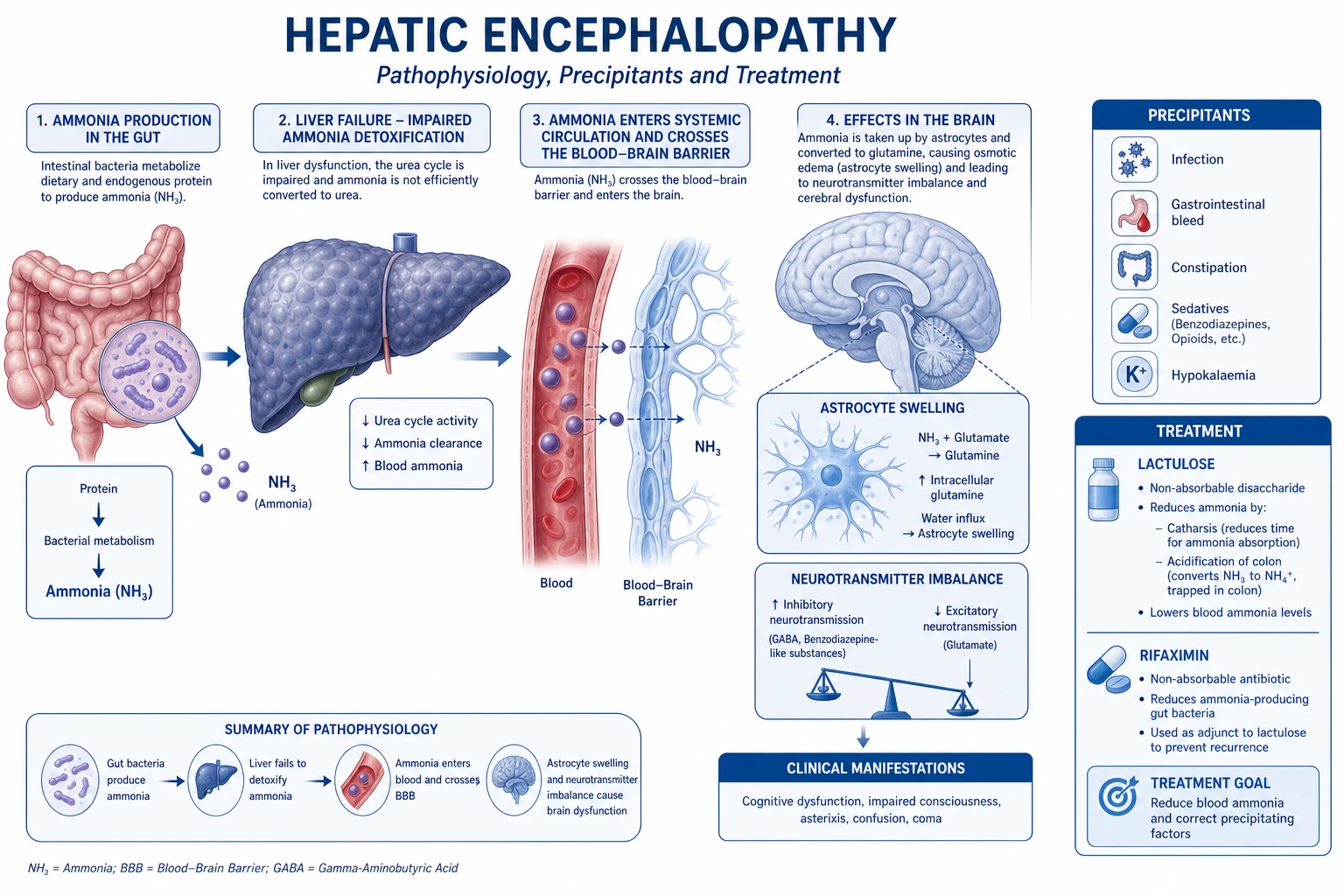
Understanding the mechanism is the key to rational therapy (lactulose and rifaximin both work by reducing gut ammonia; nutrition aims to avoid the catabolism that generates ammonia; sedatives are avoided because they act on the GABA pathway that is already up-regulated in HE).[1][5]
Ammonia -> glutamine -> astrocyte swelling -> (cerebral oedema) — the mechanistic cascade
- AMMONIA PRODUCTION (the gut) — ammonia (NH3) is generated in the colon by bacterial deamination of urea and amino acids, and by small-intestinal glutaminase. The gut is the principal source of systemic ammonia.
- IMPAIRED HEPATIC CLEARANCE + PORTOSYSTEMIC SHUNTING — in cirrhosis the failing liver cannot convert ammonia to urea (via the urea cycle), AND portosystemic shunts (collaterals + spontaneous/TIPS shunts) bypass the liver entirely. Systemic and arterial ammonia rise.
- BLOOD-BRAIN BARRIER CROSSING — ammonia (as lipophilic NH3) diffuses readily across the BBB into the brain, where it is taken up almost exclusively by astrocytes (neurons cannot detoxify ammonia).
- ASTROCYTE GLUTAMINE SYNTHETASE -> GLUTAMINE — astrocytes express glutamine synthetase, which condenses ammonia with glutamate to form glutamine. This is the brain's only ammonia-detoxification pathway. (Glutamine is normally shuttled to neurons for neurotransmitter synthesis.)
- OSMOTIC ASTROCYTE SWELLING (Alzheimer type II astrocytosis) — glutamine is osmotically active. It accumulates intracellularly, draws in water, and causes astrocyte swelling (the characteristic Alzheimer type II astrocyte of chronic HE). The system normally adapts by releasing osmolytes (myo-inositol, taurine) — this adaptation succeeds in chronic cirrhosis (so the brain does NOT swell) but is overwhelmed in acute/hyperacute ammonia loading.
- (WHEN OVERWHELMED) CYTOTOXIC CEREBRAL OEDEMA -> RAISED ICP — if glutamine accumulation outstrips osmolyte compensation (as in ALF, or occasionally in rapidly-evolving high-grade cirrhotic HE), the swollen astrocytes produce cytotoxic cerebral oedema -> raised ICP -> (rarely) uncal herniation.
- NEUROINFLAMMATION + GABA-ERGIC TONE (synergistic) — ammonia also activates microglia (neuroinflammation), increases blood-brain barrier permeability, and is associated with increased endogenous benzodiazepine-like substances and up-regulated GABA-ergic neurotransmission — which is why exogenous benzodiazepines worsen HE and why flumazenil transiently improves some patients.
The central examinable message: HE is fundamentally an AMMONIA-driven astrocyte-osmotic disorder, with inflammation and GABA-ergic tone as synergists. This explains (a) why gut-targeted therapy (lactulose, rifaximin) works, (b) why protein restriction is counterproductive (catabolism generates ammonia), (c) why benzodiazepines are harmful, and (d) why cerebral oedema is common in ALF (no time to adapt) but rare in chronic cirrhosis (the brain has adapted).[1][5]
A word on ammonia measurement: a venous (or arterial) ammonia level is supportive but neither sensitive nor specific enough to diagnose HE or to guide therapy. HE can exist with a normal ammonia (~10-20% of overt episodes), and a high ammonia can occur without HE. The 2014 guideline does NOT recommend routine ammonia measurement for diagnosis (clinical assessment + exclusion of other causes is the standard), and ammonia must NEVER be used as a treatment target (lactulose is titrated to stool output, not to ammonia). A markedly elevated ammonia (>150 umol/L) does identify a brain at risk of cerebral oedema in the ALF context, and a very high level in cirrhosis supports the diagnosis when the picture is ambiguous.[1]
Precipitants — the #1 priority (find and treat)
Episodic (acute) HE in cirrhosis almost always has a precipitant. Identifying and reversing the precipitant is the single most important intervention — without it, ammonia-lowering therapy alone is often ineffective, and recurrence is guaranteed. The mnemonic is infection, bleed, bowels, booze, benzodiazepines, (electrolytes), (TIPS).[1][5]
Precipitants of acute (episodic) HE in cirrhosis — frequency and action
| Precipitant | Frequency | Mechanism | Diagnostic action | Treatment |
|---|---|---|---|---|
| INFECTION (the #1 precipitant) | 20-50% | Sepsis -> cytokines increase BBB permeability + synergise with ammonia; infection increases catabolism -> more ammonia | Culture EVERYTHING (blood x2, urine, sputum) + diagnostic paracentesis (ascitic PMN >250/mm^3 = SBP) + CXR | Empiric broad-spectrum antibiotics immediately after cultures (e.g. ceftriaxone, piperacillin-tazobactam); + albumin 1.5 g/kg day 1 + 1 g/kg day 3 if SBP (prevents HRS) |
| Gastrointestinal bleed | 10-20% | Blood in gut = protein load -> bacterial digestion -> ammonia; also hypovolaemia -> worsens hepatic perfusion | Check Hb, haematemesis/melaena, nasogastric aspirate; urgent endoscopy | Resuscitate; vasoactive drug (terlipressin/octreotide); endoscopic band ligation; antibiotics (ceftriaxone); PPI |
| Constipation | Common | Stasis -> increased dwell time -> more ammonia absorption; also increases gut bacterial load | Bowel history; abdominal exam; plain film if severe | Lactulose (the cathartic effect treats constipation AND HE simultaneously) |
| Electrolyte derangement | Common | Hyponatraemia (the #1 electrolyte precipitant) and hypokalaemia (promotes renal ammonia generation via glutaminase); over-diuresis | U&E; review diuretic dose | Correct sodium and potassium slowly/safely; STOP/reduce diuretics; avoid rapid correction of hyponatraemia (osmotic demyelination) |
| Sedatives / benzodiazepines | Common | GABA-ergic drugs precipitate/worsen HE (GABA tone already up-regulated); also opioids | Drug history; urine drug screen | STOP the offending drug; consider flumazenil (diagnostic/therapeutic trial) for benzodiazepines; AVOID routine sedation |
| Alcohol | Variable | Alcohol intoxication or withdrawal (delirium tremens mimics HE); alcoholic hepatitis flare | History; CIWA-Ar; LFTs (AST:ALT >2, GGT) | Treat withdrawal with a SHORT-ACTING benzodiazepine (only indication for benzo in HE); prednisolone if severe alcoholic hepatitis (MELD 21-39) |
| TIPS (transjugular intrahepatic portosystemic shunt) | Post-procedure | Creates a large portosystemic shunt -> more ammonia bypasses the liver; HE in ~20-40% post-TIPS (usually within 1-3 months) | Recent TIPS history | Lactulose + rifaximin (prophylaxis post-TIPS in high-risk patients); shunt reduction/embolisation if refractory |
| Extrahepatic | Less common | Dehydration, hypovolaemia, hypoglycaemia, constipating drugs (opioids, anticholinergics), hepatocellular carcinoma, constipation from lactulose under-dosing | Targeted workup | Treat the specific cause |
The fellowship-level point: infection is the #1 precipitant and the one you must not miss. Every cirrhotic with new HE gets a full septic screen including a diagnostic paracentesis to exclude SBP — even in the absence of abdominal symptoms (SBP is frequently silent). Start empiric antibiotics after cultures if there is any suspicion. Treating the precipitant is as important as — and often more important than — the ammonia-lowering drugs.[1]
Clinical assessment and diagnosis (a diagnosis of exclusion)
HE is a diagnosis of exclusion. The confusional state of cirrhosis has a wide differential, and several mimics coexist with or masquerade as HE. The intensivist must systematically exclude alternatives before committing to a diagnosis of (only) HE.[1][3]
First-hour ICU assessment of acute hepatic encephalopathy
- GRADE THE ENCEPHALOPATHY (West Haven) — the single most important bedside assessment. Determines level of care, threshold for intubation, and urgency. Grade 2+ = overt HE = start full management. Check asterixis, orientation, sleep pattern, constructional apraxia (draw a star/clock).
- EXCLUDE HYPOGLYCAEMIA IMMEDIATELY — bedside glucose. (Severe liver failure + sepsis = hypoglycaemia; "worsening HE" may be hypoglycaemia.)
- SEPTIC SCREEN + PARACENTESIS — blood cultures x2, urine culture, sputum, CXR, and diagnostic paracentesis (ascitic cell count + culture — PMN >250/mm^3 = SBP). Start empiric antibiotics if any suspicion.
- ELECTROLYTES — U&E (sodium, potassium), creatinine, glucose, magnesium, phosphate. Hyponatraemia and hypokalaemia are key precipitants.
- EXCLUDE INTRACRANIAL PATHOLOGY — CT brain if any focal neurology, seizure, head trauma, anticoagulation, or asymmetric/unusual presentation (subdural haematoma, intracranial bleed — cirrhotics are coagulopathic and fall). HE is not focal.
- REVIEW THE DRUG CHART — benzodiazepines, opioids, anticholinergics, diuretics (over-diuresis), alcohol (CIWA-Ar for withdrawal). Send a urine drug screen.
- CHECK FOR GI BLEED — Hb, nasogastric aspirate, rectal exam; urgent endoscopy if melena/haematemesis or unexplained Hb drop.
- LIVER FUNCTION + AMMONIA — LFTs, coagulation (INR), albumin; ammonia is supportive only (NOT diagnostic, NOT a treatment target). Calculate Child-Pugh and MELD.
- CONSIDER DIFFERENTIALS — sepsis-associated encephalopathy, delirium tremens, Wernicke's (give thiamine), uraemia, hypercapnia, drug intoxication, intracranial lesion.
The high-yield message: HE is a clinical diagnosis of exclusion; treat the precipitant in parallel with the assessment, do not wait for results to start empiric antibiotics or lactulose.[1][3]
Management — the five-step framework
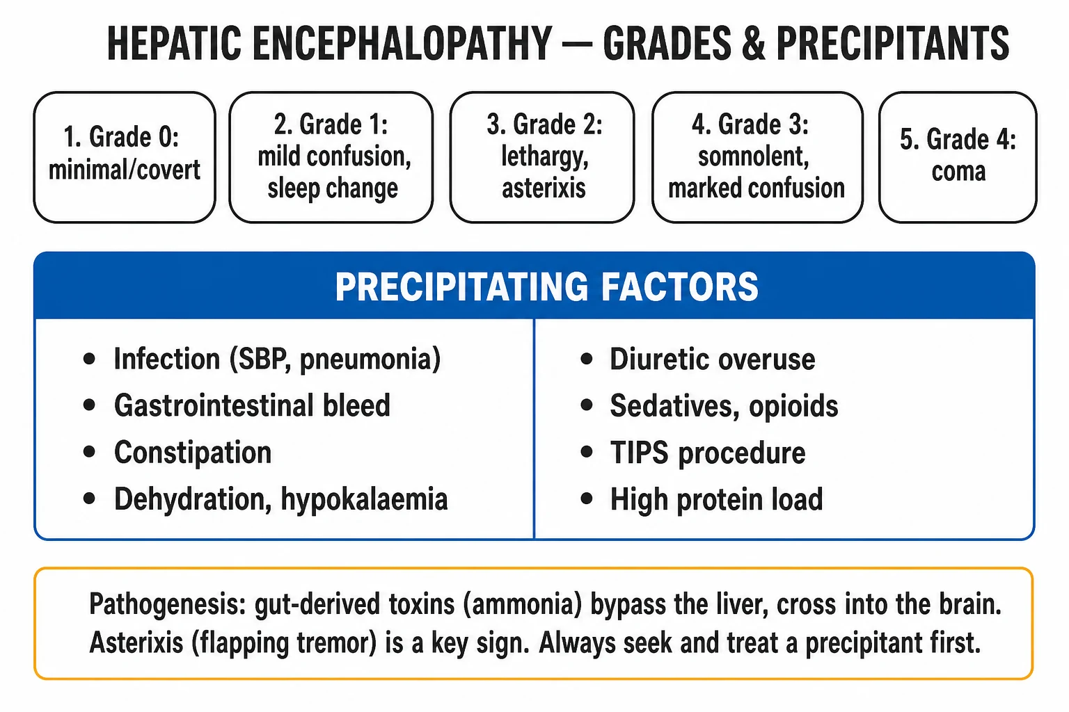
Management of acute (overt) HE — the five-step framework
- FIND AND TREAT THE PRECIPITANT (#1 priority) — septic screen + paracentesis + empiric antibiotics; treat GI bleed; correct electrolytes (Na, K); relieve constipation; stop sedatives; address alcohol withdrawal; review TIPS. Without this, ammonia-lowering therapy fails.
- LACTULOSE (first-line) — 30 mL (20 g) orally/via NGT every 1-2 hours until 2-3 soft bowel motions/day, then titrate down to a maintenance dose (typically 15-30 mL BD-TDS). The endpoint is stool frequency/consistency, NOT a target ammonia. In coma/ileus: lactulose enema (300 mL lactulose in 700 mL water retention enema q4-6h).
- RIFAXIMIN 550 mg BD (additive to lactulose) — for overt/recurrent HE or if lactulose alone fails. A minimally absorbed gut rifamycin; Bass 2010 NEJM: added to lactulose reduced breakthrough HE by 58%.
- SUPPORTIVE/AVOIDANCE — AVOID benzodiazepines unless treating alcohol withdrawal (then short-acting oxazepam/lorazepam); HIGH-PROTEIN diet 1.2-1.5 g/kg/day (do NOT restrict protein); correct electrolytes; maintain hydration/nutrition; thiamine if alcohol misuse; airway protection (intubate at grade 3 if airway unsafe).
- CEREBRAL OEDEMA (if high-grade/rapidly evolving or signs of raised ICP) — head of bed 30 degrees neutral; hypertonic saline (target Na 145-155); mannitol 0.5 g/kg if ICP monitored (osmolality <320). (Rare in cirrhotic HE but must be recognised.)
First-line therapy — lactulose
Lactulose (and its sibling lactitol) is the first-line agent for overt HE and has been for decades. It is a synthetic non-absorbable disaccharide (beta-galactosidofructose) that reaches the colon unchanged, where gut bacteria metabolise it to organic acids (lactic, acetic).[1][2]
Mechanism (two complementary actions):
- Ion-trapping of ammonia — the organic acids lower colonic pH, converting absorbable, lipophilic NH3 to the non-absorbable, charged NH4+ ion, which is trapped in the lumen and expelled in stool. (NH3 is what crosses into blood; NH4+ cannot.)
- Catharsis/laxation — the osmotic effect of the unabsorbed disaccharide produces 2-3 soft stools/day, physically removing the ammonia-laden luminal contents and shortening colonic transit (less dwell time for ammonia absorption). This also treats the precipitant of constipation. [1]
Dosing and titration — the critical examinable detail: the loading dose is 30 mL (20 g of lactulose syrup) orally or via nasogastric tube every 1-2 hours until the patient produces 2-3 soft (paste-like) bowel motions per day. Once this endpoint is reached, the dose is titrated down to a maintenance regimen (typically 15-30 mL two-to-three times daily) that sustains 2-3 soft stools/day. The endpoint is stool frequency and consistency — NOT a serum ammonia level. Over-treatment (excessive diarrhoeea) causes volume depletion, hypernatraemia, and hypokalaemia — each of which paradoxically WORSENS HE (dehydration and hypokalaemia both increase ammonia); under-treatment leaves the patient encephalopathic. In the intubated/comatose patient or in ileus where oral/NG lactulose is ineffective, give lactulose by retention enema (300 mL lactulose in 700 mL water, retained for ~1 hour, q4-6h) until the patient wakes and can take it enterally.[1][2]
Evidence: the Als-Nielsen 2004 systematic review (BMJ) of 22 randomised trials found that non-absorbable disaccharides (lactulose/lactitol) were superior to placebo/no treatment for HE and mortality benefit, and that they were as effective as (and safer than) antibiotics such as neomycin — establishing lactulose as first-line.[2]
Lactulose — what to do and what NOT to do
| Principle | Rationale |
|---|---|
| Titrate to 2-3 SOFT stools/day | The endpoint is stool output (mechanism = catharsis + ion-trapping). This is the single most tested lactulose fact. |
| Do NOT titrate to a target ammonia | Ammonia is neither diagnostic nor a treatment target; it lags and does not correlate linearly with mental state. |
| Watch for over-treatment | Excess diarrhoeea -> dehydration, hypernatraemia, hypokalaemia -> each worsens HE. Reduce the dose if >4-5 stools/day or if hypernatraemia develops. |
| Do NOT abandon lactulose for an antibiotic as monotherapy | Lactulose remains first-line; rifaximin is ADDITIVE, not a replacement. |
| Give via NGT/retention enema if obtunded | Comatose/ileus patients cannot take oral lactulose effectively — use NGT or enema (300 mL in 700 mL water). |
Rifaximin — the additive second-line agent
Rifaximin is a minimally absorbed, broad-spectrum gut rifamycin antibiotic (it inhibits bacterial DNA-dependent RNA polymerase) that, despite negligible systemic absorption (~0.4%), substantially reduces the population of ammonia-producing gut bacteria. It is recommended as an add-on to lactulose (not a replacement) for the prevention of recurrent overt HE and for episodes that are refractory to lactulose alone.[1][1]
The landmark evidence is the Bass 2010 NEJM randomised, double-blind, placebo-controlled trial. In 299 patients in remission from >=2 episodes of overt HE, the addition of rifaximin 550 mg twice daily to a background of lactulose (the majority were already on lactulose) over 6 months:
- Reduced the risk of a breakthrough HE episode by 58% (hazard ratio 0.42; 95% CI 0.28-0.64; P<0.001).
- Reduced the risk of HE-related hospitalisation by ~50% (HR 0.50; P=0.01).
- Was safe and well tolerated (the concern about Clostridium difficile / antimicrobial resistance has not materialised in long-term follow-up).[1]
Lactulose vs rifaximin for overt HE
| Feature | Lactulose | Rifaximin |
|---|---|---|
| Class | Non-absorbable disaccharide | Minimally absorbed gut rifamycin antibiotic |
| Mechanism | Lowers colonic pH (ion-traps NH3 -> NH4+) + catharsis | Reduces ammonia-producing gut bacteria |
| Role | First-line for all overt HE episodes | Add-on to lactulose for recurrent/refractory HE |
| Dose | 30 mL q1-2h to 2-3 soft stools/day, then titrate | 550 mg PO BD |
| Key trial | Als-Nielsen 2004 (BMJ) systematic review | Bass 2010 (NEJM) RCT — 58% reduction in breakthrough HE |
| Endpoint | 2-3 soft stools/day | Continued (no titration) |
| Main side effects | Diarrhoeea, flatulence, abdominal cramps, dehydration, hypernatraemia, hypokalaemia (over-use) | Generally well tolerated; theoretical C. difficile/resistance (not clinically significant) |
| Cost | Cheap | Expensive |
The examinable take-home: rifaximin is ADDITIVE to lactulose (not monotherapy) for recurrent/refractory overt HE; dose 550 mg BD; Bass 2010 showed a 58% reduction in breakthrough HE.[1][1]
Nutrition — HIGH protein, do NOT restrict
Nutritional management of HE is the source of one of the most common and most harmful errors in hepatology: the historic (and now obsolete) practice of restricting dietary protein to reduce ammonia production. Modern evidence and all current guidelines explicitly recommend AGAINST protein restriction.[1][4]
Nutrition in HE — what to do and what NOT to do
| Principle | Rationale / evidence |
|---|---|
| Do NOT restrict dietary protein | Protein restriction causes muscle (skeletal) catabolism; skeletal muscle is a major extrahepatic site of ammonia clearance (muscle glutamine synthetase), so catabolism REDUCES ammonia clearance and INCREASES ammonia. It also worsens sarcopenia, which is itself an independent predictor of HE and mortality in cirrhosis. |
| Provide a HIGH-protein diet 1.2-1.5 g/kg/day | Adequate protein preserves the muscle ammonia sink and prevents catabolism. The 2014 EASL/AASLD guideline recommends 1.2-1.5 g/kg/day protein in cirrhosis (even during HE). |
| Córdoba 2004 RCT (J Hepatol) | Randomised 30 cirrhotics with episodic HE to a low-protein diet (with progressive increments) vs a normal-protein diet: the outcome of HE was not different, but the low-protein group had higher protein breakdown (worse catabolism). Conclusion: a normal-protein diet is safe and restriction has no benefit. |
| Vegetable / dairy protein may be preferred | Vegetable and dairy proteins are richer in branched-chain amino acids (BCAA) and lower in aromatic amino acids; BCAA supplementation can be a useful adjunct in intolerant/malnourished patients. |
| Provide small frequent meals + a late-night snack | Avoids prolonged fasting (which precipitates catabolism); a complex carbohydrate snack at night reduces fasting catabolism. |
| Address sarcopenia | Sarcopenia is a strong predictor of HE (loss of the muscle ammonia sink) and mortality — resistance exercise, adequate calories (35-40 kcal/kg/day), and protein. |
The single most examinable nutrition fact: protein is NOT restricted in HE; give a HIGH-protein diet (1.2-1.5 g/kg/day) based on the Córdoba 2004 RCT (normal-protein diet safe; restriction causes catabolism that generates ammonia and worsens sarcopenia).[1][4]
Sedation in HE — avoid benzodiazepines
The GABA-ergic neurotransmitter system is up-regulated in HE (increased endogenous benzodiazepine-like substances, increased GABA-A receptor sensitivity), which is one reason patients are somnolent — and is precisely why exogenous benzodiazepines are neurotoxic in this setting and precipitate or worsen HE.[1][5]
Sedation and drugs in HE — what to do and what NOT to do
| Situation | Recommendation | Rationale |
|---|---|---|
| Routine sedation / agitation in HE | AVOID benzodiazepines. Use non-pharmacological measures first; if sedation is unavoidable, use haloperidol or a carefully titrated alpha-2 agonist (dexmedetomidine) — NOT a benzodiazepine. | Benzodiazepines are GABA-ergic and precipitate/worsen HE. |
| Alcohol withdrawal (the ONE indication) | Use a SHORT-ACTING benzodiazepine — oxazepam or lorazepam (no active metabolites; hepatic glucuronidation, not oxidative metabolism) — symptom-triggered (CIWA-Ar). | Delirium tremens mimics HE and can coexist; oxazepam/lorazepam are preferred in liver disease because they lack active metabolites. Avoid diazepam (long half-life, active metabolites, accumulates). |
| Suspected iatrogenic benzodiazepine contribution | Consider a flumazenil trial (0.2-0.5 mg IV) — a transient diagnostic/therapeutic improvement supports a benzodiazepine contribution. | Flumazenil improves some HE patients (endogenous benzodiazepines), but it is short-acting and not a treatment — it is a diagnostic clue. |
| Intubated patient requiring sedation | Prefer propofol (short half-life, allows neurological assessment) over benzodiazepines; dexmedetomidine for analgesia/sedation. | Avoids GABA-ergic load; propofol allows intermittent neurological examination. |
| Opioids | Use with caution; they accumulate in liver failure and precipitate constipation (a precipitant). | Naloxone trial if opioid contribution suspected. |
The examinable rule: AVOID benzodiazepines in HE unless treating alcohol withdrawal — and then use a SHORT-ACTING agent (oxazepam/lorazepam).[1]
Cerebral oedema — recognise and manage (rare but lethal in cirrhotic HE)
Cerebral oedema is the dominant killer in acute liver failure (Type A HE) (up to 75-80% of grade 4 patients) but is uncommon in cirrhotic (Type C) HE because the chronically diseased brain has time to osmotically adapt (releasing myo-inositol). However, it CAN occur in high-grade (grade 4), rapidly-evolving cirrhotic HE, after TIPS (acute rise in shunted ammonia), or when an acute decompensation (sepsis, variceal bleed) produces a sudden ammonia surge. The intensivist must recognise it and escalate.[1]
Cerebral oedema management in HE (when it arises)
- RECOGNISE — grade 4 coma, Cushing's triad (hypertension + bradycardia + irregular respiration — late/pre-terminal), pupillary changes, decerebrate posturing, or a sudden deterioration. A rising or very high ammonia (>150 umol/L) supports the diagnosis.
- HEAD POSITION — head of bed elevated 30 degrees and neutral (promotes jugular venous outflow, lowers ICP). Avoid tight endotracheal-tape ties and neck flexion.
- MAINTAIN CEREBRAL PERFUSION — avoid hypotension (MAP >=65), hypoxia (PaO2 >=60), and hypercapnia (target PaCO2 35-40; hypercapnia -> cerebral vasodilation -> raised ICP).
- HYPERTONIC SALINE — induce and maintain mild hypernatraemia (serum Na 145-155 mmol/L) with 30% hypertonic saline boluses or infusion (osmotically draws water out of the swollen brain).
- MANNITOL 0.5 g/kg (20% solution) bolus — for acutely raised ICP / signs of herniation, provided serum osmolality is <320 mOsm/kg and the kidneys are excreting (mannitol is a filtered osmole; it accumulates in AKI/anuria causing rebound hyperosmolality). Re-check osmolality before repeat doses. Reserve for confirmed/suspected intracranial hypertension; routine prophylactic mannitol is NOT recommended.
- ICP MONITORING — consider in grade 4 with refractory intracranial hypertension to guide osmotherapy (weigh bleeding risk against "rebalanced haemostasis").
- REFRACTORY ICP — barbiturate coma (thiopentone), induced hypothermia (32-34 degC) — bridge to recovery/transplant measures.
The key distinction to articulate: in cirrhotic HE, cerebral oedema is rare and not the focus; the focus is the precipitant + ammonia-lowering + nutrition. Cerebral oedema management is escalated only in high-grade/rapidly-evolving disease or signs of raised ICP.[1]
Second-line and adjunctive therapies
When lactulose + rifaximin + precipitant treatment are insufficient, several adjuncts exist (though none supplant first-line therapy):[1][5]
Adjunctive / second-line therapies for refractory HE
| Therapy | Mechanism | Role |
|---|---|---|
| L-ornithine L-aspartate (LOLA) | Provides substrates (ornithine) that enhance urea cycle + glutamine synthesis in residual liver and muscle -> ammonia lowering | Adjunct; evidence of benefit, used where available |
| Branched-chain amino acids (BCAA) | Correct the low BCAA:aromatic amino acid ratio; promote muscle ammonia detoxification | Adjunct in malnourished/protein-intolerant patients; oral supplementation |
| Shunt embolisation / occlusion | Reduces portosystemic shunting (esp. large spontaneous or TIPS shunts) | Refractory HE with a demonstrable large shunt (post-TIPS, congenital) |
| Flumazenil | Benzodiazepine (GABA-A) receptor antagonist | Short-acting; diagnostic/therapeutic trial where benzodiazepines contribute; not a treatment |
| Molecular Adsorbent Recirculating System (MARS) / albumin dialysis | Removes protein-bound toxins (incl. ammonia, bilirubin) | Bridge in refractory HE / ACLF; no proven survival benefit (RELIEF trial) |
| Zinc | Cofactor for urea cycle enzymes; cirrhotics are often zinc-deficient | Supplement in zinc-deficient patients (may improve ammonia clearance) |
| L-ornithine phenylacetate / glycerol phenylbutyrate | Nitrogen-scavengers (alternate pathway ammonia disposal) | Investigational/emerging for recurrent HE |
Secondary prophylaxis
After an episode of overt HE resolves, secondary prophylaxis is indicated because recurrence is high (~40% at 1 year without prophylaxis). The standard regimen is continued lactulose titrated to 2-3 soft stools/day, with rifaximin 550 mg BD added after the second episode (i.e. recurrent HE). The Bass 2010 trial is the evidence base for rifaximin prophylaxis.[1][1]
SAQ — hepatic encephalopathy in ICU
Hepatic encephalopathy — precipitant, grade, therapy
10 minutes · 10 marks
A 58-year-old with Child-Pugh C cirrhosis is somnolent but rousable, grossly disoriented, with incomprehensible speech after 5 days without stools. Na 128, K 3.0. Paracentesis excludes SBP. Outline grade, precipitants, and management.
Clinical pearls
Red flags
Prognosis
Prognosis and prognostic factors in hepatic encephalopathy
| Factor | Outcome / implication |
|---|---|
| Overt HE (any episode) — 1-year mortality | ~40-50% in cirrhosis — HE is a marker of advanced disease |
| Covert (minimal) HE | Near-normal life expectancy but impaired quality of life, driving ability, and risk of progression to overt HE |
| Recurrence after an overt episode | ~40% at 1 year without secondary prophylaxis |
| Predictors of poor outcome | Severity of underlying liver disease (high Child-Pugh / MELD), recurrent HE, refractory ascites, HRS, sarcopenia, persistent precipitant, alcohol aetiology with continued use |
| Predictors of good outcome | Treatable precipitant (esp. infection), early grade, good nutritional status (preserved muscle mass), adherence to secondary prophylaxis |
| Impact of therapy | Lactulose + rifaximin secondary prophylaxis halves recurrence (Bass 2010); nutrition preserves the muscle ammonia sink |
| Post-TIPS HE | ~20-40% develop HE; usually within 1-3 months; often manageable with lactulose + rifaximin; refractory cases need shunt reduction |
| Cerebral oedema (when it occurs) | High mortality if unrecognised; manageable with osmotherapy if caught early |
| Transplant | Liver transplantation is the definitive treatment for refractory HE in decompensated cirrhosis; HE is an indication for transplant evaluation when associated with decompensation |
The overarching prognostic message: overt HE is a sentinel event in cirrhosis — it signals advanced disease and carries a ~40-50% 1-year mortality. Each episode warrants a careful precipitant search, an escalation of liver-failure management, and (in suitable candidates) early transplant evaluation, because HE is both a manifestation of decompensation and, in its recurrent/refractory form, an indication for transplantation.[1][5]
Key trials and evidence
Bass 2010 — Rifaximin for recurrent HE (NEJM; PMID 20589374)
Source
Multicentre double-blind RCT, 6-month follow-up; 299 patients in remission from >=2 episodes of overt HE; the majority were already receiving lactulose
Intervention
Rifaximin 550 mg BD vs placebo, ADDED to background therapy (mostly lactulose)
Primary endpoint
Time to first breakthrough HE episode
Key result — breakthrough HE
Rifaximin reduced the risk by **58%** (HR 0.42; 95% CI 0.28-0.64; P<0.001) — 22.1% vs 45.9% had a breakthrough episode
Key result — HE hospitalisation
Reduced by ~50% (HR 0.50; 95% CI 0.29-0.87; P=0.01)
Safety
No increase in Clostridium difficile or antimicrobial resistance over 24-month open-label extension
Clinical bottom line
Rifaximin 550 mg BD added to lactulose is the evidence base for secondary prophylaxis of recurrent overt HE — additive, not a replacement
Als-Nielsen 2004 — Non-absorbable disaccharides meta-analysis (BMJ; PMID 15054035)
Source
Cochrane Hepato-Biliary Group systematic review; 22 randomised trials of non-absorbable disaccharides (lactulose/lactitol) in HE
Comparison 1
Disaccharides vs placebo/no treatment — beneficial effect on HE (number needed to treat ~2 for improvement); possible mortality benefit
Comparison 2
Disaccharides vs antibiotics (e.g. neomycin) — no significant difference in HE outcome, but disaccharides are SAFER (neomycin is nephrotoxic/ototoxic)
Key finding
Non-absorbable disaccharides are effective for HE and safer than antibiotics — established lactulose as FIRST-LINE
Clinical bottom line
Lactulose is the first-line agent for overt HE; antibiotics (now rifaximin) are adjuncts, not replacements
Córdoba 2004 — Normal-protein diet for episodic HE (J Hepatol; PMID 15246205)
Source
Randomised study; 30 cirrhotics admitted with an episode of episodic HE
Intervention
Low-protein diet (with progressive increments) vs normal-protein diet for 14 days, plus standard HE therapy
Primary outcome
Evolution of HE over 14 days
Key result
HE outcome was **NOT significantly different** between groups; the low-protein group had **higher protein breakdown** (worse catabolism)
Conclusion
A normal-protein diet is metabolically more adequate and SAFE; protein restriction offers NO benefit and causes catabolism
Clinical bottom line
Do NOT restrict protein in HE — give a high-protein diet (1.2-1.5 g/kg/day). This RCT overturned decades of (harmful) protein-restriction practice
References
- [1]Vilstrup H, Amodio P, Bajaj J, Cordoba J, Ferenci P, Mullen KD, Weissenborn K, Wong P Hepatic encephalopathy in chronic liver disease: 2014 Practice Guideline by the American Association for the Study of Liver Diseases and the European Association for the Study of the Liver Hepatology, 2014.PMID 25042402
- [2]Als-Nielsen B, Gluud LL, Gluud C Non-absorbable disaccharides for hepatic encephalopathy: systematic review of randomised trials BMJ, 2004.PMID 15054035
- [3]Nabi E, Bajaj JS Useful tests for hepatic encephalopathy in clinical practice Curr Gastroenterol Rep, 2014.PMID 24357348
- [4]Córdoba J, López-Hellín J, Planas M, et al. Normal protein diet for episodic hepatic encephalopathy: results of a randomized study J Hepatol, 2004.PMID 15246205
- [5]Ferenci P Hepatic encephalopathy Gastroenterol Rep (Oxf), 2017.PMID 28533911