ICU · GI & nutrition / surgical
Ileus, Bowel Obstruction & Mesenteric Ischaemia — The Acute Abdomen in ICU
Also known as Paralytic ileus · Ileus · Small bowel obstruction · SBO · Large bowel obstruction · LBO · Bowel obstruction · Strangulation · Mesenteric ischaemia · SMA embolism · Non-occlusive mesenteric ischaemia · NOMI · Pain out of proportion
The ileus, the bowel obstruction, and the mesenteric ischaemia are the three acute bowel disorders in the ICU. The ileus (paralytic) is the failure of the peristalsis (the post-op, the opioid, the electrolyte, the sepsis) — the supportive management (the NGT, the fluids, the correct the electrolytes). The bowel obstruction (mechanical) is the SBO (the adhesions the commonest) or the LBO (the malignancy, the volvulus) — the drip and suck, the surgery for the strangulation (the continuous pain, the fever, the peritonitis, the raised lactate). The mesenteric ischaemia is the vascular compromise of the bowel — the SMA embolism (the AF), the thrombosis, the venous, the non-occlusive (the low-flow). The hallmark is the PAIN OUT OF PROPORTION to the exam; the lactate rises late; the CT angiography is the diagnostic; the early surgery (the high mortality from the delay).
On this page & tools
Your progress
Saved locally on this device.
8 MCQs with explanations
Target exams
Overview & definition
The ileus, the bowel obstruction, and the mesenteric ischaemia are the three acute bowel disorders in the ICU. They present with the distension, the pain, and the constipation, but the mechanism and the management differ. The ileus is the paralytic (no peristalsis); the obstruction is the mechanical (a physical block); the mesenteric ischaemia is the vascular compromise. The mesenteric ischaemia is the most time-critical — the early surgery (the high mortality from the delay).[1]
Ileus (the paralytic)
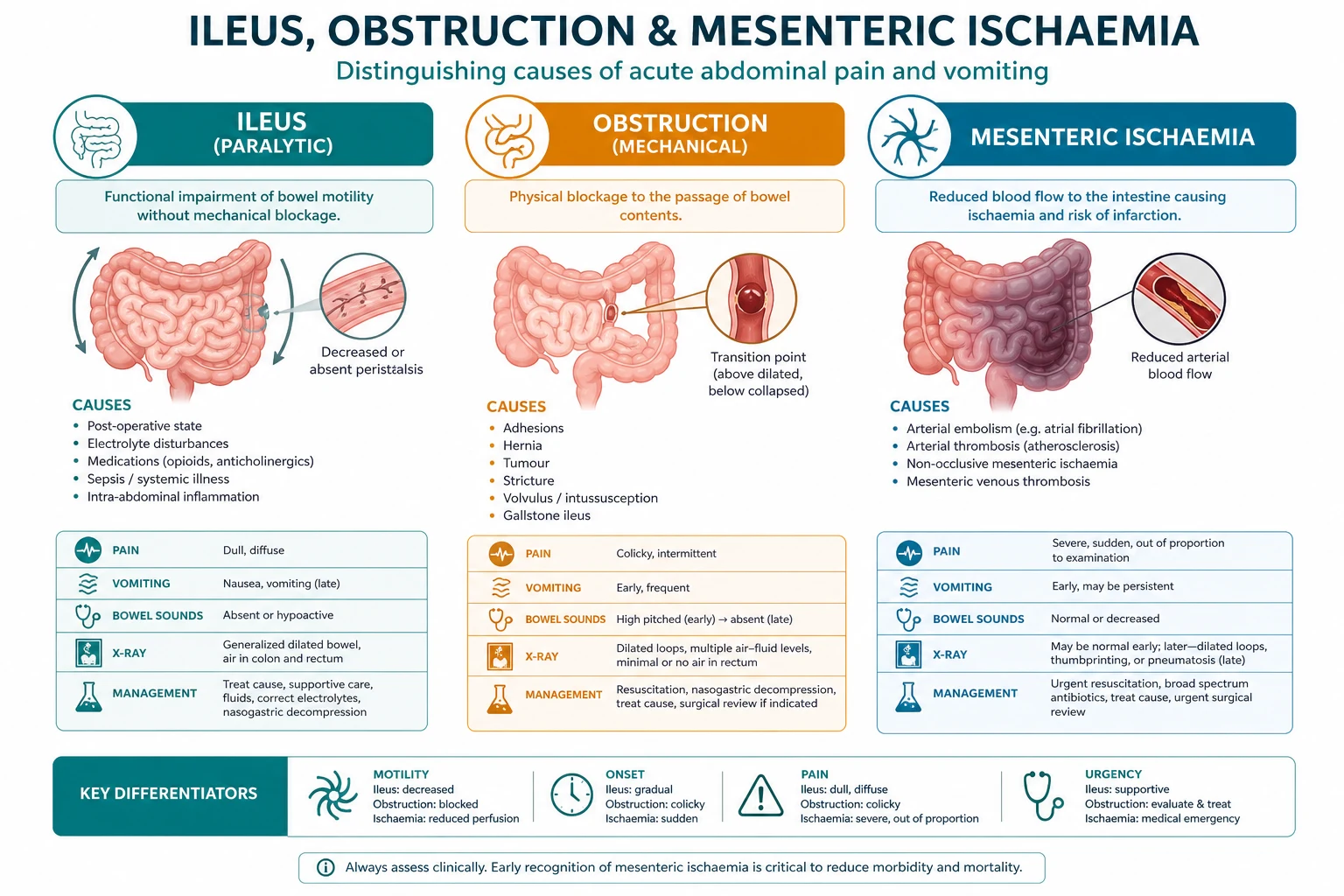
- The failure of the peristalsis (the paralytic). The bowel is structurally intact but the functionally silent.[1]
- The causes — the post-operative (the commonest), the opioids, the electrolytes (the hypokalaemia, the hypomagnesaemia), the sepsis, the retroperitoneal bleed, the spinal injury.[1]
- The clinical — the distension, the absent or the tinkling bowel sounds, the no flatus or stool, the nausea, the vomiting.[1]
- The management — the supportive: the NGT decompression, the IV fluids, the correct the electrolytes (the K, the Mg), the reduce the opioids, the ambulation. Resolves in the days. The prokinetic (the metoclopramide, the erythromycin) if the prolonged.[1]
The pathophysiology of the ileus — the neurogenic, the inflammatory, the opioid
- The postoperative ileus is the transient impairment of the coordinated myoelectric activity of the gut after the abdominal (and the non-abdominal) surgery. The three mechanisms: the neurogenic (the sympathetic inhibition, the loss of the parasympathetic tone after the visceral manipulation), the inflammatory (the macrophages and the neutrophils, the cytokines — the nitric oxide, the COX-2 prostaglandins), the opioid (the mu-receptors of the enteric nervous system — the gut the slow).[1]
- The early return of the motility — the small bowel the 24 hours, the stomach the 48 hours, the colon the 72 hours. The prolonged postoperative ileus (PPI) — the failure to tolerate the oral intake by the postoperative day 4 (the vomiting, the distension, the no flatus/stool). The PPI the risk for the prolonged length of stay, the aspiration, the malnutrition, the readmission.[1]
The differentiation of the ileus from the mechanical obstruction
- The ileus is the diffuse, the non-localised, the silent; the obstruction is the colicky, the localised, the hyperactive then silent (the transition).[1]
- On the imaging — the ileus: the gas in the large and the small bowel (the diffuse), the no cut-off, the no transition point, the peritoneal free air absent. The mechanical: the dilated loops proximal, the collapsed distal, the transition point (the CT — the transition point the diagnostic).[1]
The Ogilvie syndrome (the acute colonic pseudo-obstruction, ACPO)
- The massive dilatation of the colon WITHOUT a mechanical obstruction — the paralytic of the large bowel. Sir William Heneage Ogilvie, 1948. The caecum > 10 cm or the colon > 6 cm the diagnostic on the imaging.[2]
- The risk factors — the post-operative (the orthopaedic — the hip, the spine), the trauma, the sepsis, the electrolytes (the hypokalaemia, the hypomagnesaemia, the hypocalcaemia), the opioids, the anticholinergics, the spinal injury, the retroperitoneal pathology (the haematoma, the tumour), the critical illness.[2]
- The clinical — the marked distension, the tympanic abdomen, the no peritonism early, the absent or the tinkling bowel sounds. The ischaemia and the perforation (the caecum — the Laplace law, the wall tension rises with the radius) — the catastrophe. The caecal diameter > 12 cm for > 6 days the high risk of the perforation; the caecum > 14 cm the urgent decompression.[2]
- The management — the steps. (1) Conservative — the nothing by mouth, the NGT, the rectal tube, the correct the electrolytes (the K, the Mg, the Ca), the stop the offending drugs (the opioids, the anticholinergics), the ambulation, the position change. (2) The neostigmine — the acetylcholinesterase inhibitor, the pro-secretory, the pro-motility — for the caecum > 10 cm and the no improvement after the 48 to 72 hours of the conservative. (3) The colonoscopic decompression — for the neostigmine failure or the contraindicated. (4) The surgery (the caecostomy, the resection) — the last resort, for the ischaemia, the perforation, the peritonitis.[1][2]
The neostigmine for the Ogilvie — the landmark evidence
- The Ponec 1999 NEJM trial — the randomised, the double-blind, the placebo-controlled — the neostigmine 2 mg IV vs the placebo in the 21 patients with the ACPO (the caecum > 10 cm, the no improvement). The rapid decompression in the 91 % (10 of 11) of the neostigmine vs the 0 of 10 of the placebo (p < 0.001). The clinical decompression within the minutes. The adverse events — the bradycardia (the atropine the standby), the bronchospasm, the hypotension, the salivation, the abdominal cramping — the patient on the cardiac monitor, the atropine the ready.[1]
- The contraindications to the neostigmine — the mechanical obstruction (the absolute — exclude on the CT first), the recent MI, the bronchospasm/asthma, the bradycardia, the heart block, the renal failure (the dose adjust — the neostigmine the renal clearance), the perforation/peritonitis. The repeat dose for the recurrence — the effective (the ~30 to 40 % the recurrence).[1]
- The colonoscopic decompression — the effective (the 70 to 80 % the initial, the ~20 % the recurrence — the repeat). The risk of the perforation (the dilated, the ischaemic wall — the high risk). The Demetriou 2025 meta-analysis — the neostigmine and the colonoscopic decompression the comparable efficacy; the neostigmine the first-line (the less invasive) for the no contraindication.[10]
Bowel obstruction (the mechanical)
The small bowel obstruction (SBO)
- The adhesions (the commonest, the post-operative), the hernias, the malignancy, the Crohn's, the intussusception, the gallstone ileus.[1]
- The clinical — the colicky central-abdominal pain, the distension, the vomiting (the early, the proximal SBO), the constipation, the obstipation.[1]
- The management — the "drip and suck" (the IV fluids, the NGT decompression). The conservative trial for the uncomplicated adhesive SBO (the 48 to 72 hours). The surgery for the failed conservative, the strangulation, or the closed-loop obstruction.[1]
The large bowel obstruction (LBO)
- The malignancy (the colorectal, the commonest), the volvulus (the sigmoid, the caecal), the diverticular stricture, the hernia.[1]
- The clinical — the colicky lower-abdominal pain, the marked distension, the constipation, the obstipation. The vomiting late.[1]
- The management — the NGT, the fluids, the surgery (the resection, the stoma). The volvulus — the endoscopic detorsion (the sigmoid) then the elective resection; the caecal volvulus — the surgery.[1]
The strangulation (the emergency)
The signs of the strangulation (the ischaemic, the compromised bowel) mandate the emergency surgery:[1]
- The continuous (not colicky) pain, the tachycardia, the fever.[1]
- The peritonism or the peritonitis (the rigid abdomen, the rebound).[1]
- The raised lactate, the raised WCC, the metabolic acidosis.[1]
- The free gas (the perforation) or the pneumatosis on the imaging.[1]
The closed-loop vs the open-loop obstruction
- The open-loop — the obstruction at one point; the proximal bowel decompresses through the vomiting and the NGT; the less emergent. The closed-loop — the obstruction at two points (the "closed segment") — the bowel trapped between the two obstructions, the gas and the fluid the no escape, the rapid rise of the intraluminal pressure, the venous then the arterial compromise, the strangulation, the ischaemia, the perforation. The high risk — the surgical emergency.[3]
- The causes of the closed-loop — the hernia (the neck the two points), the volvulus (the mesentery the twist), the adhesions with the tight band, the intussusception, the gallstone ileus with the ball-valve. The CT — the "C" or the "U" — the closed segment, the "swirl sign" (the mesenteric vessels the twist), the "beak sign", the "small-bowel faeces sign", the pneumatosis, the portal gas — the signs of the ischaemia.[3]
- The hernias — the inguinal (the indirect the commoner), the femoral (the high risk of the strangulation — the narrow, the rigid neck), the incisional, the parastomal, the Richter (the anti-mesenteric border only — the no obstruction, the high risk of the strangulation and the perforation, the subtle). The incarcerated (the irreducible, the no ischaemia) becomes the strangulated (the ischaemic) — the surgery.[3]
The water-soluble contrast (the Gastrografin challenge)
- The hyperosmolar water-soluble contrast (the gastrografin, the diatrizoate) — the two roles in the adhesive SBO: the diagnostic (the passage of the contrast to the colon within the 24 hours — the ~97 % sensitivity, the ~96 % specificity — the high negative predictive value for the resolution; the high positive predictive value for the need for the surgery if the no passage) and the therapeutic (the osmotic effect — the shift of the fluid into the lumen, the reduction of the bowel-wall oedema, the stimulation of the peristalsis).[4]
- The Branco 2010 meta-analysis — the 14 studies, the 1 025 patients — the water-soluble contrast the reduced the need for the surgery (OR 0.62), the shortened the hospital stay, the reduced the recurrent admissions. The resolves the adhesive SBO in the ~60 to 80 % of the patients.[4]
- The Bologna 2017 guidelines (the WSES) — the conservative trial for the 72 hours for the uncomplicated adhesive SBO (the no signs of the strangulation, the no peritonitis, the no closed-loop, the no ischaemia). The water-soluble contrast the recommended at the 48 hours if the no resolution. The surgery — the failed conservative, the signs of the strangulation, the peritonitis, the closed-loop on the imaging.[3]
The gallstone ileus and the intussusception (the rare SBO)
- The gallstone ileus — the gallstone (the large, the > 2.5 cm) the erodes through the cholecystoduodenal fistula into the bowel, the lodges at the terminal ileum (the narrowest). The Rigler's triad — the pneumobilia, the ectopic gallstone, the small-bowel obstruction. The elderly woman. The surgery (the enterolithotomy — the milk the stone proximally, the enterotomy, the removal; the cholecystectomy the elective — the inflammation).[1]
- The Bouveret syndrome — the gallstone the lodges at the duodenum (the proximal) — the gastric outlet obstruction. The endoscopic removal (the lithotripsy) the option.[1]
- The intussusception — the telescoping. The adults — the pathological lead point (the tumour — the lipoma, the lymphoma, the adenocarcinoma; the Meckel's) — the surgery (the resection — the underlying lesion). The children — the idiopathic (the lymphoid hyperplasia) — the pneumatic/enema reduction.[1]
The large bowel obstruction — the volvulus and the pseudo-obstruction
- The sigmoid volvulus — the commoner (the ~75 % of the colonic volvulus); the elderly, the institutionalised, the chronic constipation, the high-fibre. The "coffee-bean" sign, the "bent inner tube". The endoscopic detorsion (the flexible sigmoidoscope, the flatus tube, the leave the tube the 48 to 72 hours) — the decompression; the elective resection (the recurrence the ~40 to 90 % without). The surgery — the strangulation, the peritonitis, the failed detorsion.[1]
- The caecal volvulus — the ~25 %; the younger, the mobile caecum. The "coffee-bean" pointing to the left upper quadrant. The no endoscopic detorsion (the risk of the perforation) — the surgery (the resection, the primary anastomosis).[1]
Mesenteric ischaemia
(the vascular) [1]
The vascular compromise of the bowel — the most time-critical of the three.[1]
The four types
- The acute mesenteric ARTERIAL occlusion (the SMA embolism — the AF, the thrombosis — the atherosclerosis). The pain out of proportion to the exam (the severe pain with the minimal findings early). The high mortality.[1]
- The mesenteric venous thrombosis (the hypercoagulable, the portal hypertension). The subacute presentation.[1]
- The non-occlusive mesenteric ischaemia (NOMI) — the low-flow state (the shock, the vasopressors, the dialysis). No occlusion on the angiography; the diffuse the vasospasm. Treat the underlying low-flow.[1]
- The chronic mesenteric ischaemia ("the intestinal angina") — the post-prandial pain, the weight loss, the atherosclerosis of the coeliac, the SMA, the IMA.[1]
The diagnosis and the management
- The pain out of proportion to the exam is the hallmark (the severe pain, the minimal findings early). The peritonism develops late (the infarcted bowel).[1]
- The lactate rises LATE — do NOT wait for the lactate (the early ischaemia may have the normal lactate). The high lactate is the late, the poor-prognosis sign.[1]
- The CT angiography — the diagnostic test (the vascular occlusion, the bowel-wall changes, the pneumatosis, the portal gas).[1]
- The management — the fluid resuscitation, the heparin (the anticoagulation), the early surgery (the embolectomy, the revascularisation, the resection of the infarcted bowel). The endovascular (the thrombolysis, the stenting) in the select cases. The high mortality from the delay.[1]
The SMA embolus vs the SMA thrombus (the differentiation)
The proximal arterial occlusion — distinguish the embolus from the thrombus — the different management, the different prognosis:[6]
- The SMA embolus — the commoner of the two (~50 % of the AMI). The source — the atrial fibrillation (the commonest), the mural thrombus (the recent MI), the valvular, the atheroembolism. The lodges at the point of the natural narrowing — the origin of the middle colic artery (the ~3 to 8 cm from the SMA origin). The abrupt onset — the pain out of proportion, the "severe, the sudden". The CT — the sharp, the convex, the meniscus — the filling defect at the middle colic; the no collateral; the no atherosclerosis of the origin. The management — the embolectomy (the open — the Fogarty catheter) or the endovascular thrombectomy/thrombolysis.[1]
- The SMA thrombosis — the ~25 % of the AMI. The atherosclerosis of the origin of the SMA (the pre-existing stenosis, the chronic). The superimposed thrombosis — the acute-on-chronic. The history of the intestinal angina (the post-prandial pain, the weight loss, the food fear). The CT — the tapered, the funnel-shaped occlusion at the origin of the SMA; the calcification, the atherosclerosis; the collaterals (the chronic). The management — the bypass (the aorto-mesenteric, the vein or the prosthetic), the endarterectomy, or the endovascular stenting.[1]
The mesenteric venous thrombosis (MVT)
- The thrombosis of the superior mesenteric vein — the 5 to 15 % of the AMI. The hypercoagulable (the JAK2, the myeloproliferative, the factor V Leiden, the antithrombin, the protein C/S, the malignancy, the OCP), the portal hypertension (the cirrhosis), the intra-abdominal infection/sepsis (the appendicitis, the diverticulitis, the pancreatitis), the post-operative, the trauma.[1]
- The subacute onset (the days to the weeks) — the insidious pain, the distension, the occult blood. The CT — the engorged, the thickened mesentery, the thrombus in the SMV/portal vein, the ascites, the pneumatosis late. The management — the anticoagulation (the LMWH then the warfarin/DOAC — the 3 to 6 months; the lifelong if the thrombophilia), the surgery for the peritonitis (the bowel infarction). The better prognosis than the arterial (the ~20 to 30 % mortality).[1]
The non-occlusive mesenteric ischaemia (NOMI) — the deep dive
- The diffuse vasospasm of the splanchnic vessels in the low-flow state — the no organic occlusion on the angiography. The ~20 % of the AMI. The critically-ill, the elderly, the cardiac — the ICU patient.[1]
- The mechanism — the splanchnic vasoconstriction (the alpha-adrenergic — the noradrenaline, the adrenaline, the high-dose vasopressors), the low cardiac output (the heart failure, the cardiogenic shock, the MI, the arrhythmia), the hypovolaemia (the dialysis — the intravascular depletion, the ultrafiltration), the digitalis (the historical). The territory — the SMA territory — the same as the embolus.[1]
- The diagnosis — the clinical suspicion (the ICU patient, the vasopressors, the abdominal pain/distension, the feed intolerance, the rising lactate). The CT — the no occlusion, the diffuse narrowing of the SMA branches, the poor filling of the mesenteric arcades, the alternating dilatation/narrowing (the "sausage-string"), the bowel-wall thickening. The angiography — the gold standard for the NOMI (the diffuse vasospasm).[6]
- The management — the treat the underlying low-flow — the optimise the cardiac output (the inotropes — the dobutamine, the milrinone), the reduce the vasopressors (the alpha — the noradrenaline — the worsen; the vasopressin the preferable — the no splanchnic vasoconstriction), the restore the volume, the correct the electrolytes. The selective intra-arterial papaverine (the SMA infusion — the smooth-muscle relaxant, the vasodilator) — the salvage. The surgery for the infarction/peritonitis (the resection of the necrotic bowel — the "second-look" laparotomy the 24 hours). The high mortality — the 50 to 80 %.[1]
The chronic mesenteric ischaemia (the "intestinal angina")
- The atherosclerosis of the origin of at least two of the three vessels (the coeliac, the SMA, the IMA) — the chronic, the progressive. The collateral circulation (the marginal artery of Drummond, the arc of Riolan) the protects — the reason for the "two-vessel rule" (the single-vessel disease the rarely symptomatic).[1]
- The clinical — the post-prandial pain (the crampy, the periumbilical — the 15 to 60 minutes after the meal — the "angina"), the weight loss (the food fear — the sitophobia), the food avoidance. The epigastric bruit (the ~50 %). The malabsorption late.[1]
- The diagnosis — the CT angiography (the calcified, the stenosed origins), the duplex US (the peak systolic velocity of the SMA > 275 cm/s — the stenosis), the conventional angiography (the gold standard, the pre-intervention).[1]
- The management — the revascularisation for the symptomatic (the weight loss, the pain — the risk of the acute thrombosis, the malnutrition). The endovascular (the stenting of the SMA origin — the first-line, the less invasive) or the open (the bypass — the antegrade from the supracoeliac aorta, the vein graft). The restenosis after the stent — the surveillance duplex.[1]
The biomarkers and the imaging in the AMI
- The lactate — the rises LATE (the ~76 % sensitivity, the ~93 % specificity for the infarction in the Cudnik meta-analysis). The normal lactate does NOT exclude the early AMI. The rising trend the more useful than the single value. The high lactate the poor-prognosis sign.[6]
- The D-dimer — the rises EARLY (the sensitive — the ~96 % in the Cudnik — the high negative predictive value; the low specificity — the many false positives in the ICU). The amylase/lipase — the non-specific (the bowel the source). The phosphate, the intestinal fatty acid binding protein (I-FABP), the citrulline — the investigational.[6]
- The CT angiography (the arterial phase) — the gold standard (the ~93 to 96 % sensitivity, the ~96 % specificity — the Menke meta-analysis). The findings: the vascular (the filling defect — the embolus the convex at the middle colic; the thrombus the tapered at the origin; the SMV thrombus; the diffuse narrowing — the NOMI), the bowel-wall (the thickening — the submucosal oedema, the "target"/"halo" sign; the thinning and the lack of the enhancement — the transmural infarction), the mesenteric (the fat stranding, the engorgement, the "smoke" — the venous congestion), the gas (the pneumatosis intestinalis — the intramural; the portal venous gas — the late, the poor prognosis). The free gas — the perforation.[5]
The management of the AMI — the algorithm
- The resuscitation — the ABCs, the oxygen, the IV fluids (the balanced crystalloid — the goal the MAP > 65, the lactate clearance), the NGT decompression, the broad-spectrum antibiotics (the gut flora — the cover the translocation).[1]
- The anticoagulation — the unfractionated heparin IV (the immediate — the prevent the propagation of the thrombus, the preserve the collateral). The DOAC/warfarin the after.[1]
- The Vascular Surgical / Interventional Radiology referral — the IMMEDIATE. The time is the bowel.[1]
- The revascularisation — the open (the embolectomy — the Fogarty — the embolus; the bypass/endarterectomy — the thrombus) or the endovascular (the thrombectomy, the thrombolysis, the stenting — the select cases, the no peritonitis). The papaverine intra-arterial (the NOMI, the vasospasm after the embolectomy).[1]
- The laparotomy — for the peritonitis, the pneumoperitoneum, the radiological signs of the infarction. The resection of the necrotic bowel, the primary anastomosis or the stoma (the unstable, the gross contamination — the stoma). The "second-look" laparotomy — the planned (the 24 to 48 hours) — the reassess the viability of the marginal bowel (the fluorescein, the Doppler, the gross — the colour, the peristalsis, the mesenteric pulsation).[1]
- The mortality — the 50 to 70 % overall (the SMA occlusion ~50 to 70 %, the NOMI ~50 to 80 %, the MVT ~20 to 30 %). The delay to the surgery the dominant factor — the mortality doubles for every hour of the delay in the non-peritonitic. The AF, the recent MI, the elderly, the high lactate, the renal failure, the haemodialysis the poor-prognosis.[1]
Ischaemic colitis (the "ischaemia of the colon")
The most common form of the intestinal ischaemia — the ~1 in 2 000 hospital admissions. The transient, the self-limited in the majority; the gangrene, the stricture in the minority. The separate entity from the AMI — the colonic, the less catastrophic, the often self-resolving.[7][8]
The pathophysiology and the watershed territories
- The colon ischaemic when the blood supply < the metabolic demand — the non-occlusive in the majority (the low-flow, the vasospasm), the occlusive in the minority (the embolus, the thrombosis, the iatrogenic — the ligation of the IMA in the aortic/colorectal surgery).[7]
- The watershed territories — the vulnerable:
- The Griffith's point (the splenic flexure) — the junction of the SMA (the middle colic) and the IMA (the left colic) — the poor collateral (the marginal artery of Drummond the absent in the ~5 %, the tenuous in the ~50 %). The splenic flexure the commonest site of the ischaemic colitis.[7]
- The Sudeck's point (the rectosigmoid junction) — the junction of the IMA (the superior rectal) and the internal iliac (the middle/inferior rectal). The less common — the rich dual supply of the rectum (the protected).[7]
- The right colon (the caecum, the ascending) — the SMA territory, the supplied by the ileocolic; the vulnerable in the low-flow (the NOMI); the gangrenous form the commoner — the higher mortality (~20 %).[8]
The clinical — the hallmark "the bloody diarrhoea"
- The sudden left-lower-quadrant pain, the urgency, the bloody diarrhoea (the mild, the self-limited) within the 24 hours. The NO peritonism early (the mucosa — the inner two-thirds supplied by the vasa recta — the vulnerable; the muscularis and the serosa the spared — the no rigid abdomen). The no "pain out of proportion" (the distinguish from the AMI — the AMI the small bowel, the severe pain, the no blood early; the ischaemic colitis the colon, the milder pain, the bloody diarrhoea).[7]
- The clinical subtypes (the ACG): (1) the gangrenous (the full-thickness — the infarction, the perforation, the surgery — the ~15 %); (2) the stricturing (the chronic — the fibrosis — the weeks to months later — the obstruction); (3) the fulminant (the pancolitis, the toxic megacolon, the surgery); (4) the transient, the reversible (the majority — the ~60 % — the mucosal — the resolves in the 48 to 72 hours).[7]
The causes and the risk factors
- The spontaneous (the majority) — the non-occlusive — the low-flow (the heart failure, the dehydration, the dialysis, the hypotension — the surgery, the marathon), the vasospasm, the iatrogenic (the aortic/cardiac surgery, the colonoscopy, the embolisation), the medications (the Vodusek review): the laxatives (the stimulant — the senna, the bisacodyl — the "pseudomembranous/laxative colitis"), the NSAIDs, the digoxin, the sumatriptan, the oral contraceptives, the interferon, the pseudoephedrine, the chemotherapy (the 5-FU), the cocaine.[9]
- The occlusive — the embolus (the AF), the thrombosis (the atherosclerosis of the IMA), the iatrogenic (the ligation of the IMA in the abdominoperineal resection, the aortic aneurysm repair, the coiling of the hypogastric). The hypercoagulable (the thrombotic).[7]
The diagnosis and the management
- The diagnosis — the CT (the segmental, the thickened wall — the "thumbprinting" — the submucosal oedema/haemorrhage, the pericolonic fat stranding, the pneumatosis late). The colonoscopy — the gold standard (the flexible, the gentle, the minimal insufflation — the risk of the perforation) — the haemorrhagic nodules, the ulcers, the pseudomembranes, the gangrene (the grey-green, the black — the surgery). The biopsy — the haemorrhagic, the crypt abscesses, the full-thickness the gangrene.[7]
- The management — the supportive for the majority (the transient, the no peritonism): the bowel rest, the IV fluids (the restore the perfusion), the broad-spectrum antibiotics (the gut flora — the cover the translocation), the avoid the vasoconstrictors, the correct the electrolytes. The resolve in the 48 to 72 hours. The colonoscopic follow-up (the stricture — the weeks).[7]
- The surgery — for the gangrene (the peritonitis, the pneumatosis, the portal gas, the free air), the fulminant colitis (the toxic megacolon, the perforation), the stricture (the symptomatic). The resection (the segmental), the stoma (the Hartmann's — the unstable, the contamination). The no primary anastomosis in the acute, the gross contamination.[7]
- The mortality — the transient < 5 %; the gangrenous ~20 to 50 %; the right-colon ischaemia the higher. The recurrence rare.[8]
Exam practice
SAQ — Prolonged postoperative ileus after right hemicolectomy
10 minutes · 10 marks
A 64-year-old man is reviewed in the ICU on post-operative day 5 after an open right hemicolectomy for a caecal tumour. He has not passed flatus or stool since surgery, is nauseated and distended, and vomited 200 mL of bilious fluid overnight. He remains on a morphine PCA and has not yet mobilised. Examination reveals a soft, distended, silent abdomen. Bloods: Na 136, K 3.0, Mg 0.55, CRP 85, lactate 1.4. CT abdomen shows diffusely dilated small and large bowel loops with no transition point and no free gas.
SAQ — Small bowel obstruction with strangulation
10 minutes · 10 marks
A 72-year-old woman presents with 36 hours of colicky central abdominal pain, vomiting and absolute constipation. She had an open appendicectomy 30 years ago. On examination: HR 118, BP 92/56 (MAP 67), RR 24, T 38.4°C; the abdomen is distended with severe generalised tenderness, guarding and rebound. Bloods: lactate 4.2 mmol/L, WCC 18.5, haemoglobin stable, creatinine 130. CT abdomen with IV contrast shows a closed-loop small bowel obstruction with a transition point in the mid-ileum, reduced wall enhancement, mesenteric fat stranding and a small volume of free fluid; no free gas.
Clinical pearls
Comparison tables
The ileus vs the mechanical obstruction
| Feature | The ileus (the paralytic) | The mechanical obstruction |
|---|---|---|
| The mechanism | The functional (the no peristalsis) | The physical block |
| The pain | The diffuse, the mild, the constant | The colicky, the localised |
| The bowel sounds | The absent or the tinkling | The hyperactive (early), the absent (late) |
| The distension | The generalised | The localised (the proximal to the obstruction) |
| The imaging | The gas in the large and small bowel; the no transition point | The dilated proximal, the collapsed distal; the transition point |
| The causes | The post-op, the opioids, the electrolytes, the sepsis | The adhesions, the hernia, the malignancy |
| The management | The supportive (the NGT, the fluids, the electrolytes) | The drip and suck; the surgery for the strangulation |
The SMA embolus vs the SMA thrombus vs the NOMI vs the MVT
| Feature | The SMA embolus | The SMA thrombus | The NOMI | The MVT |
|---|---|---|---|---|
| The mechanism | The embolus (the AF) | The atherosclerosis + the thrombus | The diffuse vasospasm | The venous thrombosis |
| The % of the AMI | ~50 % | ~25 % | ~20 % | ~5 to 15 % |
| The onset | The sudden | The acute-on-chronic | The gradual (the ICU) | The subacute |
| The history | The AF, the recent MI | The intestinal angina | The shock, the vasopressors, the dialysis | The hypercoagulable, the cirrhosis |
| The pain | The sudden, the severe | The post-prandial to the acute | The distension, the feed intolerance | The insidious |
| The CT | The convex defect at the middle colic | The tapered occlusion at the origin | The no occlusion; the diffuse narrowing | The SMV/portal thrombus; the engorged mesentery |
| The management | The embolectomy | The bypass/endarterectomy/stent | The optimise the flow; the papaverine | The anticoagulation |
| The mortality | ~50 to 70 % | ~50 to 70 % | ~50 to 80 % | ~20 to 30 % |
The acute mesenteric ischaemia vs the ischaemic colitis
| Feature | The acute mesenteric ischaemia (AMI) | The ischaemic colitis |
|---|---|---|
| The site | The small bowel (the SMA territory) | The colon (the watershed — the splenic flexure) |
| The mechanism | The arterial occlusion (the embolus/thrombus), the NOMI, the MVT | The non-occlusive low-flow (the majority), the occlusive |
| The hallmark | The pain out of proportion; the no blood (early) | The bloody diarrhoea; the milder pain |
| The peritonism | The late (the infarction) | The late (the gangrene — the ~15 %) |
| The diagnosis | The CTA (the arterial phase) | The CT; the colonoscopy (the gold standard) |
| The severity | The catastrophic; the 50 to 70 % mortality | The often transient; the < 5 % mortality (the gangrenous the ~20 to 50 %) |
The postoperative ileus (POI) vs the prolonged postoperative ileus (PPI)
| Feature | The POI (the normal) | The PPI (the pathological) |
|---|---|---|
| The duration | The < 4 days (the colon the slowest — the 72 hours) | The > 4 days |
| The definition | The expected after the abdominal surgery | The failure to tolerate the oral intake by the POD 4 |
| The causes | The physiological (the surgical stress) | The complication (the electrolyte, the opioid, the sepsis, the leak, the haematoma) |
| The management | The expectant | The investigate (the exclude the leak, the obstruction), the correct the cause |
| The outcome | The resolves spontaneously | The prolonged LOS, the readmission, the mortality |
Management flowcharts
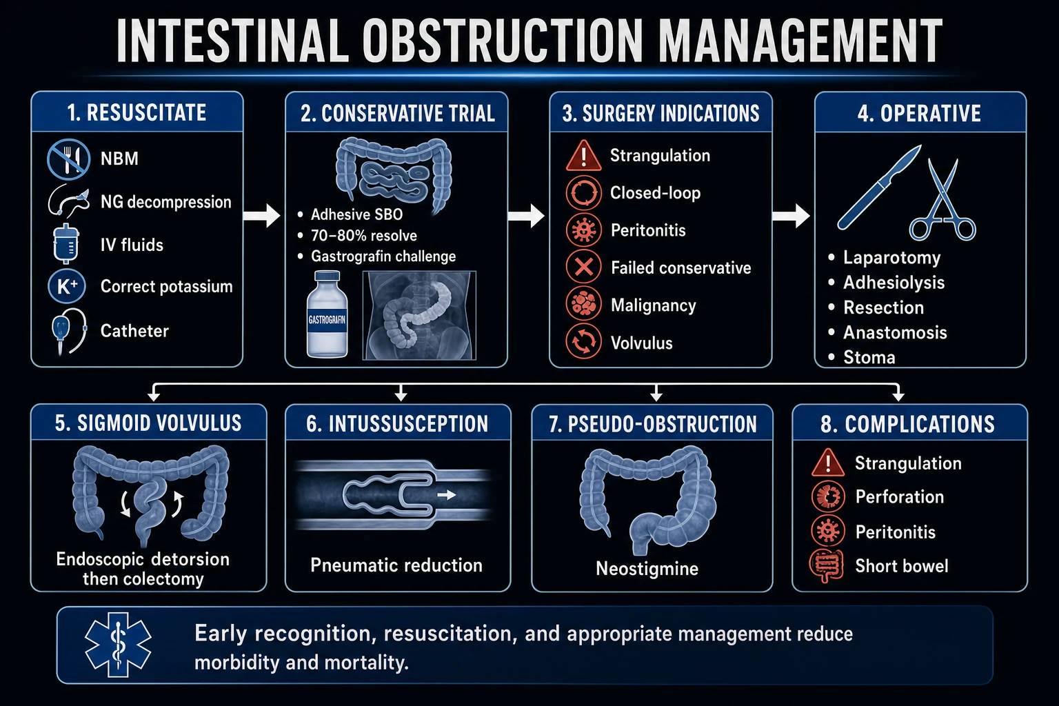
The acute abdominal pain + the distension — the ICU workup
- The ABCs — the airway, the breathing (the oxygen), the circulation (the IV access, the fluids, the vasopressors the MAP > 65).[1]
- The history — the AF, the recent MI, the vasopressors, the dialysis (the AMI); the surgery, the hernia, the malignancy (the obstruction); the post-op, the opioids, the electrolytes (the ileus).[1]
- The exam — the pain out of proportion (the AMI); the colicky + the distension (the obstruction); the diffuse, the silent (the ileus); the rigid abdomen (the strangulation/perforation — the emergency surgery).[1]
- The bloods — the lactate (the AMI — the rises late; the trend), the FBC, the CRP, the ABG (the metabolic acidosis), the amylase (the pancreatitis), the beta-HCG (the woman).[1]
- The ECG — the AF (the embolus), the recent MI.[1]
- The imaging — the CT angiography (the arterial phase) if the AMI suspected (the severe pain, the AF, the vasopressors, the rising lactate); the CT abdomen and pelvis (the contrast) for the obstruction/ileus (the transition point; the closed-loop; the pneumatosis).[1]
- The referral — the vascular surgery (the AMI), the general surgery (the obstruction, the strangulation, the perforation). The time is the bowel.[1]
The acute mesenteric ischaemia — the management algorithm
- The resuscitation — the ABCs, the oxygen, the IV fluids (the balanced crystalloid — the MAP > 65, the lactate clearance), the NGT, the broad-spectrum antibiotics.[1]
- The anticoagulation — the unfractionated heparin IV (the immediate — the prevent the propagation, the preserve the collateral).[1]
- The CTA — the arterial phase — the occlusion (the embolus the convex; the thrombus the tapered; the MVT the venous; the NOMI the diffuse narrowing).[5]
- The vascular/IR referral — the IMMEDIATE. The laparotomy for the peritonitis/pneumoperitoneum/infarction.[1]
- The revascularisation — the open (the embolectomy — the embolus; the bypass/endarterectomy — the thrombus) or the endovascular (the thrombectomy/thrombolysis/stent — the no peritonitis). The papaverine for the NOMI/vasospasm.[1]
- The laparotomy — the resection of the necrotic; the stoma in the unstable. The second-look (the 24 to 48 hours) for the marginal bowel.[1]
- The post-op — the heparin then the warfarin/DOAC; the ICU; the multi-organ failure the watch; the early nutrition (the enteral the preferred).[1]
The Ogilvie syndrome (ACPO) — the stepwise management
- The conservative (the 48 to 72 hours) — the NPO, the NGT, the rectal tube, the correct the electrolytes (the K, the Mg, the Ca), the stop the offending drugs (the opioids, the anticholinergics), the ambulation/position change.[2]
- The re-assess the diameter — the caecum < 12 cm and the no ischaemia then the continue the conservative. The caecum > 12 cm for > 6 days, the > 14 cm, or the no response then the escalate.[2]
- The neostigmine 2 mg IV (the cardiac monitor, the atropine the standby, the bedbound 15 minutes) — for the caecum > 10 cm and the no improvement. The exclude the mechanical obstruction (the CT) first.[1]
- The colonoscopic decompression — for the neostigmine failure or the contraindicated. The gentle, the minimal insufflation, the tube decompression.[2]
- The surgery — the caecostomy (the percutaneous, the surgical) or the resection — the last resort, for the ischaemia, the perforation, the peritonitis.[2]
The adhesive small bowel obstruction — the Bologna pathway
- The resuscitation — the "drip and suck" (the IV fluids, the NGT decompression), the catheter, the correct the electrolytes.[3]
- The CT — the transition point; the exclude the strangulation/closed-loop (the no conservative trial for these — the surgery).[3]
- The conservative trial (the 72 hours) — for the uncomplicated adhesive SBO. The water-soluble contrast (the gastrografin) at the 48 hours if the no resolution — the diagnostic AND the therapeutic.[4]
- The re-assess — the contrast to the colon within the 24 hours then the high likelihood of the resolution then the discharge home. The no passage / the pain / the peritonism then the surgery.[3]
- The surgery — the adhesiolysis — for the failed conservative, the signs of the strangulation, the peritonitis, the closed-loop on the imaging.[3]
The landmark trials and the guidelines
The Ponec 1999 NEJM — the neostigmine for the acute colonic pseudo-obstruction (the Ogilvie)
The design: the randomised, the double-blind, the placebo-controlled; the single-centre; the 21 patients with the ACPO (the caecum > 10 cm, the no improvement after the conservative). The intervention: the neostigmine 2 mg IV vs the saline placebo. The outcome: the clinical decompression (the passage of the flatus/stool, the reduction of the abdominal girth) within the 3 hours. The 91 % (10 of 11) of the neostigmine vs the 0 % (0 of 10) of the placebo (p < 0.001). The median time to the response ~4 minutes. The adverse: the bradycardia (the 2 — the atropine), the bronchospasm, the hypotension, the salivation, the cramping. The significance: the landmark — the established the neostigmine the first-line pharmacotherapy for the ACPO. The subsequent systematic review (Saunders 2005) and the meta-analysis (Demetriou 2025) the confirmed.[1]
The Bologna 2017 guidelines (the WSES) — the adhesive small bowel obstruction (ASBO)
The authority: the World Society of Emergency Surgery (WSES) — the ASBO working group — the evidence-based, the updated from the 2010 and the 2013. The key recommendations: the conservative trial (the 72 hours) for the uncomplicated adhesive SBO; the water-soluble contrast (the gastrografin) at the 48 hours — the diagnostic (the passage the high NPV) and the therapeutic (the osmotic effect); the early surgery for the strangulation, the peritonitis, the closed-loop; the laparoscopy the option for the experienced (the no dense adhesions, the no peritonitis). The risk factors for the strangulation — the persistent pain, the fever, the tachycardia, the raised lactate, the WCC, the peritonism.[3]
The Branco 2010 meta-analysis — the water-soluble contrast in the adhesive SBO
The design: the systematic review and the meta-analysis; the 14 studies; the 1 025 patients. The findings: the water-soluble contrast the reduced the need for the surgery (OR 0.62) and the shortened the hospital stay (~3 days). The resolution of the obstruction in the ~60 to 80 % of the patients after the contrast. The contrast to the colon within the 24 hours — the ~97 % sensitivity, the ~96 % specificity for the resolution. The significance: the established the water-soluble contrast the diagnostic AND the therapeutic in the adhesive SBO — the cornerstone of the Bologna pathway.[4]
The Menke 2010 meta-analysis — the CT in the acute mesenteric ischaemia
The design: the systematic review and the meta-analysis; the 9 studies; the 483 patients. The findings: the multidetector CT angiography — the ~93 % sensitivity, the ~96 % specificity for the acute mesenteric ischaemia. The high diagnostic accuracy. The CT angiography the first-line, the gold standard. The significance: the established the CTA the diagnostic test of the choice for the AMI — the immediate, the high accuracy, the no time for the conventional angiography.[5]
The Cudnik 2013 meta-analysis — the diagnosis of the AMI (the lactate, the D-dimer)
The design: the systematic review and the meta-analysis of the diagnostic biomarkers for the AMI. The findings: the D-dimer the high sensitivity (~96 %) — the high NPV (the rule-out in the low-risk); the low specificity. The lactate the ~76 % sensitivity, the ~93 % specificity for the infarction — the rises late (the translocation). The leucocytosis the sensitive but the non-specific. The significance: the D-dimer the rule-out; the lactate the late/poor-prognosis; the diagnosis is the clinical + the CTA — the biomarkers the adjunct.[6]
The ACG 2015 guideline — the colon ischaemia (the ischaemic colitis)
The authority: the American College of Gastroenterology — the Brandt, Feuerstadt, Longstreth. The key points: the colon ischaemia the most common form of the intestinal ischaemia; the splenic flexure (the Griffith's point) the commonest site; the bloody diarrhoea the hallmark; the CT and the colonoscopy (the gentle, the minimal insufflation) the diagnostic; the supportive the management for the majority (the antibiotics, the bowel rest, the fluids); the surgery for the gangrene, the fulminant colitis, the stricture. The medications (the laxatives, the NSAIDs, the digoxin) the precipitant.[7]
Red flags
References
- [1]Ponec LA, Saunders MD, Kimmey MB Neostigmine for the treatment of acute colonic pseudo-obstruction N Engl J Med, 1999.PMID 10403850
- [2]Saunders MD, Kimmey MB Systematic review: acute colonic pseudo-obstruction Aliment Pharmacol Ther, 2005.PMID 16268965
- [3]Ten Broek RPG, Krielen P, Di Saverio S, et al Bologna guidelines for diagnosis and management of adhesive small bowel obstruction (ASBO): 2017 update of the evidence-based guidelines from the world society of emergency surgery ASBO working group World J Emerg Surg, 2018.PMID 29946347
- [4]Branco BC, Barmparas G, Schnuriger B, et al Systematic review and meta-analysis of the diagnostic and therapeutic role of water-soluble contrast agent in adhesive small bowel obstruction Br J Surg, 2010.PMID 20205228
- [5]Menke J Diagnostic accuracy of multidetector CT in acute mesenteric ischemia: systematic review and meta-analysis Radiology, 2010.PMID 20574087
- [6]Cudnik MT, Darbha S, Jones J, et al The diagnosis of acute mesenteric ischemia: A systematic review and meta-analysis Acad Emerg Med, 2013.PMID 24238311
- [7]Brandt LJ, Feuerstadt P, Longstreth GF, et al ACG clinical guideline: epidemiology, risk factors, patterns of presentation, diagnosis, and management of colon ischemia (CI) Am J Gastroenterol, 2015.PMID 25559486
- [8]Feuerstadt P, Brandt LJ Update on Colon Ischemia: Recent Insights and Advances Curr Gastroenterol Rep, 2015.PMID 26446556
- [9]Vodusek Z, Feuerstadt P, Brandt LJ Review article: the pharmacological causes of colon ischaemia Aliment Pharmacol Ther, 2019.PMID 30467871
- [10]Demetriou G, Ghoneim A, Dattani M, et al Neostigmine Versus Colonoscopic Decompression for Acute Colonic Pseudo-Obstruction Not Resolving With Conservative Measures: A Meta-Analysis J Surg Res, 2025.PMID 40664115