ICU · GI & nutrition / surgical
Intra-abdominal Hypertension & Abdominal Compartment Syndrome — IAP, APP & Decompression
Also known as Intra-abdominal hypertension · IAH · Abdominal compartment syndrome · ACS · Intra-abdominal pressure · IAP · Abdominal perfusion pressure · APP · WSACS · Bladder pressure · Decompressive laparotomy · Open abdomen
The intra-abdominal hypertension (IAH) and the abdominal compartment syndrome (ACS) are the raised intra-abdominal pressure (IAP) and its consequences. The WSACS definitions: the IAP measured via the bladder (the 25 mL saline, the end-expiration, the supine); the IAH is the IAP over 12 mmHg (the grades I to IV); the abdominal perfusion pressure (the APP) equals the MAP minus the IAP, the target over 60; the ACS is the IAP over 20 mmHg PLUS the new organ failure. The organ effects: the reduced venous return and cardiac output, the raised airway pressure, the oliguria (the kidney compression), the splanchnic ischaemia, the raised ICP. The primary ACS (the abdominopelvic cause) vs the secondary (the sepsis, the massive fluid resuscitation, the burns, the capillary leak). The management: the measurement in the at-risk, the medical (the sedation, the neuromuscular blockade, the NGT, the paracentesis, the fluid balance, the APP over 60), the surgical decompressive laparotomy plus the open abdomen for the ACS.
On this page & tools
Your progress
Saved locally on this device.
8 MCQs with explanations
Target exams
Overview & definition
The intra-abdominal hypertension (IAH) and the abdominal compartment syndrome (ACS) are the raised intra-abdominal pressure (IAP) and its consequences. The raised IAP compresses the intra-abdominal vessels and the organs → the multi-organ failure (the cardiac, the respiratory, the renal, the splanchnic, the neurological). The ACS is the surgical emergency — the decompression reverses the organ failure. The condition is under-recognised — the measurement of the IAP in the at-risk patient is the key.[1]
The abdominal cavity is a relatively fixed, semi-rigid compartment bounded by the spine and paravertebral muscles posteriorly, the costal margin and diaphragm superiorly, and the abdominal musculature and fascia anterolaterally. Its contents (the bowel, the solid organs, the mesentery, the aorta, the IVC, the spleen, the liver, the kidneys) share a common pressure. Once the volume of the contents rises beyond the capacity of the wall to distend — the compliance exhausted — the pressure climbs steeply and the consequences cascade. The pressure-volume curve of the abdomen is non-linear: a long flat limb (compliant) followed by a steep ascending limb where small additions of volume (more oedema, more gas, more fluid) produce large rises in pressure.[1][2]
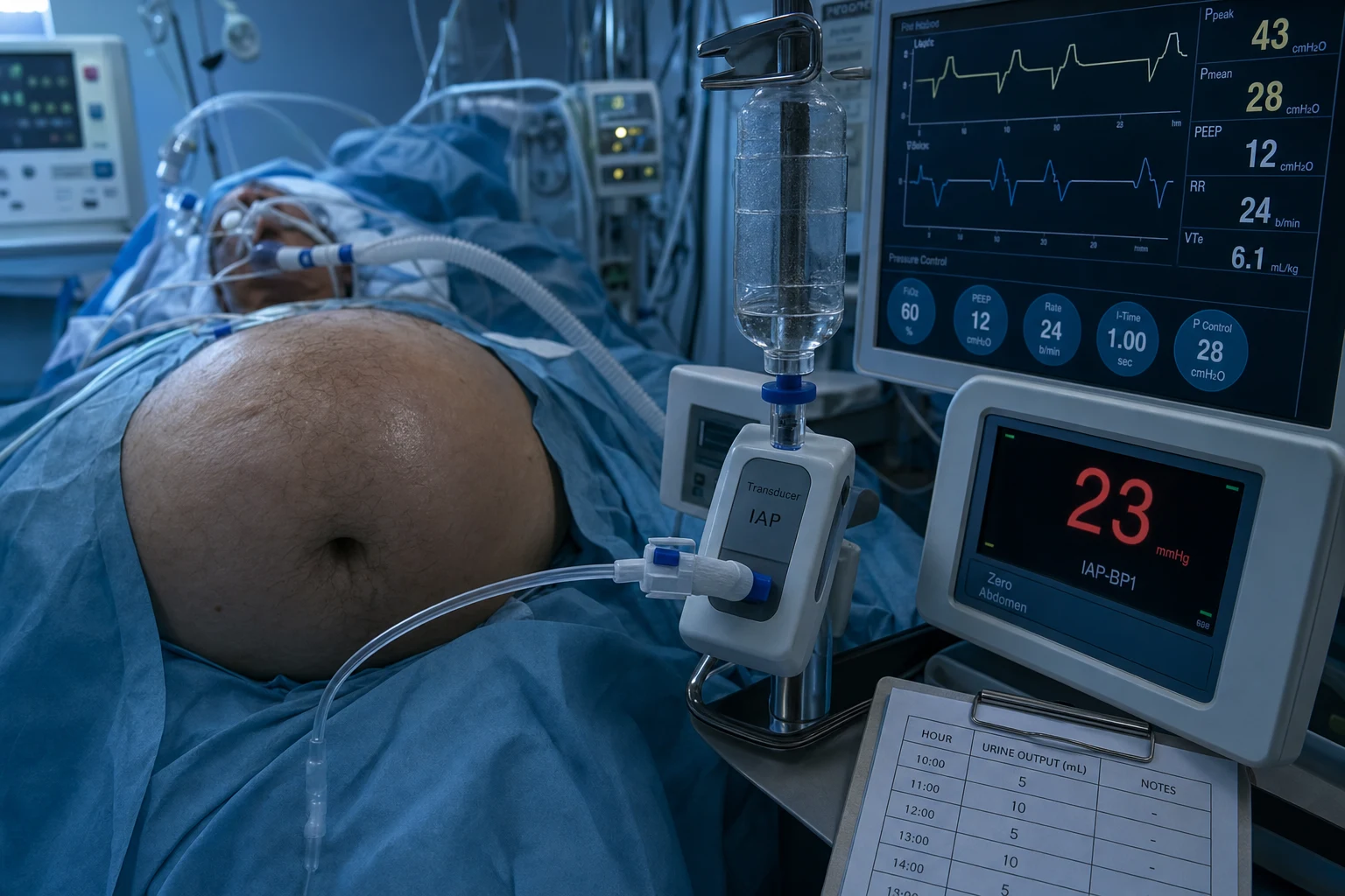
The WSACS definitions
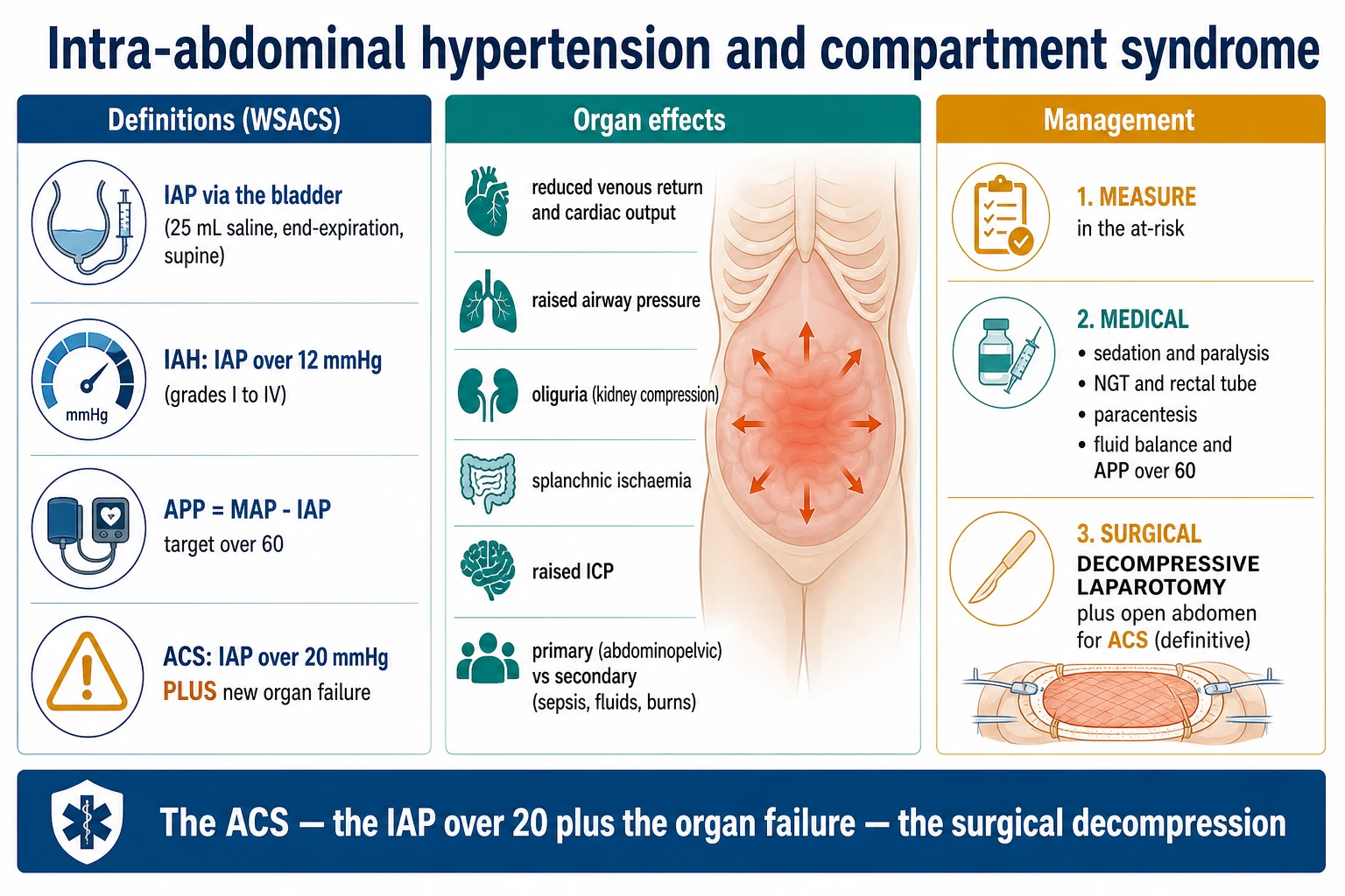
The World Society of the Abdominal Compartment Syndrome (WSACS) published its consensus definitions in 2006 and updated them in 2013; these remain the global standard and are what examiners expect you to quote.[1][14]
- The intra-abdominal pressure (IAP) — the steady-state pressure concealed within the abdominal cavity. The measured via the bladder (the transvesical) — the gold standard, after the instillation of the 25 mL of the sterile saline, at the end-expiration, the supine. The normal IAP in the healthy adult is 5 to 7 mmHg (slightly higher in the obese — 8 to 12 mmHg in the morbidly obese — and in the third trimester of pregnancy).[1][1]
- The intra-abdominal hypertension (IAH) — a sustained or repeated pathologic elevation of the IAP ≥ 12 mmHg (recorded by a minimum of two standardised measurements 1–6 hours apart). The graded:[1]
- The grade I: 12 to 15 mmHg.
- The grade II: 16 to 20 mmHg.
- The grade III: 21 to 25 mmHg.
- The grade IV: over 25 mmHg.
- The abdominal perfusion pressure (APP) = the MAP minus the IAP. The target ≥ 60 mmHg (the analogous to the cerebral perfusion pressure). A combined endpoint that pairs perfusion driving-pressure against the back-pressure squeezing the organs; it is a better resuscitation target than the MAP alone.[1][7]
- The abdominal compartment syndrome (ACS) — a sustained IAP > 20 mmHg (with or without an APP < 60 mmHg) PLUS the new-onset organ failure. The two-part definition (pressure threshold AND a new organ dysfunction) is the sine qua non — the high IAP alone, without organ failure, is the IAH and is NOT the ACS.[1][10]
- The primary ACS — from the abdominopelvic injury or the disease (the trauma, the pancreatitis, the rupture, the post-op bleeding, the ruptured AAA, the pelvic fracture). Usually requires the surgical or the radiological intervention in the abdominopelvic region.[1]
- The secondary ACS — from the conditions NOT in the region (the sepsis, the massive fluid resuscitation, the burns, the capillary leak). The over-resuscitation is the classic iatrogenic cause.[1][9]
- The recurrent ACS — the redevelopment of the ACS after the surgical or the medical decompression, or in the patient with an open abdomen. A separate category because it carries a high mortality and demands a different response (re-exploration, the escalating temporary closure).[1][8]
The IAH grades and the threshold physiology
The WSACS grade is more than a label — each band maps to a physiological transition and a management step: [1]
- Grade I (12–15 mmHg) — the early, often clinically silent IAH. The oliguria threshold is typically crossed around 15 mmHg; the renal venous outflow begins to be impeded. The management is the medical ladder (the sedation, the NGT, the fluid stewardship, the body positioning).[2]
- Grade II (16–20 mmHg) — the organ effects become measurable: the reduced urine output, the rising peak airway pressure, the fall in the cardiac output, the falling APP. The aggressive medical management; the consideration of the percutaneous drainage or the neuromuscular blockade. The close monitoring.[2][4]
- Grade III (21–25 mmHg) — the overt organ failure territory; the boundary with the ACS. The surgical decompression is the likely definitive; the open abdomen is anticipated.[8]
- Grade IV (over 25 mmHg) — the fulminant ACS; the dramatic multi-organ failure; the emergency decompressive laparotomy is the standard of care; the mortality is the high without it.[8][10]
The pathophysiology — the pressure-volume relationship and the organ cascade
The abdomen behaves as a non-linear compartment. On the flat limb of the compliance curve the wall distends and accommodates additional volume (the gas, the fluid, the oedema) with little change in the pressure. Once the elastic limit of the abdominal wall and the diaphragm is reached, the curve turns steep: each additional increment of volume (a further litre of resuscitation fluid, another loop of distended bowel) produces a disproportionate rise in IAP. The patient with the reduced abdominal-wall compliance (the obesity, the abdominal surgery, the prone position, the burns eschar, the tight fascial closure) reaches the steep limb earlier.[1][2]
The consequences are transmitted by two mechanisms: (1) direct mechanical compression of the intra-abdominal vessels and viscera, and (2) transmission of the raised intra-thoracic pressure (the diaphragm is pushed cephalad, the intra-thoracic pressure rises, and the consequences ripple through the heart, the lungs, and the brain). The cascade is self-amplifying: the raised IAP reduces the renal perfusion → the oliguria and the fluid retention → the bowel-wall oedema → the further rise in IAP. Breaking the cycle (the decompression, the fluid removal, the drainage) is the central therapeutic principle.[1][7]
The organ effects
The raised IAP affects the every organ system. The thresholds are approximate but useful and are quoted in the WSACS guidelines:[1][7]
- The cardiovascular — the compressed IVC, the reduced venous return, the reduced cardiac output, the raised CVP (the falsely raised — the central venous pressure reading ceases to reflect the true right-sided filling because the raised intra-thoracic pressure is transmitted to the intrathoracic great vessels). The compression of the arterial bed raises the systemic vascular resistance. The cardiac output falls; the wedge pressure rises; the indices of preload (the CVP, the PAOP) become unreliable. The hypotension. The functional hypovolaemia despite the apparent high filling pressures.[1][7]
- The respiratory — the raised diaphragm, the reduced thoracic compliance, the raised peak and plateau airway pressures, the atelectasis (the basilar collapse), the hypoxia, the hypercapnia (the dead space rises — the compression of the pulmonary capillaries), the right-heart strain (the raised pulmonary vascular resistance), and the precipitation or the worsening of the ARDS. The intra-thoracic pressure rises in parallel with the IAP.[1][7]
- The renal — the kidney and the renal vein compression (the renal vein outflow obstruction, the renal interstitial pressure rises, the glomerular filtration falls), the oliguria (typically at the IAP over 15 mmHg), the anuria (over 30 mmHg). The renin-angiotensin-aldosterone and the sympathetic activation. The AKI. The compression of the renal capsule and the renal veins is the dominant mechanism — the renal arterial inflow is relatively preserved until the very late stages. The low urine output in the well-resuscitated patient with the high airway pressure = the measure the IAP.[1][13]
- The splanchnic / hepatic / gut — the mesenteric and the hepatic ischaemia, the reduced hepatic blood flow, the impaired clearance of the lactate, the bacterial translocation (the mucosal ischaemia and the barrier failure), the promotion of the sepsis and the multi-organ failure. The hepatic arterial buffer response is overwhelmed. The gut is the organ that suffers earliest and longest, and the ischaemic mucosa drives the systemic inflammatory response.[1][2]
- The neurological — the raised ICP and the reduced cerebral perfusion pressure. The mechanism: the raised intra-thoracic pressure impairs the venous drainage from the brain (the jugular venous pressure rises), and the raised central venous pressure is transmitted to the intracranial venous compartment. The intra-abdominal hypertension is a recognised and reversible cause of the raised ICP in the trauma and the critically ill patient — the decompression can lower the ICP.[1]
- The abdominal wall — the reduced compliance (a vicious cycle), the impaired perfusion of the wound (the dehiscence, the infection, the hernia). The raised IAP reduces the rectus sheath and the abdominal-wall blood flow, impairing the healing and increasing the surgical-site infection.[1]
The risk factors — the four WSACS pathophysiological categories
The WSACS classifies the risk factors by mechanism — a structure that maps directly onto the medical management. The at-risk patient has typically more than one category active simultaneously.[1][2]
1. Decreased abdominal wall compliance
Anything that stiffens the abdominal wall or restricts its outward expansion:
- The obesity (the BMI over 30 — a chronic, low-grade IAH baseline of 8 to 12 mmHg in the morbidly obese).[3]
- The abdominal surgery (the tight fascial closure, the high-tension anastomosis, the packed abdomen, the abdominal wall oedema).
- The prone positioning (the patient on the abdomen — the abdomen splinted against the bed; a 5–10 mmHg rise in the IAP is typical).[11]
- The head-of-bed elevation over 30° (a 1 mmHg rise in IAP for each 10° of elevation — the practical trade-off with the VAP prophylaxis).[11]
- The burns eschar (the circumferential truncal burn — the inelastic eschar splints the wall; the escharotomy is the treatment).
- The abdominal binding (the tight binder, the military anti-shock garment).
2. Increased intraluminal contents
Gastrointestinal gas, fluid, or stool load:
- The gastroparesis, the gastric distension, the ileus, the pseudo-obstruction (Ogilvie).[2]
- The colonic pseudo-obstruction; the megacolon.
- The enteral feeding intolerance (the high gastric residual volume).
- The volvulus, the mechanical bowel obstruction.
3. Increased intra-abdominal cavity contents (capillary leak / third-space / collections)
The category that drives the secondary ACS — the classic iatrogenic form:[9]
- The massive fluid resuscitation (over 5 L of crystalloid in 24 hours; the over-resuscitation in the septic shock, the haemorrhagic shock, the burns, the pancreatitis).[9][12]
- The capillary leak syndrome (the sepsis, the burns, the pancreatitis, the major trauma, the anaphylaxis).
- The ascites, the haemoperitoneum, the haemo-ascites.
- The intra-abdominal collections (the abscess, the biloma, the pseudocyst, the urinoma).
- The bowel oedema (the ischaemia, the reperfusion, the post-operative).
- The peritonitis (the diffuse inflammatory third-space loss).
4. Miscellaneous / capillary leak and coagulopathy
The "lethal triad" amplifiers:
- The acidosis (the pH under 7.2), the hypothermia (the core under 35°C), the coagulopathy — each impairs the bowel-wall integrity and the capillary seal, the oedema worsens, the IAP rises.[1]
- The high body-mass index, the large intra-abdominal mass or tumour, the pregnancy (the third trimester), the pneumoperitoneum (the laparoscopy), the peritoneal dialysis.
A useful bedside screen — the patient at risk of IAH/ACS has two or more of: the massive resuscitation, the acidosis, the hypothermia, the coagulopathy, the abdominal distension, the oliguria disproportionate to the haemodynamics, the rising airway pressure, the obesity.[1][10]
The measurement of the IAP
The condition is under-recognised because the IAP is not measured. The index of suspicion plus the standardised measurement is the gateway to the diagnosis and the management. [1]
The transvesical (bladder) measurement — the gold standard
The bladder technique (the Kron 1984 modification of the Iberti method) is the WSACS gold standard — the safe, the cheap, the reproducible, and the bedside. The bladder wall is a passive pressure-transmitting membrane at the volumes used (the bladder acts as a passive diaphragm when the instilled volume is small).[1][13]
The standardised technique (the WSACS protocol):
- The patient supine; the absence of abdominal muscle contraction (the sedation / the analgesia; the neuromuscular blockade if needed).
- The Foley catheter connected to a pressure transducer via a three-way stopcock (or a column of saline, measured in cmH₂O and converted — 1 mmHg = 1.36 cmH₂O).
- The bladder emptied completely first.[13]
- The instillation of 25 mL (max 50 mL) of sterile warmed saline through the catheter into the bladder. The over-filling (over 50 mL) falsely elevates the reading by detrusor contraction — a recognised artefact; the under-filling dampens it.[13]
- The transducer zeroed at the level of the iliac crest (the mid-axillary line in the supine patient) — the symphysis pubis is the anatomical external landmark for the bladder.[1]
- The measurement at the end-expiration (the patient relaxed; the ventilator held at end-expiration if ventilated).
- The reading taken 30–60 seconds after the instillation (to allow the detrusor to relax).
- The average of three readings; the documented value in mmHg.
The pitfalls and the artefacts: the over-filling (the detrusor contraction — a falsely high reading); the under-filling (the damping); the patient contraction (the contraction of the abdominal wall, the cough, the bucking — a falsely high reading); the head-of-bed elevation (a 1 mmHg rise per 10° — measure in the supine for the standard reading, and document the position); the intravesical clot or the bladder tumour or the bladder surgery (the bladder wall no longer a passive membrane — the alternative method is needed).[11][13]
The other methods
- The transgastric (intragastric) — a nasogastric tube with the distal pressure port (the Spiegelberg or the Holtech catheter); the alternative when the bladder is unavailable (the cystectomy, the bladder surgery, the clot). The advantage of the continuous IAP monitoring — useful in the high-risk patient. The accuracy comparable to the bladder at the steady state.[2]
- The direct intra-peritoneal — a needle or a catheter directly into the peritoneal cavity (the Veress needle, the peritoneal dialysis catheter, the laparoscopic port). The invasive; the reference standard but the not-for-routine.
- The inferior vena cava pressure (the femoral venous catheter transduced) — a surrogate; the IVC pressure approximates the IAP below the diaphragm. Used in the research and the rare clinical case.
- The clinical estimation — the abdominal girth, the palpation, the "tense" abdomen. The correlation with the IAP is poor; the clinical estimation alone MISSES the IAH in up to half the cases — the measurement is mandatory.[1]
When to measure and how often
The at-risk patient (the criteria above) should have the baseline IAP on admission and the serial measurement every 4 to 6 hours (the continuous if the high or the rising). The threshold to escalate the monitoring: the IAP over 12 mmHg (start the every-4-hourly measurement and the medical ladder), the rising trend (the early intervention), the IAP over 20 mmHg with the organ dysfunction (the surgical decompression).[1][10]
The management
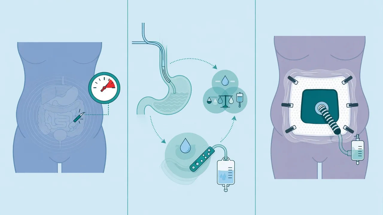
The WSACS 2013 guidelines structure the management into a graduated ladder — five intervention categories, applied in the order of the least-to-the-most invasive, escalated by the grade and the response. The overarching principle: the medical management for the IAH; the surgical decompression for the ACS.[1][2]
1. Measure the IAP in the at-risk and the serial monitoring
The at-risk: the massive fluid resuscitation, the sepsis, the pancreatitis, the peritonitis, the trauma, the burns, the acidosis, the hypothermia, the coagulopathy. The measure the IAP (the bladder) every 4 to 6 hours, or the continuous if the high. The escalation of the monitoring when the IAP rises. The maintenance of the APP over 60.[1]
2. The medical management (the grade I to II IAH; the escalating interventions of the grade III)
The five WSACS intervention categories:[1][2]
a) Improve the abdominal wall compliance:
- The sedation, the analgesia (the reduce the voluntary and the reflex muscle tone).
- The neuromuscular blockade — the short-term (the cisatracurium infusion); the reduces the muscle tone and the IAP by 5–10 mmHg; the trial of the paralysis is reasonable before the surgical decompression in the grade III IAH.[1]
- The body positioning — the supine (the head-of-bed elevation over 30° raises the IAP by ~3–5 mmHg; the prone raises it further); the trade-off with the VAP prophylaxis and the ARDS oxygenation.[11]
- The chemical relaxation of the abdominal wall (the benzodiazepines, the propofol, the dexmedetomidine).
b) Evacuate the intraluminal contents:
- The NGT (the nasogastric decompression of the stomach).
- The rectal tube (the colonic decompression), the enemas, the neostigmine (for the colonic pseudo-obstruction / the Ogilvie — the proven therapy).[2]
- The prokinetics (the metoclopramide, the erythromycin, the alvimopan).
- The reduction of the enteral feeding rate; the change to the post-pyloric feeding.
c) Reduce the intra-abdominal fluid / the percutaneous drainage:
- The paracentesis for the ascites.
- The percutaneous catheter drainage (PCD) of the intra-abdominal collections (the abscess, the haematoma, the pseudocyst) under the ultrasound or the CT guidance. The Cheatham (2011) series showed the PCD alone successfully decompressed 18 of 20 patients with the elevated IAP — a viable alternative to the laparotomy in the selected patient (the salvageable fluid collection, the coagulopathy, the high surgical risk).[5]
- The percutaneous catheter drainage is the recommended first-line intervention for the secondary ACS (the fluid-dominated) — the drains the ascites and the third-space fluid; the avoids the open abdomen.[5][6]
d) Correct the capillary leak and the fluid overload (the fluid stewardship):
- The avoid the over-resuscitation — the primary prevention of the secondary ACS.[9]
- The goal-directed fluid therapy (the balanced crystalloids — the Hartmann, the Plasma-Lyte — over the 0.9% saline; the early colloid — the albumin — for the oedema).[9]
- The aim for the net-zero or the negative fluid balance once the shock resuscitated.
- The diuretics (the furosemide) to mobilise the third-space fluid.
- The renal replacement therapy (the CRRT) for the fluid removal in the oliguric / the AKI patient.
- The avoidance of the iatrogenic fluid load — the judicious maintenance fluids, the concentration of the vasoactive infusions, the avoidance of the unnecessary flushes.[9]
e) Optimise the APP (the target over 60) and the systemic perfusion:
- The maintain the MAP (the vasopressors — the noradrenaline; the inotropes if the cardiac output low) AND the reduce the IAP.
- The APP = the MAP minus the IAP — the resuscitation target that accounts for the raised IAP; a better predictor of the survival than the MAP alone.[7]
- The rising IAP with the maintained MAP drops the APP — the organ perfusion falls even with the "normal" blood pressure. The monitor the APP.
3. The surgical decompression (the grade III to IV IAH / the ACS)
- The surgical decompressive laparotomy — the definitive treatment for the ACS. The immediate decompression reverses the organ failure (the dramatic — the blood pressure and the urine output improve within minutes; the airway pressure falls; the ICP falls). The clinical diagnosis of the ACS (the IAP over 20 plus the organ failure) is the indication; the decompression should NOT be delayed for the further investigation or the "improvement" of the electrolytes.[1][8]
- The open abdomen — the leave the abdomen open (the temporary closure — the negative-pressure dressing — the VAC / the ABThera; the Bogota bag — the sterile IV-fluid bag sutured to the skin; the Wittmann Patch — the Velcro-like closure). The planned re-closure once the swelling resolves (the 5 to 7 days, sometimes longer). The staged re-operation, the gradual approximation of the fascia, the skin grafting over the granulation tissue if the delayed closure fails (the planned ventral hernia and the late repair).[8]
- The avoid the over-resuscitation (the primary prevention of the secondary ACS — the balanced crystalloids, the goal-directed, the permissive hypotension in the trauma, the early damage-control surgery).[9]
The decision to operate is CLINICAL: the high IAP (over 20 mmHg) PLUS the new organ failure (the oliguria unresponsive to the fluids, the rising airway pressure, the worsening metabolic acidosis, the falling cardiac output). The decompressive laparotomy is the life-saving intervention; the delayed decompression increases the mortality. The chestnut: the dramatic improvement after the decompression (the immediate fall of the airway pressure, the return of the urine output, the rise of the cardiac output) is the confirmation of the diagnosis.[8][10]
IAH vs ACS — the two-part WSACS distinction that drives the management
| Feature | Intra-abdominal hypertension (IAH) | Abdominal compartment syndrome (ACS) |
|---|---|---|
| IAP | Sustained ≥ 12 mmHg | Sustained > 20 mmHg |
| Organ failure | Absent (by definition) | New-onset organ failure REQUIRED |
| APP | Often preserved | Often < 60 mmHg |
| Onset | Gradual, may be silent | Acute or acute-on-chronic, dramatic |
| Management | Medical ladder (5 WSACS categories) | Surgical decompressive laparotomy + open abdomen |
| Reversibility | Fully reversible with medical management | Reversible only with decompression |
| Mortality if untreated | Variable; progresses to ACS | Very high (50–80% without decompression) |
Primary vs secondary vs recurrent ACS — the aetiological categories
| Category | Source | Classic causes | First-line definitive treatment |
|---|---|---|---|
| Primary ACS | Abdominopelvic disease or injury; usually needs surgical/radiological intervention in the region | Trauma (blunt/penetrating), ruptured AAA, severe pancreatitis, post-op bleeding, peritonitis, pelvic fracture, delayed abdominal closure | Surgical decompression + source control in the abdomen |
| Secondary ACS | Condition NOT in the abdominopelvic region; the capillary leak / over-resuscitation | Sepsis, massive fluid resuscitation (the iatrogenic form), major burns, anaphylaxis, polytrauma | Fluid stewardship + percutaneous drainage + medical ladder; decompression if organ failure |
| Recurrent ACS | Redevelopment after surgical/medical decompression or in the open abdomen | The re-swelling, the missed collection, the on-going capillary leak | Re-exploration; escalating temporary closure; the cause addressed |
Methods of intra-abdominal pressure measurement
| Method | What is measured | Accuracy | Practical role |
|---|---|---|---|
| Transvesical (bladder) — gold standard | The bladder pressure after 25 mL saline instillation, end-expiration, supine, transducer at iliac crest | Excellent; correlates with direct intraperitoneal | The routine ICU measurement; the WSACS standard |
| Transgastric (intragastric) | The intragastric pressure via the NG tube with a pressure port | Comparable to bladder at steady state | The continuous monitoring; the alternative when the bladder unavailable |
| Direct intraperitoneal | A needle/catheter directly in the peritoneal cavity | The reference standard | The invasive; the research / the rare clinical case |
| Inferior vena cava | The femoral venous catheter transduced | A surrogate (the IVC pressure approximates the IAP below the diaphragm) | The research; the rare clinical case |
| Clinical estimation | The abdominal girth, the palpation, the "tense" abdomen | Poor — misses up to half the cases | NEVER a substitute for the measurement |
The four WSACS risk-factor categories and the matching intervention
| Risk-factor category (mechanism) | Examples | Matching medical intervention |
|---|---|---|
| 1. Decreased abdominal-wall compliance | Obesity, tight fascial closure, prone position, burns eschar, abdominal binding | Sedation, neuromuscular blockade, supine positioning, escharotomy |
| 2. Increased intraluminal contents | Gastroparesis, ileus, pseudo-obstruction (Ogilvie), enteral intolerance, volvulus | NGT, rectal tube, enemas, neostigmine, prokinetics, reduce feeding |
| 3. Increased intra-abdominal cavity contents (capillary leak / collections) | Massive resuscitation, sepsis, burns, ascites, abscess, bowel oedema | Paracentesis, percutaneous drainage, fluid removal (diuretics, CRRT) |
| 4. Amplifiers (acidosis / hypothermia / coagulopathy) | The lethal triad in the major trauma / the post-arrest | Correct the pH, rewarm, correct the coagulopathy |
FlowSteps — the clinical workflows
Standardised transvesical (bladder) IAP measurement — the WSACS protocol
Position and prepare the patient
Supine, relaxed, sedated and free of abdominal muscle contraction. Confirm the Foley catheter is patent and connected. Have a pressure transducer (or a saline column) ready. Document the position (the head-of-bed elevation raises the IAP — measure supine for the standard reading).
Empty the bladder completely
Drain the bladder fully via the catheter. Residual urine detrusor-stretches the wall and distorts the pressure; an under-distended or over-distended bladder both misrepresent the IAP.
Instil 25 mL of sterile warmed saline through the catheter
Maximum 50 mL. Over-filling beyond 50 mL provokes a detrusor contraction and a falsely HIGH reading (Malbrain 2006). Instil slowly to avoid provoking a contraction.
Zero the transducer at the level of the iliac crest
The iliac crest in the mid-axillary line is the zero-reference for the intra-abdominal cavity; the symphysis pubis is the external landmark of the bladder. A wrong zero level is the commonest source of error.
Wait 30–60 seconds for the detrusor to relax, then read at end-expiration
Read the pressure at the end of expiration in the spontaneously breathing patient; in the ventilated patient read at the end-expiratory hold. Average three readings and document the value in mmHg.
Interpret and act
Normal 5–7 mmHg; IAH ≥ 12 (grade I 12–15, II 16–20, III 21–25, IV >25); ACS if >20 PLUS new organ failure. Calculate APP = MAP − IAP; target ≥ 60 mmHg. Escalate the monitoring and the management accordingly.
The escalating management of IAH — the WSACS ladder
Recognise the at-risk patient and measure the IAP
Two or more of: massive resuscitation (>5 L crystalloid/24 h), sepsis, pancreatitis, peritonitis, trauma, burns, acidosis, hypothermia, coagulopathy, obesity, abdominal surgery, prone position. Baseline IAP on admission; serial every 4–6 h.
Grade I–II IAH (12–20 mmHg) — the medical ladder
Start all five WSACS categories: (a) sedation/analgesia, supine positioning; (b) NGT, rectal tube, prokinetics; (c) paracentesis / percutaneous drainage of any collection; (d) fluid stewardship — net-negative balance, diuretics, CRRT for fluid removal; (e) maintain APP ≥ 60 with vasopressors.
Grade III IAH (21–25 mmHg) — escalate before surgery
Add a trial of neuromuscular blockade (cisatracurium infusion — may drop IAP by 5–10 mmHg); aggressive percutaneous drainage if a collection is present; consider the body-position changes; the continuous IAP monitoring. Re-measure the APP and the organ function — the goal is to avert the surgical decompression.
ACS (IAP > 20 mmHg PLUS new organ failure) — surgical decompression
Do NOT delay. The clinical diagnosis (the high IAP plus the new oliguria, the rising airway pressure, the falling cardiac output, the worsening acidosis) is the indication. The decompressive laparotomy is the definitive. The dramatic intra-operative improvement (the fall of the airway pressure, the return of the urine output, the rise of the cardiac output) confirms the diagnosis.
Open abdomen management and the planned re-closure
Temporary closure (the negative-pressure dressing — the ABThera / the VAC; or the Bogota bag). Daily assessment of the swelling; the gradual fascial approximation; the planned re-closure at 5–7 days once the swelling resolves. The skin grafting over the granulation if the delayed closure fails (the planned ventral hernia and the late repair). The nutrition, the fistula prevention, the infection control.
Watch for the recurrent ACS
The redevelopment of the high IAP and the organ failure after the decompression — the re-exploration; the escalating temporary closure; the addressing of the cause (the on-going capillary leak, the missed collection, the on-going bleeding). The recurrent ACS carries a high mortality.
Preventing the secondary ACS — the fluid-stewardship workflow
Recognise the high-risk resuscitation
The septic shock, the haemorrhagic shock, the major burns, the severe pancreatitis — the patients who will receive litres of crystalloid and who are at the highest risk of the capillary leak and the bowel-wall oedema.
Use balanced crystalloids and goal-directed endpoints
Prefer Hartmann or Plasma-Lyte over 0.9% saline (the hyperchloraemic acidosis worsens the capillary leak). Titrate to the dynamic indices of fluid responsiveness (the PLR, the SVV), the lactate clearance, the capillary refill — NOT to a fixed MAP with escalating fluids.
Add early colloid (albumin) for the oedematous patient
The albumin draws the third-space fluid back into the intravascular space; the burns and the sepsis are the classic indications. Avoid the hydroxyethyl starch (the nephrotoxicity).
Switch to net-negative balance once the shock is resuscitated
The furosemide to mobilise the third-space fluid; the CRRT for fluid removal in the oliguric / the AKI patient. The aim for the net-zero or the negative balance from day 2–3.
Measure the IAP from the outset
The baseline on admission; the serial every 4–6 h. The rising trend is the early warning — intervene (the drainage, the diuresis, the paralysis) BEFORE the ACS develops. The fluid strategy is the single most modifiable risk factor.
The evidence and the trials
WSACS 2013 updated consensus definitions and clinical practice guidelines (Kirkpatrick 2013 — PMID 23673399)
Source
Intensive Care Medicine, 2013 — the World Society of the Abdominal Compartment Syndrome consensus, the global standard
Scope
Updated the 2006 definitions; 64 statements (graded by the GRADE methodology) covering the definitions, the measurement, the management, the surgical decompression, and the open abdomen
Key definitions
IAP normal 5–7 mmHg; IAH sustained ≥ 12 mmHg (grades I 12–15, II 16–20, III 21–25, IV >25); ACS sustained IAP > 20 mmHg (with or without APP < 60) PLUS new organ failure; APP = MAP − IAP, target ≥ 60 mmHg
Key management recommendations
Measure the IAP in the at-risk; the medical ladder (the five WSACS intervention categories) for IAH; the surgical decompression for ACS; the percutaneous catheter drainage as the first-line for the fluid-dominated secondary ACS; the open abdomen with the negative-pressure temporary closure
Bottom line
The single document to quote in the exam; the definitions are the global standard and the basis of every IAH/ACS guideline since
Malbrain 2004 — the multicentre prevalence study (PMID 14758472)
Source
Intensive Care Medicine, 2004 — the foundational epidemiology of the IAH in the critically ill
Design
Prospective multicentre study of 265 ICU patients across 14 ICUs; the IAP measured by the standardised bladder technique within 24 h of admission
Key findings
The prevalence of the IAH was over 50% on admission (and over 80% at some point during the ICU stay in the medical and the surgical patients); the IAH was independently associated with the mortality; the under-recognition was the rule — the measurement was the exception
Bottom line
Established the IAH as the common, the under-recognised, and the prognostically important problem in the ICU; the rationale for the routine measurement in the at-risk
Cheatham 2010 — is the evolving management improving survival? (PMID 20095067)
Source
Critical Care Medicine, 2010 — the before-and-after cohort from a single trauma/surgical ICU
Design
Compared the outcomes of 100 patients with ACS managed historically (1997–2002) with 100 patients managed after the institution of the WSACS protocol (2002–2007)
Key findings
The institution of the protocol (the routine IAP measurement, the medical ladder, the earlier decompression) reduced the mortality from 54% to 35%; the survivors had more medical interventions and fewer surgical decompressions; the earlier recognition and the less-invasive interventions improved the survival
Bottom line
The standardised protocol with the routine measurement and the graduated ladder improves the survival; the ACS is now a treatable disease with the protocolised care
Cheatham 2011 — percutaneous catheter decompression (PMID 21903735)
Source
Chest, 2011 — the prospective series establishing the percutaneous drainage as the alternative to the laparotomy
Design
Prospective case series of 19 patients with the elevated IAP and a drainable intra-abdominal collection (the ascites, the haematoma, the abscess, the pseudocyst); the ultrasound-guided percutaneous catheter drainage
Key findings
The drainage reduced the mean IAP from 26 to 14 mmHg immediately; 17 of 19 patients (89%) avoided the laparotomy; the APP rose from 53 to 70 mmHg; the urine output improved within hours
Bottom line
The percutaneous catheter drainage is the effective, the less-invasive alternative to the laparotomy in the patient with a drainable collection — the recommended first-line for the fluid-dominated secondary ACS
Cheatham / De Waele 2009 — the impact of body position on the IAP (PMID 19487946)
Source
Critical Care Medicine, 2009 — the multicentre analysis of the body position and the IAP measurement
Design
Multicentre study; the IAP measured by the bladder technique in the supine and then in the head-of-bed elevation of 30° and 45°
Key findings
The head-of-bed elevation raised the IAP by approximately 1 mmHg for each 10° of elevation (the 30° elevation ≈ +3 mmHg; the 45° ≈ +5 mmHg); the effect was the consistent and the reproducible across the centres
Bottom line
The standard reading is the supine; document the position. The trade-off with the VAP prophylaxis (the head-of-bed 30°) and the ARDS oxygenation (the prone) must be weighed; in the patient with the IAH, the supine and the lower head-of-bed elevation is preferred
Jacobs / Malbrain 2022 — the fluid-management narrative review (PMID 36143427)
Source
Life (Basel), 2022 — the contemporary synthesis of the fluid stewardship and the IAH/ACS
Core thesis
The fluid strategy is the single most modifiable risk factor for the secondary ACS; the over-resuscitation drives the bowel-wall oedema and the capillary leak that raise the IAP
Key recommendations
The balanced crystalloids (the Hartmann, the Plasma-Lyte) over the 0.9% saline (the hyperchloraemic acidosis worsens the leak); the early colloid (the albumin) for the oedematous patient; the avoidance of the hydroxyethyl starch (the nephrotoxicity); the goal-directed endpoints (the dynamic indices, the lactate clearance); the early switch to the net-negative balance (the furosemide, the CRRT)
Bottom line
The prevention of the secondary ACS is the fluid strategy — measure the IAP from the outset and titrate the fluid to the APP ≥ 60, not to a fixed MAP
Björck 2014 — the management of ACS and the open abdomen (PMID 24447530)
Source
European Journal of Vascular and Endovascular Surgery, 2014 — the ESVS guidelines on the open abdomen
Scope
The classification of the open abdomen (the Björck grades 1–4 — the no-fixation to the enteroatmospheric fistula); the temporary closure techniques (the negative-pressure dressing, the Bogota bag, the Wittmann Patch); the planned re-closure; the management of the enteroatmospheric fistula
Key points
The negative-pressure dressing (the VAC, the ABThera) is the preferred temporary closure — it removes the third-space fluid, the splints the wall, and the facilitates the delayed fascial closure; the early closure (within 7–10 days) reduces the morbidity; the enteroatmospheric fistula is the feared complication (the Björck grade 4)
Bottom line
The open abdomen is a managed state, not a failure — the protocolised temporary closure, the planned re-closure, and the fistula prevention are the components of the modern care
Strang 2014 — the systematic review of the IAP in the severely burned (PMID 24050978)
Source
Burns, 2014 — the systematic review of the IAH/ACS in the major burns
Findings
The IAH is common in the severely burned (the prevalence 50–70% in the >20% TBSA); the ACS develops in 5–20%; the risk factors are the large TBSA, the inhalation injury, the high-volume resuscitation (the fluid creep), the delayed resuscitation, the electrical injury
Bottom line
The burns are a classic cause of the secondary ACS; the fluid stewardship (the guideline-adherent Parkland, the early colloid) is the primary prevention; the measurement of the IAP is the part of the major-burn monitoring
Malbrain 2006 — the effect of the bladder volume on the IAP (PMID 16934130)
Source
Critical Care, 2006 — the prospective cohort on the bladder-instillation volume and the IAP accuracy
Question
How much saline should be instilled into the bladder for the accurate IAP measurement?
Findings
The instillation of 50 mL or less gave the accurate, reproducible measurement (the bladder wall acts as a passive membrane); the volumes over 50 mL provoked a detrusor contraction and a falsely HIGH reading; the under-filling (under 25 mL) damped the reading
Bottom line
The WSACS standard of 25 mL (max 50 mL) of the instillation is the evidence-based; the over-filling is the commonest source of the false-high reading
Clinical pearls — the high-yield points for the CICM / FFICM / EDIC viva
[1]Red flags
The patient with the ascites, the abscess, the haematoma, the drainable collection (the fluid-dominated secondary ACS) — the ultrasound-guided percutaneous catheter drainage first. The Cheatham (2011) series showed 89% of the patients avoided the laparotomy. The laparotomy is reserved for the source control that needs the surgery (the perforation, the ischaemia, the on-going bleeding) and for the failure of the drainage. The over-resuscitation, the capillary leak, the diffuse bowel oedema WITHOUT a focal collection — the medical ladder (the paralysis, the fluid removal, the CRRT) and the surgical decompression if the organ failure.[5][6]
SAQ — Severe acute pancreatitis with oliguria and rising airway pressures
10 minutes · 10 marks
A 54-year-old man with alcohol-related severe acute pancreatitis (modified Marshall score 4) is on day 3 of ICU care. He has received 8 L of crystalloid for his initial resuscitation. He is now oliguric (10 mL/h), his noradrenaline requirement has risen from 0.1 to 0.4 µg/kg/min, his peak airway pressure has climbed from 28 to 42 cmH₂O and his abdomen is visibly distended and tense. His serum creatinine has doubled to 220 µmol/L and his base deficit is 8 mmol/L.
SAQ — WSACS grading and resuscitation target calculation
10 minutes · 10 marks
A 38-year-old woman is 6 days post-emergent damage-control laparotomy for a Grade IV liver injury with massive transfusion. She is in ICU on 12 cmH₂O of PEEP, ventilated for ARDS. She has received 14 L of crystalloid and 6 units of packed red cells since admission. Her MAP is 64 mmHg on noradrenaline 0.25 µg/kg/min. Her transvesical IAP is measured as 22 mmHg. Her urine output over the last 4 hours is 8 mL/h. Her peak inspiratory pressure has risen from 32 to 45 cmH₂O. Her base deficit is 6 mmol/L and serum creatinine is 180 µmol/L.
References
- [1]Kirkpatrick AW, Roberts DJ, De Waele J, et al Intra-abdominal hypertension and the abdominal compartment syndrome: updated consensus definitions and clinical practice guidelines from the World Society of the Abdominal Compartment Syndrome Intensive Care Med, 2013.PMID 23673399
- [2]Malbrain ML, De Laet IE Intra-abdominal hypertension: definitions, monitoring, interpretation and management Best Pract Res Clin Anaesthesiol, 2013.PMID 24012236
- [3]Malbrain ML, Chiumello D, Pelosi P, et al Prevalence of intra-abdominal hypertension in critically ill patients: a multicentre epidemiological study Intensive Care Med, 2004.PMID 14758472
- [4]Cheatham ML, Safcsak K Is the evolving management of intra-abdominal hypertension and abdominal compartment syndrome improving survival? Crit Care Med, 2010.PMID 20095067
- [5]Cheatham ML, Safcsak K Percutaneous catheter decompression in the treatment of elevated intraabdominal pressure Chest, 2011.PMID 21903735
- [6]Ouellet JF, Leppaniemi A, De Waele JJ Alternatives to formal abdominal decompression Am Surg, 2011.PMID 21944453
- [7]Malbrain ML, Ameloot K, De Waele JJ, De Laet IE Cardiopulmonary monitoring in intra-abdominal hypertension Am Surg, 2011.PMID 21944448
- [8]Bjorck M, Wanhainen A Management of abdominal compartment syndrome and the open abdomen Eur J Vasc Endovasc Surg, 2014.PMID 24447530
- [9]Jacobs R, Wise R, Malbrain MLNG Fluid Management, Intra-Abdominal Hypertension and the Abdominal Compartment Syndrome: A Narrative Review Life (Basel), 2022.PMID 36143427
- [10]Padar M, Reintam Blaser A, Talving P, Starkopf L, Starkopf J Abdominal Compartment Syndrome: Improving Outcomes With A Multidisciplinary Approach - A Narrative Review J Multidiscip Healthc, 2019.PMID 31908470
- [11]Cheatham ML, De Waele JJ, De Laet I, et al The impact of body position on intra-abdominal pressure measurement: a multicenter analysis Crit Care Med, 2009.PMID 19487946
- [12]Strang SG, Van Lieshout EM, Van Waes OJ, Determann RM A systematic review on intra-abdominal pressure in severely burned patients Burns, 2014.PMID 24050978
- [13]Malbrain ML, Deeren DH, De Potter TJ Effect of bladder volume on measured intravesical pressure: a prospective cohort study Crit Care, 2006.PMID 16934130
- [14]Kirkpatrick AW, Roberts DJ, Faris PD, et al Methodological background and strategy for the 2012-2013 updated consensus definitions and clinical practice guidelines from the abdominal compartment society Anaesthesiol Intensive Ther, 2015.PMID 26588481