ICU · GI & nutrition / surgical
Acute lower gastrointestinal bleeding — diverticular, angiodysplasia, colitis and the resuscitate-to-surgery pathway
Also known as Lower GI bleed · LGIB · Hematochezia · Diverticular bleed · Massive rectal bleeding · Angiodysplasia · Ischaemic colitis · Oakland score · CT angiography · Tagged RBC scan · Angioembolisation · Subtotal colectomy · Heyde syndrome · Aortoenteric fistula
Acute lower gastrointestinal bleeding (LGIB) is bleeding arising distal to the ligament of Treitz, presenting most often with hematochezia (bright red blood per rectum) and less commonly with maroon stools or right-sided melena. The dominant causes are diverticular bleeding (40%, the 1 cause — painless, massive, self-limiting), colonic angiodysplasia (20%, painless and recurrent in the elderly), colitis (ischaemic, inflammatory and infectious — ~15%), anorectal sources such as haemorrhoids (~10%) and colorectal malignancy or post-polypectomy (~5%). Roughly 80% stop spontaneously. The management ladder is resuscitation with crystalloid and restrictive transfusion (Hb <70 g/L, or <80 with cardiovascular disease) → exclude an upper GI source (NG aspirate or gastroscopy, because a brisk upper bleed can present as hematochezia) → bowel preparation and colonoscopy within 24 hours (diagnostic and therapeutic — epinephrine injection, endoclips, argon plasma coagulation) → CT angiography (CTA) if bleeding is brisk and ongoing or colonoscopy is non-diagnostic (detects bleeding >0.3-0.5 mL/min) → catheter mesenteric angiography with superselective embolisation if CTA is positive (success 70-90%, ischaemia risk 5-10%) → tagged RBC scintigraphy for slow/intermittent bleeding → and segmental or subtotal colectomy as the last resort for refractory haemorrhage. The Oakland score (threshold <=8) identifies low-risk patients safe for outpatient management. Cause-specific management differs: diverticular (endoclips or APC, often self-limiting), angiodysplasia (APC ablation, consider aortic stenosis / Heyde syndrome), IBD (treat the flare, avoid over-instrumentation), ischaemic colitis (supportive — fluids, antibiotics, no endoscopic therapy unless right-sided/gangrenous).
On this page & tools
Your progress
Saved locally on this device.
Target exams
Red flags
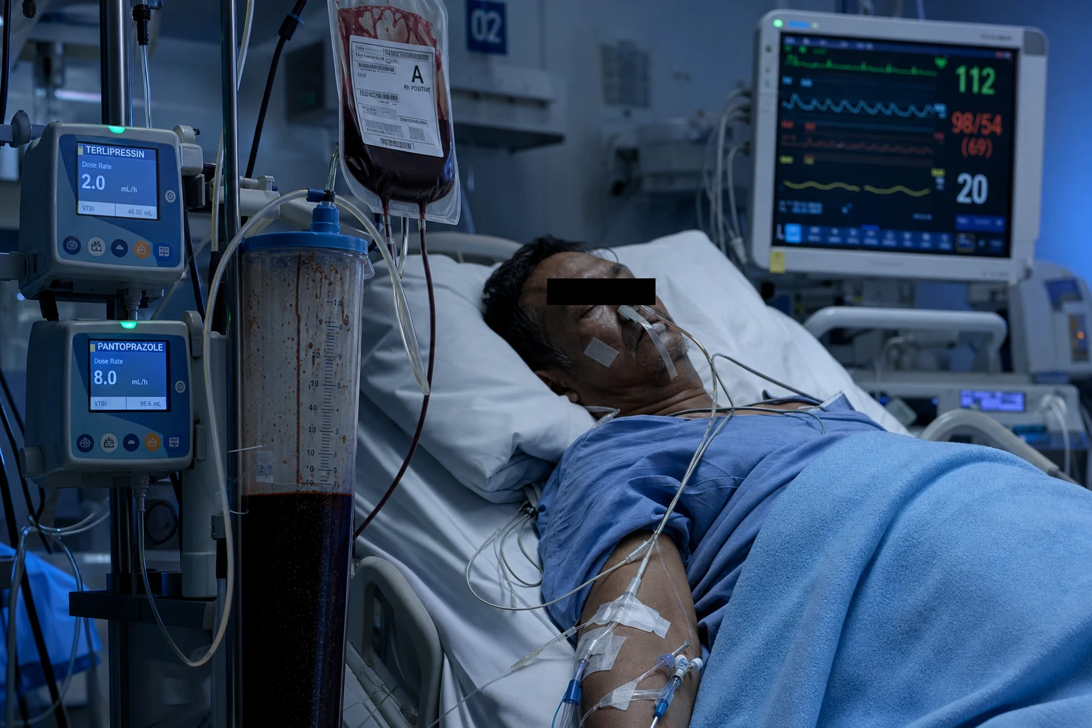
Definition and classification
A lower GI bleed (LGIB) is any haemorrhage arising from a source distal to the ligament of Treitz. It is conventionally divided into upper LGIB (from the colon, the overwhelming majority) and obscure-overt LGIB (persistent visible bleeding in which upper endoscopy and colonoscopy are both non-diagnostic — usually a small-bowel source).[1][5]
The clinical hallmark is hematochezia — the passage of bright red or maroon blood per rectum. Three features at the bedside refine the source: [1]
- Colour and volume — bright red blood suggests a left-colon or anorectal source; maroon or plum-coloured stool points to a right-colon or small-bowel source; melena (black, tarry, foul-smelling) suggests a right-colon or upper GI source.
- The presence of pain — painless massive bleeding favours diverticular disease or angiodysplasia; pain preceding the bleed favours ischaemic colitis, IBD or infection; anal pain on defecation favours a fissure or thrombosed haemorrhoid.
- Haemodynamic compromise with hematochezia — a brisk upper GI bleed (e.g. a spurting duodenal ulcer) with rapid intestinal transit can present as massive hematochezia with shock; always exclude an upper source first.[1][5]
Overt LGIB
Visible bleeding, colonoscopy diagnostic
- Bright red or maroon blood per rectum — the classic presentation
- Source in the colon in >90% of cases (diverticula, angiodysplasia, colitis, cancer)
- Colonoscopy within 24 h is the diagnostic and therapeutic mainstay
- 80% stop spontaneously; ~20% need therapeutic intervention
Obscure-overt
Visible bleeding, OGD + colonoscopy negative
- Persistent visible bleeding with a negative gastroscopy and colonoscopy
- Source is usually in the small bowel (Meckel diverticulum, small-bowel tumour, Crohn, angiodysplasia, AVM)
- Pursue capsule endoscopy, CT/MR enterography, then device-assisted enteroscopy or angiography
- ~5% of all LGIB — the diagnosis is earned after the standard workup is exhausted
Causes
The causes of acute LGIB follow a remarkably consistent distribution across large series, with diverticular disease dominant.[1][5]
Common (80%)
Major causes
- Diverticular bleed (#1 — ~40%): painless, massive, often right colon, 80% self-limiting, 10-40% recurrence
- Colonic angiodysplasia (~20%): painless, recurrent, elderly, often right colon, associated with aortic stenosis (Heyde syndrome)
- Colitis (~15%): ischaemic (elderly, pain BEFORE bleed, left-sided), IBD (ulcerative colitis > Crohn), infectious (Campylobacter, Salmonella, Shigella, E. coli O157:H7, CMV)
- Colorectal cancer / polyps (~10%): occult-to-overt, weight loss, change in bowel habit, iron-deficiency anaemia
- Post-polypectomy bleed (~5-10%): within 1-14 days of a polypectomy (delayed bleed from arterial slough in the resection bed)
Less common (20%)
Other sources
- Haemorrhoids (common but usually small-volume, on the tissue, painless unless thrombosed)
- Anal fissure (painful, small volume, bright red on the paper)
- Radiation proctopathy (history of pelvic radiotherapy — prostate, cervical; telangiectatic, chronic ooze)
- Aortoenteric fistula (rare but FATAL — history of aortic graft; the lethal mimic of LGIB)
- Small-bowel source (Meckel diverticulum in the young; small-bowel tumour; Crohn disease; NSAID enteropathy)
- Institutional / iatrogenic (post-polypectomy, post-colectomy anastomotic, rectal foreign body, trauma)
Aetiology and key numbers (large series)
Epidemiology
LGIB accounts for roughly 20-30% of all major gastrointestinal haemorrhage, with an overall incidence of about 20-30 per 100,000 adults per year rising steeply with age (over 200 per 100,000 in those >80 years).[5] The demographic is distinct from upper GI bleeding: the typical LGIB patient is elderly, comorbid, and on antiplatelet or anticoagulant therapy — which explains both the high prevalence of diverticular/angiodysplastic sources and the elevated bleeding intensity. Hospital mortality is 2-4% overall but reaches 10-20% in the subgroup requiring urgent surgery or interventional radiology, driven by comorbidity rather than exsanguination.[1][5]
Pathophysiology by cause
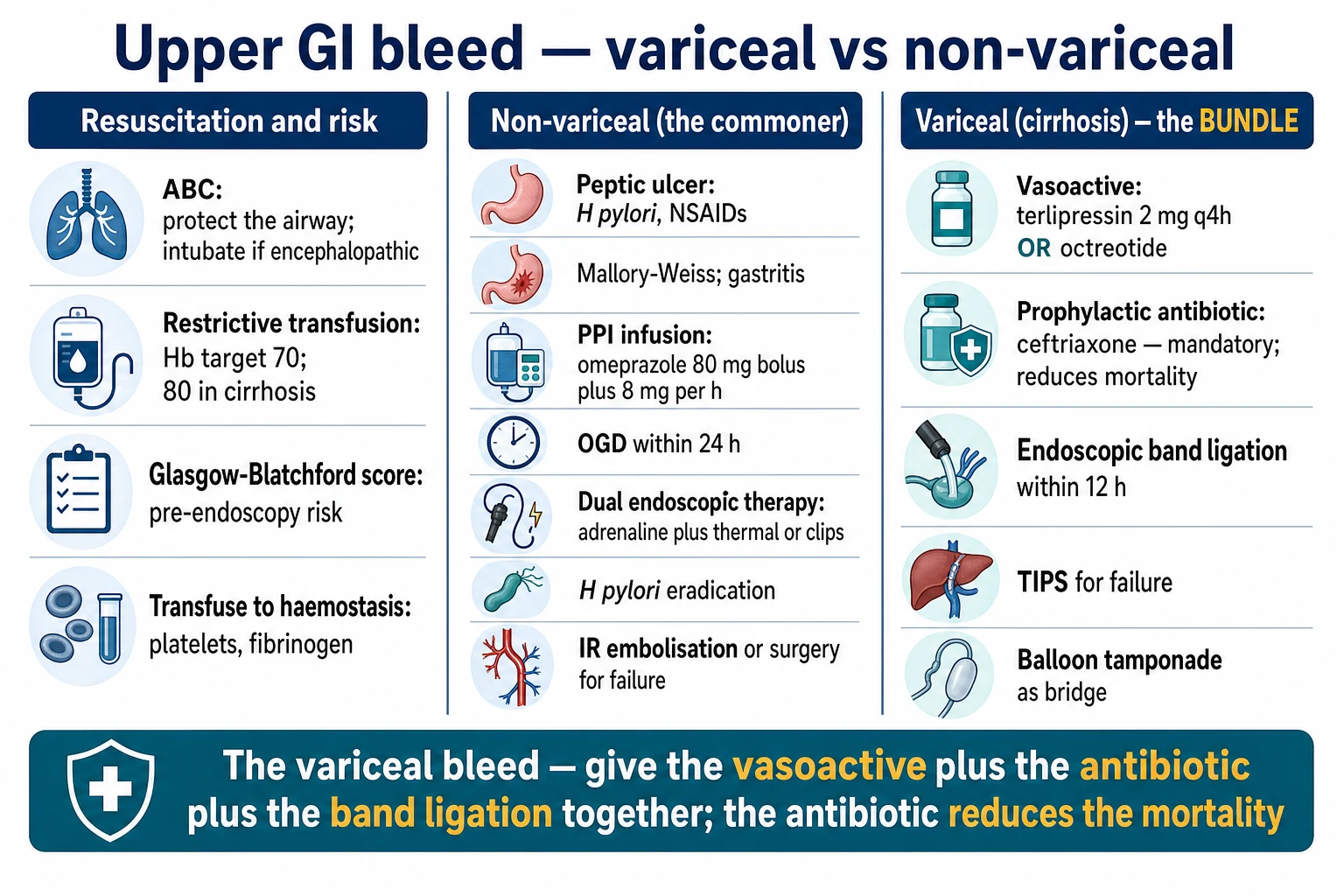
The mechanism of bleeding differs fundamentally between causes and dictates the therapeutic approach. [1]
Diverticular bleeding arises from rupture of a vasa recta artery at the dome of a diverticulum. The vasa recta penetrate the colonic wall obliquely at the points where diverticula herniate, exposing the artery to injury; the bleeding is arterial (hence brisk, painless and often massive) and tends to self-tamponade because the vessel retracts into the pericolic fat. Approximately 50-90% arise from the right colon (right-sided diverticula have wider necks exposing a longer stretch of the vasa recta), and recurrence after a first bleed is 10-40% — recurrent diverticular bleeding is an indication for elective segmental colectomy.[1]
Colonic angiodysplasia (arteriovenous malformation) consists of ectatic, thin-walled submucosal vessels that are prone to rupture. Lesions are typically small (<10 mm), multiple, right-sided and painless, and present with recurrent minor or major bleeds in the elderly. The association with aortic stenosis (Heyde syndrome) reflects an acquired type 2A von Willebrand deficiency — high shear across a stenotic aortic valve proteolyses the largest von Willebrand multimers that normally mediate platelet adhesion under shear in the angiodysplastic vessels; aortic valve replacement can abolish the bleeding.[5]
Ischaemic colitis results from a non-occlusive reduction in mesenteric perfusion (hypotension, dehydration, low-flow state, recent vascular surgery or embolisation) affecting the watershed regions — classically Griffith point (splenic flexure, at the junction of SMA and IMA territories) and Sudeck point (rectosigmoid junction, at the junction of IMA and hypogastric arteries). The bleed reflects mucosal sloughing and is preceded by cramping abdominal pain, then bloody diarrhoea. Right-sided ischaemic colitis (SMA territory) is the high-mortality phenotype, often progressing to transmural gangrene.[5]
Inflammatory bowel disease bleeds from friable, inflamed mucosa — far more often in ulcerative colitis (continuous, distal, mucosal) than Crohn disease (patchy, transmural). Severe acute UC can present with massive colonic haemorrhage as the index presentation of a flare; management combines resuscitation with treatment of the underlying inflammation (IV steroids, cyclosporin/infliximab) rather than endoscopic haemostasis.[5]
Colorectal malignancy bleeds from a friable, ulcerated tumour surface — typically low-volume, chronic, occult, producing iron-deficiency anaemia, but occasionally presenting with overt hematochezia. Post-polypectomy bleeding is arterial, from sloughing of the electrocoagulated pedicle or resection bed, typically 1-14 days after the index procedure.[1]
Clinical presentation and examination
A focused assessment at first contact establishes severity, hints at the source, and excludes mimics.[1][5]
- History — colour/volume/duration of bleeding; associated abdominal pain (and its timing relative to the bleed — pain before favours ischaemic colitis/IBD/infection); prior episodes; anticoagulant/antiplatelet and NSAID use; abdominal or vascular surgery (aortic graft → aortoenteric fistula); pelvic radiotherapy; recent colonoscopy or polypectomy; change in bowel habit/weight loss (malignancy); and comorbidity (renal, cardiac, hepatic).
- Vital signs and shock index — tachycardia, postural drop, hypotension, oliguria, altered mentation. A shock index (HR/SBP) >1 signals haemodynamic compromise.
- Abdominal examination — usually unremarkable in diverticular/angiodysplastic bleeding; tenderness or peritonism favours ischaemia, IBD, infection or perforation; a palpable mass or hepatomegaly raises concern for malignancy; surgical scars and a pulsatile abdominal mass (AAA/aortic graft) are critical.
- Digital rectal examination — assess the character and colour of the blood (bright red vs maroon vs melaena), look for anorectal sources (haemorrhoids, fissure, mass), and palpate for a rectal tumour. Frank melaena on DRE excludes a purely distal colonic source and demands consideration of a right-sided or upper GI bleed.
- Bedside anoscopy/sigmoidoscopy — a rapid bedside rigid sigmoidoscopy can diagnose a distal anorectal source and, if blood is seen above the rectosigmoid, confirms a more proximal colonic bleed. [1]
Differentiation from an upper GI bleed
This is the single most important early fork, because the management pathways diverge. A brisk upper GI bleed (e.g. a posterior duodenal ulcer eroding the gastroduodenal artery) with rapid intestinal transit can present with massive hematochezia and shock — the haematochezia is not proof of a colonic source.[1][5]
Favours upper GI source
Pursue gastroscopy first
- Shock with massive hematochezia (rapid upper transit can mimic a colonic bleed)
- Melaena on rectal exam or a history of melaena/haematemesis
- Raised urea with a NORMAL creatinine (digested blood as protein load)
- Prior peptic ulcer disease, known varices, chronic liver disease, recent NSAID use
- Bile-stained NG aspirate with NO blood → upper source unlikely (high NPV)
- Blood-stained or coffee-ground NG aspirate → upper source confirmed (pursue gastroscopy)
Favours lower GI source
Pursue colonoscopy first
- Bright red blood with stable observations (classic LGIB phenotype)
- Painless massive bleed in a known diverticular patient (right colon)
- Pain preceding the bleed in an elderly patient (ischaemic colitis, left-sided)
- Bile-stained NG aspirate with no blood (effectively excludes an actively bleeding upper source)
- Normal urea with normal creatinine, and a colonoscopy demonstrating a lesion
Risk stratification — the Oakland score
Unlike upper GI bleeding (where the Glasgow-Blatchford and Rockall scores dominate), the validated risk-stratification tool for LGIB is the Oakland score, derived from the UK National Comparative Audit and designed to identify patients at low risk of adverse outcome who are safe for discharge.[3][7]
The Oakland score uses seven variables — age, sex, previous admission for LGIB, digital rectal examination findings (blood), heart rate, systolic blood pressure and haemoglobin — to generate a score from 0 to 35. A threshold of <=8 identifies patients at very low risk of rebleeding, transfusion, intervention, readmission or death, with a sensitivity above 95% for adverse outcomes and a C-statistic of 0.84 in the derivation cohort and 0.79 in external validation.[3]
Risk stratification at first contact (click each)
Shock + brisk ongoing bleed — the haemorrhagic-control pathway
Haemodynamic instability with brisk ongoing bleeding: two large-bore cannulae, activate massive transfusion protocol, restrictive transfusion target, exclude upper source (NG aspirate / gastroscopy), and proceed DIRECTLY to CT angiography (do not wait for bowel preparation). If CTA is positive, proceed to mesenteric angiography and superselective embolisation; surgery if refractory.
Other scores have been described but are less widely used in the CICM/FFICM/EDIC context: the SHAKE score (Systolic BP, Heart rate, Antiplatelets, Ketorolac, comorbidity) predicts severe bleeding, and the ABC score (Age, Blood tests, Comorbidities) was developed for mortality across all GI bleeding. The Oakland score is the single most exam-relevant and is now embedded in UK and many international guidelines.[2][3]
Investigations
A focused, parallel panel is sent at the same time as resuscitation.[1][5]
- Bloods — full blood count (haemoglobin may be normal early — equilibration takes hours; rely on the trend, the venous lactate and the shock index), group and cross-match (2-4 units, or activate the massive transfusion protocol if shocked), coagulation (INR, fibrinogen), urea and electrolytes (a raised urea with normal creatinine points to an upper source — digested blood as a protein load), liver function tests, and a venous gas for base excess/lactate (perfusion and resuscitation marker).
- ECG and troponin — in the elderly comorbid patient, massive bleeding precipitates type 2 myocardial infarction; an ECG also flags occult ischaemic heart disease (relevant to terlipressin/vasopressors).
- Computed tomography angiography (CTA) — the first-line modality in the unstable patient with brisk ongoing bleeding who cannot be bowel-prepped for colonoscopy. CTA detects active extravasation at bleeding rates >0.3-0.5 mL/min, is rapid (minutes), is non-invasive, surveys the entire bowel, and maps mesenteric vascular anatomy for embolisation. Sensitivity ~85-90% for active bleeding. A negative CTA does not exclude intermittent or slow bleeding.[2][5]
- Colonoscopy — the diagnostic and therapeutic mainstay once the patient is resuscitated and bowel-prepared (polyethylene glycol 4 L over 4 h, via NG if needed). Identifies a source in 70-90% and offers therapy (epinephrine injection, endoclips, argon plasma coagulation, band ligation). Performed within 24 h (urgent) in higher-risk patients; a fully prepped bowel is essential for both diagnosis and safe therapy.[1]
- Tagged RBC scintigraphy (Tc-99m-labelled red cell scan) — sensitive for slow or intermittent bleeding (>0.1-0.5 mL/min) and can be imaged over hours, but is slow, offers poor anatomical localisation, and has largely been supplanted by CTA. May still be used when CTA and colonoscopy are repeatedly negative in a patient with ongoing obscure-overt bleeding.[5]
- Capsule endoscopy and CT/MR enterography — for obscure-overt bleeding (negative gastroscopy and colonoscopy), to localise a small-bowel source prior to device-assisted enteroscopy or surgery.
Management — the general ladder
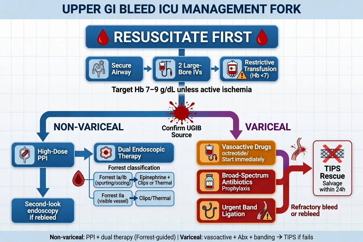
The first priority in every LGIB is airway, breathing, circulation, then resuscitation, then risk stratification, then localisation and haemostasis in the order dictated by the haemodynamic state.[1][2]
Lower GI bleed management protocol (stable-to-moderate)
ABCDE + resuscitation
Airway and breathing first. Two large-bore cannulae (16 G or larger). Crystalloid resuscitation (20 mL/kg bolus of balanced crystalloid, titrated to perfusion). Restrictive transfusion target: Hb <70 g/L (or <80 g/L with cardiovascular disease) — transfuse one unit at a time and reassess, per the TRICC principle. Cross-match 2-4 units. Monitor BP, HR, capillary refill, urine output and venous lactate trend.
Exclude an upper GI source
Massive hematochezia can come from an upper source with rapid transit. Pass a wide-bore NG tube: bile-stained aspirate with no blood has a high negative predictive value for an upper source; blood or coffee-grounds confirms it. In a shocked patient with an ambiguous source, perform gastroscopy BEFORE colonoscopy and apply the upper-GI bundle as indicated.
Calculate the Oakland score and risk-stratify
Oakland <=8 in a stable patient may permit discharge with urgent outpatient colonoscopy. Higher scores mandate admission. The Oakland score supersedes the older "blatchford-style" guesswork for LGIB and is now embedded in UK and many international guidelines.
Bowel preparation + colonoscopy within 24 h
Give polyethylene glycol (PEG) 4 L over ~4 h via NG if needed until the effluent is clear. Urgent colonoscopy within 24 h is diagnostic (source found in 70-90%) and therapeutic: epinephrine injection (1:10,000 in 1-2 mL aliquots), through-the-scope endoclips, argon plasma coagulation (APC) for angiodysplasia and oozing, and band ligation for haemorrhoids.
CT angiography (for brisk ongoing bleeding or negative colonoscopy)
If the patient is actively bleeding briskly and cannot be safely prepped for colonoscopy, OR colonoscopy is non-diagnostic, proceed to CTA. It detects extravasation at bleeding rates >0.3-0.5 mL/min, localises the bleeding segment, and maps mesenteric anatomy for embolisation. CTA also identifies the rare lethal mimics (aortoenteric fistula, ischaemic colitis with pneumatosis).
Catheter angiography + superselective embolisation
If CTA is positive (contrast extravasation), proceed to mesenteric (IMA/SMA) angiography and superselective embolisation of the bleeding vessel with microcoils or cyanoacrylate. Success rate 70-90%. Complications: post-embolisation ischaemia (5-10%), contrast nephropathy, groin access. Indicated also after failed endoscopic therapy or early rebleeding.
Surgery (last resort)
Reserved for: massive uncontrolled bleeding, failed endoscopy AND embolisation, or recurrent refractory bleeding. Segmental colectomy if the source is localised (e.g. a known diverticular segment); subtotal colectomy if the source cannot be localised and bleeding is ongoing. Emergency colectomy carries mortality 10-30%. Pre-operative localisation of the bleeding site is critical — blind surgery is disastrous.
The massive LGI bleed algorithm
A massive LGIB — ongoing brisk bleeding with haemodynamic compromise — bypasses the elective colonoscopic pathway and goes straight to resuscitation, source exclusion, and CTA-driven haemorrhage control.[1][2]
Massive lower GI bleed — the haemorrhagic-control pathway
Activate the massive transfusion protocol and resuscitate
Two large-bore cannulae; rapid transfusion of packed red cells, fresh frozen plasma and platelets in a ~1:1:1 ratio (per massive transfusion principles); correct fibrinogen to >1.5 g/L with cryoprecipitate; calcium replacement (citrate chelation in massive transfusion causes hypocalcaemia); aim for restrictive but adequate transfusion and a normalising lactate. Treat hypothermia, acidosis and coagulopathy (the lethal triad).
Exclude an upper source — gastroscopy if any doubt
In a massively bleeding patient with hematochezia, gastroscopy to exclude an upper source comes BEFORE colonoscopy. NG aspirate is a useful triage tool but in massive bleeding with shock, proceed directly to gastroscopy. Do not delay definitive upper-GI therapy (band ligation, ulcer clipping) on the assumption the source is colonic.
Proceed directly to CT angiography (skip bowel prep)
In the unstable patient with brisk bleeding, do NOT wait for bowel preparation for colonoscopy. CTA is rapid, identifies the bleeding segment and extravasation, and maps the mesenteric vessels. CTA also screens for ischaemic colitis, aortoenteric fistula and other surgical pathologies.
Mesenteric angiography + superselective embolisation
If CTA shows extravasation, proceed immediately to catheter angiography and embolisation (microcoils or glue) of the culprit artery (commonly the IMA, specifically the marginal artery of Drummond or a vasa rectum). Embolisation is the preferred haemostatic modality in the massive-bleeding patient, avoiding the morbidity of emergency surgery.
Bowel prep + targeted colonoscopy after stabilisation
Once bleeding is controlled (or slowed), bowel-prep the patient and perform colonoscopy within 24 h to confirm the source and deliver definitive endoscopic therapy, and to exclude synchronous lesions (e.g. a cancer proximal to a bleeding diverticulum).
Surgery for refractory haemorrhage
If embolisation fails, is unavailable, or bleeding is exsanguinating, proceed to surgery. Segmental colectomy if the bleeding site has been localised; subtotal colectomy (with end ileostomy) if localisation has failed and bleeding is ongoing. Bridge unstable patients to theatre with ongoing transfusion and vasopressor support.
Cause-specific management
The endoscopic, radiological and surgical therapy differs by underlying cause, and the Fellowship exam rewards a structured cause-by-cause approach.[1][2][5]
Diverticular bleed
#1 cause — arterial, painless, self-limiting
- 80% stop spontaneously — resuscitate and observe
- Endoscopic therapy if active bleeding or a visible vessel: epinephrine + through-the-scope clips, or band ligation
- Embolisation for brisk bleeding not controlled endoscopically (or pre-colonoscopy if unstable)
- Recurrent diverticular bleeding (10-40% after a first bleed) is an indication for elective segmental colectomy
- Avoid elective surgery after a single self-limiting episode
Angiodysplasia
Painless, recurrent, elderly, right colon
- Endoscopic ablation with argon plasma coagulation (APC) is first-line therapy
- Multiple right-sided lesions are common — examine the caecum and ascending colon carefully
- Exclude Heyde syndrome: aortic stenosis + acquired type 2A von Willebrand deficiency; aortic valve replacement may cure the bleeding
- Refractory diffuse angiodysplasia: consider octreotide, thalidomide, or tamoxifen in selected patients; TIPS is NOT indicated
- Repeated therapeutic colonoscopy reduces recurrence
IBD (ulcerative colitis)
Treat the flare, not the bleeding vessel
- Massive colonic haemorrhage can be the index presentation of severe acute UC
- Treat the underlying inflammation: IV hydrocortisone 100 mg q6h, consider IV cyclosporin (2 mg/kg/day) or infliximab (5 mg/kg) for steroid-refractory disease
- Endoscopic therapy has a limited role — bleeding reflects diffuse mucosal inflammation, not a discrete vessel
- Subtotal colectomy for refractory massive haemorrhage in fulminant colitis (also excludes colitis-related cancer)
- Avoid over-instrumentation in an inflamed, thin-walled colon (perforation risk)
Ischaemic colitis
Pain BEFORE the bleed — a different pathway
- Usually a non-occlusive low-flow insult affecting the Griffith (splenic flexure) and Sudeck (rectosigmoid) watershed regions
- Supportive management: bowel rest, IV fluids to restore perfusion, broad-spectrum antibiotics for translocation, treat precipitant (cardiac failure, hypotension, dehydration)
- Endoscopic therapy has NO role — bleeding reflects mucosal sloughing, not a discrete vessel
- Surgery reserved for transmural infarction, perforation or refractory bleeding — right-sided ischaemic colitis has the highest mortality and most often needs surgery
- Colonoscopy (within 48-72 h if stable) characterises the distribution and severity (thumbprinting, haemorrhagic nodules, gangrene)
Anticoagulation and antiplatelet management
The decision to stop, reverse or continue antithrombotics balances the severity of bleeding against the thrombotic risk of the underlying indication. There is no blanket rule — each decision is individualised.[1][2]
- Aspirin (secondary prevention) — for established cardiovascular or cerebrovascular disease, continue aspirin through an LGIB admission; the thrombotic risk of stopping outweighs the rebleeding risk. Withhold only during active uncontrolled haemorrhage and restart within 7 days once haemostasis is secured.
- Dual antiplatelet therapy (DAPT) — after a recent coronary stent (especially <6 months for DES), continue at least one antiplatelet (aspirin preferred); interrupt the second (clopidoggel/ticagrelor) only during active bleeding and restart as soon as possible. Coordinate with cardiology.
- Warfarin — reverse slowly with vitamin K (5-10 mg IV) and/or prothrombin complex concentrate (PCC) only if bleeding is severe; do NOT normalise the INR in patients with mechanical valves without haematology/cardiology input. Prothrombin complex concentrate is preferred over fresh frozen plasma for rapid reversal.
- Direct oral anticoagulants (DOACs) — time since last dose and drug half-life matter. Idarucizumab reverses dabigatran; andexanet alfa or PCC reverses factor Xa inhibitors (apixaban, rivaroxaban). Activated charcoal if ingestion within 2-4 h. Discuss with haematology; restart once haemostasis is secure and the thrombotic indication persists.
- Heparin infusions — typically held during active bleeding and resumed once haemostasis is achieved. [1]
Key evidence and guidelines
ACG LGIB Guideline 2016 (Strate & Gralnek)
Am J Gastroenterol 2016
US clinical guideline for the management of acute lower GI bleeding
Key finding
Resuscitate; exclude upper source; colonoscopy within 24 h; CTA for brisk/ongoing bleeding; angiographic embolisation for failed endoscopy; surgery as last resort. Oakland score for risk stratification.
Practice change
Standardised the colonoscopy → CTA → embolisation → surgery ladder
ACG LGIB Guideline Update 2023
Am J Gastroenterol 2023
Updated US clinical guideline incorporating CTA-first strategies for unstable patients
Key finding
CTA is first-line in the haemodynamically unstable patient; colonoscopy within 24 h for stable/controlled patients; superselective embolisation preferred over empiric surgery; aspirin for secondary prevention should be continued.
Practice change
Reaffirmed CTA-first in unstable LGIB and continuation of aspirin
Oakland Score (Oakland 2016)
PLoS One 2016
Derivation (n=2336) and external validation (n=288) of a risk score for safe discharge after LGIB
Key finding
A score <=8 identifies patients safe for discharge with urgent outpatient colonoscopy; sensitivity >95% for adverse outcomes; C-statistic 0.84 (derivation) and 0.79 (validation).
Practice change
Oakland <=8 is the validated threshold for considering outpatient management
Green urgent colonoscopy RCT (2005)
Am J Gastroenterol 2005
RCT, n=100, urgent colonoscopy (rapid purge) vs standard care for acute LGIB
Key finding
Urgent colonoscopy identified a definitive bleeding source significantly more often (OR 2.6), but did NOT improve mortality, length of stay, transfusion, rebleeding or surgery rates.
Practice change
Urgent colonoscopy improves diagnostic yield but not outcomes; supports a measured approach with bowel preparation
TRICC (Hébert 1999)
NEJM 1999
Multicentre RCT, n=838 euvolaemic critically ill patients, restrictive (Hb 70-90) vs liberal (Hb 100-120) transfusion
Key finding
Restrictive strategy was at least as effective and possibly superior, with fewer complications; hospital mortality lower in the restrictive group among less acutely ill and younger patients.
Practice change
Restrictive transfusion (Hb <70) is the standard of care in the critically ill, including LGIB
UK National LGIB Audit (Oakland 2019, Gut)
Gut 2019
National audit of LGIB management across UK hospitals and operationalisation of the Oakland score
Key finding
Confirmed wide variation in practice; validated the Oakland score in routine care; supported standardised risk-stratified pathways and reduced unnecessary admissions.
Practice change
Embedded the Oakland score in national LGIB pathways
Complications
- Rebleeding — the dominant early complication; diverticular bleeds recur in 10-40% within 1-4 years. Predicted by active bleeding or a visible vessel at colonoscopy; managed by repeat endoscopy, embolisation or surgery.
- Post-embolisation ischaemia — occurs in 5-10% of patients undergoing superselective mesenteric embolisation; usually self-limiting (mucosal ischaemia) but may progress to transmural infarction requiring surgery.
- Contrast nephropathy — from CTA and catheter angiography, especially in the elderly comorbid patient; pre-hydrate and minimise contrast where possible.
- Bowel preparation complications — aspiration (especially in the elderly or those with reduced consciousness), fluid overload, electrolyte disturbance; intubate the encephalopathic patient before bowel prep.
- Surgical complications — anastomotic leak, stoma-related morbidity, incisional hernia, and the high mortality (10-30%) of emergency colectomy in the elderly comorbid patient.
- Transfusion-related complications — TACO, TRALI, infection and immunomodulation; minimised by the restrictive strategy.
- Thrombotic events — from inappropriate cessation of antiplatelets/anticoagulants; stent thrombosis and stroke are preventable by restarting these agents early once haemostasis is secured. [1]
Prognosis
Outcomes and key numbers
Overall mortality is 2-4%, driven by comorbidity rather than exsanguination. Outcome is worst in the elderly, the anticoagulated with strong thrombotic indications, and those requiring emergency surgery or interventional radiology. The Oakland score identifies the low-risk outpatient group; the remainder follow the colonoscopy → CTA → embolisation → surgery ladder. Patients with right-sided ischaemic colitis, aortoenteric fistula and fulminant colitis are the highest-mortality subgroups and require early surgical input.[1][2][5]
Exam practice
SAQ — Massive lower GI bleeding in an elderly man
10 minutes · 10 marks
A 78-year-old man with known diverticular disease, hypertension and a mechanical aortic valve replacement (on warfarin) presents with two episodes of massive bright red blood per rectum over four hours. On arrival he is pale and clammy, GCS 15, heart rate 122/min, blood pressure 86/52 mmHg, respiratory rate 24, oxygen saturation 95% on room air. Abdomen is soft and non-tender. Initial haemoglobin 96 g/L, platelets 210 x 10^9/L, INR 3.4, fibrinogen 2.1 g/L, urea 6.2, creatinine 88, lactate 3.6. An NG aspirate is bile-stained with no blood. A diagnosis of acute massive lower GI bleeding (presumed diverticular) is made.
Clinical pearls
Red flags
References
- [1]Strate LL, Gralnek IM. ACG Clinical Guideline: Management of Patients With Acute Lower Gastrointestinal Bleeding Am J Gastroenterol, 2016.PMID 26925883
- [2]Strate LL, Gralnek IM, Ahsan M. Management of Patients With Acute Lower Gastrointestinal Bleeding: An Updated ACG Guideline Am J Gastroenterol, 2023.PMID 36735555
- [3]Oakland K, Jairath V, Uebersax D, et al. MicroRNA-15b regulates mitochondrial ROS production and the senescence-associated secretory phenotype through sirtuin 4/SIRT4 Aging (Albany NY), 2016.PMID 26959556
- [4]Green BT, Rockey DC, Portwood G, et al. Urgent colonoscopy for evaluation and management of acute lower gastrointestinal hemorrhage: a randomized controlled trial Am J Gastroenterol, 2005.PMID 16279891
- [5]Huang CS, Lęski M, Lichtenstein DR. Notum palmitoleoyl-protein carboxylesterase regulates Fas cell surface death receptor-mediated apoptosis via the Wnt signaling pathway in colon adenocarcinoma Bioengineered, 2021.PMID 34402722
- [6]Hébert PC, Wells G, Blajchman MA, et al. Reliability of self-measured blood pressure for research purposes Arch Intern Med, 1999.PMID 10547179
- [7]Oakland K, Guy R, Uebersax D, et al. Metabolomics evaluation of the impact of smokeless tobacco exposure on the oral bacterium Capnocytophaga sputigena Toxicol In Vitro, 2016.PMID 27480511