ICU ·
Ogilvie syndrome (acute colonic pseudo-obstruction)
Also known as Acute colonic pseudo-obstruction (ACPO) · Ogilvie syndrome · Acute adynamic ileus of the colon · Colonic pseudo-obstruction · Non-mechanical colonic obstruction · Neostigmine-responsive colonic ileus · Caecal perforation risk (ACPO) · Percutaneous caecostomy · Ponec neostigmine trial · ASCRS ACPO guideline
Ogilvie syndrome (acute colonic pseudo-obstruction, ACPO) is massive colonic dilation WITHOUT mechanical obstruction, caused by an imbalance between sympathetic (inhibitory, excess) and parasympathetic (excitatory, deficit) autonomic input to the colon, producing colonic atony. It is a disease of the hospitalised: precipitants include recent surgery (orthopaedic, cardiac, caesarean, abdominal), critical illness/sepsis, electrolyte disturbance (hypokalaemia, hypomagnesaemia, hypocalcaemia), opioids and anticholinergics, immobility, retroperitoneal pathology (haematoma, malignancy), trauma, burns, and metabolic disease. Presentation is progressive, often painless, abdominal distension over 3-7 days with tympani, minimal or absent bowel sounds, and nausea; pain disproportionate to the picture or peritonism signals ischaemia or perforation. Diagnosis is clinical plus imaging: plain AXR (and confirmatory CT with contrast) showing caecal dilation, with explicit exclusion of mechanical obstruction. Perforation risk climbs with caecal diameter greater than 12 cm AND duration greater than 6 days. Management is staged: conservative measures first (NPO, NG and rectal tubes, aggressive electrolyte correction, stop opioids/anticholinergics, mobilise, position changes) for 48-72 hours; if refractory, neostigmine 2-2.5 mg IV slow push (acetylcholinesterase inhibitor, 91% response in the Ponec trial) with continuous ECG and atropine at the bedside; colonoscopic decompression for neostigmine failure or contraindication; surgery (percutaneous tube caecostomy, or resection) for refractory or perforated cases.
On this page & tools
Your progress
Saved locally on this device.
Target exams
Red flags
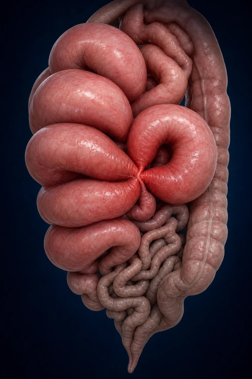
Overview & definition
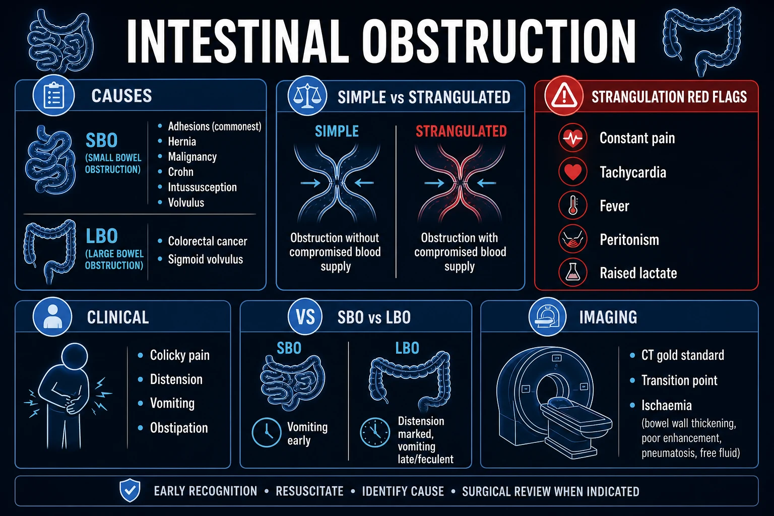
Ogilvie syndrome — acute colonic pseudo-obstruction (ACPO) — is massive colonic dilation in the absence of any mechanical obstruction. Sir William Heneage Ogilvie described it in 1948 in patients with retroperitoneal malignancy infiltrating the splanchnic nerves; the eponym persists although the modern understanding is of an autonomic imbalance producing colonic atony rather than a discrete anatomical lesion.[3][4]
The hallmark is caecal and right-colonic dilation (often >10 cm, sometimes >15-20 cm) with preserved but dysmotile bowel — there is no stricture, tumour, volvulus, or hernia to account for it. By contrast with mechanical obstruction, the patient is remarkably pain-free for the degree of distension, and by contrast with toxic megacolon there is no underlying mucosal inflammation and (initially) no systemic toxicity.[1][6]
ACPO is almost exclusively a disease of the hospitalised, sick, and immobile: roughly 95% of cases occur in inpatients with one or more identifiable precipitants. Untreated, the dilated caecum is at risk of ischaemia and perforation, which carries a mortality of 40-50%; the entire strategy is to recognise the syndrome, exclude mechanical obstruction, reverse precipitants, and decompress the colon before it ruptures.[1][3]
[1]Pathophysiology
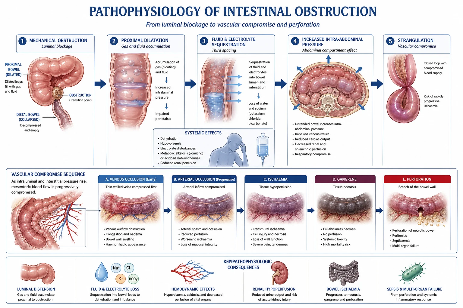
The mechanism explains every management decision and is a common exam question.[3][4][8]
-
Autonomic imbalance. The colon receives sympathetic inhibitory input (T5-L2 via the splanchnic nerves) and parasympathetic excitatory input (vagus to the splenic flexure; sacral S2-S4 via the pelvic splanchnic nerves to the distal colon). ACPO reflects an excess of sympathetic (inhibitory) tone and/or a deficit of parasympathetic (excitatory) tone, tipping the balance toward colonic atony. The proposed triggers include retroperitoneal irritation (haematoma, tumour, pancreatitis) directly interrupting the splanchnic plexus, and the systemic stress response of critical illness increasing sympathetic outflow.[3][4]
-
Why the caecum dilates first and most. By the Law of Laplace (wall tension = intraluminal pressure × radius / [2 × wall thickness]), the caecum — the widest segment of the colon — develops the greatest wall tension for any given intraluminal pressure. The adult caecum also has the thinnest wall relative to its radius. It is therefore both the segment most prone to dilate and the segment at greatest risk of ischaemic perforation once the diameter exceeds ~12 cm.[3][5]
-
Functional, not anatomical, obstruction. There is no mechanical blockage — gas and liquid accumulate because the atonic colon cannot generate the propagating motor complexes needed to move them. The proximal colon distends while the distal colon often remains decompressed, producing the characteristic radiographic pattern of massive right-sided dilation with a collapsed distal colon.[6]
-
The vicious cycle. Each precipitant feeds the others: opioids and anticholinergics directly suppress motility; electrolyte disturbance (especially hypokalaemia and hypomagnesaemia) impairs smooth-muscle excitability; immobility abolishes the postural and ambulatory stimuli to gut motility; and the growing colonic girth itself stretches and ischaemias the wall, further depressing local neuromuscular function. Reversing every reversible precipitant in parallel is the foundation of conservative therapy.[4][8]
Risk factors and precipitants
ACPO is multifactorial — the average patient has three or more precipitants stacked together. Recognising and reversing each one is itself treatment. The landmark 400-case Vanek & Al-Salti analysis catalogued the precipitant profile that still holds today.[3][4]
Surgery / trauma
Most common setting
- Orthopaedic (hip/knee replacement, pelvic surgery, spinal) — the classic trigger; immobility, opioids, and retroperitoneal/pelvic dissection combine
- Cardiac/thoracic surgery (CABG, valve) — bypass-related splanchnic hypoperfusion plus post-op opioids
- Caesarean section and postpartum state — up to 50% of obstetric ACPO; pelvic/retroperitoneal autonomic disruption
- Abdominal/pelvic surgery, burns, polytrauma, retroperitoneal haematoma or malignancy
Critical illness
The ICU substrate
- Sepsis and systemic inflammatory response — sympathetic surge, splanchnic vasoconstriction, ileus
- Electrolyte disturbance: HYPOKALAEMIA, HYPOMAGNESAEMIA, HYPONATRAEMIA, hypocalcaemia, hypophosphataemia — all impair smooth-muscle excitability
- Metabolic: uraemia, hypothyroidism, diabetes, hypoxia, acid-base disturbance
- Immobility and prolonged bed-rest abolish the ambulatory stimulus to colonic motility
Drugs
Stop the cause
- OPIOIDS — the single most important reversible drug precipitant; act on mu-receptors in the enteric nervous system
- Anticholinergics (atropine, hyoscine, tricyclics, antipsychotics) — directly oppose parasympathetic tone
- Calcium-channel blockers, clonidine, dexmedetomidine, high-dose opioids
- Sedatives, antihistamines, high-dose opioids; recent cessation of prokinetics
Neurological / retroperitoneal
Interrupt the plexus
- Retroperitoneal malignancy, haematoma, abscess, pancreatitis — mechanical/chemical interruption of the splanchnic plexus
- Spinal cord injury, stroke, Parkinson disease, multiple sclerosis, dementia
- Herpes zoster, electrolyte-related autonomic neuropathy, solid-organ or stem-cell transplant
- Radiation, cytomegalovirus (in immunocompromised), and other causes of autonomic denervation
Clinical presentation
The clinical picture is distension out of proportion to distress — the phrase to remember for the exam.[5][6]
- Abdominal distension — progressive over 3-7 days, often noticed first by nursing staff as increasing girth; tympanic on percussion, most marked in the right iliac fossa and upper abdomen.
- Minimal or no pain — the cardinal distinguishing feature. Cramping may be present but is mild; severe pain, especially new pain, or peritonism is a red flag for ischaemia or perforation and mandates urgent surgical review.
- Nausea and vomiting are common; bowel sounds are usually absent or hypoactive (occasionally high-pitched and tinkling, but not the obstructive succession-splash of mechanical obstruction).
- Constipation and failure to pass flatus are usual, though some patients paradoxically continue to pass small amounts of stool and gas because the distal colon is not mechanically blocked.
- Respiratory compromise — massive abdominal distension splints the diaphragm and may cause baseline hypoxia or difficulty ventilating an intubated patient; decompression improves compliance.
- Signs of the precipitant — fever, hypotension, oliguria from sepsis; the fresh surgical wound; the post-caesarean patient; profound weakness from hypokalaemia. [1]
Diagnosis
Diagnosis rests on three pillars: (1) clinical suspicion in an at-risk patient, (2) radiographic confirmation of massive colonic dilation, and (3) explicit exclusion of mechanical obstruction — the last is non-negotiable, because treating a mechanical obstruction with a prokinetic (neostigmine) or endoscopic insufflation risks blow-out perforation.[1][6]
Imaging approach
Imaging pathway in suspected Ogilvie syndrome
Plain abdominal X-ray (AXR) — first and follow-up test
Look for: MASSIVE COLONIC DILATION (caecum often >10 cm; right and transverse colon dilated, distal colon typically collapsed/normal), loss of haustra in the dilated segment, and FREE INTRAPERITONEAL GAS (perforation). Cheap, bedside, repeatable every 12-24 h to track the caecal diameter trend. Measure the caecal diameter at its widest point — this single number drives escalation.
CT abdomen/pelvis with IV contrast — confirmatory and to exclude obstruction
Confirms dilation, excludes a mechanical cause (stricture, tumour, volvulus, internal hernia), assesses wall thickening, pneumatosis or portal venous gas (ischaemia), free gas (perforation), and any retroperitoneal precipitant (haematoma, mass, pancreatitis). CT is now the gold-standard single test where available.
Water-soluble contrast enema — when CT is equivocal
A water-soluble (NOT barium) contrast enema definitively distinguishes pseudo-obstruction (column of contrast freely refluxes around the dilated colon with no cut-off) from mechanical obstruction (contrast stops at the lesion). The hyperosmolar contrast (e.g. gastrografin) may itself have a modest prokinetic effect. Avoid barium — risks impaction and peritonitis if perforated.
Erect CXR or lateral decubitus film
To detect free subdiaphragmatic gas (perforation) in the patient who cannot stand for an erect film. A small amount of free gas in the setting of ACPO is an emergency — urgent surgical review.
Radiographic thresholds and perforation risk
Laboratory workup
- U&E, Mg, Ca, phosphate, LFTs, glucose, TFTs, VBG/lactate — quantify and correct every electrolyte deficit; lactate elevation suggests ischaemia.
- FBC, CRP — leucocytosis and rising CRP suggest ischaemia, perforation, or supervening sepsis.
- ECG — baseline before neostigmine (bradycardia, heart block); and to detect hypokalaemic/hypomagnesaemic arrhythmias.
- Group and save / crossmatch — if surgery or colonoscopy is contemplated. [1]
When to perform endoscopy for diagnosis
Diagnostic flexible sigmoidoscopy is not required if CT confirms pseudo-obstruction and excludes mechanical obstruction. Limited endoscopy is reserved for the patient in whom imaging is equivocal or in whom an alternative diagnosis (ischaemic colitis, CMV) must be biopsied — and even then only with minimal insufflation and CO₂ rather than air.[7]
Differential diagnosis
The decisive distinction is from mechanical obstruction (which must be excluded before neostigmine) and from toxic megacolon (in which the colon is inflamed and neostigmine is contraindicated).[1][6]
Ogilvie (ACPO)
Atonic, non-toxic
- Massive colonic dilation WITHOUT mechanical obstruction and WITHOUT mucosal inflammation
- Hospitalised, elderly, post-op, electrolyte-disordered, opioid-exposed patient; minimal pain
- Neostigmine IS the treatment (after excluding obstruction); colonoscopic decompression if refractory
- Caecum >12 cm or >6 days defines high perforation risk
Toxic megacolon
Inflamed + toxic
- Non-obstructive dilation + systemic toxicity + mucosal inflammation (C. difficile, UC, CMV, ischaemic)
- Bloody diarrhoea, fever, tachycardia, leucocytosis; thumbprinting and loss of haustra on imaging
- NEOSTIGMINE CONTRAINDICATED — inflamed, paper-thin colon perforates; treat the cause (steroids/antibiotics) and prepare for colectomy
Mechanical obstruction
Must be excluded
- Volvulus, tumour, stricture, hernia, adhesions — dilation is proximal to a discrete cut-off point
- Colicky pain, distended proximal bowel with the step-ladder pattern and absent distal gas; no passage of flatus or stool
- CT with contrast (or water-soluble enema) defines the level and cause; NEVER give neostigmine until excluded
Hirschsprung / chronic pseudo-obstruction
Subacute/chronic
- Chronic intestinal pseudo-obstruction (CIP) — neuromuscular gut disorder, recurrent over months-years
- Congenital (Hirschsprung) or acquired (mitochondrial, paraneoplastic, scleroderma); different natural history
- Not the acute ICU syndrome — managed with chronic prokinetics, nutrition support, and selective surgery
Caecal volvulus
A specific mechanical cause
- Twisted mobile caecum producing a closed-loop obstruction — a "coffee bean" sign pointing to the left upper quadrant
- Is a MECHANICAL obstruction — needs endoscopic or surgical detorsion, NOT neostigmine
- Often confused radiographically with ACPO; the key is the closed-loop appearance and the point of torsion
Management
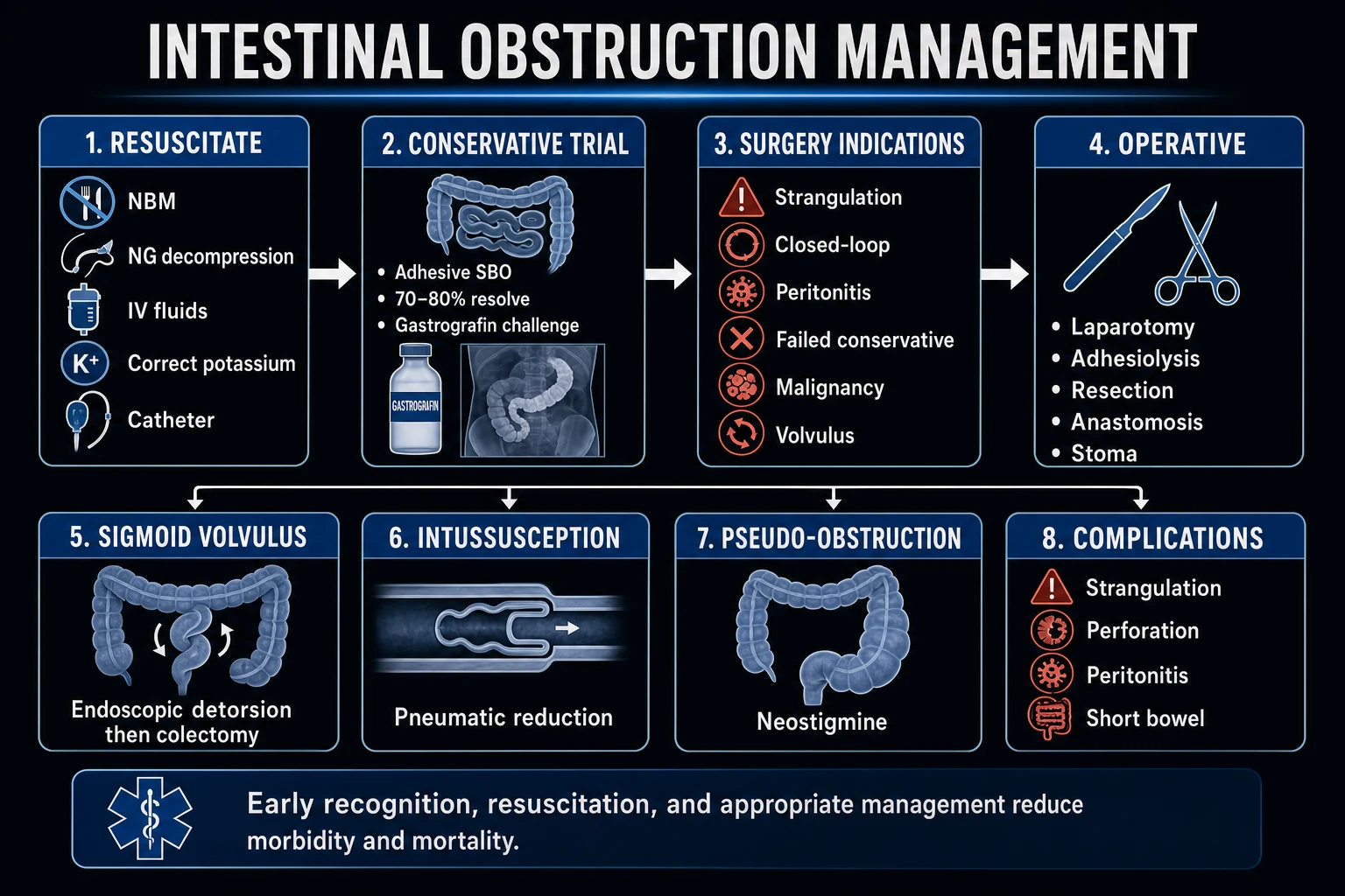
Management is a staged ladder, with each rung reserved for patients who fail the rung below or who present with high-risk features. The aims are: (1) reverse every reversible precipitant, (2) decompress the colon before it perforates, and (3) keep the patient surgically safe throughout.[1][4]
Ogilvie syndrome management protocol
1. Conservative bundle (48-72 h trial — if low-risk features)
Nil by mouth (bowel rest). NASOGASTRIC TUBE for upper GI decompression (reduces swallowed air reaching the colon). RECTAL TUBE for lower GI decompression (vents the rectum and distal colon). CORRECT ALL ELECTROLYTES: potassium to >4.0 mmol/L, magnesium to >0.8 mmol/L, calcium and phosphate normal. STOP ALL causative medications: opioids (substitute multimodal non-opioid analgesia), anticholinergics, calcium-channel blockers, sedatives. MOBILISE (ambulate if possible) and frequent position changes / knee-to-chest posture. Serial AXRs every 12-24 h to track the caecal diameter. ~one-third of patients decompress on this alone.
2. Neostigmine — first-line pharmacological decompression
If conservative therapy fails at 48-72 h OR the patient is high-risk at presentation. NEOSTIGMINE 2-2.5 mg IV slow push over 3-5 min. Acetylcholinesterase inhibitor → increases acetylcholine at muscarinic receptors → stimulates colonic peristalsis → decompression. Ponec NEJM 1999: 91% clinical response (flatus/stool and reduced caecal diameter) vs 0% placebo. Place the patient on a BEDPAN/COMMODE before injecting — rapid decompression (median 4 min) is expected. Recurrence ~11% — may repeat dose.
3. Colonoscopic decompression — if neostigmine fails or is contraindicated
Therapeutic colonoscopy: advance gently into the dilated right colon, aspirate gas and liquid stool, and (ideally) leave a DECOMPRESSION TUBE in the caecum to prevent re-accumulation. Success ~70-80%; recurrence 20-40% (lower if a tube is left). Use CO2 INSUFFLATION, NOT air — CO2 is absorbed and re-distends less. Minimal or no bowel prep (gentle saline enema only — full prep in an obstructed colon risks perforation). Risks: perforation (higher if ischaemic; quoted 1-3%). Repeat colonoscopy may be needed for recurrence.
4. Surgery — for refractory, ischaemic, or perforated cases
Options by scenario: (a) PERCUTANEOUS TUBE CAECOSTOMY — radiological or surgical, vents the caecum; preferred for refractory but viable colon in a poor surgical candidate; (b) SURGICAL CAECOSTOMY — open placement of a decompressive tube; (c) SEGMENTAL OR SUBTOTAL COLECTOMY if the colon is ischaemic or non-viable; (d) EMERGENCY LAPAROTOMY + RESECTION for perforation with peritonitis (often with stoma rather than primary anastomosis). Surgical mortality is high (10-50%) — these are sick patients, and the operation is a salvage manoeuvre.
5. Continuous monitoring throughout
Serial abdominal X-rays every 12-24 h to track caecal diameter (target <10 cm). Serial abdominal examination — new pain, guarding, or rebound = ischaemia/perforation until proven otherwise. Serial bloods (K, Mg, Ca, lactate, WCC). Once decompressed, prevent recurrence: maintain electrolyte correction, minimise opioids, mobilise, and feed early.
Neostigmine — the deep dive
Neostigmine is the pharmacological core of ACPO management and a very high-yield exam topic. It is an acetylcholinesterase inhibitor: by preventing breakdown of acetylcholine at the synapse it augments the deficient parasympathetic drive to the colon, restoring peristalsis and producing rapid decompression.[2][6]
Evidence
The Ponec trial (NEJM 1999) is the pivotal randomised, double-blind, placebo-controlled study: 21 patients with caecal diameter ≥10 cm and no mechanical obstruction were randomised to neostigmine 2 mg IV vs placebo. 10 of 11 (91%) neostigmine patients had prompt clinical decompression (median 4 min) vs 0 of 11 placebo; 8 of 9 placebo non-responders then decompressed with open-label neostigmine. Symptomatic bradycardia needing atropine occurred in 2 of 11. This single trial established neostigmine as first-line pharmacological therapy.[2]
Ponec (NEJM 1999)
NEJM 1999
21 patients with ACPO (caecum ≥10 cm, no mechanical obstruction) — neostigmine 2 mg IV vs placebo, double-blind RCT
Key finding
91% (10/11) neostigmine decompressed (median 4 min) vs 0% placebo; 8/9 placebo non-responders then responded to open-label neostigmine
Practice change
Established IV neostigmine as first-line pharmacological therapy for ACPO
Dosing and administration
Contraindications
Absolute
Do not give
- Mechanical obstruction not yet excluded — prokinetic against a blockage risks blow-out perforation
- Suspected or confirmed colonic perforation
- Generalised peritonitis
- Severe active bronchospasm / asthma (cholinergic bronchoconstriction)
- High-grade AV block or severe bradycardia not paced
Relative
Weigh risk
- Recent myocardial infarction (bradycardia poorly tolerated)
- Heart rate <60 at baseline or on rate-control agents
- Renal insufficiency (neostigmine is renally excreted; reduce dose)
- Active peptic ulcer disease or recent GI surgery (increased motility/scretions)
- Urinary obstruction, mechanical — cholinergic stimulation worsens
Adverse effects
Bradycardia and hypotension (most common; usually transient, atropine-reversible); bronchospasm; excessive salivation and lacrimation; nausea and abdominal cramping; miosis and sweating; rarely AV block or asystole requiring atropine and resuscitation.[2]
Colonoscopic decompression
Reserved for neostigmine failure or contraindication, or for the high-risk patient who is deteriorating. It is both therapeutic and (if needed) diagnostic.[7][6]
- Technique: gentle advancement to the caecum (or as far as safely possible), aspiration of gas and liquid stool, leaving a decompression tube in the right colon to maintain venting.
- Insufflation: use CO2, not air — CO2 is rapidly absorbed and re-distension is less; minimise insufflation volume.
- Bowel prep: minimal or none — a gentle saline enema at most. Never give full mechanical prep to an obstructed, dilated colon (perforation, fluid overload).
- Success and recurrence: initial success ~70-80%; recurrence 20-40%, lower if a decompression tube is left and kept patent.
- Risks: perforation (quoted 1-3%; higher if the wall is ischaemic — abort if the mucosa looks dusky, haemorrhagic, or non-viable), sedation-related hypotension/hypoxia.
- Repeat: a second decompression is reasonable for recurrence, after re-addressing precipitants; further recurrence usually warrants surgery.[7]
Surgical management
Surgery is reserved for refractory ACPO, ischaemic/non-viable colon, or perforation, and is a salvage operation in a high-risk patient — mortality is 10-50%.[1][4]
Surgical decision-making in Ogilvie syndrome
Absolute indications for surgery
CONFIRMED PERFORATION (free gas, peritonitis, pneumoperitoneum), GENERALISED PERITONITIS, or ISCHAEMIC/NON-VIABLE COLON (seen at colonoscopy or suggested by pneumatosis, portal venous gas, rising lactate). These are not negotiable — operate.
Refractory ACPO (relative indication)
Failure to decompress despite conservative therapy, neostigmine, and colonoscopic decompression; or repeated recurrences after successful decompression. The rising caecal diameter on serial imaging despite maximal therapy is the trigger.
Operation: percutaneous tube caecostomy (first surgical choice for viable colon)
A tube is placed into the caecum — percutaneously (radiological or endoscopic) or via a mini-laparotomy — to vent the gas and decompress the colon over days. Preferred in the poor surgical candidate with viable bowel; can be removed once the colon has decompressed and motility returned. Lower morbidity than resection.
Operation: resection for non-viable or perforated colon
Caecal/segmental or SUBTOTAL COLECTOMY with stoma (primary anastomosis is avoided in the obstructed, ischaemic, malnourished patient). For frank perforation with contamination, an emergency laparotomy with resection and stoma (often end ileostomy / Hartmann-type) is performed. Damage-control principles apply in the unstable.
Postoperative and recurrence prevention
Maintain electrolyte correction, minimise opioids, mobilise early, and address the underlying precipitant (sepsis, metabolic, drug). Reconstructive surgery (stoma reversal) is planned electively weeks-months later once recovered.
Monitoring and complications
[4]Recognised complications
- Colonic ischaemia — the dilated, tense wall outstrips its blood supply; suggested by new pain, peritonism, rising lactate, pneumatosis or portal venous gas. Mandates urgent surgical review.
- Colonic perforation — the dominant fatal complication; risk rises with caecal diameter >12 cm and duration >6 days. May be clinically silent in the sedated/ventilated patient — maintain a high index of suspicion and rely on AXR free gas and lactate. Mortality of perforated ACPO is 40-50%.[3]
- Abdominal compartment syndrome — massive girth can raise intra-abdominal pressure, compromising renal, respiratory, and gut perfusion. Measure bladder pressure if suspected; decompression helps.
- Aspiration — reduced gastric emptying and stasis increase aspiration risk; keep NPO with NG decompression.
- Neostigmine adverse effects — bradycardia, hypotension, bronchospasm, AV block (see above).
- Recurrence — ~11% after neostigmine, 20-40% after colonoscopy; prevented by addressing precipitants.[2]
Ogilvie syndrome — the numbers to know
Special scenarios
Prognosis
Outcomes hinge on three things: the reversibility of the precipitants, the speed of decompression, and whether perforation occurs. Roughly a third of patients decompress with conservative measures alone; neostigmine resolves another large fraction; colonoscopic decompression rescues most of the remainder. Surgical patients are a selected, sicker group with mortality of 10-50%. Perforated ACPO carries a 40-50% mortality — the entire strategy is to decompress before the caecum ruptures. Recurrence (after neostigmine ~11%; after colonoscopy 20-40%) is prevented by sustaining electrolyte correction, minimising opioids, and mobilising.[1][3][4]
SAQ — Ogilvie syndrome: staged management ladder
10 minutes · 10 marks
A 67-year-old man is day 5 after a left total hip replacement. He has progressive abdominal distension over 3 days with intermittent nausea but minimal pain. He is on oxycodone SR 20 mg BD and is largely bed-bound. He has not passed flatus or stool for 48 hours. Observations: HR 88, BP 132/78, RR 18, SpO2 96%, T 37.4, abdominal girth 102 cm, tympanic on percussion, soft, mildly tender but no peritonism. Bowel sounds are high-pitched and occasional. Bloods: K 3.2 mmol/L, Mg 0.65 mmol/L, Hb 112, WCC 9.8, CRP 78, creatinine 92, lactate 1.4. AXR shows massive caecal and right-colonic dilation (caecum 11.5 cm), distended transverse colon, and minimal rectal gas. CT with IV and rectal contrast shows dilation from caecum to mid-transverse colon with NO transition point, NO obstructing lesion, and no free gas.
SAQ — Neostigmine: pharmacology, evidence, and adverse effects
10 minutes · 10 marks
A 72-year-old woman has been in ICU for 8 days following a ruptured AAA repair. She is ventilated, on noradrenaline 0.15 mcg/kg/min, fentanyl 100 mcg/h, and has been progressively distended over 5 days. Abdomen is grossly distended and tympanic with minimal pain. AXR shows caecal diameter 13.5 cm with distended right colon, no free gas, no transition point on CT. K 3.8, Mg 0.7, lactate 1.6. Conservative measures (NG, rectal tube, K/Mg correction, fentanyl wean) have failed over 48 hours. The ICU consultant asks you to write up neostigmine and justify the choice to the reg.
Clinical pearls [1]
Red flags
One-paragraph exam answer
Ogilvie syndrome (acute colonic pseudo-obstruction, ACPO) is massive colonic dilation without mechanical obstruction, produced by an imbalance of autonomic input — sympathetic (inhibitory) excess and parasympathetic (excitatory) deficit — causing colonic atony. By the Law of Laplace the wide, thin-walled caecum dilates first and is at greatest risk of ischaemic perforation. It is a disease of the hospitalised: precipitants are recent surgery (orthopaedic, cardiac, caesarean, abdominal), critical illness/sepsis, electrolyte disturbance (K/Mg/Ca/phosphate), opioids and anticholinergics, immobility, and retroperitoneal pathology. Presentation is progressive, painless distension over 3-7 days with tympani and reduced bowel sounds. Diagnosis is clinical plus imaging — plain AXR and confirmatory CT with contrast, with explicit exclusion of mechanical obstruction (a water-soluble contrast enema if CT is equivocal). Perforation risk rises with caecal diameter >12 cm OR duration >6 days, and is highest when both are present. Management is staged: (1) conservative for 48-72 h (NPO, NG and rectal tubes, aggressive K/Mg/Ca correction, stop opioids/anticholinergics, mobilise, position changes) — resolves ~one-third; (2) neostigmine 2-2.5 mg IV slow (acetylcholinesterase inhibitor, 91% response in Ponec NEJM 1999) with continuous ECG and atropine drawn up; (3) colonoscopic decompression (CO2 insufflation, leave a tube) for neostigmine failure or contraindication; (4) surgery — percutaneous tube caecostomy for refractory-but-viable colon, resection for ischaemia or perforation. Never give neostigmine until mechanical obstruction is excluded or for toxic megacolon (where the inflamed colon perforates). Perforated ACPO carries a 40-50% mortality — the whole strategy is to decompress before the caecum ruptures.[1][2][3][4]
References
- [1]Vogel JD, Feingold DL, Stewart DB, Turner JS, Boutros M, Chun J, Steele SR Clinical Practice Guidelines for Colon Volvulus and Acute Colonic Pseudo-Obstruction Dis Colon Rectum, 2016.PMID 27270510
- [2]Ponec RJ, Saunders MD, Kimmey MB Neostigmine for the treatment of acute colonic pseudo-obstruction N Engl J Med, 1999.PMID 10403850
- [3]Vanek VW, Al-Salti M Acute pseudo-obstruction of the colon (Ogilvie's syndrome). An analysis of 400 cases Dis Colon Rectum, 1986.PMID 3753674
- [4]Underhill J, Munding E, Hayden D Acute Colonic Pseudo-obstruction and Volvulus: Pathophysiology, Evaluation, and Treatment Clin Colon Rectal Surg, 2021.PMID 34305473
- [5]Arthur T, Burgess A Acute Colonic Pseudo-Obstruction Clin Colon Rectal Surg, 2022.PMID 35966377
- [6]Saunders MD Acute colonic pseudo-obstruction Gastrointest Endosc Clin N Am, 2007.PMID 17556152
- [7]Naveed M, Jamil LH, Fujii-Lau LL, et al American Society for Gastrointestinal Endoscopy guideline on the role of endoscopy in the management of acute colonic pseudo-obstruction and colonic volvulus Gastrointest Endosc, 2020.PMID 31791596
- [8]Keller J, Layer P [Acute colonic pseudo-obstruction: Ogilvie syndrome] Med Klin Intensivmed Notfmed, 2015.PMID 26400054
- [9]Jayaram P, Mohan M, Lindow S Postpartum Acute Colonic Pseudo-Obstruction (Ogilvie's Syndrome): A systematic review of case reports and case series Eur J Obstet Gynecol Reprod Biol, 2017.PMID 28531835
- [10]Bernardi MP, Warrier S, Lynch AC Acute and chronic pseudo-obstruction: a current update ANZ J Surg, 2015.PMID 25943300