ICU · GI/Nutrition
Acute pancreatitis complications and management
Also known as Severe acute pancreatitis · Infected pancreatic necrosis · APACHE II / Ranson / BISAP scores · Walled-off necrosis · Step-up approach (PANTER/TENSION) · Revised Atlanta Classification 2012
Severe acute pancreatitis (15-20% of cases) causes local (necrosis, pseudocyst, walled-off necrosis, abscess) and systemic (SIRS → MODS, ARDS, AKI, shock, DIC) complications. Severity scoring: APACHE II (8 = severe), Ranson (3+ = severe), BISAP (3+ = severe), Revised Atlanta Classification (persistent organ failure 48h = severe). Management: aggressive IV fluids (Lactated Ringer preferred — reduces SIRS, WATERFALL trial), early enteral nutrition (within 48h — reduces infection), pain control (opioids), ERCP if gallstone obstruction/cholangitis. Antibiotics ONLY for proven infection (infected necrosis, cholangitis) — NOT prophylactic (Cochrane shows no mortality benefit). Infected necrosis: drain first (percutaneous/endoscopic — step-up approach, PANTER & TENSION trials), delay surgery 4 weeks for demarcation (POINTER trial).
On this page & tools
Your progress
Saved locally on this device.
Target exams
Red flags
Severity classification
Revised Atlanta Classification (click each)
Persistent organ failure (>48h)
Persistent organ failure (>48h): respiratory (PaO2/FiO2 <300), cardiovascular (SBP <90 after fluids), renal (creatinine >170 µmol/L). Single or multiple organ failure. ICU admission required. High mortality (30-50% if multi-organ, >48h).
Management
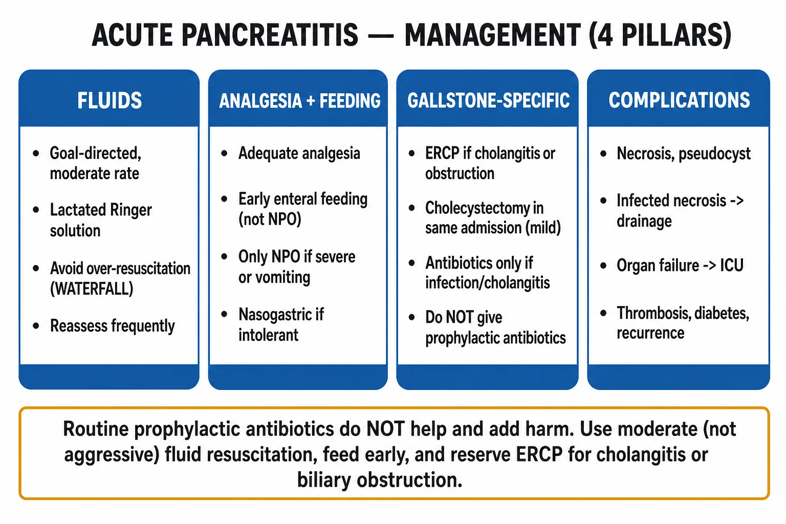
Severe pancreatitis management protocol
Aggressive IV fluid resuscitation
Lactated Ringer solution preferred (reduces SIRS and CRP vs saline — WATERFALL trial). Rate: 5-10 mL/kg/h for first 24-48h. Goal: urine output 0.5-1 mL/kg/h, haematocrit 35-44%. Monitor for fluid overload (can cause compartment syndrome). WATERFALL trial: aggressive (20 mL/kg bolus then 3 mL/kg/h) had MORE fluid overload than moderate (1.5 mL/kg/h) with no outcome benefit — avoid over-resuscitation. Reduce rate once goals met.
Early enteral nutrition (within 48h)
START EARLY. Old practice of NPO to "rest the pancreas" is WRONG and harmful. Enteral nutrition within 48h reduces: infection, complications, mortality, length of stay. NG or oral feeding both effective (meta-analyses show no advantage of nasojejunal over NG). Do NOT wait for bowel sounds or flatus. Pancreatitis is NOT a contraindication to feeding. TPN only if enteral feeding fails after 5-7 days (TPN increases infection, cost).
Pain control
Opioids: morphine, fentanyl, or oxycodone. Traditionally avoided morphine (sphincter of Oddi spasm — but evidence shows no significant effect). Use whatever controls pain. Patient-controlled analgesia (PCA) for severe pain. Avoid NSAIDs (bleeding risk if necrosis, AKI risk).
Do NOT give prophylactic antibiotics
Multiple RCTs and Cochrane meta-analyses: prophylactic antibiotics do NOT reduce infected necrosis or mortality. Increase: fungal infections, antibiotic resistance, C. difficile. Give antibiotics ONLY if: proven infection (positive cultures, infected necrosis on imaging), cholangitis, pneumonia, UTI. Cover: carbapenem (meropenem — good pancreatic penetration). ACG/IAP guidelines strongly recommend AGAINST prophylactic antibiotics.
Do NOT give prophylactic probiotics
PROPATRIA trial (Besselink, Lancet 2008): probiotic prophylaxis INCREASED mortality (16% vs 6%, p=0.01) — bowel ischaemia in 9 patients, some requiring surgery. NEVER give probiotics for predicted severe pancreatitis. This was a major change in practice.
ERCP if gallstone obstruction
Indications for urgent ERCP (<24-72h): cholangitis (fever + jaundice + RUQ pain — Charcot triad), biliary obstruction (persistent jaundice), suspected ampullary obstruction. NOT indicated for: gallstones without obstruction, mild pancreatitis without cholangitis. Cholecystectomy during same admission (after recovery) to prevent recurrence in mild cases; delayed in severe/necrotising until inflammation settles (~6 weeks).
Manage complications
ARDS: lung-protective ventilation (6 mL/kg, plateau <30). AKI: RRT if indicated. Shock: noradrenaline, careful fluids (fluid overload worsens outcome). Abdominal compartment syndrome: measure bladder pressure, decompress if >20 mmHg + organ failure. Hyperglycaemia: insulin infusion (target 6-10). Hypocalcaemia: only treat if symptomatic (tetany, seizures). Adjust magnesium first.
Atlanta 2012 classification — morphology of local complications
The Revised Atlanta Classification (2012) standardises terminology based on timing (<4 weeks vs >4 weeks) and content (fluid vs necrotic). This is heavily examined — memorise the matrix. [1]
[3] [1]Local complications — detail
Peripancreatic fluid collection (APFC) & pseudocyst
- APFC (<4 weeks, fluid only): occurs in ~50% of acute pancreatitis. Usually resolves spontaneously within weeks. No intervention needed unless infected (rare). Do NOT drain sterile APFC.
- Pseudocyst (>4 weeks, fluid only, encapsulated wall): forms when APFC persists and develops a fibrous/inflammatory wall. Most remain asymptomatic and resorb. Indications for drainage: infected, symptomatic (>6 cm and persistent pain, gastric outlet obstruction, biliary obstruction, rupture). Route: endoscopic (transgastric — preferred, lumen-apposing metal stent/LAMS), percutaneous, or surgical cystgastrostomy.
Pancreatic necrosis — sterile vs infected
- Sterile necrosis: managed conservatively with supportive care. Do NOT debride sterile necrosis — even if extensive. Most patients recover without intervention. Antibiotics NOT indicated.
- Infected necrosis: the dominant cause of late mortality in severe pancreatitis. Suspect with: clinical deterioration (fever, rising WBC/CRP, new sepsis/shock) after day 7-14. CT sign: gas within necrotic tissue (pathognomonic — requires no FNA). Fine needle aspiration (FNA) confirms (Gram stain/culture) but is NOT required if gas is present or clinical picture convincing.
Walled-off necrosis (WON)
- WON (>4 weeks, necrotic content, encapsulated wall): the "mature" form of necrosis. Intervention is safer after wall maturation (~4 weeks) — reduces bleeding risk, defines plane for debridement. Symptoms: pain, early satiety, gastric outlet obstruction, biliary obstruction, infection. Endoscopic drainage with LAMS + direct endoscopic necrosectomy (DEN) is now first-line (TENSION trial).
Local complication — decision pathway
Day 1-4: initial phase
No CT needed unless diagnosis unclear or patient deteriorating. Early CT can UNDERESTIMATE necrosis (necrosis demarcates over 3-5 days). Manage supportively: fluids, analgesia, early EN. Measure severity scores.
Day 5-7: first CT if severe
Contrast-enhanced CT (CECT) if persistent organ failure, worsening pain, fever, or rising inflammatory markers. Assess: extent of necrosis (<30%, 30-50%, >50% — >30% predicts complications), peripancreatic fluid, vascular complications (splenic vein thrombosis, pseudoaneurysm).
Identify infection (day 7-14+)
Suspect infected necrosis if: fever, rising CRP/WBC, new/worsening sepsis, clinical deterioration. CT: gas in necrotic tissue = infected (pathognomonic). FNA optional. Sterile necrosis: continue conservative management. Infected: antibiotics + consider drainage.
Delay intervention if possible (≥4 weeks)
POINTER trial: postponed drainage (median 14 days later than immediate) gave similar complication rates, fewer fistulas, less pancreatic fistula surgery. Delay lets necrosis demarcate into WON — safer drainage/necrosectomy, less bleeding. If patient stable on antibiotics, WAIT.
Choose drainage route (step-up)
PANTER trial: percutaneous catheter drainage (PCD) first, with endoscopic/laparoscopic step-up if needed, superior to primary open necrosectomy. TENSION trial: endoscopic step-up superior to surgical step-up (fewer fistulas, shorter LOS). Route chosen by anatomy + local expertise. Endoscopic preferred when collection abuts stomach/duodenum.
Necrosectomy only if drainage insufficient
If drainage fails (ongoing sepsis, large solid burden): add minimally invasive necrosectomy. Endoscopic DEN via LAMS, percutaneous, or VARD (video-assisted retroperitoneal debridement). Open necrosectomy = LAST resort (highest mortality, fistula rate).
Systemic complications — SIRS to MODS
Severe pancreatitis is a systemic inflammatory disease. Pancreatic injury releases pro-inflammatory mediators (cytokines IL-6, IL-8, TNF-α, phospholipase A2, trypsin, platelet-activating factor) that drive SIRS → distant organ dysfunction. [1]
Mechanism of distant organ injury
Acinar cell injury → systemic inflammation
Premature intracellular trypsinogen activation → acinar cell necrosis → release of damage-associated molecular patterns (DAMPs) and cytokines. Activated neutrophils/macrophages amplify inflammation (IL-6, IL-8, TNF-α). This systemic cytokine storm is the substrate for SIRS and MODS.
Endothelial dysfunction + capillary leak
Inflammatory mediators cause widespread endothelial injury → increased vascular permeability ("third spacing"), vasodilation, hypovolaemia, microvascular thrombosis. Contributes to: hypotension/shock, pulmonary oedema, abdominal compartment syndrome.
Organ-specific injury
Lungs: phospholipase A2 degrades surfactant + cytokine-mediated alveolar-capillary leak → ARDS. Kidneys: hypovolaemia + cytokine-induced tubular injury + intra-abdominal hypertension → AKI. Coagulation: tissue factor release + endothelial injury → DIC. Heart: myocardial depressant factor + cytokines → cardiomyopathy.
Severity scoring systems
Four scoring systems are examinable. Each has strengths/weaknesses — understand WHEN each applies and its cut-offs. [1]
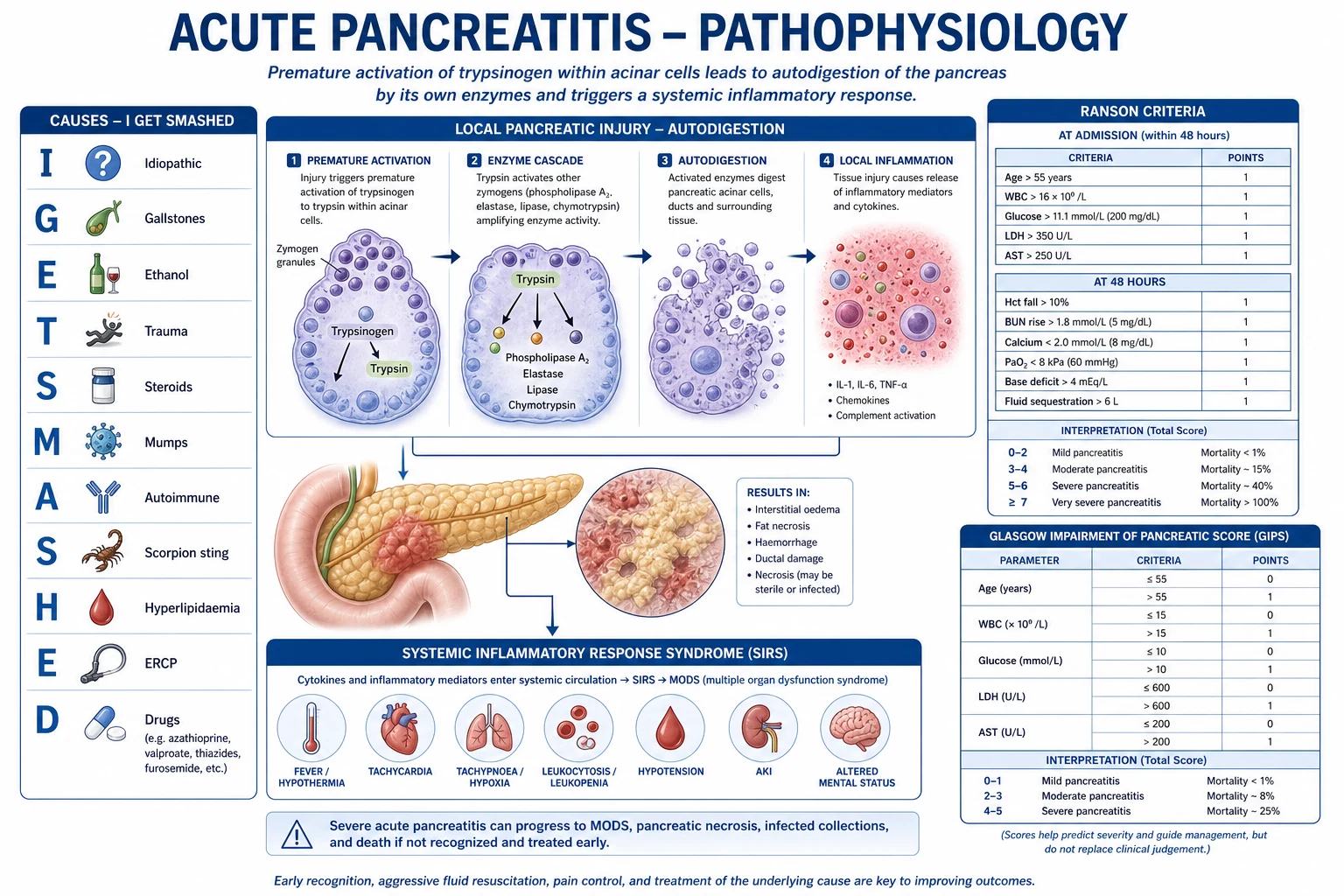
[12] [11] [10] [3]Ranson criteria — the 11 criteria (memorise)
At admission (5): age >55, WBC >16, glucose >11 mmol/L, AST >250, LDH >350. At 48h (6): Ca <2.0 mmol/L, haematocrit drop >10%, PaO2 <60 mmHg, base deficit >4, fluid sequestration >6 L, BUN rise >1.8 mmol/L. [1]
Mnemonic for non-gallstone Ranson: GA LAW (Glucose, Age, LDH, AST, WBC) at admission; at 48h: C HOBBS F (Calcium, Hematocrit, Oxygen, Base deficit, BUN, Fluid sequestration). [1]
BISAP score (5 components)
BUN >25 mg/dL (8.9 mmol/L), Impaired mental status (GCS <15), SIRS (≥2 criteria), Age >60, Pleural effusion. Score 1 point each; 3+ = severe. Validated in Wu et al 2008 (large population-based study). [1]
CT Severity Index (Balthazar)
Combines Balthazar grade (A-E: normal to extensive necrosis) with necrosis percentage (0/3/6/8 points for none/<30%/30-50%/>50%). Modified CTSI (Mortele) is simpler and correlates better with outcome. CTSI >7 = severe.
Infected necrosis — step-up approach
The step-up approach revolutionised management of infected necrotising pancreatitis. Before 2010, primary open necrosectomy was standard (mortality 20-40%). The PANTER and TENSION trials established that drain first, debride only if needed reduces morbidity and mortality. [1]
Infected necrosis management (step-up approach)
Diagnose infection
Clinical deterioration (fever, rising WBC/CRP, new sepsis) + CT findings (gas in necrotic tissue = pathognomonic, rim enhancement). Fine needle aspiration (FNA) can confirm but is NOT necessary if gas present or clinical picture convincing. Sterile necrosis does NOT require antibiotics or drainage.
Antibiotics
Broad-spectrum with good pancreatic penetration: carbapenem (meropenem 1g IV TDS), OR piperacillin-tazobactam 4.5g IV TDS, OR quinolone (ciprofloxacin) + metronidazole. Carbapenems achieve highest pancreatic tissue levels. De-escalate once cultures available. Continue until infection controlled + source managed. Antifungal prophylaxis NOT routine (CONSORT/Addenbrooke's studies show fluconazole does not reduce fungal infection).
Drain first (percutaneous or endoscopic)
Percutaneous catheter drainage (PCD, CT/US-guided) or endoscopic transluminal drainage (via stomach/duodenum using LAMS). PANTER trial: drain-first step-up approach superior to primary open surgery — fewer major complications/deaths (35% vs 69%), fewer fistulas, lower cost. ~35% of patients need ONLY drainage (no further intervention). Delay intervention for at least 4 weeks if haemodynamically stable (POINTER trial).
Step-up to necrosectomy if drainage insufficient
If drainage alone insufficient (ongoing sepsis, large necrotic burden): add necrosectomy. Minimally invasive routes: endoscopic DEN (via LAMS), percutaneous (VARD — video-assisted retroperitoneal debridement). TENSION trial: endoscopic step-up superior to surgical step-up (lower fistula rate, shorter LOS). Open necrosectomy = LAST resort (highest mortality, fistula, diabetes, exocrine insufficiency).
Timing — delay 4 weeks if possible
POINTER trial (2024): immediate vs postponed (median 14 days) drainage. Postponed group had similar complications, fewer pancreatic fistulas, less pancreatic surgery. Delay lets necrosis demarcate into WON (defined wall) — safer drainage, less bleeding, fewer procedures. Indications for earlier (immediate) drainage: septic shock unresponsive to antibiotics, peritonitis, bowel ischaemia/perforation.
Pseudocyst management
Peripancreatic fluid collection: usually resolves spontaneously. Pseudocyst (mature wall, >4 weeks, pure fluid): observe unless symptomatic. Drain if: infected, >6 cm and persistent, causing symptoms (pain, gastric outlet obstruction, biliary obstruction). Drainage route: endoscopic (transgastric — preferred), percutaneous, or surgical cystgastrostomy.
Landmark trials
PANTER — Step-up vs open necrosectomy (NEJM 2010)
Multicentre RCT, 88 patients with necrotising pancreatitis + suspected infection. Step-up approach (PCD ± minimally invasive retroperitoneal necrosectomy) vs primary open necrosectomy.
Key finding
Primary endpoint (major complications/death): 35% (step-up) vs 69% (open). Step-up reduced: new-onset multi-organ failure (12% vs 40%), incisional hernias, new-onset diabetes. ~35% treated with drainage alone — no surgery needed.
TENSION — Endoscopic vs surgical step-up (Lancet 2018)
Multicentre RCT, 98 patients with infected necrotising pancreatitis. Endoscopic step-up (endoscopic transluminal drainage ± endoscopic necrosectomy, with percutaneous fallback) vs surgical step-up (PCD ± VARD).
Key finding
Primary endpoint (composite: major complications/death): not significantly different (43% endoscopic vs 45% surgical). BUT endoscopic group: significantly fewer pancreatic fistulas (5% vs 28%), shorter length of stay, fewer interventions.
POINTER — Immediate vs postponed drainage (Ann Surg 2024)
Multicentre RCT, 42 patients with infected necrotising pancreatitis. Immediate drainage vs postponed (antibiotics + wait until clinical deterioration or >4 weeks).
Key finding
Median postponement 14 days. No significant difference in composite endpoint (death/major complications). Postponed group: fewer pancreatic fistulas, fewer drainage-related complications, less pancreatic surgery. ~60% of postponed group eventually needed drainage.
WATERFALL — Aggressive vs moderate fluid resuscitation (NEJM 2022)
Multicentre RCT, 249 patients with acute pancreatitis. Aggressive (20 mL/kg bolus, then 3 mL/kg/h) vs moderate (1.5 mL/kg/h, bolus 10 mL/kg only if hypovolaemic) Lactated Ringer for 72-120h.
Key finding
Primary endpoint (MODS at 72h): no significant difference. Aggressive group had significantly MORE fluid overload (20.5% vs 6.3%, p=0.004). No difference in SIRS, ARDS, or death.
PROPATRIA — Probiotic prophylaxis (Lancet 2008)
Multicentre RCT, 298 patients with predicted severe acute pancreatitis. Probiotic (multispecies) vs placebo to prevent infectious complications.
Key finding
NO reduction in infectious complications. INCREASED mortality: 16% (probiotic) vs 6% (placebo, p=0.01). 9 patients in probiotic group developed bowel ischaemia (4 died), vs none in placebo.
Antibiotics — what to give and when
[1] [4] [15] [1]Vascular and other complications
[3] [4]SAQ — Infected pancreatic necrosis and the step-up approach
10 minutes · 10 marks
A 54-year-old man is day 14 in ICU with severe necrotising gallstone pancreatitis (gallstone aetiology; BISAP 4; APACHE II 14 at admission). He has been receiving NG feeding, moderate goal-directed lactated Ringer resuscitation and supportive care. Over 48 hours he develops spiking fevers (39.2 degrees C), new atrial fibrillation with rate 138, rising vasopressor requirement (noradrenaline 0.45 mcg/kg/min), lactate 3.6 mmol/L, WBC 21 and CRP 290 mg/L. Contrast-enhanced CT shows >50% pancreatic necrosis with new gas locules within the collection, partially encapsulated. He has not been on antibiotics.
SAQ — Abdominal compartment syndrome in severe acute pancreatitis
10 minutes · 10 marks
A 46-year-old woman is ICU day 5 with severe alcohol-related necrotising pancreatitis (APACHE II 16, BISAP 4, >50% necrosis on day-7 CT). Cumulative fluid balance is +9.4 L. Over 12 hours she becomes oliguric (12 mL/h despite frusemide), her plateau pressure has risen from 29 to 41 cmH2O at unchanged tidal volume (6 mL/kg), her lactate has risen from 1.4 to 3.6 mmol/L and she requires noradrenaline 0.4 mcg/kg/min for a MAP of 66. The abdomen is grossly distended and tense. Bladder pressure (instilled 25 mL saline, supine, end-expiratory) is 28 mmHg. Recent creatinine 215 micromol/L, PaO2/FiO2 180.
Clinical pearls
Red flags
Common exam questions
[1] [1] [1] [1]Aetiology — comprehensive mnemonic
[1]Disposition and follow-up
Post-ICU and discharge planning
Resolve acute episode
Symptoms settled, tolerating oral diet, inflammatory markers trending down, no ongoing organ failure. Cholecystectomy before discharge if gallstone aetiology and mild-moderate episode.
Address local complications
Pseudocysts/WON: arrange outpatient follow-up imaging. Most resorb; persistent symptomatic collections need elective drainage. Pancreatic duct disruption/fistula: ERCP ± stenting.
Screen for long-term sequelae
Diabetes (HbA1c at 3 months), exocrine insufficiency (faecal elastase — enzyme replacement if low), recurrent symptoms. Counselling on alcohol cessation if applicable. Lipid-lowering (fibrate) for hypertriglyceridaemia.
Prevent recurrence
Gallstone: cholecystectomy. Alcohol: cessation programme. Hypertriglyceridaemia: fibrate ± omega-3, diabetic control. Drugs: cease offending agent. ERCP: avoid if possible. Autoimmune: corticosteroids (type 1). Tumour: surgical referral.
References
- [1]Tenner S, Vege SS, Sheth SG, Sauer BG. American College of Gastroenterology Guidelines: Management of Acute Pancreatitis Am J Gastroenterol, 2024.PMID 38857482
- [2]International Association of Pancreatology. International Association of Pancreatology Revised Guidelines on Acute Pancreatitis 2025: Supported and Endorsed by the American Pancreatic Association, European Pancreatic Club, Indian Pancreas Club, and Japan Pancreas Society Pancreatology, 2025.PMID 40651900
- [3]Banks PA, Bollen TL, Dervenis C, Gooszen HG, Johnson CD, Sarr MG, et al. Classification of acute pancreatitis--2012: revision of the Atlanta classification and definitions by international consensus Gut, 2013.PMID 23100216
- [4]Working Group IAP/APA Acute Pancreatitis Guidelines. IAP/APA evidence-based guidelines for the management of acute pancreatitis Pancreatology, 2013.PMID 24054878
- [5]de-Madaria E, Buxbaum JL, Maisonneuve P, Singh VK, Sáez-González A, Zhou Y, et al.; WATERFALL trial group. Aggressive or Moderate Fluid Resuscitation in Acute Pancreatitis N Engl J Med, 2022.PMID 36103415
- [6]van Santvoort HC, Besselink MG, Bakker OJ, Hofker HS, Boermeester MA, Dejong CH, et al.; Dutch Pancreatitis Study Group. A step-up approach or open necrosectomy for necrotizing pancreatitis N Engl J Med, 2010.PMID 20410514
- [7]van Brunschot S, van Grinsven J, van Santvoort HC, Bakker OJ, Besselink MG, Boermeester MA, et al.; TENSION trial group. Endoscopic or surgical step-up approach for infected necrotising pancreatitis: a multicentre randomised trial Lancet, 2018.PMID 29108721
- [8]Van Veldhuisen CL, Sissingh NJ, Boxhoorn L, van Dijk SM, van Brunschot S, Belgers H, et al.; POINTER study group. Long-Term Outcome of Immediate Versus Postponed Intervention in Patients With Infected Necrotizing Pancreatitis (POINTER): Multicenter Randomized Trial Ann Surg, 2024.PMID 37450701
- [9]Besselink MG, van Santvoort HC, Buskens E, Boermeester MA, van Goor H, Timmerman HM, et al.; PROPATRIA study group. Probiotic prophylaxis in predicted severe acute pancreatitis: a randomised, double-blind, placebo-controlled trial Lancet, 2008.PMID 18279948
- [10]Wu BU, Johannes RS, Sun X, Conwell DL, Banks PA. The early prediction of mortality in acute pancreatitis: a large population-based study Gut, 2008.PMID 18519429
- [11]Ranson JH, Rifkind KM, Roses DF, Fink SD, Eng K, Spencer FC. Objective early identification of severe acute pancreatitis Am J Gastroenterol, 1974.PMID 4835417
- [12]Knaus WA, Draper EA, Wagner DP, Zimmerman JE. APACHE II: a severity of disease classification system Crit Care Med, 1985.PMID 3928249
- [13]van Grinsven J, van Santvoort HC, Boermeester MA, Besselink MG, Dutch Pancreatitis Study Group. Superiority of Step-up Approach vs Open Necrosectomy in Long-term Follow-up of Patients With Necrotizing Pancreatitis Gastroenterology, 2019.PMID 30391468
- [14]Moggia E, Koti R, Belgaumkar AP, Fazio F, et al. Pharmacological interventions for acute pancreatitis Cochrane Database Syst Rev, 2017.PMID 28431202
- [15]Min L, He F, Bi B, Jiang S, Zhu L, Zhang Z. Assessment of Prophylactic Carbapenem Antibiotics Administration for Severe Acute Pancreatitis: An Updated Systematic Review and Meta-Analysis Digestion, 2022.PMID 35026770