ICU · gi-nutrition
Acute Severe Intra-Abdominal Infection and Peritonitis — Comprehensive (Primary/SBP, Secondary, Tertiary, Source Control, Open Abdomen)
Also known as Spontaneous bacterial peritonitis · SBP · Secondary peritonitis · Tertiary peritonitis · Intra-abdominal infection · Complicated intra-abdominal infection · Source control · Damage control laparotomy · Open abdomen · Candida peritonitis · Anastomotic leak
Acute severe intra-abdominal infection with peritonitis is one of the most common and most lethal surgical emergencies managed in the ICU, with mortality ranging from <5% for uncomplicated community-acquired infection up to 30-50% for tertiary (persistent/recurrent) peritonitis in the critically ill. Peritonitis is classified into THREE pathophysiologically and therapeutically distinct categories. PRIMARY (SPONTANEOUS BACTERIAL) PERITONITIS (SBP): infection of cirrhotic ascites WITHOUT an identifiable intra-abdominal surgical source; monomicrobial (E. coli ~70%, Klebsiella, then gram-positives); diagnosed by ascitic polymorphonuclear (PMN) count >250 cells/mm3 (do NOT wait for culture — ~50% culture-negative); treated with a THIRD-GENERATION CEPHALOSPORIN (cefotaxime 2 g IV q8h for 5 days) PLUS ALBUMIN 1.5 g/kg day 1 then 1 g/kg day 3 (the landmark Sort 1999 NEJM trial proved albumin REDUCES hepatorenal syndrome from 33% to 10% and in-hospital mortality from 29% to 10%); NO surgery/source control required. SECONDARY PERITONITIS: infection from an intra-abdominal SOURCE requiring operative or interventional control — perforated viscus (appendicitis, diverticulitis, peptic ulcer, ischaemic bowel), anastomotic leak, bowel infarction, post-surgical leak; POLYMICROBIAL (gram-negative enteric bacilli + anaerobes (Bacteroides) + Enterococcus); management is SOURCE CONTROL + broad-spectrum antibiotics — source control is MANDATORY and antibiotics alone cannot cure secondary peritonitis. SOURCE CONTROL = all physical measures to eliminate a source and restore anatomy: DRAIN (pus/fluid), DEBRIDE (necrotic/infected tissue), DIVERT (defunction the bowel), and in the critically ill/physiologically exhausted patient a DAMAGE-CONTROL laparotomy (control contamination, temporary abdominal closure, resuscitate in ICU, planned re-operation). EMPIRIC ANTIBIOTICS: piperacillin-tazobactam OR a carbapenem (meropenem) + metronidazole for broad gram-negative + anaerobic cover; add Enterococcus cover (esp. healthcare-associated); add an antifungal (an echinocandin or amphotericin/fluconazole) for fungal (Candida) peritonitis. DURATION is SHORT: 4-7 days after adequate source control (STOP-IT trial, NEJM 2015 — ~4 days as good as ~8 days). TERTIARY PERITONITIS: persistent or recurrent peritonitis after >48h of apparently appropriate treatment of secondary peritonitis, or persistent intra-abdominal infection in the critically ill without a clear drainable source; reflects immune dysregulation/immune paresis as much as infection; organisms are MDR — Enterococcus (incl. VRE), Pseudomonas, Acinetobacter, MRSA, and Candida; management is CULTURE-DIRECTED, broad-spectrum, anti-fungal-inclusive therapy, repeat source-control assessment, and aggressive ICU support; mortality 30-50%. OPEN ABDOMEN management (negative pressure wound therapy/VAC, planned re-laparotomy 'on-demand' vs 'planned', and delayed primary fascial closure) is the strategy for the physiologically deranged patient in whom closure would cause abdominal compartment syndrome or where a second-look is required. DIAGNOSIS rests on CT abdomen with contrast (free intraperitoneal gas = perforation; fluid collections/abscess; fat stranding; ischaemia), diagnostic paracentesis (for SBP), and — selectively — diagnostic laparoscopy or diagnostic peritoneal lavage (now largely supplanted by CT and FAST ultrasound).
On this page & tools
Your progress
Saved locally on this device.
Target exams
Red flags
Overview
Acute severe intra-abdominal infection sits at the interface of surgery and intensive care, and it is the paradigm disease of source control: the principle that infection driven by a physical source (a hole in the bowel, a collection of pus, a segment of dead gut) cannot be cured by antibiotics alone — the source must be found and physically corrected. The fellowship-level challenge is to (1) recognise which type of peritonitis the patient has, because SBP, secondary, and tertiary peritonitis demand fundamentally different strategies; (2) execute source control with the right timing and the right technique, including the disciplined use of damage-control surgery and the open abdomen in the physiologically deranged patient; (3) choose, time, and DURATION-limit antibiotics rationally; and (4) anticipate the special problems of the cirrhotic (hepatorenal syndrome), the immunoparalysed critically-ill patient (tertiary peritonitis and MDR organisms), and the patient with fungal (Candida) peritonitis. This topic covers the full fellowship-exhaustive syllabus, anchored in the current WSES (2017), SIS (2017), IDSA/SIS (2010), AASLD (2012), WSACS (2013) and Montravers (2016) guidance.[1][2][4][6]
Classification — the single most examinable framework
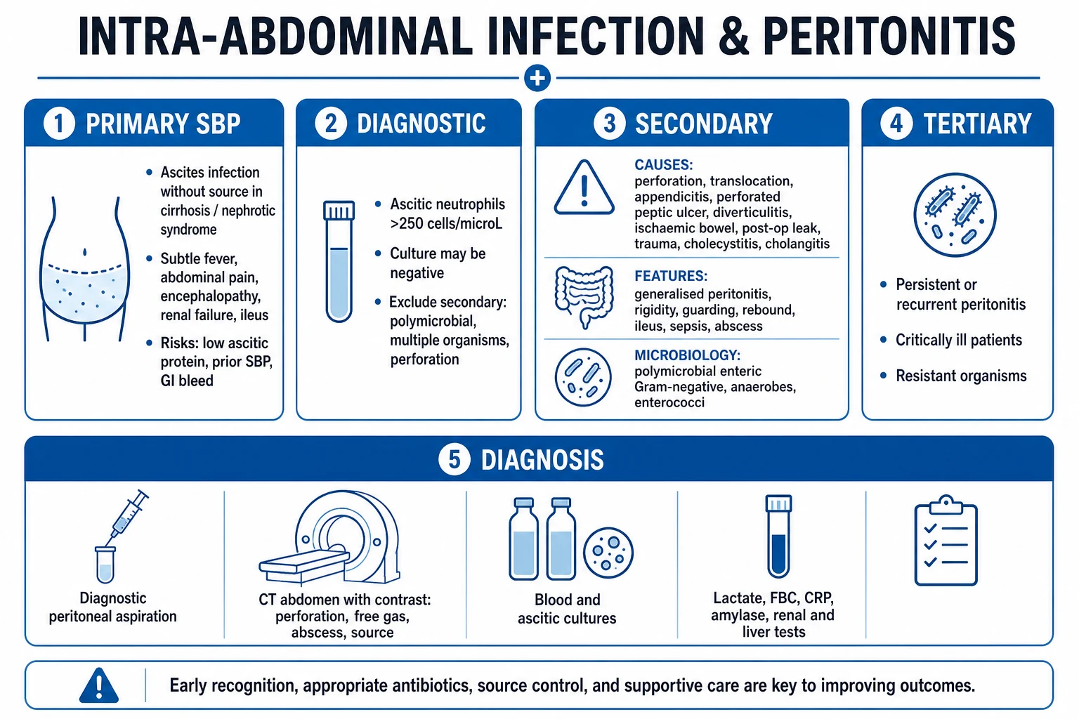
Peritonitis is classified by whether there is an intra-abdominal source requiring physical control, by the temporal relationship to prior treatment, and by the microbial pattern. The three categories — primary (SBP), secondary, and tertiary — have different aetiologies, organisms, diagnostic tests, and treatments, and confusing them is a classic and dangerous error.[1][2][6]
Primary (SBP) vs secondary vs tertiary peritonitis — the defining comparison
| Feature | Primary (SBP) | Secondary | Tertiary |
|---|---|---|---|
| Definition | Infection of ascites WITHOUT an intra-abdominal source | Infection FROM an intra-abdominal source requiring physical control | Persistent/recurrent peritonitis after >48h of treatment of secondary, or persistent IAI in the critically ill |
| Population | Cirrhosis with ascites (also nephrotic syndrome, CAPD) | Any patient with abdominal pathology (perforation, ischaemia, leak) | Critically ill ICU patients after surgery for secondary peritonitis |
| Source | None (bacterial translocation across oedematous gut/lymphatics) | Perforated viscus, leak, abscess, infarcted bowel | Often no single drainable source; diffuse, low-grade |
| Microbiology | MONOMICROBIAL: E. coli (~70%), Klebsiella, then S. pneumoniae, Enterococcus; anaerobes rare | POLYMICROBIAL: gram-negative bacilli + anaerobes (Bacteroides) + Enterococcus | MDR: Enterococcus (incl. VRE), Pseudomonas, Acinetobacter, MRSA, Stenotrophomonas, Candida |
| Diagnosis | Ascitic PMN >250/mm3 (cell count first; culture in blood-culture bottles) | CT abdomen (free gas, collection, fat stranding); diagnostic laparoscopy/laparotomy | Persistent sepsis + positive cultures from peritoneal fluid/drain; CT to exclude new source |
| Source control | NOT required (no source) | MANDATORY (drain/debride/divert; surgery or IR) | Re-assess for occult source; often none drainable |
| Antibiotics | Cefotaxime/ceftriaxone x5 days (+ albumin) | Broad-spectrum (piptazo OR carbapenem + metronidazole); short course | Culture-directed, broad + antifungal; de-escalate to sensitivities |
| Mortality | 10-30% (lower with albumin) | 5-20% (community); up to 25-30% (high-risk/healthcare-associated) | 30-50% |
The essential triage question at the bedside is: Is there an intra-abdominal source that requires physical control? If yes (and the patient is not cirrhotic with ascites) this is secondary peritonitis and the patient needs source control in addition to antibiotics. If the patient is cirrhotic with ascites and there is no evidence of a perforation, it is SBP and surgery is not only unnecessary but harmful. If the patient has already been treated for secondary peritonitis for >48h and is not improving, suspect tertiary peritonitis with MDR organisms.[2][6]
Spontaneous bacterial peritonitis (SBP)
SBP is the infection of ascitic fluid in a cirrhotic patient (or, rarely, a patient with nephrotic syndrome or heart-failure ascites) in the absence of an intra-abdominal surgical source. It is the archetype of primary peritonitis and is one of the highest-yield, most distinctively-managed topics in the ICU syllabus.[3]
Pathophysiology
Cirrhosis produces portal hypertension, splanchnic congestion, and a profoundly dysfunctional gut barrier. Bacterial translocation — the migration of enteric bacteria across the oedematous, permeable intestinal wall into mesenteric lymph nodes and the systemic circulation — seeds the ascitic fluid. The ascites itself is a poor defensive medium: it has low opsonic/complement activity (low ascitic protein), so bacteria that would be cleared in normal peritoneum proliferate unchecked. The result is a monomicrobial infection, overwhelmingly by enteric gram-negative aerobic rods. Impaired reticuloendothelial (Kupffer cell) clearance and cirrhotic immune paresis complete the setup.[3]
Diagnosis — the cell count, not the culture
The diagnostic test is diagnostic paracentesis with a cell count. The threshold is an ascitic polymorphonuclear (PMN) leucocyte count >250 cells/mm3. This is the single most important number in SBP. Treat on the cell count — do NOT wait for culture, because approximately half of genuine SBP episodes are culture-negative (low bacterial load, fastidious organisms, or prior partial antibiotic exposure). When SBP is suspected, inoculate ascitic fluid into blood culture bottles (aerobic and anaerobic) at the bedside before giving antibiotics — this roughly doubles culture yield and guides later de-escalation.[3]
Diagnostic thresholds and terminology in cirrhotic ascites
| Condition | Ascitic PMN count | Ascitic culture | Action |
|---|---|---|---|
| SBP | >250/mm3 | Positive (often) | Cefotaxime + albumin |
| Culture-negative neutrocytic ascites (CNNA) | >250/mm3 | Negative | Treat IDENTICALLY to SBP (same organisms, same outcomes) |
| Bacterascites | <250/mm3 | Positive (single organism) | Repeat tap; treat if PMN rises or symptoms persist |
| Secondary bacterial peritonitis (a surgical source) | Very high (often >1000, but variable), polymicrobial | Polymicrobial | Search for a source (CT); source control + broad antibiotics |
| Poly-microbial bacterascites | Variable | Multiple organisms / anaerobes | Suspect bowel perforation — imaging + surgery |
Clinical triggers for paracentesis: perform a diagnostic tap in ANY cirrhotic with ascites who has new abdominal pain, fever, fever of unknown source, hepatic encephalopathy, worsening renal function, leucocytosis, or unexplained clinical deterioration. A low threshold is essential — SBP can be subtle and the consequences of missing it (hepatorenal syndrome, death) are severe. Routine prophylactic fresh frozen plasma or platelets before a paracentesis are NOT recommended — coagulopathy does not meaningfully increase bleeding risk from a tap.[3]
Treatment — cephalosporin + albumin
SBP treatment — the evidence-based regimen
| Element | Drug / dose | Rationale |
|---|---|---|
| Antibiotic | Cefotaxime 2 g IV q8h (or ceftriaxone 2 g IV daily) for 5 days | Third-generation cephalosporin covers >95% of causative organisms (E. coli, Klebsiella, S. pneumoniae); 5 days is as effective as 10 |
| Albumin | 1.5 g/kg on day 1, then 1 g/kg on day 3 | Prevents hepatorenal syndrome; reduces mortality (Sort 1999, NEJM) |
| Alternative antibiotic (if septic/healthcare-exposure/resistance) | Piperacillin-tazobactam or carbapenem | For nosocomial SBP or known ESBL/resistant organisms |
| Oral option (uncomplicated, non-septic, no prior quinolone prophylaxis) | Ofloxacin 400 mg BD | Only for carefully selected uncomplicated cases — still give albumin |
| Repeat tap | At 48h | PMN should fall by >25% if on appropriate therapy; if not, reconsider organism/resistance/source |
Albumin is the pivotal adjuvant. SBP provokes a marked decrease in systemic vascular resistance and effective arterial blood volume, which in the cirrhotic precipitates the feared complication of hepatorenal syndrome (HRS). The landmark trial by Sort and colleagues (NEJM 1999) randomised SBP patients to cefotaxime + albumin vs cefotaxime alone: albumin reduced HRS from 33% to 10% (a 67% relative reduction) and in-hospital mortality from 29% to 10% (a 65% relative reduction). Albumin is therefore standard of care for all SBP patients, and is non-negotiable in those with risk factors for renal deterioration (bilirubin >68 umol/L, creatinine >88 umol/L, blood urea nitrogen >30 mg/dL).[3]
Who gets albumin in SBP — risk-stratified approach
| Group | Recommendation | Reasoning |
|---|---|---|
| All SBP patients | Give albumin 1.5 g/kg day 1 + 1 g/kg day 3 | Sort 1999 trial evidence; reduced HRS and mortality |
| Bilirubin >68 umol/L (4 mg/dL) OR creatinine >88 umol/L (1 mg/dL) | Definitely give — highest HRS risk | These were the highest-risk subgroup in the trial |
| BUN >30 mg/dL | Definitely give | Marker of impending HRS |
| Very low-risk SBP (normal renal function, normal bilirubin) | Still recommended (evidence-based), but benefit is smaller | Harm is minimal; consistency is safest |
SBP prophylaxis
Secondary prophylaxis (after a first SBP episode) is lifelong — SBP recurs in ~70% within a year without it. Use norfloxacin 400 mg daily or ciprofloxacin 500 mg daily. Primary prophylaxis is indicated for ascitic protein <15 g/L with concomitant renal/liver dysfunction, and short-term prophylaxis is mandatory after a gastrointestinal bleed in cirrhosis (7 days of norfloxacin reduces infection and bacterial translocation). Long-term quinolone prophylaxis selects quinolone-resistant organisms and shifts the microbiology toward gram-positives — a reason to remain vigilant.[3]
Secondary peritonitis — the disease of source control
Secondary peritonitis is by far the commonest form in surgical ICU practice. It arises whenever the integrity of the gastrointestinal tract is breached and endogenous bacteria contaminate the peritoneum. The cardinal principle — repeated in every guideline — is that source control is mandatory and antibiotics are an adjunct, not a substitute.[1][2][4]
Aetiology — the sources you must find and fix
Causes of secondary peritonitis and their source-control specifics
| Source | Presentation | Source control |
|---|---|---|
| Appendicitis (perforated) | RLQ pain, fever, sepsis; free gas if perforated | Appendicectomy ± peritoneal lavage |
| Diverticulitis (Hinchey III-IV perforation) | LLQ pain, elderly; purulent/faeculent peritonitis | Hartmann's procedure (or resection+anastomosis in selected); laparoscopic lavage for selected purulent (Ladies trial — caution) |
| Perforated peptic ulcer | Sudden severe epigastric pain, 'board-like' rigidity, free gas under diaphragm | Omental (Graham) patch repair; consider non-op management in sealed, stable perforations |
| Ischaemic bowel / infarction (mesenteric ischaemia) | Pain out of proportion, metabolic acidosis, high lactate, risk factors (AF, vascular disease) | Resection of necrotic bowel ± second-look; treat cause (embolectomy, revascularisation) |
| Anastomotic leak (post-operative, day 3-7) | New fever, ileus, sepsis, drain output (faeculent/bilious/purulent) after surgery | Re-operation (repair/defunction/stoma); IR drainage if contained; consider faecal diversion |
| Bowel obstruction with strangulation/perforation | Colicky pain, distension, peritonism, rising lactate | Resection of ischaemic/ perforated segment ± stoma |
| Post-traumatic bowel injury | Blunt/penetrating abdominal trauma | Repair/resection at laparotomy/laparoscopy |
| Gallbladder/biliary (perforation, bile leak) | RUQ sepsis; bilious peritoneal fluid | Cholecystectomy; bile duct repair/stent for duct injury |
| Abscess (post-operative or de novo) | Localised collection, swinging fever, raised inflammatory markers | Percutaneous (IR) drainage first-line if accessible; surgery if not |
The intensivist's role in secondary peritonitis is to recognise the sick patient, resuscitate, get broad-spectrum antibiotics in EARLY (within one hour of septic shock), localise the source by imaging, and ensure the surgeon/interventional radiologist achieves definitive source control without delay — delay beyond 24 hours from onset independently worsens mortality.[1][2]
Diagnosis — find the source
Diagnostic pathway in suspected severe intra-abdominal infection
- RECOGNISE AND RESUSCITATE — septic peritonitis presents with fever (or hypothermia), tachycardia, tachypnoea, abdominal pain/distension, peritonism (rigidity, guarding, rebound), and evolving septic shock. ABC; IV access; blood cultures; broad-spectrum antibiotics WITHIN 1 HOUR of shock; lactate; crystalloid resuscitation; noradrenaline for vasopressor-dependent shock.
- CT ABDOMEN WITH IV CONTRAST is the diagnostic workhorse — look for (a) FREE INTRAPERITONEAL GAS (perforation of a viscus — often best seen over the liver on an erect chest X-ray or on CT), (b) FREE FLUID or a FLUID COLLECTION/ABSCESS (rim-enhancing, gas-containing), (c) FAT STRANDING and bowel-wall thickening (inflammation/ischaemia), (d) lack of bowel-wall enhancement (ischaemia/infarction), (e) the responsible lesion (appendicolith, diverticula, stricture, mass). CT guides whether the source is amenable to IR drainage or requires surgery.
- DIAGNOSTIC PARACENTESIS if ascites is present (cirrhotic) — cell count and culture to diagnose/exclude SBP and to distinguish monomicrobial SBP from polymicrobial secondary peritonitis.
- ERECT CHEST X-RAY — free air under the diaphragm (pneumoperitoneum) is the classic sign of a perforated viscus, though CT is more sensitive.
- BEDSIDE ULTRASOUND / FAST — useful in the unstable trauma patient and to identify free fluid or a target for drainage; CT is preferred for definitive source localisation when the patient is stable enough to travel.
- DIAGNOSTIC LAPAROSCOPY / LAPAROTOMY — both diagnostic and therapeutic; laparoscopy is increasingly first-line for perforated appendicitis, perforated ulcer, and selected perforations in the stable patient; laparotomy for the unstable or when laparoscopy is non-diagnostic.
- DIAGNOSTIC PERITONEAL LAVAGE (DPL) — now largely supplanted by CT and FAST ultrasound, but still occasionally used in trauma when CT is unavailable and FAST is negative yet suspicion is high: positive if RBC >100,000/mL (hollow viscus injury) or bile/food/faeces (bowel injury). Microscopic DPL (>5,000/mm3 and a benign FAST) is less reliable.
- REPEAT ASSESSMENT — a patient who is not improving despite 'adequate' treatment at 48-72h warrants repeat imaging to look for an undrained collection, ongoing leak, or the evolution to tertiary peritonitis.
The microbiological principle: obtain cultures BEFORE antibiotics when feasible, but never delay antibiotics in septic shock. Blood cultures, peritoneal fluid cultures (in blood-culture bottles), drain/pus cultures, and swabs guide de-escalation. Specimens from the operative field are especially valuable.[2][6]
Source control — the defining principle (drain, debride, divert)
Source control is defined as 'all physical measures undertaken to eliminate a source of infection and to restore anatomy and function'. Its four components are best remembered as the '3 Ds plus a second look':[1][6]
The components of source control
| Component | What it means | Examples |
|---|---|---|
| DRAIN | Drain pus, contaminated fluid, and gas from the peritoneal cavity or a collection | Peritoneal lavage at laparotomy; percutaneous catheter drainage of an abscess; surgical drains |
| DEBRIDE | Remove necrotic and non-viable tissue that sustains infection | Resection of necrotic bowel; debridement of necrotic tissue |
| DIVERT | Defunction the gastrointestinal tract to stop ongoing contamination | Resection with stoma (Hartmann's) rather than primary anastomosis in the septic/contaminated patient; defunctioning ileostomy |
| (Re-)ASSESS / SECOND LOOK | Confirm the source is controlled; detect persistent/recurrent infection | Planned re-laparotomy; repeat CT; open abdomen for re-look |
The choice between surgery and interventional radiology (IR) depends on the source, accessibility, stability, and local expertise. IR-guided percutaneous drainage is first-line for a discrete, accessible abscess (post-operative collection, peri-appendiceal abscess), often avoiding surgery entirely. Surgery is required for diffuse peritonitis (free perforation), ischaemic/infarcted bowel, uncontrolled leak, failed IR drainage, or haemodynamic instability without a drainable target.[1][2]
Damage control — for the deranged patient
When the patient is physiologically deranged — the lethal triad of hypothermia, acidosis, and coagulopathy — a prolonged definitive operation will kill them. The principle of damage control surgery is to perform the minimum necessary to control contamination and bleeding, then temporarily close the abdomen, move to the ICU for resuscitation/correction of physiology, and return to theatre for definitive repair once stabilised.[6]
Damage control laparotomy for severe intra-abdominal infection
- RECOGNISE the deranged physiology — acidosis (pH <7.2), hypothermia (<35 C), coagulopathy, massive transfusion, lactate rising, vasopressor-dependent shock. These patients cannot tolerate a long definitive operation.
- CONTROL CONTAMINATION FAST — rapidly find and control the perforation/leak (oversew, staple off, resect); control haemorrhage (packing). Do NOT attempt a delicate primary anastomosis in a shocked, acidotic patient — it will leak.
- DIVERT, do not anastomose — resect ischaemic/infarcted bowel and bring out a stoma (e.g. Hartmann's for sigmoid perforation); this minimises the chance of a second leak in a hostile, contaminated abdomen.
- PERITONEAL LAVAGE — wash out the contamination with several litres of warm saline to reduce bacterial and particulate load.
- TEMPORARY ABDOMINAL CLOSURE (open abdomen) — apply a negative-pressure wound therapy (VAC) dressing or other temporary closure; do NOT force a tight primary closure that would cause abdominal compartment syndrome.
- ICU RESUSCITATION — correct acidosis (restore perfusion, fluids/blood, vasopressors, inotropes), warm the patient, correct coagulopathy (blood products, calcium), support organs (ventilation, renal replacement therapy).
- PLANNED RE-LAPAROTOMY (24-48h) — return to theatre once physiology corrected for definitive repair, washout, and inspection for missed injuries/necrosis; adopt an 'on-demand' or 'planned' re-look strategy depending on the situation.
- DELAYED DEFINITIVE CLOSURE — aim for primary fascial closure during the same admission once the abdomen is clean and physiology restored; manage the open abdomen with NPWT in the interim.
The damage-control philosophy explicitly accepts an incomplete initial operation in exchange for survival — the operation is resuscitative, not definitive. The intensivist and surgeon must communicate continuously, because the decision to re-operate (planned vs on-demand) and the timing of definitive closure are made jointly.[1][6]
Empiric antibiotics — broad gram-negative + anaerobic cover
Empiric antibiotic therapy must cover enteric gram-negative bacilli, anaerobes (Bacteroides), and — depending on setting and severity — Enterococcus and Candida. Therapy is started EARLY (within one hour of septic shock), broad initially, then DE-ESCALATED to culture sensitivities. Choice is stratified by community-acquired vs healthcare-associated and by severity.[2][4]
Empiric antibiotic regimens by severity and setting
| Setting / severity | First-line regimen | Notes |
|---|---|---|
| Community-acquired, low-moderate severity | Cefuroxime + metronidazole OR co-amoxiclav + metronidazole OR ceftriaxone + metronidazole | Cover enteric GNB + anaerobes; Enterococcus cover not usually needed for community source |
| Community-acquired, high severity / septic shock | Piperacillin-tazobactam OR cefepime/ceftazidime + metronidazole | Broad GNB (incl. Pseudomonas) + anaerobes |
| Healthcare-associated / nosocomial / post-operative / high-risk | Piperacillin-tazobactam OR carbapenem (meropenem) + metronidazole; add vancomycin/linezolid if Enterococcus/VRE risk | Must cover Enterococcus and resistant GNB; add antifungal if Candida risk |
| Severe sepsis/shock, ESBL/resistance risk | Carbapenem (meropenem) ± aminoglycoside; + metronidazole; + antifungal (echinocandin) | Maximal broad cover; de-escalate aggressively to cultures |
| Suspected/confirmed fungal peritonitis | ADD echinocandin (caspofungin/micafungin) initially; fluconazole/amphotericin if susceptible/stable | See fungal section |
When to extend cover — Enterococcus and antifungal
| Add cover for… | Indications | Agent |
|---|---|---|
| Enterococcus | Healthcare-associated/post-operative infection, prior cephalosporins/fluoroquinolones, immunocompromised, severe sepsis, valvular heart disease | Ampicillin (if susceptible) or vancomycin; linezolid/daptomycin for VRE |
| Candida (antifungal) | Recurrent/ongoing intra-abdominal infection, recent broad-spectrum antibiotics, multiple abdominal surgeries, severe necrotising pancreatitis, immunosuppression, prolonged ICU stay, Candida colonisation at multiple sites | Echinocandin (caspofungin/micafungin) if critically ill/unstable; fluconazole if stable and likely susceptible; amphotericin B as alternative; de-escalate to susceptibility and source control |
| MRSA | Known colonisation, healthcare-associated, severe sepsis, line/SSI risk | Vancomycin or linezolid |
| MDR gram-negatives (ESBL, CRE, Acinetobacter, Pseudomonas) | Healthcare-associated, recent antibiotics/healthcare exposure, immunocompromised, known colonisation | Carbapenem (meropenem/imipenem); consider polymyxin/colistin, aminoglycoside, tigecycline per sensitivities and ID advice |
The recurring mistake is to continue the broad empiric regimen for days once cultures are back. The disciplined approach is start broad within the hour, obtain good cultures and source control, then DE-ESCALATE to the narrowest effective agent guided by sensitivities — this is antimicrobial stewardship applied to intra-abdominal infection.[2]
Antibiotic duration — short is standard (STOP-IT)
Prolonged antibiotic courses were once routine after intra-abdominal infection; they are no longer justified. The pivotal trial is STOP-IT (Sawyer et al., NEJM 2015), which randomised patients with complicated intra-abdominal infection and adequate source control to approximately 4 days vs ~8 days of antibiotics.[2]
Duration of antibiotics after adequate source control — the evidence
| Source / guideline | Recommended duration | Basis |
|---|---|---|
| STOP-IT trial (NEJM 2015) | ~4 days after adequate source control | No difference in surgical-site infections, recurrent IAI, or mortality vs ~8 days |
| SIS 2017 / IDSA-SIS 2010 | Until source control achieved + a defined SHORT course (~4-7 days) | Stewardship; prolonged courses increase resistance, Candida, C. difficile |
| WSES 2017 | Generally 4-7 days after adequate source control; stop sooner if clinical recovery | Duration tied to clinical response, not a fixed number |
| Exceptions needing LONGER courses | (1) Source control inadequate/unachieved; (2) persistent bacteraemia; (3) fungaemia; (4) immunocompromise; (5) some specific sources | Clinical judgement; re-evaluate daily |
Stop antibiotics when: the patient is clinically improving (afebrile, haemodynamically stable, resolving ileus, falling inflammatory markers, tolerating diet) AND source control is adequate. Do NOT treat a falling but elevated CRP or a residual collection on imaging in a recovering patient — clinical recovery is the endpoint, not a number.[2]
Tertiary peritonitis — the MDR, immune-paresis syndrome
Tertiary peritonitis is persistent or recurrent peritonitis that develops after >48 hours of apparently appropriate treatment of secondary peritonitis, or persistent intra-abdominal infection in the critically ill without an obvious drainable source. It reflects a combination of MDR infection and the immune paralysis (immunoparesis) of critical illness more than a simple failure of source control.[6]
Tertiary peritonitis — the defining features
| Feature | Tertiary peritonitis |
|---|---|
| Definition | Persistent/recurrent peritonitis >48h after treatment of secondary, or persistent IAI in critically ill |
| Pathophysiology | MDR organisms + immune paresis of critical illness (low-grade, diffuse, often no single drainable focus) |
| Organisms | MDR — Enterococcus (incl. VRE), Pseudomonas aeruginosa, Acinetobacter, MRSA, Stenotrophomonas, and Candida |
| Diagnosis | Persistent/recurrent sepsis despite source control + positive cultures from peritoneal fluid/drain; CT to exclude a new/missed source |
| Management | (1) Re-assess source control (repeat imaging, re-operation if a source is found); (2) CULTURE-DIRECTED broad-spectrum therapy targeting MDR — vancomycin/linezolid (Enterococcus/MRSA), anti-pseudomonal carbapenem (Pseudomonas/Acinetobacter), echinocandin (Candida); (3) aggressive ICU organ support; (4) de-escalate to sensitivities |
| Mortality | 30-50% |
The intensivist must distinguish failure of source control (an undrained collection, ongoing leak, ischaemic bowel — a SURGICAL problem needing re-operation) from true tertiary peritonitis (MDR/immunoparesis with no single drainable source — a MEDICAL/stewardship problem needing culture-directed antibiotics and organ support). The first is fixed in theatre; the second is not. Both may coexist. Re-imaging (CT) and repeated microbial sampling are the tools to separate them.[6]
Fungal (Candida) peritonitis
Candida peritonitis is most often encountered after gastrointestinal perforation or recurrent abdominal surgery, where Candida (which colonises the gut) contaminates the peritoneum along with bacteria. It is NOT an indication to treat every patient with Candida isolated from a peritoneal swab — transient colonisation is common and self-limiting after source control. Treatment is indicated when Candida is isolated from a sterile intra-abdominal specimen in a patient with intra-abdominal infection (especially critically ill, recurrent, or with risk factors) or with candidaemia.[2]
Candida intra-abdominal infection — who to treat and how
| Issue | Recommendation |
|---|---|
| When to treat | Candida from a sterile intra-abdominal specimen in a patient with clinical IAI (esp. post-operative peritonitis, recurrent/necrotising pancreatitis, immunocompromised); candidaemia |
| Risk factors | Prolonged broad-spectrum antibacterials, recent abdominal surgery (especially multiple), severe necrotising pancreatitis, immunosuppression, prolonged ICU stay, total parenteral nutrition, Candida colonisation at multiple sites |
| Initial therapy (critically ill / unstable) | Echinocandin (caspofungin loading 70 mg then 50 mg daily; micafungin 100 mg daily) |
| Step-down (stable, susceptible) | Fluconazole (after clinical response and if C. albicans / susceptible species) |
| Alternatives | Amphotericin B (liposomal preferred); voriconazole for selected species |
| Duration | Until source control achieved, clinical recovery, and (if bacteraemic/fungaemic) cultures negative |
| Source control | Essential — remove infected/necrotic tissue and foreign material; antifungals fail without it |
Amphotericin B (or its liposomal formulation) remains a valid alternative for fungal peritonitis, especially where echinocandins are unavailable or for resistant species, but echinocandins are now preferred first-line in the critically ill because of their better safety profile. Fluconazole is a reasonable choice in the stable patient with a likely-susceptible organism (C. albicans).[2][4]
Open abdomen management
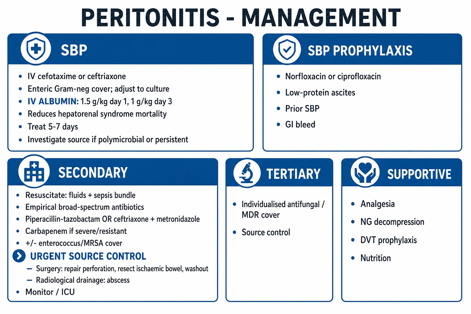
Leaving the abdomen open (laparostomy) is a deliberate strategy in severe intra-abdominal infection when (a) primary closure would cause abdominal compartment syndrome (the swollen, oedematous bowel cannot be contained without raising intra-abdominal pressure to harmful levels), (b) a planned second-look is required (to check for bowel viability after ischaemia, or to confirm source control), or (c) the patient is too unstable for a definitive closure (damage control). The WSACS 2013 consensus defined the open abdomen, proposed a classification system, and made specific recommendations for its management.[5]
Techniques for temporary abdominal closure / open abdomen management
| Technique | Description | Role |
|---|---|---|
| Negative-pressure wound therapy (NPWT / VAC) | A porous sponge/foam dressing over the exposed viscera, sealed with an adhesive drape, connected to continuous negative pressure (~125 mmHg) | Preferred temporary closure; removes fluid, reduces oedema, promotes granulation, facilitates delayed fascial closure; WSACS RECOMMENDATION |
| Bogota bag | A sterile plastic (IV-fluid-bag) sheet sewn to the skin over the viscera | Historical/simple temporary cover; less effective than NPWT |
| Wittmann patch / dynamic retention sutures | Synthetic sheet with hook-and-loop Velcro-like layers allowing progressive tensioning | Aids progressive fascial closure over days |
| Absorbable / biological mesh | Bridge the fascial defect | Usually for when primary closure impossible; NOT for routine early use (WSACS suggests avoiding routine early biologic mesh) |
| Skin-only closure | Close the skin but not the fascia | Temporary; leaves a planned ventral hernia to repair later |
Goals and milestones of open abdomen management
- PROTECT THE VISCERA from desiccation, fistulation, and infection — cover exposed bowel with a non-adherent interface beneath the NPWT sponge (a key modification to reduce enteric fistula risk).
- CONTROL FLUID and REDUCE OEDEMA — NPWT removes exudate; avoid positive fluid balance after resuscitation (a WSACS suggestion to limit IAH).
- MONITOR INTRA-ABDOMINAL PRESSURE (bladder pressure) — the open abdomen treats overt ACS; if the abdomen is closed, IAP must be monitored to detect recurrent IAH/ACS.
- PLAN RE-LAPAROTOMY — decide between PLANNED (mandatory scheduled return, e.g. for ischaemia second-look) and ON-DEMAND (return only if not improving) re-look; modern practice favours on-demand re-look in most peritonitis (fewer operations, no outcome detriment).
- ACHIEVE DELAYED PRIMARY FASCIAL CLOSURE — aim to close the fascia during the SAME admission (WSACS recommendation); use progressive tensioning or NPWT-assisted closure; the longer the abdomen stays open, the higher the fistula, infection, and hernia rates.
- AVOID / MANAGE ENTERIC FISTULA — the most feared complication of the open abdomen; prevent with visceral protection and early closure; manage with NPWT modification, sepsis control, nutritional optimisation, and definitive surgery later.
- NUTRITION — early enteral nutrition is the default (maintains gut barrier, reduces translocation); the open abdomen is NOT a contraindication to enteral feeding.
The WSACS 2013 definitions are worth knowing: intra-abdominal hypertension (IAH) = sustained IAP >12 mmHg; abdominal compartment syndrome (ACS) = sustained IAP >20 mmHg WITH new organ dysfunction. The open abdomen is both a consequence (decompression for ACS) and a planned strategy (to prevent ACS in the at-risk patient).[5]
Abdominal compartment syndrome (ACS) — when the abdomen itself fails
ACS is defined as a sustained intra-abdominal pressure >20 mmHg with new-onset organ failure. It complicates severe intra-abdominal infection (and severe pancreatitis, trauma, massive fluid resuscitation) when oedematous, fluid-loaded viscera raise IAP to the point where perfusion of the kidneys, gut, and lungs collapses.[5]
Effects of raised intra-abdominal pressure — organ by organ
| System | Effect of raised IAP | Clinical sign |
|---|---|---|
| Renal | Reduced renal perfusion / venous congestion | Oliguria, rising creatinine (mimics and precipitates AKI) |
| Cardiovascular | Reduced venous return, raised intrathoracic pressure, reduced cardiac output | Hypotension, raised CVP/PAP (misleadingly), poor perfusion |
| Respiratory | Elevated diaphragm, reduced thoracic compliance | Raised peak airway pressures, hypoxia, hypercapnia, difficulty ventilating |
| Gut | Splanchnic ischaemia, bacterial translocation | Ileus, worsening acidosis, liver dysfunction |
| Cerebral | Raised intracranial pressure (via raised intrathoracic pressure) | Worsened cerebral perfusion in brain-injured patients |
Monitor bladder pressure in any at-risk patient (severe intra-abdominal infection/pancreatitis with massive fluid resuscitation, oliguria unresponsive to fluid, refractory hypoxaemia/raised airway pressures). A staged approach to management: reduce intra-abdominal volume (NG/rectal decompression, diurese/reduce fluids, neuromuscular blockade), and surgical decompressive laparotomy (open abdomen) if refractory — decompression can be dramatically life-saving.[5]
Special situations
Post-operative peritonitis (anastomotic leak): presents typically day 3-7 after surgery with new fever, ileus, abdominal pain, sepsis, and drain output (faeculent, bilious, or purulent). CT with WATER-SOLUBLE contrast (never barium — risk of chemical peritonitis if extravasation) confirms the leak. Management is source control (re-operation: repair/defunction/stoma; or IR drainage if contained and stable), broad antibiotics, and ICU support. A leak after a low rectal anastomosis may be managed with diversion +/- drainage rather than take-down if contained.[2]
CAPD peritonitis: in continuous ambulatory peritoneal dialysis, peritonitis presents with cloudy dialysate and abdominal pain; diagnosis is dialysate WBC >100/mm3 (after a dwell) with >50% PMNs. Organisms are coagulase-negative Staph (~40%, touch contamination), Staph aureus (~15%, exit-site infection), gram-negatives (~15-20%), and fungal (~2%, needs catheter removal). Treatment is intraperitoneal antibiotics (e.g. vancomycin/cefazolin + ceftazidime/gentamicin, per ISPD guidelines) with prompt catheter removal for fungal, refractory, recurrent, or Pseudomonas peritonitis.[6]
Severe intra-abdominal infection in septic shock: apply the Surviving Sepsis Hour-1 bundle — measure lactate, obtain blood cultures, give broad-spectrum antibiotics and crystalloid, and start vasopressors (noradrenaline) for vasopressor-dependent shock — but recognise that source control is the sepsis-specific intervention and should be achieved as early as feasible (ideally within 6-12 hours of diagnosis). Antibiotics without source control will not resolve septic shock from a perforation.[1]
Clinical pearls
Red flags
[1] [1] [1] [1] [1] [1] [1]Prognosis
Outcomes and prognostic factors in severe intra-abdominal infection and peritonitis
| Factor | Outcome / mortality | Notes |
|---|---|---|
| SBP (with albumin) | 10-20% in-hospital | Higher with renal dysfunction, advanced liver disease; 1-year mortality ~50-70% (underlying cirrhosis) |
| SBP (without albumin) | ~30% in-hospital | HRS 33%; markedly worse |
| Community-acquired secondary peritonitis | <5-10% | Low severity, prompt source control |
| High-severity / healthcare-associated secondary | 20-30% | APACHE II high, age, comorbidity, delay |
| Tertiary peritonitis | 30-50% | MDR organisms, immune paresis |
| Candida peritonitis (critically ill) | 25-40% | Worse with unresolved source, candidemia |
| Predictors of poor outcome | — | Age, APACHE II, comorbidity, delay to source control >24h, inadequate initial source control, inappropriate initial antibiotics, bacteraemia/candidaemia, malnutrition, immunosuppression |
| Recurrent SBP | ~70% within 1 year without prophylaxis | Lifelong secondary prophylaxis (norfloxacin/ciprofloxacin) |
| Open abdomen — same-admission fascial closure | Achieved in ~60-80% with NPWT | Higher fistula/hernia rates if prolonged |
Mortality is driven by three modifiable factors the intensivist controls: (1) the speed and adequacy of source control (the single most important determinant); (2) the appropriateness and timing of antibiotics (early, broad, then de-escalated); and (3) the quality of organ support (resuscitation, ventilation, renal replacement, nutrition). Non-modifiable risk (age, comorbidity, severity at presentation) sets the baseline, but optimal source control, stewardship, and ICU care substantially shift outcome. The patient with SBP additionally depends on albumin to prevent the otherwise near-inevitable progression to hepatorenal syndrome.[1][3][6]
Key trials and evidence
STOP-IT — Short vs Prolonged Antibiotic Course after Source Control (NEJM 2015, Sawyer; PMID 25376890)
Source
Multicentre randomised non-inferiority trial; 518 adults with complicated intra-abdominal infection and adequate source control, 23 US centres
Intervention
Control: antibiotics until ~4-7 days (a defined SHORT course after source control). Intervention: ~8 days (prolonged course)
Primary composite (surgical-site infection + recurrent IAI + death)
21.8% (short) vs 22.3% (prolonged) — non-inferior (within pre-specified margin)
Key finding
A SHORT (~4-day) course after adequate source control is as good as a prolonged (~8-day) course
Clinical bottom line
Stop antibiotics once the patient is clinically improving and source control is adequate — duration tied to recovery, not a fixed number; prolonged courses cause harm (resistance, Candida, C. difficile)
Sort et al — Albumin in SBP (NEJM 1999; PMID 10588909)
Source
Randomised trial; 126 cirrhotic patients with SBP, single centre (Barcelona)
Intervention
Cefotaxime + ALBUMIN (1.5 g/kg day 1, then 1 g/kg day 3) vs cefotaxime alone
Hepatorenal syndrome
10% (albumin) vs 33% (control) — p=0.002 (67% relative reduction)
In-hospital mortality
10% (albumin) vs 29% (control) — p=0.01 (65% relative reduction)
3-month mortality
22% vs 41% — p=0.03
Clinical bottom line
Albumin is STANDARD OF CARE for all SBP (1.5 g/kg day 1 + 1 g/kg day 3), especially with bilirubin >68 umol/L or creatinine >88 umol/L — it prevents HRS and halves mortality
WSACS 2013 — Open Abdomen and Abdominal Compartment Syndrome Consensus (Intensive Care Med 2013, Kirkpatrick; PMID 23673399)
Source
Updated consensus definitions and clinical practice guidelines from the World Society of the Abdominal Compartment Syndrome (modified Delphi + GRADE)
Definitions
IAH = sustained IAP >12 mmHg; ACS = sustained IAP >20 mmHg with new organ failure; defined the open abdomen and an open-abdomen classification system
Key recommendations
Measure IAP in at-risk patients; avoid sustained IAH; protocolised IAP monitoring/management; decompressive laparotomy for overt ACS; negative-pressure wound therapy and efforts to achieve same-admission fascial closure in the open abdomen
Key suggestions
Avoid positive fluid balance after resuscitation; percutaneous catheter drainage for IAH/ACS; avoid routine early biologic mesh in open abdominal wounds
Clinical bottom line
The authoritative reference for IAH/ACS and open-abdomen management — defines when to leave the abdomen open, how to manage it (NPWT/VAC), and how to achieve delayed primary fascial closure
SAQ — Secondary peritonitis and source control
10 minutes · 10 marks
A 68-year-old with perforated diverticulitis is in septic shock on noradrenaline. CT shows free gas and a pelvic collection. The surgical registrar asks whether antibiotics alone for 48 hours are acceptable.
References
- [1]Sartelli M, Chichom-Mefire A, Labricciosa FM, et al. The management of intra-abdominal infections from a global perspective: 2017 WSES guidelines for management of intra-abdominal infections World J Emerg Surg, 2017.PMID 28702076
- [2]Mazuski JE, Tessier JM, May AK, et al. The Surgical Infection Society Revised Guidelines on the Management of Intra-Abdominal Infection Surg Infect (Larchmt), 2017.PMID 28085573
- [3]Runyon BA; AASLD Practice Guidelines Committee. [Efficacy and safety of domestic biofragmentable anastomotic ring in the intestinal anastomosis] Zhonghua Wei Chang Wai Ke Za Zhi, 2012.PMID 22941691
- [4]Solomkin JS, Mazuski JE, Bradley JS, et al. Diagnosis and management of complicated intra-abdominal infection in adults and children: guidelines by the Surgical Infection Society and the Infectious Diseases Society of America Clin Infect Dis, 2010.PMID 20034345
- [5]Kirkpatrick AW, Roberts DJ, De Waele J, et al. Intra-abdominal hypertension and the abdominal compartment syndrome: updated consensus definitions and clinical practice guidelines from the World Society of the Abdominal Compartment Syndrome Intensive Care Med, 2013.PMID 23673399
- [6]Montravers P, Blot S, Dimopoulos G, et al. Therapeutic management of peritonitis: a comprehensive guide for intensivists Intensive Care Med, 2016.PMID 26984317