ICU · GI & nutrition / infection
Severe Diarrhoea & C. difficile — Toxins, Severity & the Vancomycin/Fidaxomicin Era
Also known as C. difficile · Clostridioides difficile · Antibiotic-associated diarrhoea · Pseudomembranous colitis · Toxic megacolon · NAP1 · Ribotype 027 · Fidaxomicin · Oral vancomycin · Bezlotoxumab · Faecal microbiota transplant · FMT
The C. difficile is the key ICU cause of the severe diarrhoea — the antibiotic-associated (the clindamycin, the fluoroquinolones, the cephalosporins), the spore-forming, the toxin A and B producer, the hypervirulent NAP1 or ribotype 027. The spectrum: the diarrhoea to the pseudomembranous colitis to the toxic megacolon to the perforation. The severe (the WCC over 15, the AKI creatinine over 1.5 times, the hypoalbuminaemia, the ileus or the megacolon); the fulminant (the shock, the megacolon, the perforation). The diagnosis: the GDH plus the toxin (the two-step); the PCR detects the colonisation, not the active disease (the over-diagnosis). The treatment (IDSA 2021): the non-severe the fidaxomicin OR the oral vancomycin 125 mg QID; the severe the oral vancomycin; the FULMINANT the vancomycin 500 mg QID plus the IV metronidazole plus-or-minus the rectal vancomycin plus the surgery (the subtotal colectomy for the megacolon). The recurrence: the bezlotoxumab, the faecal microbiota transplant. The stop the antibiotics and the PPI; the soap-and-water hand hygiene (the alcohol gel does not kill the spores); the isolate.
On this page & tools
Your progress
Saved locally on this device.
8 MCQs with explanations
Target exams
Overview & definition
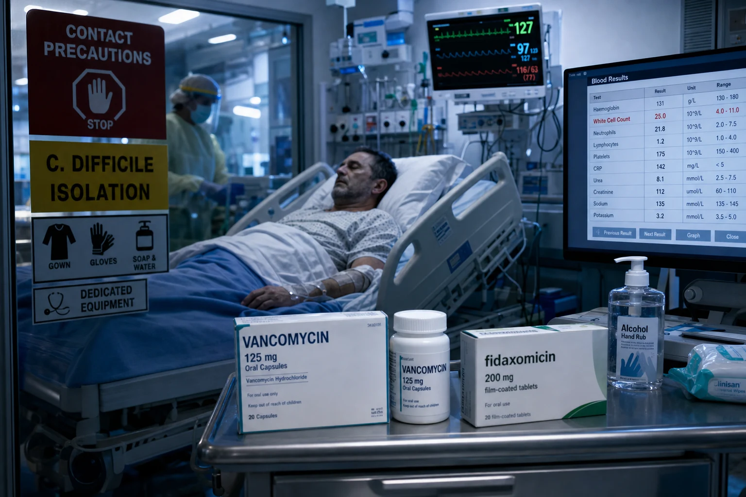
The C. difficile (the Clostridioides difficile) is the key ICU cause of the severe diarrhoea. The antibiotic-associated, the spore-forming, the toxin-producing Gram-positive anaerobe. The spectrum: the mild diarrhoea → the pseudomembranous colitis → the toxic megacolon → the perforation and the sepsis. The fulminant disease (the shock, the megacolon, the perforation) is the ICU emergency. The two priorities: (1) the severity-stratified treatment (the fidaxomicin or the oral vancomycin; the IV metronidazole + the surgery for the fulminant) and (2) the infection control (the isolate, the soap-and-water, the stop the offending antibiotic).[1]
The pathogen and the spectrum
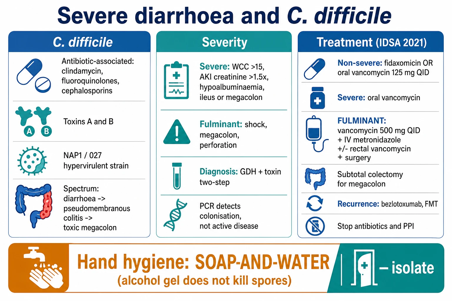
- The antibiotic-associated — the clindamycin (the classic), the fluoroquinolones, the cephalosporins, the broad-spectrum. The antibiotic disrupts the gut flora → the C. difficile overgrowth.[1]
- The toxins A (the enterotoxic) and B (the cytotoxic) — the mediators of the disease. The hypervirulent NAP1 / ribotype 027 strain produces more toxin (the fluoroquinolone-resistant, the more severe, the higher recurrence).[1]
- The spectrum — the mild diarrhoea → the pseudomembranous colitis (the yellow plaques on the mucosa, the "pseudomembranes") → the toxic megacolon (the markedly dilated colon, the perforation risk) → the perforation and the sepsis.[1]
Pathophysiology — the toxin cascade
The chain of events from a healthy colon to pseudomembranous colitis has four linked steps; every management decision targets a specific link.[1][1]
[1]The hypervirulent NAP1 / ribotype 027 strain is the epidemic clone: it produces MORE toxin A and B (a deletion in the tcdC repressor gene removes the brake on toxin production), produces binary toxin, is FLUOROQUINOLONE-RESISTANT (selected out by widespread fluoroquinolone use), and is associated with more severe disease, higher recurrence, and higher mortality. The 2003–2010 North American and European NAP1/027 epidemic reshaped CDI from a clindamycin-associated nuisance into a hypervirulent ICU killer.[9]
Antibiotics — the #1 risk
Highest to lowest risk
- **Clindamycin** — the classic, first-identified association; historically the highest-risk single agent
- **Fluoroquinolones** (ciprofloxacin, levofloxacin, moxifloxacin) — drove the NAP1/027 epidemic by selecting the resistant clone
- **Cephalosporins** (2nd/3rd generation, especially ceftriaxone) — very common ICU culprit
- **Broad-spectrum penicillins** (amoxycillin-clavulanate, piperacillin-tazobactam) — moderate risk
- **Carbapenems** (meropenem, imipenem) — high risk via profound anaerobe kill
- Risk is HIGHEST during antibiotic therapy and for ~4 weeks AFTER cessation; persists at lower level for ~90 days
Host factors
Non-antibiotic
- **Age > 65 years** — the single strongest host risk factor; 10-fold increased incidence vs younger adults
- **PPI use** — reduced gastric acidity lets ingested spores survive into the colon; ~50–60% increased risk; the most modifiable non-antibiotic risk
- **ICU/healthcare exposure & long hospital stay** — environmental spore pressure; the longer the stay, the higher the risk
- **Chemotherapy / malignancy / neutropenia** — mucosal damage plus immune compromise; stem-cell transplant recipients are highest-risk
- **Immunosuppression** — transplant, HIV, chronic steroids, IBD on biologics
- **Severe comorbidity / renal failure / GI surgery / tube feeding** — all independent risk factors
Protective factors
Reduce risk
- **Antimicrobial stewardship** — the single most effective intervention; narrows spectrum and shortens duration
- **Intact gastric acidity** — PPI avoidance where possible
- **Healthy obligate-anaerobe microbiota** — the basis of colonization resistance
- **Bezlotoxumab** — passive anti-toxin B immunity in high-risk patients
- **Vaccine** ( investigational; not yet in routine use)
The severity
- The severe — the WCC over 15, the AKI (the creatinine over 1.5 times the baseline), the hypoalbuminaemia, the fever, the abdominal pain, the ileus or the megacolon.[1]
- The fulminant — the hypotension or the shock, the ileus, the toxic megacolon, the perforation. The ICU emergency.[1]
Non-severe CDI
WBC <15 AND Cr <1.5x baseline
- Definition: WBC <15 × 10^9/L AND serum creatinine <1.5 × baseline
- Clinical: watery diarrhoea (≥3 unformed stools/24 h), mild abdominal cramping, low-grade or no fever
- No signs of shock, no megacolon, no peritonism
- Treatment: fidaxomicin 200 mg BD OR oral vancomycin 125 mg QID x 10 days
Severe CDI
WBC ≥15 OR Cr ≥1.5x baseline
- Definition: WBC ≥15 × 10^9/L OR serum creatinine ≥1.5 × baseline (IDSA/SHEA criteria)
- Additional markers: serum albumin <30 g/L, marked leucocytosis, fever, abdominal pain
- A leucocytoid reaction with WBC >30 or a bandemia is a classic clue to severe CDI in the ICU
- Treatment: oral vancomycin 125–500 mg QID (fidaxomicin also acceptable); add IV metronidazole if fulminant features develop
Fulminant CDI
Shock / ileus / megacolon / perforation
- Definition: hypotension or shock (need for vasopressors), ileus, toxic megacolon, or perforation
- PARADOX: fulminant CDI may have LITTLE OR NO DIARRHOEA (ileus masks it) — suspect in any ICU patient with unexplained marked leucocytosis and abdominal distension
- Imaging: caecum >9 cm or transverse colon >6 cm = toxic megacolon; free gas = perforation
- Treatment: oral/NG vancomycin 500 mg QID + IV metronidazole 500 mg TDS + rectal vancomycin if ileus + urgent surgical review
The diagnosis
- The two-step algorithm — the GDH (the glutamate dehydrogenase, the antigen) plus the toxin A and B (the EIA). The GDH-positive and the toxin-positive = the active disease. The GDH-positive and the toxin-negative = the colonisation or the low-toxin disease; the confirm with the NAAT/PCR.[1]
- The PCR/NAAT — detects the gene (the colonisation), not the active toxin disease → the over-diagnosis. The use to resolve the discordant GDH-toxin, NOT as the first-line.[1]
- The sigmoidoscopy/colonoscopy — the pseudomembranes (the diagnostic of the pseudomembranous colitis), but the avoided in the severe (the perforation risk). The CT — the colonic wall thickening, the "handprint" oedema, the megacolon.[1]
The two-step diagnostic algorithm — step-by-step
Step 0 — WHO to test
Test only patients with ≥3 unformed stools in 24 h who are NOT on a laxative, and who are symptomatic. DO NOT test formed stool. DO NOT test asymptomatic patients (colonisation in 5–20% of inpatients). DO NOT perform a "test of cure" — toxin may persist for weeks after clinical cure.
Step 1 — GDH screening (the antigen)
Glutamate dehydrogenase (GDH) EIA detects a C. difficile antigen produced by ALL strains (toxigenic and non-toxigenic). High negative predictive value: GDH-negative → CDI effectively excluded. Fast (15–45 min), cheap. GDH detects COLONISATION or infection — does not prove toxin production.
Step 2 — Toxin A/B EIA (if GDH positive)
Enzyme immunoassay for toxin A and B in the SAME stool sample. Toxin-positive = active CDI (treat). GDH+/toxin+ → diagnose and treat. GDH+/toxin− → discordant; this reflects either colonisation with a low-level-toxin producer OR genuine CDI below the toxin assay detection threshold. Go to step 3.
Step 3 — NAAT/PCR (resolve discordance)
Nucleic acid amplification for the toxin gene (tcdA/tcdB). NAAT+ in a GDH+/toxin− patient: confirms toxigenic C. difficile is present but cannot distinguish colonisation from active disease. Treat if clinical syndrome fits (esp. if high WBC, AKI, or characteristic imaging). NAAT− in a GDH+/toxin− patient: colonisation with a non-toxigenic strain; look for another cause of the diarrhoea.
Step 4 — Endoscopy / imaging (selected cases)
Flexible sigmoidoscopy for pseudomembranes is diagnostic of pseudomembranous colitis but is reserved for atypical presentations or when stool testing is delayed. AVOID full colonoscopy in severe/filmfulminant disease (perforation risk). CT abdomen shows colonic wall thickening, "handprint"/"thumbprinting" oedema, ascites, and megacolon — useful in the ileus/filmfulminant patient where stool cannot be obtained.
GDH EIA
Antigen screen
- Detects glutamate dehydrogenase — present in ALL C. difficile strains (toxigenic and non-toxigenic)
- High sensitivity (~95%) and high NPV — excellent rule-OUT
- Cannot distinguish colonisation from active disease
- Cheap, fast (15–45 min); first step in the two-step algorithm
Toxin A/B EIA
Active toxin
- Detects active toxin A and B — the marker of disease, not just colonisation
- Moderate sensitivity (~63–94%), high specificity — a positive proves active CDI
- GDH+ / toxin+ → active CDI; treat. GDH+ / toxin− → discordant, proceed to NAAT
- Fast, cheap; the second step in the two-step algorithm
NAAT / PCR
Gene detection
- Detects toxin gene (tcdA/tcdB) — highly sensitive and specific for the organism
- Detects COLONISATION as well as disease → over-diagnoses if used as first-line
- Use ONLY to resolve a discordant GDH+/toxin− result, NOT as a stand-alone test
- Do NOT use for test-of-cure; toxin gene may persist for weeks after cure
The treatment
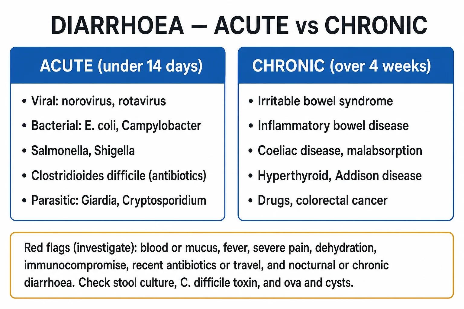
(IDSA 2021) [1]
The non-severe initial episode
- The fidaxomicin (the preferred — the lower recurrence) OR the oral vancomycin 125 mg QID for the 10 days.[1]
- The metronidazole is the demoted (only if the fidaxomicin and the vancomycin unavailable, and for the non-severe).[1]
The severe initial episode
- The oral vancomycin 125 mg QID for the 10 days (or the fidaxomicin).[1]
The fulminant (the shock, the megacolon, the perforation)
- The oral vancomycin 500 mg QID PLUS the IV metronidazole 500 mg TDS.[1]
- The rectal vancomycin (the enema) if the ileus (the oral route not reaching the colon).[1]
- The urgent surgical review — the subtotal colectomy for the toxic megacolon, the perforation, or the failure to improve. The high mortality.[1]
The recurrence
- The bezlotoxumab (the monoclonal antibody against the toxin B) — for the recurrence prevention in the high-risk (the elderly, the severe, the multiple recurrences). The single dose.[1]
- The faecal microbiota transplant (FMT) — for the multiple recurrences; the restores the gut flora.[1]
- The fidaxomicin or the tapered-pulsed vancomycin for the recurrent.[1]
Fidaxomicin (PREFERRED)
Macrocyclic, narrow spectrum
- Dose: 200 mg PO BD x 10 days (extended-pulsed regimen 200 mg BD x 5 days then 200 mg OD every 48 h x 5 doses is also effective)
- Mechanism: inhibits bacterial RNA polymerase — different target from vancomycin
- Narrow spectrum: spares the protective obligate anaerobes (preserves colonization resistance)
- Clinical cure ~88%; RECURRENCE ~12–15% (vs ~25% with vancomycin) — the key advantage
- IDSA/SHEA 2021 PREFERRED for an initial episode and first recurrence
Oral vancomycin
Glycopeptide, luminal
- Dose: 125 mg PO QID x 10 days (non-severe); 500 mg PO/NG QID for severe/fulminant
- Mechanism: cell-wall synthesis inhibition; minimal systemic absorption → high luminal levels
- Must be given ORALLY (or via NG) — IV vancomycin does NOT reach the gut lumen
- Clinical cure ~86%; recurrence ~25%
- For the fulminant/ileus patient: add RECTAL vancomycin 500 mg in 100 mL saline QID retention enema
- Taper/pulse regimen for first recurrence: 125 mg QID x 7d → BD x 7d → OD x 7d → q48h x 8d → q72h x 14d
IV metronidazole
Nitroimidazole, systemic
- Dose: 500 mg IV TDS (or 500 mg PO TDS for non-severe if no alternative)
- Mechanism: DNA strand breakage via reactive intermediates
- Reaches colonic lumen via biliary and vascular exudation — useful adjunct in ileus/fulminant
- Now THIRD-LINE for CDI: inferior to vancomycin in severe disease; cumulative neurotoxicity (peripheral neuropathy) with repeated courses
- Reserved for fulminant CDI (always combined with oral/rectal vancomycin) and for true non-availability of fidaxomicin/vancomycin
Fulminant CDI (shock / ileus / megacolon / perforation) — step-by-step
1. Recognise fulminant CDI
Any of: hypotension/shock requiring vasopressors, ileus (absent or reduced diarrhoea despite high WBC), toxic megacolon (caecum >9 cm or transverse colon >6 cm on imaging), perforation, peritonism. PARADOX: fulminant CDI may have NO diarrhoea — suspect in any ICU patient with unexplained marked leucocytosis and abdominal distension.
2. Multi-route antibiotic therapy
Vancomycin 500 mg PO/NG QID (oral luminal) + IV metronidazole 500 mg TDS (systemic + luminal via exudation) + rectal vancomycin 500 mg in 100 mL normal saline QID retention enema IF ileus prevents oral delivery. Triple-route therapy ensures toxin-suppressing drug reaches the colon even when gut motility is absent. IV vancomycin alone is USELESS — it does not reach the lumen.
3. ICU supportive care
Aggressive IV fluid resuscitation, vasopressors (noradrenaline first-line) for shock, lactate clearance monitoring, serial abdominal exams, serial WBC/lactate/creatinine. AVOID opioids and anti-diarrhoeals (worsen ileus/megacolon). Deprescribe the PPI where possible. Consider IV tigecycline (case series/observational) as salvage. Continue ventilation/supportive care as needed.
4. Early surgical review + imaging
Daily (or twice-daily) abdominal X-ray for colonic diameter trend and perforation. CT abdomen if deterioration or diagnostic uncertainty. The general surgeon/colorectal team should be involved at the POINT of fulminant diagnosis, not at the point of perforation — surgical mortality rises steeply with delay.
5. Surgery — subtotal colectomy
Indications: perforation (free gas), peritonitis, worsening megacolon despite 48–72 h maximal medical therapy, or clinical deterioration with multi-organ failure. Operation: SUBTOTAL COLECTOMY with end ileostomy (Hartmann-style). Avoid primary anastomosis in the acutely inflamed, malnourished, shocked patient — leak risk is prohibitive. Mortality 25–45%.
Recurrence risk
Who recurs
- After a FIRST CDI episode: recurrence risk ~20–25% (vancomycin) or ~12% (fidaxomicin)
- After a FIRST recurrence: risk of further recurrence jumps to ~40%
- After ≥2 recurrences: risk of further recurrence ~50–65% — a vicious cycle
- Risk factors: age >65, ongoing antibiotic need, severe initial disease, PPI use, immunosuppression, NAP1/027 strain, hypoalbuminaemia
First recurrence
Antibiotic
- Fidaxomicin 200 mg BD x 10 days (PREFERRED — lowers further recurrence)
- OR a tapered/pulsed vancomycin course (125 mg QID x 7d → BD x 7d → OD x 7d → q48h x 8d → q72h x 14d)
- A prolonged vancomycin taper rests the spore–vegetative cycle and reduces re-seeding
Multiple recurrence (≥2)
FMT
- Faecal microbiota transplant (FMT): ~80–94% cure — the single most effective therapy for multiple-recurrent CDI
- Routes: colonoscopic, nasoduodenal, capsule — colonoscopic has the highest single-dose cure rate
- Given AFTER a short vancomycin "runway" (3–4 days) to suppress vegetative C. difficile before the microbiota is restored
- Screen donors for enteric pathogens, multiresistant organisms, SARS-CoV-2, and SARS-related viruses
Adjunct: bezlotoxumab
Anti-toxin B mAb
- Single IV dose of bezlotoxumab 10 mg/kg alongside standard antibiotic therapy for an INDEX or recurrent episode
- Halves the 12-week recurrence rate (~17% vs ~28% placebo; MODIFY I & II)
- Target HIGH-recurrence-risk patients: age >65, ≥2 prior CDI, severe CDI, immunocompromise, ongoing antibiotics
- NOT a substitute for fidaxomicin/vancomycin — an ADJUNCT given in addition
The adjunctive and the infection control
- The stop the offending antibiotic and the PPI (the PPI is the risk factor) where possible.[1]
- The isolate (the contact precautions).[1]
- The soap-and-water hand hygiene — the alcohol gel does NOT kill the spores (the soap-and-water physically removes them).[1]
- The bleach the environmental cleaning (the spores resistant to the routine disinfectants).[1]
- The avoid the anti-motility agents (the loperamide) in the severe (the worsens the megacolon).[1]
The CDI prevention bundle in the ICU
1. Antimicrobial stewardship — the single most effective intervention
Narrowest-spectrum antibiotic for the shortest effective duration. Antibiotic "TIME-OUT" at 48–72 h: re-evaluate the need, narrow based on cultures, set a stop date. Stewardship programmes cut CDI rates by 30–50%. Target the high-risk agents: fluoroquinolones, clindamycin, cephalosporins, broad-spectrum penicillins, carbapenems.
2. Contact precautions + soap-and-water hand hygiene
Place suspected/confirmed CDI in a SINGLE room with dedicated equipment. GLOVES and GOWNS for ALL contact. CRITICAL: use SOAP AND WATER for hand hygiene (alcohol-based hand rub does NOT kill spores — spores are resistant). Wash before AND after patient contact, after removing gloves. Continue precautions until at least 48 h after diarrhoea resolves.
3. Environmental cleaning with a sporicidal agent
Clean the room and shared equipment with a SPORICIDAL agent — diluted bleach (sodium hypochlorite 1,000–5,000 ppm) or a sporicidal peroxide-based product. Quaternary ammonium compounds do NOT kill spores. Daily AND terminal cleaning of CDI rooms. Dedicate equipment (stethoscope, BP cuff, thermometer).
4. Deprescribe the PPI
Proton pump inhibitors reduce gastric acidity and increase CDI risk by ~50–60%. Review every ICU admission: stop the PPI unless there is a clear ongoing indication (stress-ulcer prophylaxis in the ventilated/coagulopathic, established Barrett, documented ulcer). Do NOT start a PPI as GI prophylaxis reflexively.
5. Bezlotoxumab in the high-risk
Single IV dose alongside standard therapy for an index or recurrent episode in patients at high recurrence risk: age >65, ≥2 prior CDI, severe CDI, immunocompromise, ongoing antibiotics. Halves recurrence at 12 weeks.
6. Diagnostic stewardship
Test only symptomatic patients (≥3 unformed stools/24 h). Do NOT test formed stool, asymptomatic patients, or perform test-of-cure. A positive result triggers isolation and treatment — over-testing leads to over-treatment and inflated rates.
C. difficile in numbers — the exam facts
The landmark trials
MODIFY I & MODIFY II — bezlotoxumab for prevention of recurrent CDI (Wilcox 2017, NEJM; PMID 28121498)
Design
Two identical phase 3, double-blind, placebo-controlled RCTs; ~2,650 patients combined, receiving bezlotoxumab (anti-toxin B monoclonal antibody) or placebo DURING standard-of-care antibiotic therapy for a primary or recurrent CDI episode
Intervention
Single IV infusion of bezlotoxumab 10 mg/kg vs placebo, given alongside vancomycin, fidaxomicin, or metronidazole
Primary outcome
12-week recurrent CDI: bezlotoxumab ~17% vs placebo ~28% (absolute reduction ~10%, relative reduction ~40%). Pooled across both trials and consistent.
High-risk benefit
In patients aged >65, ≥2 prior CDI, or severe disease, the absolute reduction was larger (~15–20%); these are the patients in whom bezlotoxumab is most cost-effective.
Clinical bottom line
Bezlotoxumab is an adjunctive single-dose therapy that HALVES recurrence in high-risk CDI patients. Reserve for those at highest recurrence risk. It is NOT a substitute for standard antibiotic therapy — it is given IN ADDITION to it.
Study 003 (Louie 2011) + Study 002 (Cornely 2012) — fidaxomicin vs vancomycin for CDI (NEJM; PMID 21288078)
Design
Two paired phase 3, prospective, randomised, double-blind trials; ~1,100 patients each, comparing fidaxomicin 200 mg BD vs vancomycin 125 mg QID for 10 days
Primary outcome
Clinical cure: fidaxomicin non-inferior (slightly superior) to vancomycin (~88% vs ~86%)
Recurrence
Recurrence at 4 weeks: fidaxomicin ~13% vs vancomycin ~24% (significant). Sustained clinical response (cure without recurrence) significantly higher with fidaxomicin.
Strain effect
In NAP1/027 infection both drugs had higher recurrence; fidaxomicin still had lower recurrence than vancomycin
Clinical bottom line
Fidaxomicin is PREFERRED over vancomycin for an initial CDI episode — similar cure rate but significantly LOWER recurrence and a narrower microbiological footprint (spares protective anaerobes). These two trials are the foundation of the IDSA/SHEA 2021 preference for fidaxomicin.
Zhao 2024 — fidaxomicin vs vancomycin meta-analysis (Medicine; PMID 39121324)
Design
Systematic review and meta-analysis of randomised controlled trials comparing fidaxomicin vs vancomycin for C. difficile infection, across all published RCTs
Key findings
Fidaxomicin gave a similar clinical cure rate to vancomycin but a significantly LOWER sustained recurrence rate. The recurrence advantage was consistent across subgroups, including initial episodes and first recurrences. The narrower microbiological footprint (sparing protective obligate anaerobes) is the proposed mechanism.
Clinical bottom line
The highest-quality contemporary meta-analytic confirmation that fidaxomicin reduces sustained recurrence vs vancomycin — the basis for the IDSA/SHEA 2021 preference for fidaxomicin as first-line. Extended-pulsed fidaxomicin is an option when drug cost/availability is a concern.
van Nood 2013 — first randomised FMT trial for recurrent CDI (NEJM; PMID 23323867)
Design
Open-label, randomised controlled trial; 43 patients with ≥2 recurrences. Three arms: (1) donor FMT via nasoduodenal tube after 4 days vancomycin + bowel lavage, (2) vancomycin alone, (3) vancomycin + bowel lavage
Primary outcome
Cure without relapse at 10 weeks: FMT 94% vs vancomycin 31% vs vancomycin + lavage 23%. Trial STOPPED EARLY for efficacy of FMT.
Clinical bottom line
The landmark trial establishing FMT as dramatically effective for multiple-recurrent CDI. Cure rates ~80–94% transformed recurrent CDI from a debilitating cycle into a curable condition.
Hvas 2019 — FMT vs fidaxomicin for recurrent CDI (Gastroenterology; PMID 30610862)
Design
Open-label, randomised controlled trial in patients with recurrent CDI; faecal microbiota transplantation (FMT) vs fidaxomicin, with the option of rescue FMT for non-responders
Primary outcome
FMT was superior to fidaxomicin for sustained cure of recurrent CDI; cure rates with FMT approximated 90%, well above fidaxomicin monotherapy. Rescue FMT raised overall FMT-group cure into the 90% range.
Clinical bottom line
Established FMT as the most effective therapy for multiple-recurrent CDI and directly compared it favourably against the best available antibiotic (fidaxomicin). Together with van Nood 2013, underpins FMT as standard of care for ≥2 recurrences.
IDSA/SHEA 2021 Focused Update — CDI management guideline (Johnson/Lavergne/Skinner 2021, CID; PMID 34164674)
Type
Clinical practice guideline — focused update (replaces 2017/2018 guidance)
Key recommendations
(1) Fidaxomicin PREFERRED over vancomycin for initial episode and first recurrence. (2) Vancomycin 125 mg QID acceptable alternative (non-severe); 500 mg QID for severe. (3) Metronidazole only if both fidaxomicin and vancomycin unavailable. (4) Bezlotoxumab as adjunct for high-recurrence-risk patients. (5) FMT for ≥2 recurrences. (6) Test unformed stool only; no test-of-cure; no testing asymptomatic patients.
Clinical bottom line
The definitive contemporary CDI guideline for adults and children — sets fidaxomicin first, vancomycin second, and positions bezlotoxumab and FMT for recurrence prevention and multiple recurrence respectively.
Johnson 2014 — vancomycin vs metronidazole vs tolevamer (POLY-CDI; Clin Infect Dis; PMID 24799326)
Design
Two parallel phase 3 RCTs; ~1,100 patients, comparing vancomycin, metronidazole, and the (now abandoned) toxin-binder tolevamer
Key finding
Vancomycin was superior to metronidazole for clinical cure in MODERATE-SEVERE CDI; similar in mild disease. Metronidazole cure rates lower in severe disease. Tolevamer inferior.
Clinical bottom line
Established vancomycin as superior to metronidazole for severe CDI, and demoted metronidazole to a third-line/limited role. Underpins the modern move away from metronidazole as first-line.
Loo 2011 — the NAP1/027 epidemic cohort (NEJM; PMID 22047560)
Design
Prospective multicentre cohort during the 2003–2010 NAP1/027 epidemic in Quebec and the USA; characterised host and pathogen risk factors for CDI and colonisation
Key findings
Fluoroquinolone use was the dominant antibiotic driver of the NAP1/027 epidemic (the clone is fluoroquinolone-resistant). Older age and prolonged hospital stay were the dominant host factors. NAP1/027 strains caused more severe disease, higher recurrence, and higher mortality than historical ribotypes.
Clinical bottom line
Explains why CDI severity and incidence rose sharply after 2000 — the convergence of widespread fluoroquinolone use and the emergence of a hypervirulent, resistant clone. Underpins the modern emphasis on fluoroquinolone stewardship.
SAQ — Fulminant C. difficile colitis with toxic megacolon
10 minutes · 10 marks
A 72-year-old man, day 14 of piperacillin-tazobactam for hospital-acquired pneumonia and on a PPI for stress-ulcer prophylaxis, develops marked leucocytosis (WBC 38 × 10⁹/L), abdominal distension, and vasopressor-requiring shock (noradrenaline 0.4 mcg/kg/min) with lactate 4.2 mmol/L. He has passed no stool for 36 hours. CT abdomen shows a caecal diameter of 11 cm with marked colonic wall thickening and pericolonic stranding.
SAQ — Fidaxomicin versus vancomycin for an initial CDI episode
10 minutes · 10 marks
A 68-year-old woman presents with a first episode of C. difficile infection: four days of watery diarrhoea after a 7-day course of ceftriaxone for pyelonephritis. WBC 12 × 10⁹/L, creatinine 90 µmol/L (baseline 80), albumin 32 g/L, apyrexial, no abdominal distension, no shock. She asks whether she should receive fidaxomicin or vancomycin.
Clinical pearls
[1]Red flags
References
- [1]Johnson S, Lavergne V, Skinner AM, et al. Clinical Practice Guideline by the Infectious Diseases Society of America (IDSA) and Society for Healthcare Epidemiology of America (SHEA): 2021 Focused Update Guidelines on Management of Clostridioides difficile Infection in Adults Clin Infect Dis, 2021.PMID 34164674
- [2]Wilcox MH, Gerding DN, Poxton IR, et al.; MODIFY I and MODIFY II Investigators. Bezlotoxumab for Prevention of Recurrent Clostridium difficile Infection N Engl J Med, 2017.PMID 28121498
- [3]Louie TJ, Miller MA, Mullane KM, et al.; OPT-80-003 Clinical Study Group. Fidaxomicin versus vancomycin for Clostridium difficile infection N Engl J Med, 2011.PMID 21288078
- [4]van Nood E, Vrieze A, Nieuwdorp M, et al. Duodenal infusion of donor feces for recurrent Clostridium difficile N Engl J Med, 2013.PMID 23323867
- [5]Hvas CL, Dahl Jorgensen SM, Jorgensen SP, et al. Fecal Microbiota Transplantation Is Superior to Fidaxomicin for Treatment of Recurrent Clostridium difficile Infection Gastroenterology, 2019.PMID 30610862
- [6]Zhao Z, Wu Y, Geng X, et al. Efficacy of fidaxomicin versus vancomycin in the treatment of Clostridium difficile infection: A systematic meta-analysis Medicine (Baltimore), 2024.PMID 39121324
- [7]Juo YY, Sanaiha Y, Jabaji Z, et al. Trends in Diverting Loop Ileostomy vs Total Abdominal Colectomy as Surgical Management for Clostridium difficile Colitis JAMA Surg, 2019.PMID 31268492
- [8]Johnson S, Louie TJ, Gerding DN, et al.; POLY-CDI study. Vancomycin, metronidazole, or tolevamer for Clostridium difficile infection: results from two multinational, randomized, controlled trials Clin Infect Dis, 2014.PMID 24799326
- [9]Loo VG, Brassard P, Poirier L, et al. Host and pathogen factors for Clostridium difficile infection and colonization N Engl J Med, 2011.PMID 22047560
- [10]Oshima T, Wu L, Li M, et al. Magnitude and direction of the association between Clostridium difficile infection and proton pump inhibitors in adults and pediatric patients: a systematic review and meta-analysis J Gastroenterol, 2018.PMID 28744822
- [11]Kwon JH, Olsen MA, Dubberke ER. The morbidity, mortality, and costs associated with Clostridium difficile infection Infect Dis Clin North Am, 2015.PMID 25677706