ICU · GI & nutrition / surgical
Upper GI Bleed — Variceal & Non-Variceal — Resuscitation, Risk Scores & the Endoscopic Bundle
Also known as Upper GI bleed · UGIB · Haematemesis · Melaena · Variceal bleed · Non-variceal bleed · Peptic ulcer bleed · Glasgow-Blatchford score · Rockall score · Terlipressin · Octreotide · Band ligation · TIPS · Proton pump inhibitor infusion · TRIGGER trial · Ceftriaxone prophylaxis
Upper GI bleeding is the haematemesis or melaena from a source proximal to the ligament of Treitz. The first decision is resuscitation (airway protection with early intubation in the encephalopathic, restrictive transfusion to haemoglobin 70 g/L or 80 g/L in cirrhosis per the TRIGGER trial) followed by the variceal versus non-variceal fork. Non-variceal (peptic ulcer, Mallory-Weiss) — PPI infusion (pantoprazole/omeprazole 80 mg bolus then 8 mg/h), OGD within 24 h with dual endoscopic therapy (adrenaline injection plus thermal coagulation or clips), H. pylori eradication. Variceal (cirrhosis) — the time-limited BUNDLE of vasoactive drug (terlipressin 2 mg IV q4h OR octreotide 50 mcg bolus then 50 mcg/h) PLUS prophylactic antibiotic (ceftriaxone 1 g — mandatory, reduces mortality) PLUS endoscopic band ligation within 12 h, with early pre-emptive TIPS for high-risk (Child-Pugh C 10-13 or Child-Pugh B with active bleeding) and balloon tamponade as a bridge.
On this page & tools
Your progress
Saved locally on this device.
Target exams
Red flags
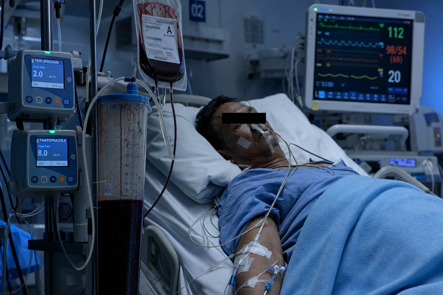
Definition and classification
An upper GI bleed (UGIB) is defined as any bleeding from a source proximal to the ligament of Treitz. It accounts for the majority of GI bleeding presentations and carries an overall mortality of approximately 10%.[5] The single most important early decision is the variceal versus non-variceal fork, because the management bundles differ fundamentally.[2][5]
Non-variceal
~80% of UGIB
- Peptic ulcer disease (the majority — H. pylori, NSAIDs, stress ulceration)
- Mallory-Weiss tear (forceful vomiting/retching, usually self-limiting)
- Erosive gastritis / oesophagitis (alcohol, NSAIDs, critical illness)
- Upper-GI malignancy (gastric, oesophageal)
- Dieulafoy lesion, AVM, watermelon stomach (GAVE)
- Aortoenteric fistula (history of AAA graft — the lethal mimic)
Variceal
~20% of UGIB but higher mortality
- Oesophageal varices (the classic variceal source)
- Gastric varices (often from left gastric or short veins)
- Portal hypertensive gastropathy (diffuse oozing)
- Underlying cirrhosis with clinically significant portal hypertension
- Mortality 15-25% at 6 weeks; risk of early rebleeding
- Distinct bundle: vasoactive + antibiotic + band ligation
Severity of presentation at first contact (click each)
Shock + suspected variceal — the bundle
Suspected variceal (cirrhosis, signs of portal hypertension): start vasoactive + antibiotic BEFORE endoscopy; protect the airway (intubate if encephalopathic); band ligation within 12 h; consider early pre-emptive TIPS for Child-Pugh C or Child-Pugh B with active bleeding.
Pathophysiology
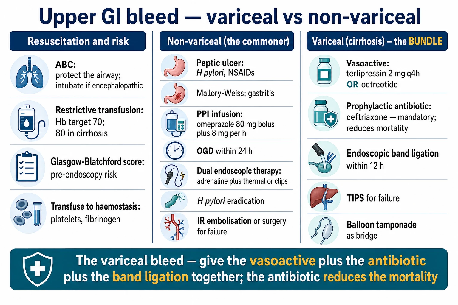
The pathophysiology of UGIB differs by source. Peptic ulcer bleeds occur when an ulcer erodes into a submucosal artery (classically the gastroduodenal or left gastric). The Forrest classification stratifies the bleeding stigmata: Forrest Ia/Ib (active spurting/oozing), IIa (visible vessel), IIb (adherent clot), IIc (black spot), and III (clean base) — the risk of rebleeding falls steeply across these categories and dictates the need for endoscopic therapy and PPI infusion.[5][6]
Variceal bleeding arises from clinically significant portal hypertension (hepatic venous pressure gradient above 10-12 mmHg). The porto-systemic pressure gradient drives blood into the collateral circulation, dilating submucosal oesophageal and gastric veins until a varix ruptures. Bleeding tends to be torrential and intermittent, and is worsened by anything that raises portal pressure — including over-transfusion, which is why the restrictive strategy matters most in this group.[1][2]
Clinical presentation
- Haematemesis — red blood (active, brisk) or coffee-ground vomit (partially digested).
- Melaena — black, tarry, foul-smelling stool; indicates at least 50-100 mL of blood in the upper GI tract. The higher the source, the more likely melaena; the brisker the bleed, the more likely haematochezia (maroon/red stool, which can masquerade as a lower GI bleed).
- Symptoms of hypovolaemia — presyncope, dizziness, dyspnoea; chest pain in the patient with coronary disease. Hypovolaemic shock (tachycardia, hypotension, cold peripheries, oliguria, altered conscious state) signals a major bleed (class I-IV shock).
- Signs of cirrhosis and portal hypertension — spider naevi, palmar erythema, jaundice, ascites, caput medusae, hepatic encephalopathy, asterixis, fetor hepaticus. Their presence shifts the working diagnosis to a variceal source and triggers the bundle immediately. [1]
Investigations
A focused, parallel panel is taken at the same time as resuscitation:[5][8]
- Bloods — full blood count and group & cross-match (2-4 units, or a massive transfusion protocol activation in shock); coagulation (INR, fibrinogen); urea and electrolytes (a raised urea with normal creatinine suggests an upper GI source — blood digested as protein); liver function tests (hypoalbuminaemia, derangement suggest cirrhosis); lactate (perfusion marker).
- Venous gas — metabolic acidosis in shock; base excess tracks the resuscitation.
- ECG — silent myocardial ischaemia is common in the elderly; troponin if indicated.
- Coagulation screen — INR is often abnormal in cirrhosis and with warfarin/DOAC; do NOT delay endoscopy for normalisation of INR alone — give prothrombin complex concentrate (PCC) for life-threatening bleeding on warfarin.
- Risk scores — Glasgow-Blatchford at presentation; Rockall after endoscopy. [1]
Management — resuscitation first
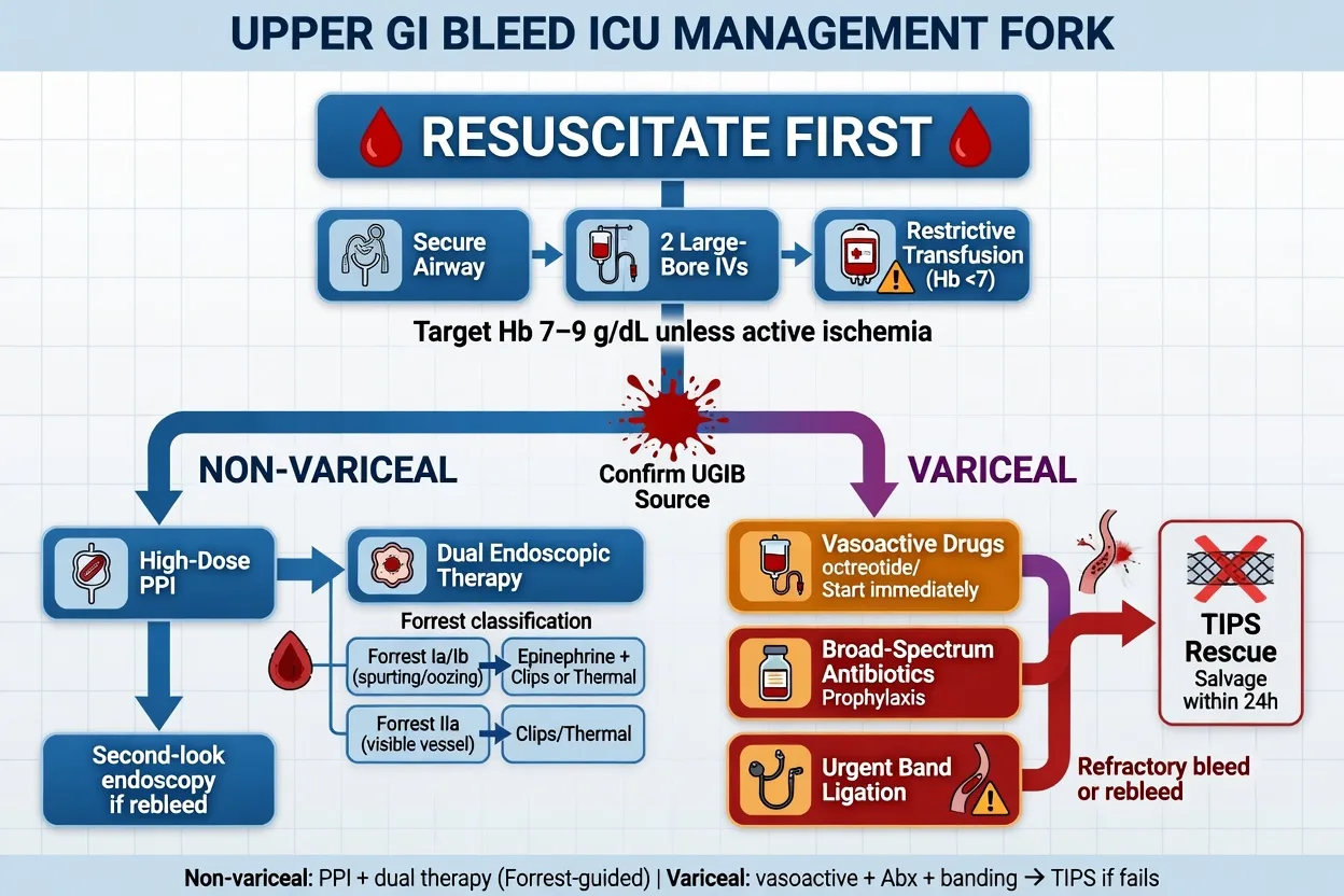
The first priority in any UGIB is airway, breathing, circulation, then transfusion strategy and risk stratification. Vasoactive drugs and antibiotic prophylaxis are started empirically in any patient with suspected or known cirrhosis and a UGIB, before endoscopy.[2][8]
Initial resuscitation (the first 60 minutes)
Airway — intubate the encephalopathic or the massive bleeder
Elective RSI and intubation BEFORE endoscopy for the encephalopathic cirrhotic or the patient with torrential haematemesis. Aspiration of blood is an avoidable cause of death. Notify anaesthetics and ICU early.
Two large-bore cannulae (16 G or larger) and send bloods
FBC, U&E, LFT, coagulation (INR, fibrinogen), lactate, group & cross-match 2-4 units. Activate the massive transfusion protocol if shocked. Take venous gas for base excess.
Restrictive transfusion — Hb 70 g/L, or 80 g/L in cirrhosis
Transfuse packed red cells to a restrictive target of Hb 70 g/L (TRIGGER trial). In cirrhosis with variceal bleeding, use a target of 80 g/L. Over-transfusion raises portal pressure and worsens variceal bleeding. Transfuse individualised units, not arbitrary targets.
Calculate the Glasgow-Blatchford Score and trigger the bundle if variceal
GBS at presentation to triage outpatient vs inpatient. If cirrhosis suspected, START the vasoactive drug (terlipressin 2 mg IV q4h OR octreotide 50 mcg bolus then 50 mcg/h) AND prophylactic antibiotic (ceftriaxone 1 g IV) BEFORE endoscopy. Do not wait for endoscopic confirmation.
Correction of coagulopathy to haemostasis — not normalisation
Platelets if below 50 x 10^9/L (target above 50 in active bleeding); fibrinogen with cryoprecipitate if below 1.5 g/L. In cirrhosis, aim to rebalance rather than normalise the INR — the INR overestimates bleeding risk in cirrhosis. Use viscoelastic testing (TEG/ROTEM) to guide component therapy.
Transfusion strategy — the TRIGGER trial
TRIGGER (Villanueva 2013)
NEJM 2013
Multicentre RCT — restrictive (Hb 70 g/L) vs liberal (Hb 90 g/L) transfusion in 921 patients with severe acute UGIB
Key finding
Restrictive strategy improved 6-week survival (95% vs 91%; hazard ratio 0.55), lower rebleeding, and fewer adverse events. Subgroup with cirrhosis and Child-Pugh A/B: survival benefit was significant; the small Child-Pugh C subgroup did not reach significance.
Practice change
Restrictive transfusion (Hb 70 g/L; 80 g/L in cirrhosis) is the standard of care for acute UGIB
The mechanism underpinning the benefit is twofold. First, in variceal bleeding, a higher haematocrit and blood volume raise splanchnic and portal venous pressure, counteracting the haemostatic effect of the vasoactive drug and the band ligation — bleeding resumes. Second, transfused red cells and stored plasma impair coagulation through dilution of clotting factors and the storage lesion, paradoxically promoting rebleeding. The restrictive strategy also reduces transfusion-related complications (TACO, TRALI, infection).[1]
Coagulation support — rebalance, do not normalise
In cirrhosis, the INR is a poor reflection of bleeding risk because the liver under-produces both procoagulant and anticoagulant factors. Viscoelastic testing (TEG or ROTEM) is the preferred tool to guide component therapy: give fibrinogen (cryoprecipitate) when the clot firmness is low, platelets when the MA (maximum amplitude) is reduced, and prothrombin complex concentrate (PCC) — not fresh frozen plasma as first line — for a prolonged clotting time. Avoid normalising the INR in cirrhosis; aim for haemostasis, not normalisation.[2]
Management — the non-variceal bleed
The non-variceal ulcer bleed is the commonest UGIB. The bundle is PPI infusion + dual endoscopic therapy + H. pylori eradication.[5][6]
1. Proton pump inhibitor infusion
The PPI infusion is given once an ulcer with high-risk stigmata is confirmed at endoscopy (or empirically if OGD will be delayed). The 80 mg bolus plus 8 mg/h infusion for 72 hours regime was validated by Lau et al. in 2000: it raised intragastric pH above 6, stabilising the clot and reducing rebleeding after endoscopic therapy.[6]
PPI infusion protocol (after endoscopic therapy for high-risk stigmata)
Pantoprazole (or omeprazole) 80 mg IV bolus
Given after endoscopic confirmation of an ulcer with active bleeding, visible vessel, or adherent clot (Forrest Ia/Ib/IIa). The bolus rapidly raises intragastric pH above 6, stabilising the platelet plug.
Then 8 mg/h continuous infusion for 72 hours
Maintains pH above 6 to prevent pepsin-mediated clot lysis. After 72 hours, switch to oral PPI twice daily, then once daily. The infusion is most beneficial after endoscopic therapy in Asian populations; benefit in Western populations is smaller but the regimen is still recommended.
Pre-endoscopy PPI — downscope endoscopy, not a substitute
If OGD is delayed beyond 24 h, an IV PPI infusion may downstage the bleeding stigmata and reduce the need for endoscopic therapy, but it does NOT reduce mortality. Do not let a PPI infusion delay urgent OGD in a variceal or unstable patient.
2. Dual endoscopic therapy
For ulcers with high-risk stigmata (Forrest Ia, Ib, IIa), dual endoscopic therapy is standard: adrenaline (epinephrine) injection PLUS a thermal (heater probe, bipolar) or mechanical (through-the-scope clip) modality. Adrenaline alone is inferior — it provides tamponade and vasoconstriction but does not seal the vessel, so rebleeding rates are high without a second modality.[5]
Adrenaline injection
The first half of dual therapy
- 1:10,000 epinephrine injected in 1-2 mL aliquots around the bleeding point
- Provides vasoconstriction and tamponade
- Reduces active bleeding and improves visualisation
- Monotherapy has HIGH rebleeding rate — must combine with thermal or clip
Thermal or clip
The second half — seals the vessel
- Thermal: heater probe or bipolar coagulation — direct vessel sealing
- Mechanical: through-the-scope (TTS) clips — physically oppose vessel walls
- Argon plasma coagulation for superficial oozing (Forrest IIc)
- Combine with adrenaline for the best rebleeding/mortality outcomes
3. H. pylori eradication and secondary prevention
Test for Helicobacter pylori (rapid urease test at endoscopy, histology, or urea breath test/stool antigen after PPI cessation) and eradicate with triple or quadruple therapy. Stop NSAIDs; give PPI-based stress ulcer prophylaxis if the patient remains critically ill. Restart secondary cardiovascular antiplatelet/anticoagulant therapy early (within 7 days for aspirin) once haemostasis is secured — the thrombotic risk of stopping outweighs the rebleeding risk in most patients.[5]
4. Rescue therapy for failed endoscopic control
If endoscopic therapy fails to control bleeding or rebleeding occurs, interventional radiology (IR) embolisation of the feeding artery (left gastric for gastric ulcers, gastroduodenal for duodenal) is the preferred rescue therapy. Surgery is reserved for failed or unavailable IR. A second-look endoscopy is not routinely recommended.[5]
Management — the variceal bleed (the BUNDLE)
The variceal bleed requires a time-limited bundle delivered together within 12 hours: vasoactive drug + prophylactic antibiotic + endoscopic band ligation. No component can be omitted.[2][8]
The variceal bundle — three drugs/procedures within 12 h
Vasoactive drug — START BEFORE endoscopy
Terlipressin 2 mg IV every 4 hours (a vasopressin V1 analogue) OR octreotide 50 mcg bolus then 50 mcg/h infusion (a somatostatin analogue). Both produce splanchnic vasoconstriction and lower portal pressure. Continue for 2-5 days. Terlipressin may cause ischaemia (use cautiously in coronary disease). In Australia octreotide is more commonly used; terlipressin is preferred where available.
Prophylactic antibiotic — ceftriaxone 1 g IV daily for up to 7 days
Antibiotic prophylaxis is MANDATORY in cirrhosis with a UGIB. It reduces bacterial translocation, infection, rebleeding and mortality (number needed to treat ~11 for mortality). Third-generation cephalosporin (ceftriaxone) is preferred over oral quinolones due to rising quinolone resistance.
Endoscopic band ligation within 12 hours
Definitive endoscopic therapy for oesophageal varices. The varix is suctioned into the cap and a rubber band is deployed, strangulating the vessel. Sclerotherapy is inferior and reserved if banding is technically impossible. For isolated gastric varices, cyanoacrylate (glue) injection is preferred.
Consider early pre-emptive TIPS within 72 h for high-risk patients
Child-Pugh C (score 10-13) or Child-Pugh B with active bleeding at endoscopy. These patients fail medical therapy and benefit from early TIPS placement within 72 h, which reduces treatment failure and mortality. Do NOT wait for a second bleed to refer.
Vasoactive drugs — terlipressin vs octreotide
Terlipressin
Vasopressin V1 analogue
- Dose: 2 mg IV every 4 hours for the first 48 h, then 1 mg q4h (reduce after control)
- Network meta-analyses suggest it is the most effective vasoactive for control of variceal bleeding
- Causes ischaemia (coronary, mesenteric, digital) — avoid/monitor in ischaemic heart disease
- Can cause hyponatraemia — monitor sodium
Octreotide / somatostatin
Somatostatin analogues
- Octreotide: 50 mcg bolus then 50 mcg/h infusion for 2-5 days
- Somatostatin: 250 mcg bolus then 250 mcg/h infusion
- Fewer ischaemic side effects; widely available in ANZ
- Equivalent or slightly inferior to terlipressin in some meta-analyses; both acceptable
Antibiotic prophylaxis — the highest-yield single intervention
Early pre-emptive TIPS — for the high-risk subgroup
Early TIPS IPD meta-analysis (Nicoară-Farcău 2021)
Gastroenterology 2021
Individual-patient-data meta-analysis of 4 RCTs (n=963) of early pre-emptive TIPS within 72 h vs standard therapy in high-risk variceal bleeding (Child-Pugh C 10-13 or Child-Pugh B with active bleeding)
Key finding
Pre-emptive TIPS reduced treatment failure (HR 0.55) and improved 1-year survival (HR 0.65) without increasing hepatic encephalopathy. The benefit was restricted to the high-risk subgroup.
Practice change
Pre-emptive TIPS within 72 h is indicated for Child-Pugh C 10-13 or Child-Pugh B with active bleeding at endoscopy
Rescue therapy for refractory variceal bleeding
Rescue therapy if the variceal bundle fails
Balloon tamponade (Sengstaken-Blakemore or Linton) as a temporary bridge
A gastric balloon inflated against the gastro-oesophageal junction tamponades variceal bleeding. Maximum dwell time 24 h (risk of aspiration, pressure necrosis, oesophageal perforation). It is a BRIDGE to definitive therapy (TIPS), not a treatment. Intubate before insertion.
Self-expanding covered metal oesophageal stent
An alternative bridge for refractory oesophageal variceal bleeding — fewer complications than balloon tamponade and can stay in up to 7 days. Less widely available.
Salvage TIPS — the definitive rescue
Transjugular intrahepatic portosystemic shunt lowers the porto-systemic gradient below the bleeding threshold and is the definitive rescue for refractory variceal bleeding. Covered stents have improved patency. Hepatic encephalopathy is the principal complication.
Adjust the vasoactive drug and re-endoscope
If the band has slipped or a new varix is bleeding, re-endoscopy with repeat band ligation is appropriate; ensure the vasoactive infusion is running and the antibiotic has been given. Activate massive transfusion protocol for ongoing torrential bleeding.
Evidence and guidelines
TRIGGER (Villanueva 2013)
NEJM 2013
RCT, n=921, restrictive (Hb 70) vs liberal (Hb 90) transfusion in severe acute UGIB
Key finding
Restrictive strategy improved 6-week survival (HR 0.55), lower rebleeding and fewer adverse events; benefit preserved in cirrhosis subgroup.
Practice change
Restrictive transfusion (Hb 70; 80 in cirrhosis) is the standard of care
Baveno VII (2022)
J Hepatol 2022
International consensus on portal hypertension
Key finding
Vasoactive + antibiotic + band ligation bundle within 12 h; pre-emptive TIPS for high-risk; restrictive transfusion (Hb 80 in cirrhosis).
Practice change
Standardised the variceal bundle and pre-emptive TIPS criteria
ACG UGIB Guideline (2021)
Am J Gastroenterol 2021
US clinical guideline for upper GI and ulcer bleeding
Key finding
Risk stratification with GBS; OGD within 24 h; dual endoscopic therapy for high-risk stigmata; PPI infusion 80 mg bolus + 8 mg/h x 72 h.
Practice change
Reaffirmed dual endoscopic therapy and PPI infusion regime
ESGE Variceal Guideline (2022)
Endoscopy 2022
European Society of GI Endoscopy consensus
Key finding
Vasoactive started at admission; band ligation within 12 h; pre-emptive TIPS within 72 h for high-risk; balloon tamponade as bridge only.
Practice change
Operationalised the variceal bundle timing
Antibiotic prophylaxis Cochrane (2011)
Aliment Pharmacol Ther 2011
Updated Cochrane meta-analysis of antibiotic prophylaxis in cirrhosis with UGIB
Key finding
Reduced all-cause mortality (NNT ~11), bacterial infection and rebleeding. Cephalosporins superior to quinolones where resistance is high.
Practice change
Antibiotic prophylaxis is mandatory in cirrhosis with any UGIB
Terlipressin meta-analysis (2018)
Medicine 2018
Systematic review and meta-analysis of RCTs of terlipressin in acute variceal bleeding
Key finding
Terlipressin improved control of bleeding and reduced 7-day mortality vs placebo; comparable to somatostatin/octreotide for control.
Practice change
Terlipressin a first-line vasoactive option where available
Complications
- Rebleeding — the dominant early complication; highest with variceal bleeds (within 5 days) and ulcers with high-risk stigmata. Predicted by the Rockall score; managed by repeat endoscopy, IR embolisation (non-variceal) or salvage TIPS (variceal).
- Aspiration pneumonia — particularly in the encephalopathic cirrhotic; prevented by early intubation before endoscopy.
- Hepatic encephalopathy — precipitated by the protein load from digested blood plus infection; treated with lactulose, rifaximin, and correction of the precipitant. TIPS itself causes encephalopathy in 20-50%.
- Acute kidney injury — hypovolaemia, hepatorenal syndrome (in cirrhosis with bacterial infection), contrast (TIPS). Avoid nephrotoxins; albumin resuscitation in cirrhosis.
- Coagulopathy — dilutional and disease-related (cirrhosis, warfarin/DOAC); managed by component therapy guided by viscoelastic testing, PCC for warfarin, and specific reversal agents for DOACs.
- TIPS-related — hepatic encephalopathy, heart failure (increased venous return), haemolysis, contrast-induced AKI.
- Terlipressin ischaemia — coronary, mesenteric, digital; monitor closely. [1]
Prognosis
Outcomes and key numbers
Prognosis depends on the source (variceal worse than non-variceal), the patient's comorbidity (age, cardiac and hepatic reserve), and the speed and completeness of the bundle. The Rockall score predicts mortality; the Glasgow-Blatchford predicts the need for intervention. Cirrhotics who receive the full bundle (vasoactive + antibiotic + band ligation) and an early pre-emptive TIPS where indicated have substantially better outcomes than those who receive only one or two components.[2][7][8]
Exam practice
SAQ — Acute variceal bleed in a cirrhotic
10 minutes · 10 marks
A 56-year-old man with known alcohol-related cirrhosis (Child-Pugh B, score 9) presents to the emergency department with two episodes of large-volume haematemesis. He is drowsy but rousable (GCS 13), with a temperature of 36.4°C, heart rate 118/min, blood pressure 84/50 mmHg, respiratory rate 22, oxygen saturation 95% on room air. He has spider naevi, icteric sclerae and a palpable spleen. Initial haemoglobin 72 g/L, platelets 65 x 10^9/L, INR 1.7, fibrinogen 1.3 g/L, albumin 28 g/L, bilirubin 62, urea 14, creatinine 95. Venous gas: pH 7.28, lactate 3.2. A diagnosis of suspected acute variceal bleeding is made.
Clinical pearls
Red flags
References
- [1]Villanueva C, Colomo A, Bosch A, et al. Transfusion strategies for acute upper gastrointestinal bleeding N Engl J Med, 2013.PMID 23281973
- [2]de Franchis R, Bosch J, Garcia-Tsao G, et al. Baveno VII - Renewing consensus in portal hypertension J Hepatol, 2022.PMID 35120736
- [3]Blatchford O, Murray WR, Blatchford M. A risk score to predict need for treatment for upper-gastrointestinal haemorrhage Lancet, 2000.PMID 11073021
- [4]Rockall TA, Logan RF, Devlin HB, Northfield TC. Selection of patients for early discharge or outpatient care after acute upper gastrointestinal haemorrhage. National Audit of Acute Upper Gastrointestinal Haemorrhage Lancet, 1996.PMID 8609747
- [5]Laine L, Barkun AN, Saltzman JR, Martel M, Enestvedt BK. ACG Clinical Guideline: Upper Gastrointestinal and Ulcer Bleeding Am J Gastroenterol, 2021.PMID 33929377
- [6]Lau JY, Sung JJ, Lee KK, et al. Effect of intravenous omeprazole on recurrent bleeding after endoscopic treatment of bleeding peptic ulcers N Engl J Med, 2000.PMID 10922420
- [7]Nicoară-Farcău O, Han G, Rudler M, et al. Effects of Early Placement of Transjugular Portosystemic Shunts in Patients With High-Risk Acute Variceal Bleeding: a Meta-analysis of Individual Patient Data Gastroenterology, 2021.PMID 32980344
- [8]Gralnek IM, Camus Duboc M, Garcia-Pagan JC, et al. Endoscopic diagnosis and management of esophagogastric variceal hemorrhage: European Society of Gastrointestinal Endoscopy (ESGE) Guideline Endoscopy, 2022.PMID 36174643
- [9]Chavez-Tapia NC, Barrientos-Gutierrez T, Tellez-Avila FI, et al. Meta-analysis: antibiotic prophylaxis for cirrhotic patients with upper gastrointestinal bleeding - an updated Cochrane review Aliment Pharmacol Ther, 2011.PMID 21707680
- [10]Zhou X, Tripathi D, Song T, et al. Terlipressin for the treatment of acute variceal bleeding: A systematic review and meta-analysis of randomized controlled trials Medicine (Baltimore), 2018.PMID 30508958
- [11]Zou Z, Yan X, Lu H, et al. Comparison of drugs facilitating endoscopy for patients with acute variceal bleeding: a systematic review and network meta-analysis Ann Transl Med, 2019.PMID 32042733
- [12]García-Pagán JC, Saffo S, Mandorfer M, Valla DC, Gracia-Sancho J, Bosch J. Where does TIPS fit in the management of patients with cirrhosis? JHEP Rep, 2020.PMID 32671331