ICU · GI/Nutrition
Acute variceal haemorrhage
Also known as Variceal bleed · Oesophageal varices · Gastric varices · Band ligation · Terlipressin · Sengstaken-Blakemore · TIPS · TIPSS
Acute variceal haemorrhage is life-threatening bleeding from oesophageal or gastric varices that form as a consequence of portal hypertension (cirrhosis 1 cause). Mortality 15-25% per episode and 60% recurrence within one year without secondary prophylaxis. PATHOPHYSIOLOGY: portal venous pressure >10 mmHg (HVPG >12 mmHg clinically significant) → collateral portosystemic circulation → thin-walled varices at the gastro-oesophageal junction and gastric fundus → wall tension rises with radius (Laplace) → rupture when wall stress exceeds tensile strength. MANAGEMENT cascade: resuscitate (ABCDE, restrictive transfusion Hb 70-80) → start vasoactive drug (terlipressin 2 mg IV Q4H or octreotide) AND prophylactic antibiotic (ceftriaxone 1g IV daily — reduces mortality, NNT ~10) BEFORE endoscopy → endoscopic band ligation within 12h → balloon tamponade (Sengstaken-Blakemore) as a temporary bridge → salvage TIPS for refractory bleeding; early pre-emptive TIPS within 72h for high-risk patients (Child-Pugh C 10-13 or Child-Pugh B with active bleeding) per Baveno VII.
On this page & tools
Your progress
Saved locally on this device.
Target exams
Red flags
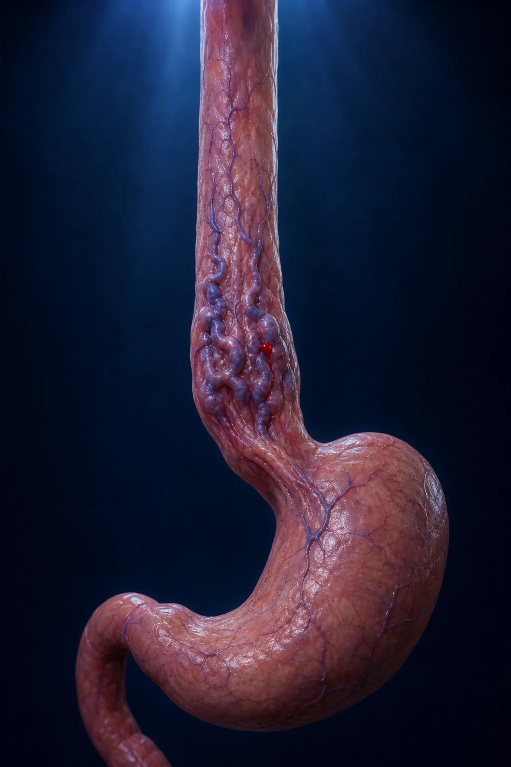
Pathophysiology — portal hypertension to varix rupture
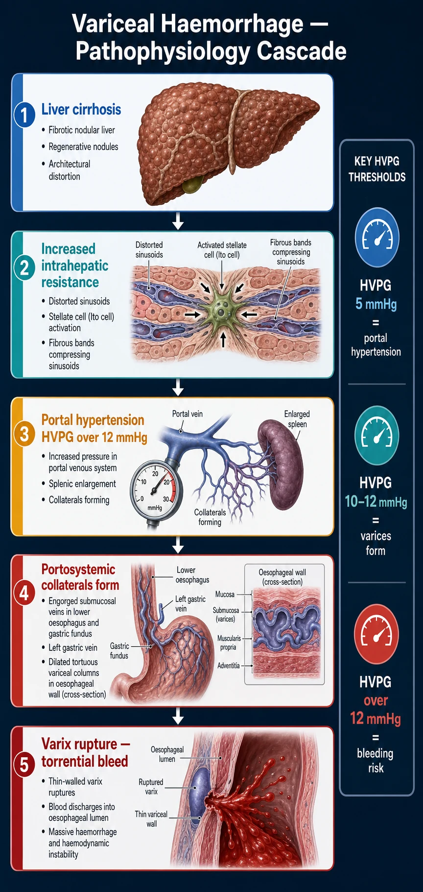
Variceal haemorrhage is the terminal event of a haemodynamic chain that begins in the liver sinusoid and ends in a thin-walled collateral at the gastro-oesophageal junction. Understanding the chain is the key to understanding why each step of the management cascade works: every intervention — vasoactive drug, band ligation, balloon tamponade, TIPS — is a physical or pharmacological attack on portal venous pressure or on the varix wall itself. [1]
Step 1 — increased intrahepatic resistance. Cirrhosis distorts the hepatic architecture with fibrous septa and regenerative nodules, mechanically compressing sinusoids. The static component of resistance is amplified by a dynamic component: activated hepatic stellate cells contract around the sinusoid under the influence of vasoconstrictors (endothelin, thromboxane, angiotensin II) whose effect is unopposed because the cirrhotic liver under-produces the vasodilator nitric oxide. The net result is a rise in portal venous pressure, quantified by the hepatic venous pressure gradient (HVPG) — the gradient between the wedged (occluded) hepatic venous pressure and the free hepatic venous pressure, which estimates portal pressure. [1]
Step 2 — clinically significant portal hypertension. HVPG is the single most important physiological variable in this disease. The normal value is 3-5 mmHg. Clinically significant portal hypertension (CSPH) is defined as HVPG ≥10 mmHg: above this threshold varices form, ascites accumulates, and the risk of bleeding begins. Varices do not bleed until HVPG exceeds 12 mmHg, and the risk of bleeding and rebleeding climbs steeply as HVPG rises above 16 mmHg and above 20 mmHg. The corollary used at the bedside (Baveno VII): an HVPG reduction of ≥10% from baseline, or to below 12 mmHg, achieved by beta-blockade (or by TIPS), confers substantial protection from first and recurrent bleeding. [1]
Step 3 — portosystemic collateral formation and varix development. The hypertensive portal circulation decompresses through pre-existing embryonic channels that re-open under pressure, forming portosystemic collaterals at three principal sites: (i) the gastro-oesophageal junction (oesophageal and gastric varices, the clinically important ones), (ii) the rectum (anorectal varices), and (iii) the retroperitoneum and umbilicus (caput medusae). The varices at the gastro-oesophageal junction are dangerous because they lie in the submucosa, supported only by a thin connective tissue plane, and drain into the azygos system against a relatively low pressure — the pressure drop across them is large. [1]
Step 4 — wall tension rises and the varix ruptures. The wall tension of a varix obeys Laplace's law (T = P × r / 2w): tension rises with intraluminal pressure (P), rises with the varix radius (r), and falls with wall thickness (w). Three features therefore predict rupture: (a) high portal pressure (high P), (b) large varix size (high r — the endoscopic "red wale signs", "cherry-red spots" and "red spots" mark thinned wall regions), and (c) thin varix wall (low w). A precipitating transient rise in intra-abdominal pressure — vomiting, retching, coughing, or a Valsalva — is frequently the immediate trigger, as is a meal (splanchnic hyperaemia raises portal inflow). Rupture is a mechanical event, not a coagulopathic one: that is why the definitive therapy is mechanical (band, balloon, shunt), and why correcting coagulopathy alone will never control a variceal bleed. [1]
Why cirrhosis begets infection, and infection begets bleeding. Bacterial translocation from a gut made permeable by portal hypertension seeds the circulation with endotoxin; this provokes a systemic inflammatory response that impairs coagulation, raises portal pressure through splanchnic vasodilation (NO-mediated), and is the single strongest predictor of failure to control bleeding and of early rebleeding. This is the pathophysiological rationale for mandatory prophylactic antibiotics: treating translocation lowers portal pressure and restores haemostatic competence, which is why antibiotics — uniquely among the bundle components — reduce mortality.[1][5]
Oesophageal vs gastric varices — sites, classification and therapy
| Feature | Oesophageal varices | Gastric varices |
|---|---|---|
| Location | Distal oesophagus / gastro-oesophageal junction | Cardia (GOV1/2), fundus (IGV1), isolated (IGV2) — Sarin classification |
| Prevalence | ~80% of variceal bleeds | ~10-15% (fundal IGV1 bleeds are the most dangerous) |
| Bleeding tendency | Common; HVPG >12 mmHg threshold | Less common but bleed at lower pressures and are more torrential |
| Endoscopic therapy (first line) | Endoscopic band ligation (EBL) — rubber band strangulates the varix | Cyanoacrylate (glue) injection — N-butyl-2-cyanoacrylate polymerises on contact with blood; band ligation is inferior for fundal varices |
| Vasoactive + antibiotics | Yes — standard bundle | Yes — standard bundle |
| Refractory / first-line if large | Balloon tamponade (oesophageal balloon) → TIPS | TIPS often first-line for isolated fundal varices; BRTO if a spontaneous splenorenal shunt exists |
| Rebleeding risk | 60% at 1 year without secondary prophylaxis (beta-blocker + serial banding) | Higher than oesophageal; TIPS or BRTO often needed |
| Aspiration risk at endoscopy | High | High |
Risk stratification — Child-Pugh, MELD, and the high-risk patient
The severity of underlying liver disease, not the volume of blood lost, is the dominant determinant of mortality in variceal bleeding. Two scores are used in tandem: Child-Pugh (the traditional bedside score, and the one that defines the high-risk group for early TIPS) and MELD (the objective, laboratory-only score that drives transplant allocation and is preferred for prognosis). Both must be calculated at admission. [1]
Child-Pugh score — class predicts mortality and TIPS candidacy (click each)
10-15 — decompensated
Decompensated. Highest mortality from a variceal bleed. Child-Pugh C with score 10-13 also qualifies for EARLY pre-emptive TIPS (within 72h). Child-Pugh C with score >13 is generally EXCLUDED from TIPS — the liver cannot tolerate the diversion of portal blood, and TIPS precipitates liver failure and death.
Child-Pugh uses five variables — bilirubin, albumin, INR, ascites, encephalopathy — each scored 1-3. Its weakness is the subjectivity of the two clinical variables (ascites and encephalopathy grade) and its coarse resolution. MELD (Model for End-stage Liver Disease) uses only objective laboratories — bilirubin, creatinine, INR, and sodium (MELD-Na) — and was designed to predict 90-day mortality. It is the basis of transplant allocation in most regions. [1]
Child-Pugh vs MELD — which to use and when
| Feature | Child-Pugh score | MELD (MELD-Na) score |
|---|---|---|
| Variables | Bilirubin, albumin, INR, ascites, encephalopathy | Bilirubin, creatinine, INR, sodium (MELD-Na) — laboratory only |
| Subjectivity | Ascites and encephalopathy grades are subjective | Fully objective — reproducible between observers |
| Range | 5-15, classes A/B/C | Continuous; MELD-Na up to 40 |
| Primary role | Bedside severity; defines the early-TIPS high-risk group (Child C 10-13 or Child B + active bleeding) | Transplant allocation; predicts 90-day mortality |
| Mortality calibration | Coarse (class-level) | Fine — each point ≈ 1-2% 90-day mortality |
| Limitations | Subjective clinical variables | Underestimates severity in infection/SBP; sodium excluded in original MELD (added in MELD-Na) |
| Practical use in variceal bleed | Calculate at admission → identifies the early-TIPS candidate | Calculate at admission → informs transplant referral and goals-of-care discussion |
The clinically actionable point: a cirrhotic who bleeds and falls into Child-Pugh C 10-13 OR Child-Pugh B with active bleeding at endoscopy has failed medical therapy in advance. Do not wait for a rebleed — refer for early pre-emptive TIPS within 72h of admission. This is the Baveno VII position and the single most exam-testable decision point in the disease.[1][4]
Management cascade
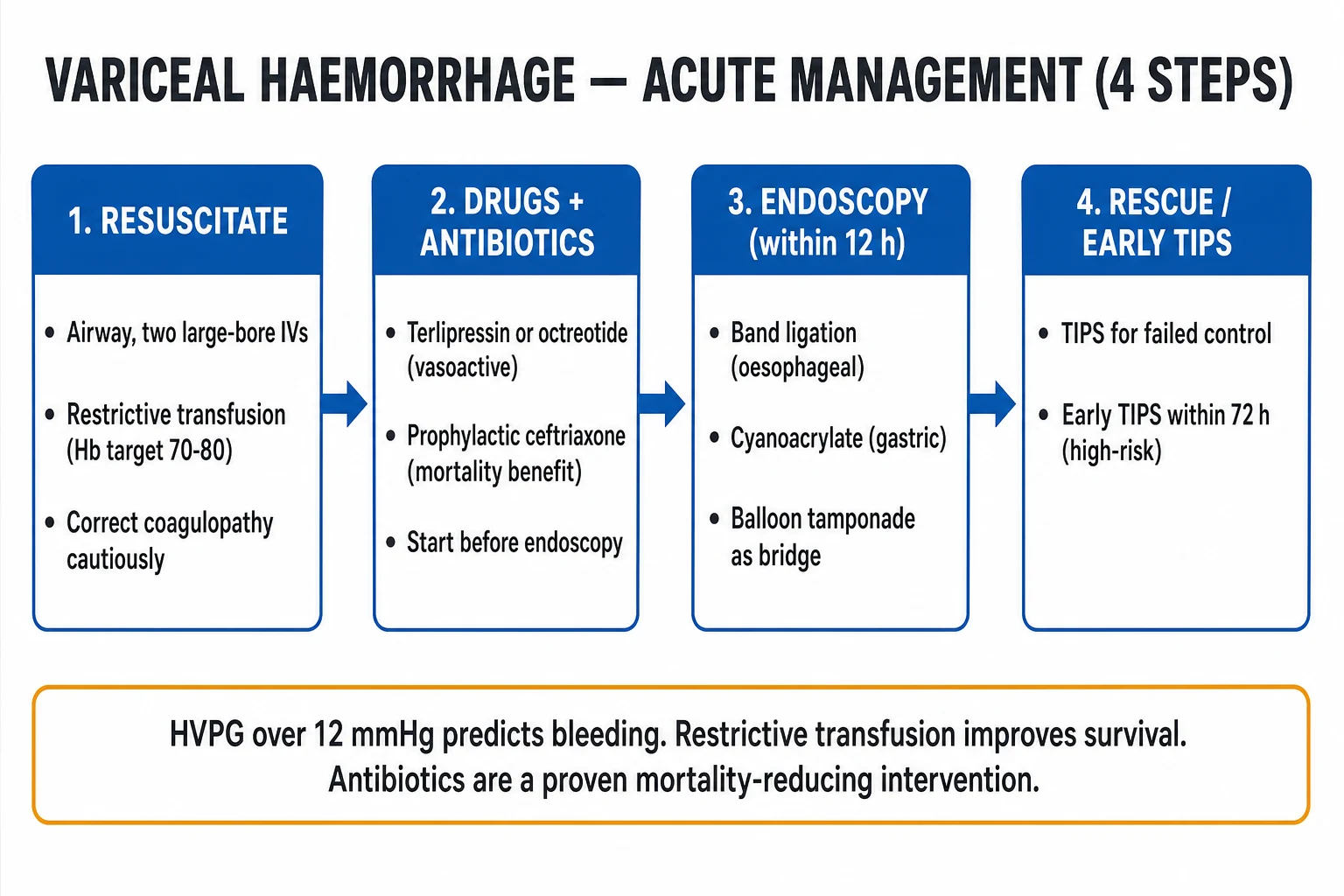
The cascade is a sequence in which each tier is started before the previous one has failed: the vasoactive drug and antibiotic are given before endoscopy, endoscopy is performed before tamponade is needed, and a salvage TIPS plan is in place before the balloon goes in. The bundle components are mutually reinforcing — omitting any one worsens control of bleeding, rebleeding, and mortality. [1]
Variceal haemorrhage management — the full cascade
Resuscitation + restrictive transfusion (first 60 min)
ABCDE. Two large-bore (14-16 G) cannulae. Intubate EARLY if encephalopathic or has ongoing torrential haematemesis (aspiration of blood is an avoidable cause of death — RSI, large-bore suction, head-down tilt, left lateral). Resuscitate with crystalloid to restore perfusion, then **restrictive transfusion**: transfuse to Hb 70-80 g/L (TRIGGER trial — over-transfusion raises splanchnic and portal venous pressure, disrupts the clot, and increases rebleeding and mortality). Send FBC, coagulation (INR, fibrinogen), U&E, LFTs, albumin, lactate, group and cross-match. Correct coagulopathy to **haemostasis, not normalisation** (the cirrhotic INR overestimates bleeding risk — use TEG/ROTEM): platelets if <50, cryoprecipitate if fibrinogen <1.5 g/L, PCC (not FFP as first line) for a prolonged clotting time.
Vasoactive drug BEFORE endoscopy
**Terlipressin 2 mg IV Q4H** (a vasopressin V1 analogue with a longer half-life than vasopressin and fewer side effects) OR **octreotide 50 mcg bolus then 50 mcg/h infusion** (a somatostatin analogue). Mechanism: splanchnic vasoconstriction → reduces portal venous inflow → reduces portal and variceal pressure → slows/stops bleeding → improves endoscopic visualisation. START IMMEDIATELY on suspicion of variceal bleeding (known cirrhosis, signs of chronic liver disease) — do NOT wait for endoscopic confirmation. Continue for 2-5 days. Terlipressin side effects: peripheral/visceral ischaemia (coronary, mesenteric, digital), hyponatraemia; contraindicated in ongoing cardiac/mesenteric ischaemia.<Cite id="2" />
Prophylactic antibiotics BEFORE endoscopy
**Ceftriaxone 1 g IV daily for (max) 7 days** to ALL cirrhotic patients with a GI bleed. Antibiotics reduce bacterial infection (translocation → SBP, bacteraemia), reduce rebleeding, and are the ONLY bundle component that reduces MORTALITY (NNT ~10). Preferred over oral quinolones (norfloxacin/ciprofloxacin) because of rising Gram-negative quinolone resistance. Works by treating subclinical bacterial translocation that drives splanchnic vasodilation and coagulopathy — a haemodynamic, not just anti-infective, effect.<Cite id="5" />
Endoscopic therapy within 12 hours
Upper GI endoscopy within 12 h (after resuscitation + vasoactive + antibiotic are running). **Oesophageal varices**: endoscopic band ligation (EBL) — the varix is suctioned into the cap and a rubber band strangulates it, causing thrombosis then sloughing. Sclerotherapy is inferior (higher rebleeding, complications) and is reserved for when banding is technically impossible. **Gastric (especially fundal) varices**: cyanoacrylate ("superglue") injection — N-butyl-2-cyanoacrylate polymerises on contact with blood and obliterates the varix; EBL is less effective for fundal varices. Erythromycin 250 mg IV 30-90 min before endoscopy (prokinetic) clears blood from the stomach and improves the field of view in active bleeding.
Balloon tamponade — BRIDGE only (if uncontrolled)
**Sengstaken-Blakemore** (or Minnesota) tube: gastric balloon inflated with 250-350 mL air/water (tamponades gastric variceal and distal oesophageal bleeding) ± oesophageal balloon inflated to 30-40 mmHg (tamponades oesophageal varices). It is a TEMPORARY BRIDGE to definitive therapy (TIPS), never a destination — **maximum 24 h** (pressure necrosis, aspiration, oesophageal perforation/rupture). **Intubate BEFORE insertion** (airway protection — aspiration risk). Deflate the oesophageal balloon every 8-12 h to limit ischaemia. An alternative bridge is a **self-expanding covered metal oesophageal stent (SEMS)** — easier to place, less traumatic, may be left longer.
Early pre-emptive TIPS (within 72 h) for high-risk
For **Child-Pugh C 10-13** OR **Child-Pugh B with active bleeding at endoscopy** — the patient has effectively failed medical therapy in advance. Place a covered TIPS (transjugular intrahepatic portosystemic shunt) within 72 h of admission. Reduces treatment failure and mortality without increasing hepatic encephalopathy (Garcia-Pagan 2010; Baveno VII). Do NOT wait for a rebleed to refer.<Cite id="1" /><Cite id="4" />
Salvage TIPS for refractory bleeding
For bleeding uncontrolled by vasoactive drug + band ligation, or that recurs despite a second endoscopic attempt: salvage TIPS. Interventional radiology cannulates the internal jugular vein → hepatic vein → creates a stented channel through the liver parenchyma to the portal vein → shunts portal blood to the systemic circulation → decompresses the portal system → stops variceal bleeding. Excluded from TIPS: Child-Pugh C >13, severe encephalopathy, severe cardiopulmonary disease, portal vein thrombosis, active sepsis. Complications: hepatic encephalopathy (portosystemic shunt bypasses hepatic detoxification), liver failure (diverts nutrient portal blood from hepatocytes), cardiac decompensation (increased venous return).
Secondary prophylaxis (after the bleed is controlled)
After successful control, the rebleeding risk is ~60% at 1 year without secondary prophylaxis. Combine: (1) a **non-selective beta-blocker** (propranolol, nadolol, carvedilol — titrate to HR 55-60; carvedilol additionally lowers portal pressure via alpha-1 blockade) AND (2) **serial endoscopic band ligation** until the varices are eradicated (every 2-4 weeks). If beta-blockers are not tolerated or contraindicated, band ligation alone. TIPS for recurrent bleeding despite optimal combination prophylaxis.
Vasoactive drugs — choosing between terlipressin and octreotide
Both reduce splanchnic blood flow and portal pressure, but through different receptors and with different pharmacology. The choice is governed by availability, contraindications, and local practice. [1]
Terlipressin vs octreotide vs somatostatin — the vasoactive agents
| Feature | Terlipressin | Octreotide | Somatostatin |
|---|---|---|---|
| Class | Vasopressin V1 receptor analogue | Somatostatin analogue | Native hormone |
| Dose | 2 mg IV Q4H (bolus) | 50 mcg bolus then 50 mcg/h infusion | 250 mcg bolus then 250 mcg/h infusion |
| Onset / half-life | Slow onset, long t½ (~6 h) — bolus dosing | Rapid onset, short t½ (~1-2 h) — infusion | Very short t½ (~2-3 min) — infusion |
| Efficacy | Best evidence for bleeding control and trend toward mortality benefit (CONSORT) | Equivalent for bleeding control; widely used in US | Equivalent; limited by cost and short half-life |
| Key side effects | Ischaemia (coronary, mesenteric, digital, skin), hyponatraemia, hypertension | Bradycardia, hyperglycaemia, abdominal cramps (less ischaemia than terlipressin) | Bradycardia, hyperglycaemia |
| Contraindications | Active cardiac/mesenteric/digital ischaemia | Caution in heart block | Caution in heart block |
| ANZ / UK first line | Terlipressin (preferred) | Octreotide (alternative) | Used in some centres |
The pragmatic rule: give whichever is available before endoscopy, and continue it for 2-5 days (Baveno VII recommends up to 5 days, with the peak rebleeding risk in the first 5 days).[1][2]
Key trials and evidence
TRIGGER (Villanueva 2013)
NEJM 2013
Multicentre RCT — restrictive (transfuse at Hb 70 g/L) vs liberal (transfuse at Hb 90 g/L) in 921 patients with severe acute upper GI bleeding, ~33% cirrhotic.
Key finding
Restrictive strategy improved 6-week survival (95% vs 91%; HR 0.55), reduced rebleeding (HR 0.68) and adverse events. In the cirrhosis/Child-Pugh A-B subgroup, the survival benefit was significant; the small Child-Pugh C subgroup did not reach significance.
Practice change
Restrictive transfusion (Hb 70 g/L; up to 80 g/L in cirrhosis) is the standard of care. Over-transfusion raises portal pressure and disrupts the variceal clot.
Bernard meta-analysis (1999)
Hepatology 1999
Meta-analysis of RCTs of antibiotic prophylaxis vs no prophylaxis in cirrhotic patients with GI bleeding.
Key finding
Antibiotic prophylaxis significantly reduced bacterial infections. Subsequent meta-analyses (Cochrane) extended this to a reduction in all-cause mortality, infection-related mortality, and rebleeding — making antibiotics the only bundle component with a proven mortality benefit (NNT ~10).
Practice change
Antibiotic prophylaxis (ceftriaxone 1 g IV daily) is MANDATORY in every cirrhotic with a GI bleed, started before endoscopy.
Garcia-Pagan early TIPS (2010)
NEJM 2010
RCT of early pre-emptive covered TIPS within 72 h vs standard therapy (vasoactive + band ligation) in high-risk cirrhotics (Child-Pugh C 10-13 or Child-Pugh B with active bleeding).
Key finding
Early TIPS dramatically reduced treatment failure (12% vs 0% at 1 year in the early-TIPS group) and improved 1-year survival (86% vs 61%). No increase in hepatic encephalopathy.
Practice change
Early pre-emptive TIPS within 72 h is the standard of care for the high-risk group (Child-Pugh C 10-13 or Child-Pugh B + active bleeding). Do NOT wait for a rebleed to refer. Confirmed in an individual-patient-data meta-analysis of four RCTs.
Balloon tamponade — technique and pitfalls
The Sengstaken-Blakemore (or four-lumen Minnesota) tube is a physical tamponade, not a treatment. It buys hours — typically to arrange a salvage TIPS — and is dangerous if left too long. [1]
Insertion. Intubate first (RSI, large-bore suction, head-down). Pass the tube orally (nasogastric passage risks nasal necrosis and is harder), confirm gastric placement (auscultate, then X-ray). Inflate the gastric balloon first with 250-350 mL of air/water with contrast; apply gentle traction (≈0.5-1 kg) to seat it against the gastro-oesophageal junction. If oesophageal variceal bleeding continues, inflate the oesophageal balloon to 30-40 mmHg (use a manometer). Secure traction with a helmet/football helmet setup or tape. [1]
Duration and complications. Maximum 24 h — and ideally much shorter, because pressure necrosis of the oesophagus, aspiration pneumonia, and airway occlusion (tube displacement) all rise steeply with time. Deflate the oesophageal balloon every 8-12 h for 30 min. Re-bleeding on deflation confirms the need for definitive therapy. The self-expanding covered metal stent (SEMS) is a less traumatic alternative bridge that may be left in place for up to 7 days and does not require traction.[1][2]
TIPS — indications, contraindications, complications
Indications. (1) Early pre-emptive TIPS within 72 h for Child-Pugh C 10-13 or Child-Pugh B with active bleeding. (2) Salvage TIPS for refractory or recurrent bleeding uncontrolled by the bundle. (3) Other portal-hypertension indications beyond bleeding: refractory ascites, hepatic hydrothorax, Budd-Chiari syndrome, hepatorenal syndrome (selected patients). [1]
Contraindications. Absolute: severe congestive heart failure, severe tricuspid regurgitation, severe pulmonary hypertension, active sepsis, polycystic liver disease, unrelieved biliary obstruction. Relative (and relevant to variceal bleeding): Child-Pugh C >13 (TIPS precipitates fatal liver failure), severe encephalopathy, portal vein thrombosis (relative — may be recanalised), severe hepatocellular carcinoma. [1]
Complications. (1) Hepatic encephalopathy — 20-40% post-TIPS (the shunt diverts ammonia-laden portal blood past the liver); highest in the elderly and those with pre-existing encephalopathy; managed with lactulose ± rifaximin, and refractory cases may need shunt reduction/embolisation. (2) Liver failure — diversion of portal nutrient flow from hepatocytes; worst in advanced disease (hence the Child-Pugh >13 exclusion). (3) Cardiac decompensation — the sudden increase in venous return can precipitate heart failure in borderline cardiac function. (4) Shunt dysfunction — stenosis/thrombosis requiring revision. [1]
Gastric varices and BRTO. For isolated gastric fundal varices with a spontaneous splenorenal (or gastrorenal) shunt, balloon-occluded retrograde transvenous obliteration (BRTO) is an alternative to TIPS: it occludes the draining shunt (and thus the varix) without decompressing the portal system — so it does NOT worsen encephalopathy and may even improve it (it preserves hepatic portal flow). The trade-off is that BRTO can worsen oesophageal varices and ascites by raising portal pressure.[1][2]
Coagulopathy in cirrhosis — rebalance, do not normalise
The cirrhotic INR is a poor marker of bleeding risk because the liver under-produces both procoagulant factors (II, V, VII, IX, X, XI, fibrinogen) and anticoagulant factors (protein C, protein S, antithrombin). The net haemostatic balance is "rebalanced" — and the INR, which measures only the procoagulant limb, systematically overestimates bleeding risk. Three practical consequences: (1) do NOT delay endoscopy to normalise the INR with FFP; (2) guide component therapy with viscoelastic testing (TEG/ROTEM), not the INR — give fibrinogen (cryoprecipitate) for low clot firmness, platelets for a low maximum amplitude, and prothrombin complex concentrate (PCC) rather than FFP as first line for a prolonged clotting time; (3) the INR remains a prognostic marker (it is the backbone of Child-Pugh and MELD), not a transfusion trigger.[2]
Exam practice
SAQ — Acute variceal bleed: resuscitation, bundle and rescue
10 minutes · 10 marks
A 58-year-old man with known alcohol-related cirrhosis (Child-Pugh B, score 9) is brought to the emergency department after two large-volume haematemesis episodes. He is drowsy but rousable (GCS 13), heart rate 122/min, blood pressure 82/48 mmHg, respiratory rate 24, SpO2 94% on room air. He has spider naevi, icteric sclerae, palmar erythema and a palpable spleen. Initial Hb 69 g/L, platelets 58 x 10^9/L, INR 1.8, fibrinogen 1.2 g/L, albumin 26 g/L, bilirubin 64, urea 16, creatinine 102. Venous gas: pH 7.27, lactate 3.6. A diagnosis of suspected acute variceal bleeding is made.
SAQ — Terlipressin and endoscopic band ligation in acute variceal bleeding
10 minutes · 10 marks
A 64-year-old woman with hepatitis-C-related cirrhosis (Child-Pugh B, score 8) presents with a single large haematemesis. She is alert (GCS 15), heart rate 108/min, blood pressure 94/60 mmHg, respiratory rate 22, SpO2 96% on room air. Hb 74 g/L, platelets 72 x 10^9/L, INR 1.7, albumin 28 g/L, bilirubin 52, creatinine 88. Endoscopy is scheduled in approximately 90 minutes. The diagnosis is suspected acute variceal haemorrhage.
SAQ — Refractory variceal bleeding: balloon tamponade and salvage TIPS
10 minutes · 10 marks
A 55-year-old man with alcohol-related cirrhosis (Child-Pugh C, score 11) continues to have torrential haematemesis despite terlipressin, ceftriaxone, and two attempts at endoscopic band ligation. He is intubated and ventilated, heart rate 128/min, blood pressure 70/40 mmHg, Hb 52 g/L, lactate 5.8. The massive transfusion protocol is running. The gastroenterology registrar asks for your help with temporary haemostasis while interventional radiology is mobilised.
Clinical pearls
Red flags
Prognosis
Variceal bleed evidence and outcomes
Mortality: 15-25% per episode (the dominant determinant is underlying liver severity — Child-Pugh/MELD — not the volume of blood lost). Six-week mortality rises sharply with Child-Pugh C, bacterial infection, renal failure, hepatocellular carcinoma and active bleeding at endoscopy. Rebleeding: ~60% at 1 year without secondary prophylaxis; reduced to ~30-40% with beta-blocker + serial band ligation, and further with early pre-emptive TIPS in high-risk patients. HVPG thresholds: CSPH ≥10 mmHg; varices bleed >12 mmHg; risk climbs above 16-20 mmHg. A ≥10% HVPG reduction (or to <12) on therapy protects from bleeding. Key trials: TRIGGER (Villanueva 2013) — restrictive transfusion (Hb 70-80) beats liberal; Bernard (1999) and Cochrane — antibiotics reduce mortality; Garcia-Pagan (2010) — early pre-emptive TIPS for high-risk (Child C 10-13 / Child B + active bleeding) improves survival. Baveno VII (2022): the governing consensus — start vasoactive + antibiotic before endoscopy; band ligation within 12 h; early pre-emptive TIPS within 72 h for the high-risk group; carvedilol for CSPH prophylaxis.[1]
Secondary prophylaxis and long-term ICU exit plan
Once the index bleed is controlled, fellowship answers must pivot to secondary prophylaxis — untreated, rebleeding rates approach 60% within a year and each rebleed carries high mortality. The evidence-based dual strategy is: [1]
- Non-selective beta-blockade (preferably carvedilol, or propranolol/nadolol where carvedilol is unavailable) to reduce portal inflow and achieve an HVPG reduction ≥10% or to <12 mmHg when measurable.
- Serial endoscopic band ligation until varices are eradicated, then surveillance endoscopy per local protocol. [1]
Combination therapy outperforms either alone for rebleeding prevention. Patients who received early pre-emptive TIPS already have portal decompression; beta-blockers are less central but hepatic encephalopathy surveillance, nutrition, alcohol abstinence support, and transplant assessment become the ICU exit tasks.[1][2]
Secondary prophylaxis options after index control
| Strategy | Mechanism | Notes |
|---|---|---|
| Carvedilol / NSBB | Reduce cardiac output + splanchnic vasoconstriction → lower portal inflow | First-line pharmacological; watch hypotension, bradycardia, asthma |
| Serial EBL | Mechanical obliteration of varices | Continue until eradication; combine with NSBB |
| TIPS (selected) | Portal decompression by stent shunt | Already done if pre-emptive/salvage; watch HE and heart failure |
| Transplant listing | Definitive therapy for decompensated cirrhosis | Discuss early in Child-Pugh C / ACLF survivors |
ICU checklist — first 12 hours
Variceal bleed first-12-hour ICU checklist
0–15 min
ABCDE; early intubation if encephalopathy or torrential haematemesis; two large-bore IV lines; activate massive transfusion if shocked; send FBC, coag/fibrinogen, group and cross-match, lactate, LFTs, cultures if septic.
0–30 min
Start terlipressin (or octreotide) AND ceftriaxone before endoscopy; restrictive transfusion target Hb 70–80 g/L unless exsanguinating; correct platelets/fibrinogen to haemostasis targets; do not chase a normal INR with FFP alone.
Within 12 h
Endoscopy with band ligation (oesophageal) or appropriate gastric-variceal therapy; keep vasoactive and antibiotic running; document high-risk status for early pre-emptive TIPS referral.
If uncontrolled
Intubate if not already; balloon tamponade or covered SEMS as a ≤24 h bridge; urgent salvage TIPS; family update on high mortality and possible transplant pathway.
After control
Continue vasoactive 2–5 days; antibiotics up to 7 days; secondary prophylaxis plan; nutrition, thiamine if alcohol, HE prevention, and HDU/ward step-down criteria.
Exam viva anchors
[1]Gastric varices — the one important exception
Most ICU stems concern oesophageal varices treated with band ligation. Gastric varices (especially GOV2 and IGV1) bleed less often but more severely, and endoscopic cyanoacrylate glue injection (or thrombin in some centres) is preferred over banding. Balloon-occluded retrograde transvenous obliteration (BRTO) is an alternative where expertise exists. TIPS remains appropriate for refractory gastric variceal bleeding, sometimes combined with embolisation of the gastrorenal shunt. Do not answer every variceal stem with “band ligation only” — ask the examiner whether the endoscopist found oesophageal or gastric varices.[1][2]
Differential of UGIB in the cirrhotic patient
Not every haematemesis in cirrhosis is variceal. Keep a short differential for the viva: [1]
- Oesophageal/gastric varices (portal hypertensive)
- Portal hypertensive gastropathy
- Peptic ulcer disease (still common; treat as non-variceal if confirmed)
- Mallory–Weiss tear after vomiting
- Dieulafoy lesion
- Post-banding ulcer bleeding [1]
Endoscopy is both diagnostic and therapeutic; the pre-endoscopy bundle (vasoactive + antibiotic + restrictive resuscitation) remains correct while the source is unconfirmed in a known cirrhotic.[2]
Disposition after control
Step-down from ICU when haemostasis is secure for ≥24 h, vasoactive therapy is weaning or complete, encephalopathy is manageable, airway protection is assured, and a secondary-prophylaxis plan (NSBB + banding schedule or post-TIPS pathway) is documented. Involve hepatology early for transplant candidacy after any Child-Pugh C or ACLF-associated bleed. [1]
Document the index HVPG discussion (if measured), the TIPS decision rationale, and alcohol-cessation / thiamine plan in the discharge summary — examiners treat incomplete secondary-prophylaxis planning as a fail on the long-case. [1]
References
- [1]de Franchis R, Bosch J, Garcia-Tsao G, et al. Baveno VII - Renewing consensus in portal hypertension J Hepatol, 2022.PMID 35120736
- [2]Tripathi D, Stanley AJ, Hayes PC, et al. Hyperbaric Oxygen in Lower Limb Trauma (HOLLT); protocol for a randomised controlled trial BMJ Open, 2015.PMID 26068515
- [3]Villanueva C, Colomo A, Bosch A, et al. Transfusion strategies for acute upper gastrointestinal bleeding N Engl J Med, 2013.PMID 23281973
- [4]Garcia-Pagan JC, Caca K, Bureau C, et al. Early use of TIPS in patients with cirrhosis and variceal bleeding N Engl J Med, 2010.PMID 20573925
- [5]Bernard B, Grange JD, Khac EN, Amiot X, Opolon P, Poynard T. Inhibition of system A amino acid transport and hepatocyte proliferation following partial hepatectomy in the rat Hepatology, 1999.PMID 10421652