ICU · Haematology / transfusion
Anaemia in the ICU — Phlebotomy, Functional Iron & Restrictive Transfusion
Also known as Anaemia in the ICU · ICU-acquired anaemia · Phlebotomy · Blood conservation · Functional iron deficiency · Hepcidin · Anaemia of chronic disease · IV iron · Erythropoietin · EPO · Restrictive transfusion · TRICC trial · TRISS trial · TRALI · TACO · Iron studies · Transfusion thresholds
Anaemia is near-universal in ICU (over 90 per cent by the day 3). The number 1 MODIFIABLE cause is the phlebotomy (the blood draws — 40 to 70 mL per day from the routine blood sampling). The other causes: the inflammation (anemia of chronic disease — hepcidin-mediated functional iron deficiency), the blood loss (GI, surgical, coagulopathy), the haemodilution (fluids), the marrow suppression (sepsis, CKD, low EPO), the nutritional (B12, folate, iron). The management: reduce the phlebotomy (the blood-conservation bundle — small-volume tubes, point-of-care testing, reduce the routine bloods, the arterial-line sampling), the restrictive transfusion (Hb under 70 — TRICC, TRISS; one unit at a time), the IV iron for the functional iron deficiency (NOT oral — unreliable absorption), the EPO NOT routine (the thrombosis risk; CKD only), and the treat the underlying cause.
On this page & tools
Your progress
Saved locally on this device.
8 MCQs with explanations
Target exams
Red flags
Overview & definition
Anaemia is near-universal in the ICU (over 90 per cent by the day 3). It worsens the oxygen delivery, the weaning, the mortality. The number 1 MODIFIABLE cause is the phlebotomy (the blood draws) — the ICU patient loses 40 to 70 mL per day from the routine blood sampling (the equivalent of 1 unit of blood per week). The management: (1) reduce the phlebotomy (the blood-conservation bundle), (2) the restrictive transfusion (Hb under 70), (3) the IV iron for the functional iron deficiency, and (4) the treat the cause.[1]
The causes
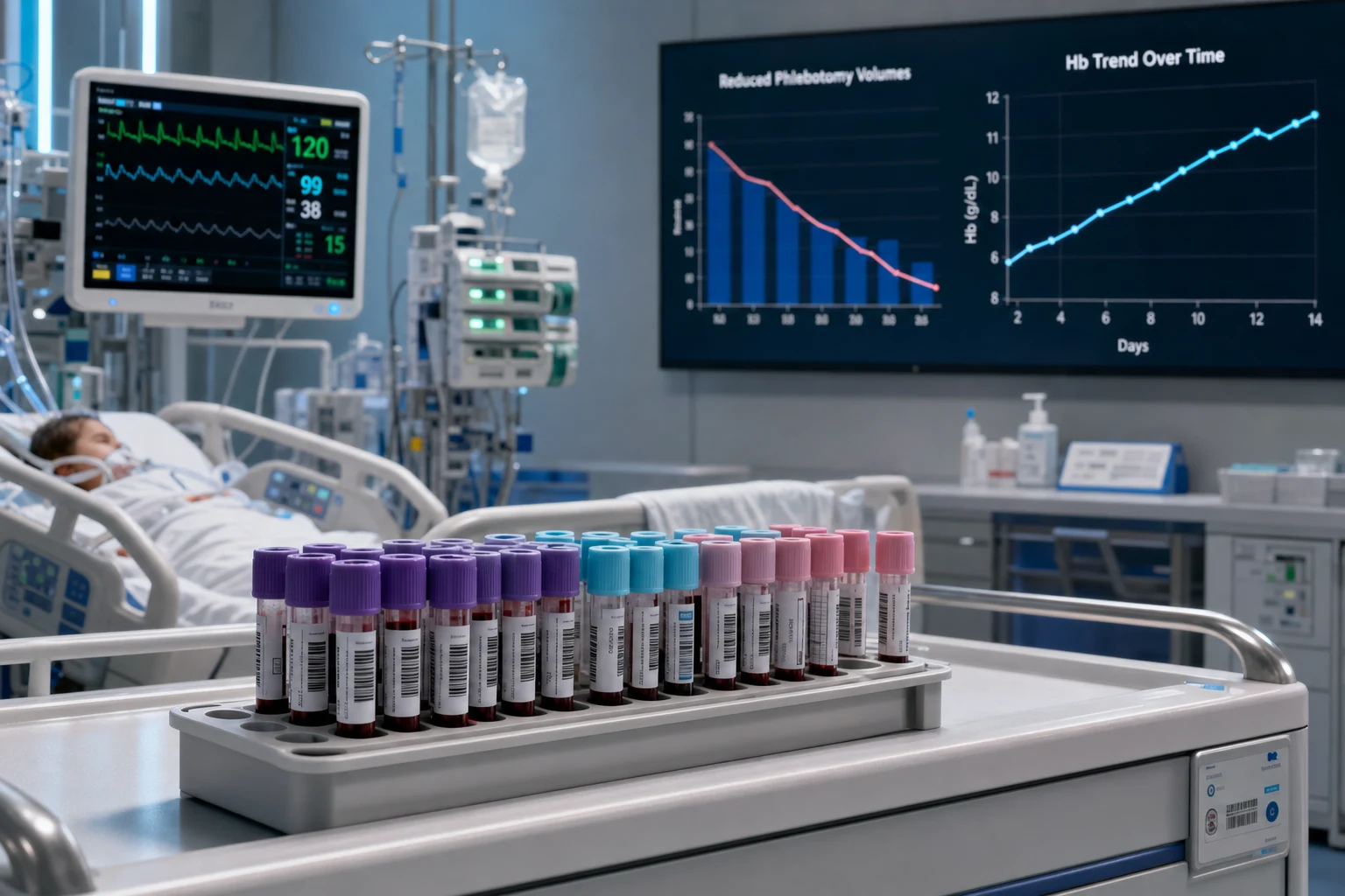
- Phlebotomy (the number 1 modifiable cause) — 40 to 70 mL per day from the routine blood sampling (the 1 mL per tube; the ABG, the daily bloods, the cultures, the coagulation).[1]
- Inflammation (anemia of chronic disease) — the hepcidin the upregulated → the iron the sequestered in the macrophages → the functional iron deficiency (the ferritin high but the iron the unavailable).[1]
- Blood loss — the GI, the surgical, the line sites, the coagulopathy.[1]
- Haemodilution — the fluid the resuscitation → the apparent anemia (the Hb the diluted).[1]
- Marrow suppression — the sepsis, the CKD (the low EPO), the drugs.[1]
- Nutritional — the B12, the folate, the iron (the pre-existing or the ICU-acquired).[1]
The blood-conservation bundle (the key)
- The reduce the phlebotomy — the number 1 modifiable cause. The measures:[1]
- The small-volume tubes (the paediatric — the 2 mL instead of the 5 to 10 mL).[1]
- The point-of-care testing (the blood-gas analyser — the smaller the sample, the faster the result).[1]
- The reduce the routine bloods (the not the daily bloods if the stable; the question-driven the testing).[1]
- The arterial-line sampling (the less the waste — the dead-space the returned).[1]
- The blood-conservation the device (the closed-loop the sampling).[1]
- The restrictive transfusion (Hb under 70 — the TRICC, TRISS; the one unit at a time).[1]
The treatment
1. Reduce the phlebotomy (the definitive modifiable)
The blood-conservation bundle (above). The single most effective measure to reduce the ICU anemia.[1]
2. The IV iron
- For the functional iron deficiency (the hepcidin-mediated — the ferritin high but the iron the unavailable).[1]
- The IV (NOT the oral — the unreliable absorption in the ICU; the hepcidin the blocks the GI absorption).[1]
- The ferric carboxymaltose or the ferric derisomaltose (the single the large dose; the lower the anaphylaxis risk than the older IV iron).[1]
- The timing — the early (the not the wait for the recovery); the benefit the in the post-ICU the rehabilitation.[1]
3. The EPO (erythropoietin)
- The NOT routine (the mixed the trials; the thrombosis risk — the VTE, the stroke; the no the mortality benefit).[1]
- The for the CKD (the confirmed EPO deficiency — the EPO the appropriate).[1]
4. Treat the underlying cause
- The GI bleed, the coagulopathy, the sepsis, the CKD, the nutritional deficiency.[1]
- The minimise the iatrogenic blood loss (the phlebotomy, the line the insertion, the dialysis circuit).[1]
Exam practice — SAQs
SAQ — ICU-acquired anaemia, functional iron deficiency and the transfusion decision
10 minutes · 10 marks
A 68-year-old man is on ICU day 9 with severe necrotising pancreatitis. He has been ventilated for 7 days and on CRRT for AKI since day 3. His haemoglobin has drifted from 112 g/L on admission to 67 g/L today. He is afebrile, haemodynamically stable on no vasopressors, lactate 1.1 mmol/L, with no evidence of active bleeding. Ferritin 680 ug/L, transferrin saturation 14 per cent, CRP 95 mg/L, reticulocyte count low. The team asks whether to transfuse two units of packed red cells.
SAQ — Transfusion threshold in acute coronary syndrome
10 minutes · 10 marks
A 74-year-old woman is admitted with an acute non-ST-elevation myocardial infarction. She has an Hb of 68 g/L on admission (iron studies confirm mixed iron deficiency). Troponin is elevated, the ECG shows anterolateral T-wave inversion, and she has ongoing chest pain. The registrar asks whether the restrictive transfusion threshold (Hb under 70) applies here.
SAQ — Anaemia of critical illness: the hepcidin axis and iron-studies interpretation
10 minutes · 10 marks
A 62-year-old woman is on ICU day 14 after severe ARDS from pneumococcal pneumonia. She was extubated 48 hours ago and is now in the recovery phase. Her Hb has drifted from 105 g/L on admission to 76 g/L today. She is afebrile, haemodynamically stable on no vasopressors, lactate 1.0 mmol/L, with no evidence of bleeding. Ferritin 720 ug/L, transferrin saturation 12 per cent, CRP 60 mg/L, reticulocyte count inappropriately low, MCV 86 fL. The physiotherapy team report she is profoundly fatigued and struggling to mobilise. The registrar asks whether she should receive oral iron, IV iron, EPO, or a transfusion.
SAQ — Transfusion threshold in septic shock: the TRISS trial
10 minutes · 10 marks
A 58-year-old man is admitted to ICU with septic shock from a urinary-tract source. He requires noradrenaline 0.3 mcg/kg/min, is ventilated for ARDS, and has a lactate of 3.2 mmol/L that is slowly falling. His Hb is 74 g/L. There is no active bleeding, no acute coronary syndrome, and his SpO2 is 96 per cent on PEEP 10 and FiO2 0.5. The registrar proposes transfusing two units of packed red cells to improve oxygen delivery and reduce the lactate.
Red flags
The causes of ICU anaemia — quantified and mechanistic
ICU anaemia is multifactorial — almost never a single cause. The dominant contributors change over the admission: haemodilution and bleeding dominate the first 48 h; phlebotomy and inflammation dominate day 3 onwards. The clinical task is to identify which mechanisms are operant in this patient and which are reversible.[1]
The six mechanisms of ICU anaemia — and which are fixable
| Mechanism | How it produces anaemia | Typical ICU setting | Modifiable? | Intervention |
|---|---|---|---|---|
| Phlebotomy (iatrogenic) | Cumulative diagnostic blood loss — 40-70 mL/day, ~1 unit/week | Every ICU patient; worse with arterial lines, frequent gases, research sampling | YES — the #1 modifiable | Small-volume tubes, POC testing, question-driven testing, closed-loop sampling |
| Inflammation (anaemia of chronic disease) | IL-6 → hepcidin ↑ → ferroportin internalised → iron trapped in macrophages AND GI absorption blocked → functional iron deficiency; plus EPO resistance and reduced RBC lifespan | Sepsis, pancreatitis, burns, prolonged ventilation, post-major-surgery | PARTIALLY | IV iron bypasses hepcidin blockade; treat the source of inflammation |
| Blood loss | Overt (GI, surgical, line-site, haemoptysis) or occult (coagulopathy, retroperitoneal) | Trauma, GI bleed, post-op, DIC, line insertion | YES | Source control, correction of coagulopathy, minimise procedural loss |
| Haemodilution | Crystalloid/colloid resuscitation expands plasma volume faster than RBC mass → the measured Hb falls though no RBC lost | Early sepsis, trauma, DKA resuscitation, post-arrest | YES | Treat the cause, allow a negative fluid balance as the patient recovers (the Hb will "rise" as fluid mobilises) |
| Bone-marrow suppression / reduced erythropoiesis | Sepsis (pro-inflammatory cytokines suppress progenitors), CKD (low EPO), drugs (chemotherapy, linezolid, ganciclovir, phenytoin), critical illness itself | Long-stay ICU, CKD, oncology, drug-induced | PARTIALLY | Treat sepsis, give EPO only for confirmed CKD deficiency, review drug chart |
| Nutritional deficiency | Pre-existing or ICU-acquired iron, B12, folate deficiency; refeeding can unmask | Malnourished, alcoholic, post-bariatric, long-stay | YES | Replace the specific deficiency (B12/folate if megaloblastic; IV iron if iron-deficient) |
The timeline — which cause dominates when
| Time from admission | Dominant mechanism(s) | Practical implication |
|---|---|---|
| 0-48 h | Haemodilution (resuscitation fluids), overt blood loss (bleed, surgery) | The falling Hb is largely dilutional — do NOT chase the number; reassess after fluid balance turns negative |
| Day 2-7 | Phlebotomy (cumulative, now ~200-400 mL), ongoing inflammation, continued bleeding | This is where the blood-conservation bundle earns its keep; check ferritin/TsAT for functional iron deficiency |
| Day 7+ | Anaemia of chronic disease entrenched (hepcidin high), marrow suppression, nutritional depletion, EPO blunting | IV iron if TsAT low with high ferritin; consider the reversible contributors before any transfusion |
Haemodilution — apparent versus true anaemia
After aggressive crystalloid resuscitation the haematocrit falls even though no red cells have been lost — the plasma volume has simply expanded. This is the commonest reason for an "anaemia" consult in the first 24 h of sepsis or trauma. Two principles:[1]
- Dilutional anaemia is not hypovolaemia — the patient is typically fluid-positive, not shocked. Transfusing to a number here causes TACO and worsening pulmonary oedema.
- The Hb will rise as fluid mobilises — once the source is controlled and the patient enters a negative fluid balance (often with diuresis or RRT), the Hb typically climbs 10-20 g/L. Reassess at that point before transfusing. [1]
The exam trap: a septic patient resuscitated with 6 L of crystalloid has an Hb of 72 g/L on day 2 with a normal lactate and no ongoing bleed. The "anaemia" is mostly dilutional. Restrictive threshold (Hb <70) plus observation is correct; a 2-unit transfusion is not. [1]
Inflammation of chronic disease — the hepcidin axis
The molecular driver of ICU-acquired anaemia is hepcidin, the liver-derived master regulator of iron homeostasis. The cascade:[1]
The hepcidin cascade in ICU anaemia
| Step | Event | Consequence |
|---|---|---|
| 1. Trigger | Any inflammatory stimulus (sepsis, surgery, trauma, pancreatitis, malignancy) → IL-6 release | IL-6 is the dominant hepcidin inducer |
| 2. Hepcidin upregulation | IL-6 → hepatocyte → hepcidin synthesis ↑ (hepcidin is also an acute-phase peptide) | Hepcidin binds ferroportin on enterocytes and macrophages |
| 3. Iron sequestration | Hepcidin-ferroportin complex → ferroportin internalised and degraded → iron CANNOT exit the cell | Iron trapped in macrophages (recycled from senescent RBCs) and in duodenal enterocytes (absorbed iron) |
| 4. Functional iron deficiency | Serum iron FALLS despite normal/raised iron stores; transferrin saturation LOW; ferritin HIGH (ferritin is an acute-phase reactant) | Marrow sees no iron → erythropoiesis impaired |
| 5. Additional hits | Inflammation also: blunts EPO production, causes EPO resistance, shortens RBC lifespan (premature clearance), and suppresses erythroid progenitors | A multifactorial anaemia, not just iron blockade |
Why this matters at the bedside: oral iron is futile — hepcidin blocks enterocyte iron export, so the absorbed iron never reaches the marrow. IV iron bypasses the gut entirely and delivers iron directly to transferrin, partially overcoming the blockade. This is the rationale for IV (not oral) iron in the ICU patient with functional iron deficiency.[1]
Iron studies interpretation — the five patterns
Iron studies are among the most over-ordered and misinterpreted tests in the ICU. The key is to interpret ferritin + transferrin saturation (TsAT) + CRP together, never in isolation, because ferritin is an acute-phase reactant (a "normal" ferritin of 100 in a septic, inflamed patient is actually low — the inflammation should have driven it much higher).[1]
Iron studies — the five patterns you must distinguish in ICU
| Pattern | Ferritin | Transferrin saturation | TIBC/transferrin | CRP | Interpretation | Action |
|---|---|---|---|---|---|---|
| Iron-deficiency anaemia (true, absolute) | LOW (<30) | LOW (<20%) | HIGH (TIBC raised) | Normal | Absolute iron deficiency — no iron stores | Iron replacement (IV in ICU); investigate source of loss |
| Anaemia of chronic disease (functional) | HIGH (often 100-700+, even >1000) | LOW (<20%) | LOW (TIBC reduced) | Raised | Iron trapped by hepcidin — stores full, iron unavailable | Treat the inflammation; IV iron may help; do NOT use oral |
| Combined ACD + true iron deficiency | LOW-NORMAL but inappropriately low for the inflammation (e.g. ferritin 50 in a septic patient — should be >300) | VERY LOW (<15%) | Normal-low | Raised | Both problems coexist — the common ICU reality | IV iron is indicated; the inappropriately low ferritin unmasks true deficiency |
| Thalassaemia trait (microcytic mimic) | NORMAL-HIGH | NORMAL-HIGH | NORMAL | Normal | Not iron deficiency at all — globin chain defect; microcytosis without iron lack | Do NOT give iron (no deficiency, risk overload); diagnose with Hb electrophoresis |
| Sepsis / critical illness (non-specific) | Markedly HIGH (acute-phase) | LOW | Low | Very raised | A combination of ACD, dilution, marrow suppression | Treat the cause; iron studies are unhelpful in acute sepsis — do not chase them in the first 48 h |
The two calculations that rescue the iron studies — the sTfR/log-ferritin ratio and TsAT
| Tool | Formula / threshold | What it tells you | Why it helps in ICU |
|---|---|---|---|
| Transferrin saturation (TsAT) | Serum iron ÷ TIBC × 100 (or iron ÷ transferrin × 1.25). <20% = iron-deficient erythropoiesis | The proportion of iron-binding sites actually carrying iron — the most direct measure of iron DELIVERY to the marrow | Works even when ferritin is confounded by inflammation; a TsAT <20% with a high ferritin = functional deficiency (give IV iron) |
| Soluble transferrin receptor (sTfR) to log(ferritin) ratio | sTfR / log(ferritin). Raised ratio (>~2) suggests TRUE iron deficiency even with inflammation | sTfR rises when the marrow is starved of iron (transferrin receptors shed into plasma); it is NOT an acute-phase reactant | Distinguishes combined deficiency from pure ACD when ferritin is misleading |
Clinical rule of thumb: in an inflamed ICU patient, a TsAT <20% (with ferritin anywhere from 30 to 800) indicates functional/absolute iron deficiency and is the trigger for IV iron. Do not wait for ferritin to fall below 30 — in inflammation it never will. [1]
Haemolysis in the ICU patient
Haemolysis is an under-recognised contributor to ICU anaemia. Suspect it when the reticulocyte count is high (the marrow is responding), LDH is raised, haptoglobin is undetectable, and there is unconjugated hyperbilirubinaemia. The blood film shows polychromasia (reticulocytes) and, in microangiopathic causes, schistocytes/fragmented red cells.[1]
Causes of haemolysis in the ICU — by mechanism
| Mechanism | Examples | Key clue | Management |
|---|---|---|---|
| Mechanical (microangiopathic) | DIC, TTP/HUS, HELLP, malignant hypertension, mechanical valves, ECMO circuit, VAD | SCHISTOCYTES on film, thrombocytopenia | Treat the cause (TTP → PLEX; DIC → source; ECMO/VAD → circuit haemolysis, optimise flow) |
| Immune (autoimmune haemolytic anaemia) | Warm AIHA (IgG, idiopathic or SLE/CLL/drug), cold agglutinin (Mycoplasma, EBV, lymphoma) | Positive direct antiglobulin (Coombs) test, spherocytes (warm) | Steroids (warm); rituximab; avoid cold (cold agglutinin); transfuse ONLY if life-threatening (cross-match is difficult) |
| Infection-related | Severe malaria (blackwater fever), clostridial sepsis (C. perfringens α-toxin), babesiosis | Travel/exposure history, parasites on film, massive intravascular haemolysis | Antimalarials/antibiotics; exchange transfusion for severe malaria |
| Drug-induced | G6PD-deficient patient given oxidant drug (dapsone, primaquine, sulfa, rasburicase), drug-induced immune haemolysis | G6PD assay, temporal link to drug | Stop the drug; supportive; avoid oxidants in G6PD deficiency |
| Haemoglobinopathy | Sickle-cell crisis, thalassaemia intermedia | Known diagnosis, sickled cells/target cells on film | Disease-specific (see SCD topic) |
| Hypersplenism | Chronic liver disease, portal hypertension, haematological malignancy | Splenomegaly, pancytopenia, sequestered RBCs | Treat underlying; rarely splenectomy |
The exam trap: a falling Hb with schistocytes on the film is NOT autoimmune haemolysis — it is microangiopathic (TTP until proven otherwise if platelets low and Coombs negative). The direct Coombs test separates the immune from the mechanical: Coombs-positive = immune; Coombs-negative with schistocytes = microangiopathic. [1]
Bone-marrow suppression in the ICU
Critical illness itself suppresses erythropoiesis through several converging mechanisms — this is part of why the Hb keeps falling even after bleeding and phlebotomy are controlled:[1]
Mechanisms of marrow suppression in critical illness
| Mechanism | Detail |
|---|---|
| Blunted EPO production | The inflammatory cytokine milieu (TNF-α, IL-1) suppresses renal EPO synthesis — the EPO response to anaemia is inappropriately low for the degree of anaemia |
| EPO resistance | Even the EPO that is produced acts less effectively — marrow progenitors are relatively resistant (this is why simply giving EPO has disappointing results — see EPO section) |
| Direct progenitor suppression | Pro-inflammatory cytokines (IFN-γ, TNF-α) induce apoptosis of erythroid progenitors (BFU-E/CFU-E) in the marrow |
| Reduced RBC lifespan | The RBC membrane is damaged by oxidative stress and the activated reticuloendothelial system clears cells early — the ICU RBC lives ~90 days instead of 120 |
| Drug effects | Chemotherapy, linezolid (myelosuppression, lactic acidosis), ganciclovir, valganciclovir, trimethoprim, phenytoin, chloramphenicol, sulphonamides |
| CKD | Reduced renal EPO production (the kidney is the source); the higher the CKD stage, the lower the EPO |
| Parvovirus B19 | Transient aplastic crisis (especially in sickle cell, hereditary spherocytosis) — sudden absence of reticulocytes |
When to transfuse — threshold by scenario
The single most important transfusion principle in ICU: transfuse the patient, not the number — but use the Hb as the trigger within a scenario-specific threshold. The restrictive strategy (Hb <70) is safe and superior for most ICU patients; specific contexts raise the threshold.[1][2]
Transfusion thresholds by clinical scenario — the ICU guide
| Scenario | Hb threshold (g/L) | Rationale / evidence | Notes |
|---|---|---|---|
| General ICU (stable) | <70 | TRICC (Hébert 1999) — restrictive as safe as liberal, fewer complications; TRISS extended this to septic shock | Transfuse to target 70-90; ONE unit then reassess |
| Septic shock | <70 | TRISS (Holst 2014) — 7 vs 9, no mortality difference, half the transfusions | Even in septic shock the restrictive threshold wins |
| Acute coronary syndrome (ACS) | <80 | The ischaemic myocardium cannot compensate by extracting more oxygen (already near-maximal extraction at rest) | REALITY and MINT suggest the threshold may be rising again — individualise; some now use <80-90 in active ACS |
| Active bleeding / massive haemorrhage | <80 (permissive, with massive-transfusion protocol) | In massive bleeding, transfuse per MTP (RBC:plasma:platelets ~1:1:1) — do NOT wait for a number | Target Hb 80-100 during active bleeding; the target is driven by haemodynamics, not a single Hb |
| Cardiac surgery (perioperative) | <75 | TRICS III (Mazer 2017) — restrictive (Hb <75) non-inferior to liberal even in moderate-to-high-risk cardiac surgery | Confirms restrictive safety in the cardiac surgical population |
| Paediatric ICU (stable) | <70 | TRIPICU (Lacroix 2007) — restrictive as safe as liberal in stable critically-ill children | Does NOT apply to unstable children or those with severe hypoxaemia/cyanotic heart disease |
| Acute upper GI bleed (non-massive) | <70 | Restrictive strategy improves survival and reduces rebleeding (lower portal pressure) | BUT transfuse earlier (<90) if ACS, stroke, or massive exsanguinating bleed coexist |
| Traumatic brain injury | Controversial — avoid Hb <80-90 in the acute phase | The injured brain is exquisitely sensitive to secondary ischaemia; some advocate <90 | Balance against the (weak) signal of harm from liberal transfusion |
| Chronic, stable anaemia (no acute bleed) | Often no transfusion — treat the cause | E.g. CKD anaemia — give EPO/iron, not RBCs | Transfusion is a bridge, not a treatment, for chronic anaemia |
Deciding whether to transfuse — the practical ICU algorithm
- Is the patient actively bleeding or in shock? — if YES, this is a massive-transfusion / haemorrhage-control problem, NOT a threshold problem. Activate the MTP, achieve haemostasis, and transfuse to haemodynamic and haemostatic targets (see Massive transfusion topic).
- Is there an ACS, acute ischaemia, or severe hypoxaemia the anaemia is worsening? — if YES, use the higher threshold (Hb <80, some units <90 in active ACS). The ischaemic tissue cannot compensate for a low oxygen content.
- Otherwise apply the restrictive threshold — Hb <70 g/L. Transfuse ONE unit of packed red cells.
- Reassess after each unit — recheck Hb, the symptoms, the lactate, the perfusion. Do not transfuse by prescription ("2 units"); transfuse by response. Each unit should raise the Hb ~10 g/L (or ~0.03 haematocrit) in a non-bleeding adult.
- Ask 'why is this patient anaemic?' before the next unit — phlebotomy? bleeding? dilution? ACD? Treat the cause in parallel so the transfusion is not merely repeated daily.
- Document the indication — every unit needs a recorded indication. Over-transfusion (TACO, TRALI, infection, MODS) is a leading cause of preventable ICU harm.
The TRICC trial and the restrictive transfusion evidence
The modern era of ICU transfusion began with TRICC (1999), which demolished the old "10/30" rule (transfuse to keep Hb >100). The cumulative evidence across critical illness, sepsis, cardiac surgery, paediatrics, and acute MI now firmly supports a restrictive strategy.[1]
The restrictive-transfusion landmark trials — what each showed
| Trial (year) | Population | Restrictive vs Liberal threshold | Primary result | Take-home |
|---|---|---|---|---|
| TRICC (Hébert 1999) | 838 general ICU | Hb <70 (target 70-90) vs <100 (target 100-120) | 30-day mortality 18.7% vs 23.3% (NS, p=0.11); hospital mortality significantly lower with restrictive | Restrictive safe; lower-APACHE and younger patients did BETTER restrictive; the 10/30 rule was dead |
| TRISS (Holst 2014) | 998 septic shock | Hb <70 vs <90 | 90-day mortality 43% vs 45% (NS); half the transfusions | Restrictive safe even in septic shock — the classic exemption was disproven |
| TRIPICU (Lacroix 2007) | 637 stable paediatric ICU | Hb <70 vs <95 | New/progressive MODS no different; 44% fewer transfusions | Restrictive safe in stable critically-ill children |
| TRICS III (Mazer 2017) | 5243 moderate-to-high-risk cardiac surgery | Hb <75 vs <95 (OR/ICU) / <85 (ward) | Composite (death/MI/stroke/RRT) non-inferior with restrictive | Restrictive safe even in cardiac surgical patients — the cardiac "exemption" is shrinking |
| REALITY (Ducrocq 2021) | 668 acute MI + anaemia | Hb <80 vs <100 | Met non-inferiority for 30-day MACE; but signal of harm at 6 months (higher all-cause mortality) | Restrictive probably acceptable in MI but not definitively safe — individualise |
| MINT (Carson 2023) | 3504 ACS or stable ischaemic heart disease + anaemia | Hb <80 (restrictive) vs <100 (liberal) | Numerically MORE death/MI with restrictive (did not meet the pre-specified significance bar but trend concerning) | In ACS the restrictive strategy is NOT clearly safe — many now transfuse at <80 (or higher) in active ACS |
Restrictive vs liberal transfusion — the balance of harm
| Factor | Restrictive (Hb <70) | Liberal (Hb <90-100) |
|---|---|---|
| Transfusion-related complications (TRALI, TACO, allergy, haemolysis) | FEWER (less exposure) | MORE (each unit carries risk) |
| Infection / immunomodulation (TRIM) | FEWER nosocomial infections | MORE (transfusion-related immunomodulation; transfusion is independently associated with infection) |
| Mortality (general ICU, sepsis, cardiac surgery, paediatrics) | EQUIVALENT or better | No benefit; sometimes worse |
| Mortality (acute MI / ACS) | POSSIBLY worse — the one context where liberal may be favoured | MINT trended toward benefit in ACS |
| Cost / blood inventory | Lower consumption, conserves supply | Higher consumption |
| Tissue oxygenation in ischaemia | Adequate in most; borderline in the severely ischaemic | Theoretical reserve in ischaemic tissue |
Hébert 1999 — TRICC: restrictive vs liberal RBC transfusion in critical care (PMID 9971864)
Source
New England Journal of Medicine — multicentre RCT, 838 euvolaemic critically-ill patients
Intervention
Restrictive (transfuse if Hb <70 g/L, maintain 70-90) vs Liberal (transfuse if Hb <100 g/L, maintain 100-120)
Primary outcome
30-day all-cause mortality 18.7% (restrictive) vs 23.3% (liberal) — not statistically different (p=0.11)
Secondary
Hospital mortality significantly LOWER with restrictive (22.2% vs 28.1%, p=0.05); patients with APACHE II ≤20 and age <55 did significantly BETTER restrictive
Cardiac subgroup
No significant difference in known cardiac disease; underpowered for acute MI (a signal that the cardiac question was not closed)
Legacy
Killed the '10/30' transfusion rule; the foundation of every modern restrictive guideline
Holst 2014 — TRISS: lower vs higher Hb threshold in septic shock (PMID 25270275)
Source
New England Journal of Medicine — multicentre RCT, 998 patients with septic shock, 32 Scandinavian ICUs
Intervention
Transfuse at Hb <70 g/L (restrictive) vs <90 g/L (liberal); all leukoreduced
Primary outcome
90-day mortality 43.0% (restrictive) vs 45.0% (liberal) — no difference (RR 0.94, 95% CI 0.78-1.09)
Secondary
No difference in ischaemic events, severe adverse reactions, mechanical-ventilation, vasopressor or RRT days; restrictive group received median 1 unit vs 4 units
Take-home
The 'sepsis exemption' to the restrictive strategy was disproven — transfuse septic shock at Hb <70
Lacroix 2007 — TRIPICU: transfusion strategies in paediatric ICU (PMID 17442904)
Source
New England Journal of Medicine — multicentre non-inferiority RCT, 637 stable critically-ill children
Intervention
Restrictive (transfuse if Hb <70 g/L) vs Liberal (transfuse if Hb <95 g/L)
Primary outcome
New or progressive multiple-organ dysfunction syndrome — NO significant difference
Secondary
44% fewer patients exposed to transfusion in the restrictive group; 46% fewer total transfusions
Take-home
Restrictive strategy (Hb <70) is safe in stable critically-ill children — extends the TRICC principle to paediatrics
Mazer 2017 — TRICS III: restrictive vs liberal transfusion in cardiac surgery (PMID 29188999)
Source
New England Journal of Medicine — international non-inferiority RCT, 5243 patients undergoing cardiac surgery with moderate-to-high mortality risk (EuroSCORE I ≥6)
Intervention
Restrictive (transfuse if Hb <75 g/L intra-op/post-op ICU) vs Liberal (Hb <95 in OR/ICU, <85 on ward)
Primary outcome
Composite of death, MI, stroke, or new RRT — restrictive NON-INFERIOR
Take-home
The 'cardiac disease needs higher Hb' belief is not supported — even in cardiac surgery the restrictive threshold is safe
Ducrocq 2021 — REALITY: restrictive vs liberal transfusion in acute MI (PMID 33560322)
Source
JAMA — multicentre non-inferiority RCT, 668 patients with acute MI and anaemia
Intervention
Restrictive (transfuse at Hb ≤80 g/L) vs Liberal (transfuse at Hb ≤100 g/L)
Primary outcome
30-day composite of MACE — restrictive MET the non-inferiority margin
Cautionary signal
At 6 months, all-cause mortality was HIGHER in the restrictive group — a warning that the restrictive strategy may not be uniformly safe in MI
Carson 2023 — MINT: liberal vs restrictive transfusion in ACS (PMID 37952219)
Source
New England Journal of Medicine — multicentre RCT, 3504 patients with ACS or stable ischaemic heart disease and anaemia
Intervention
Restrictive (Hb <80 g/L) vs Liberal (Hb <100 g/L) transfusion threshold
Primary outcome
Composite of death or MI at 30 days — 17.3% (restrictive) vs 15.9% (liberal); did NOT meet the pre-specified significance threshold but the trend favoured liberal
Take-home
In ACS the restrictive strategy is NOT proven safe — the one setting where a higher transfusion threshold (Hb <80, some units higher) remains defensible
Complications of transfusion — TRALI, TACO, infections, immunomodulation
Every unit of blood is a liquid organ transplant with real hazards. The two pulmonary reactions — TRALI and TACO — are the highest-stakes and most-tested, but infection and immunomodulation matter too.[1][7]
TRALI vs TACO vs acute haemolytic — the three high-stakes transfusion reactions
| Feature | TRALI (transfusion-related acute lung injury) | TACO (transfusion-associated circulatory overload) | Acute haemolytic (ABO incompatibility) |
|---|---|---|---|
| Mechanism | Donor anti-leukocyte (anti-HLA/anti-HNA) antibodies → neutrophil activation → pulmonary capillary leak → ARDS-like | Circulatory OVERLOAD from the transfusion volume → hydrostatic pulmonary oedema | ABO incompatibility (wrong blood to wrong patient) → intravascular haemolysis → DIC, renal failure |
| Timing | Within 6 hours of transfusion (2019 consensus: Type I = no ARDS risk factor; Type II = with ARDS risk factor) | During or up to 12 hours after transfusion | During / very soon after the start of the unit |
| Fluid balance | EUVOLAEMIC — NO overload (the key distinguishing feature) | Fluid-POSITIVE — the hallmark | Variable |
| BNP / NT-proBNP | Normal/low | Raised (cardiac stretch) | Not diagnostic |
| Chest X-ray | Bilateral infiltrates (white-out) | Bilateral infiltrates ± pleural effusions, enlarged cardiac silhouette | Not specific |
| Hypertension | Absent (often hypotension) | Present (helps distinguish from TRALI) | Hypotension/shock |
| Treatment | SUPPORTIVE (oxygen, ventilation) — NO diuretics (TRALI is capillary leak, not overload) | Diuretics (furosemide), slow/stop the transfusion, reduce volume | STOP the transfusion IMMEDIATELY; supportive (fluids, vasopressors), treat DIC, renal protection |
| Mortality | ~5-10% (the leading cause of transfusion-related death where reported) | Lower than TRALI but common | High |
| Prevention | Male-donor / never-pregnant-donor plasma (fewer antibodies); plasma from male donors preferred | Slow rate (2-4 h/unit), one unit at a time, diurese high-risk patients (elderly, cardiac) | Two-person bedside identity check; clerical error is the usual cause |
Infectious risks of transfusion — current magnitude
| Pathogen | Estimated residual risk (per unit, high-income countries) | Notes |
|---|---|---|
| HIV | ~1 in 1-2 million | Nucleic-acid testing (NAT) has driven this down |
| Hepatitis C | ~1 in 1-2 million | NAT screening |
| Hepatitis B | ~1 in 100,000-300,000 | Lower than HCV; window period |
| Bacterial contamination (platelets) | ~1 in 1000-3000 (severe sepsis ~1 in 100,000) | Platelets stored at room temperature — the highest infectious risk product; screening/pooled testing now routine |
| West Nile virus | Rare; seasonal screening | NAT in endemic areas |
| Cytomegalovirus (CMV) | Reduced by leukodepletion (WBCs carry CMV) | Leukodepletion standard; CMV-seronegative units for transplant/immunocompromised |
| Prion (vCJD) | Exceedingly rare; leukodepletion reduces | Historical concern, now near-zero |
| Parasites (malaria, Chagas) | Rare in non-endemic donors; travel/triage screening | Relevant in returned travellers / endemic regions |
Transfusion-related immunomodulation (TRIM) is a separate, subtle harm: transfusion transiently suppresses recipient cellular immunity (mediated by donor leukocytes and soluble factors), which is associated with a higher rate of nosocomial infection, possible cancer recurrence, and (in theory) reduced transfusion-reaction immunogenicity. Leukodepletion (now near-universal) mitigates but does not abolish TRIM. The clinical bottom line: every avoidable transfusion exposes the patient to infection risk — another reason the restrictive strategy wins.[1]
The common, non-life-threatening transfusion reactions
| Reaction | Mechanism | Features | Management |
|---|---|---|---|
| Febrile non-haemolytic | Cytokines from donor white cells; recipient anti-leukocyte antibodies | Fever, rigors during/after transfusion; normal haemoglobin (no haemolysis) | Slow/stop; antipyretic; prevented by leukodepletion (now standard) |
| Mild allergic (urticaria) | Donor plasma proteins → IgE-mediated histamine | Itch, urticaria, no fever, stable observations | Slow/stop; antihistamine; usually can restart cautiously |
| Anaphylactoid / severe allergic | Often anti-IgA in IgA-deficient recipient | Hypotension, bronchospasm, angioedema, gastrointestinal symptoms | STOP; adrenaline, fluids, intubation if needed; washed/deglycerolised products or IgA-deficient donor units in future |
| Delayed haemolytic | Anamnestic response to previously immunised antigen (e.g. Kidd, Duffy) 3-14 days post-transfusion | Falling Hb, mild jaundice, positive DAT, spherocytes | Usually self-limiting; supportive; identify the antibody for future cross-matching |
| Post-transfusion purpura | Antibody (usually anti-HPA-1a) destroys recipient AND transfused platelets | Severe thrombocytopenia 5-10 days post-transfusion | IVIG; platelet antigen-matched units |
| Transfusion-associated graft-vs-host disease | Donor T-lymphocytes engraft in an immunocompromised recipient (or one-way HLA match) | Fever, rash, diarrhoea, pancytopenia 4-30 days later — almost universally fatal | Irreversible; PREVENT with irradiation of cellular products for immunocompromised/related-donor units |
IV iron — formulations, evidence and the ICU place
IV iron is the rational therapy for functional iron deficiency because it bypasses the hepcidin-mediated gut blockade. The newer formulations (ferric carboxymaltose, ferric derisomaltose, iron isomaltoside) allow large single-dose administration with a far lower anaphylaxis risk than the older high-molecular-weight iron dextran (withdrawn).[1]
The IV iron formulations used in ICU
| Formulation | Typical dose | Test dose | Anaphylaxis risk | Notes |
|---|---|---|---|---|
| Ferric carboxymaltose | 1000 mg IV over ~15 min (single dose); can repeat | Not required | Low (modern) | The most-studied modern agent; large single dose; widely available in ANZ/UK |
| Ferric derisomaltose | 1000 mg IV (can go up to 20 mg/kg single dose) | Not required | Low | Single large dose; favourable safety profile |
| Iron isomaltoside (monoferric) | Up to 20 mg/kg single dose | Not required | Low | Allows full repletion in one visit |
| Iron sucrose | 100-200 mg per dose (multiple doses) | Not required | Low | Lower per-dose iron content; commonly used in dialysis/CKD |
| Low-molecular-weight iron dextran | Variable | Often required | Lower than high-MW dextran | Rarely first-line now |
| (High-molecular-weight iron dextran) | — | — | WITHDRAWN (high anaphylaxis) | Historical; the source of the old "iron anaphylaxis" fear |
The ICU evidence on IV iron is modest and mixed — trials (IRONMAN and others) have shown reduced transfusion in some populations but no consistent mortality benefit, and the optimal timing (early vs recovery phase) is debated. The pragmatic position: give IV iron to ICU patients with confirmed functional iron deficiency (low TsAT) who are not acutely septicaemic (iron can feed pathogens in active bacteraemia), with the aim of improving recovery-phase haematocrit and reducing later transfusion rather than as an acute rescue.[1]
Erythropoietin — the controversy
EPO was once a promising "anaemia fix" for ICU patients (it raises the reticulocyte count and reduces transfusion requirement in trials). It has fallen out of favour because:[1]
EPO in ICU — why it is NOT routine
| Issue | Detail |
|---|---|
| No consistent mortality benefit | Multiple RCTs and meta-analyses (e.g. the large Corwin-era trials) showed EPO reduces the number of units transfused but does NOT improve survival, length of stay, or organ failure |
| Thrombosis risk | EPO increases viscosity and platelet activity — a real signal of VTE, stroke, and vascular access thrombosis in ICU trials |
| EPO resistance in critical illness | The inflamed marrow is relatively resistant to EPO, so even high doses have a blunted effect (the "EPO resistance" phenomenon) |
| Delayed onset | EPO takes days to weeks to raise the Hb — useless for the acute anaemia the intensivist is managing |
| Cost and complexity | A costly therapy with marginal benefit and real harm |
| The one niche — CKD | Patients with confirmed CKD-associated EPO deficiency (the kidney is not making enough) are the appropriate candidates; even here, correct iron first |
The bottom line: restrictive transfusion + IV iron + blood conservation is the modern bundle. EPO is reserved for the CKD patient with proven EPO deficiency, ideally after iron repletion. The exam answer: "EPO is not routinely recommended in ICU anaemia because trials show reduced transfusion but no mortality benefit and a real thrombosis risk; it is reserved for CKD." [1]
Clinical pearls
Additional red flags
References
- [1]Hébert PC, Wells G, Blajchman MA, et al A multicenter, randomized, controlled clinical trial of transfusion requirements in critical care. Transfusion Requirements in Critical Care Investigators, Canadian Critical Care Trials Group N Engl J Med, 1999.PMID 9971864
- [2]Holst LB, Haase N, Wetterslev J, et al Lower versus higher hemoglobin threshold for transfusion in septic shock N Engl J Med, 2014.PMID 25270275
- [3]Lacroix J, Hébert PC, Hutchison JS, et al Transfusion strategies for patients in pediatric intensive care units N Engl J Med, 2007.PMID 17442904
- [4]Mazer CD, Whitlock RP, Fergusson DA, et al Studies on Chemical Reactivity and Electrocatalysis of Two Acylmethyl(hydroxymethyl)pyridine Ligand-Containing [Fe]-Hydrogenase Models (2-COCH(2)-6-HOCH(2)C(5)H(3)N)Fe(CO)(2)L (L = η(1)-SCOMe, η(1)-2-SC(5)H(4)N) Inorg Chem, 2017.PMID 29188999
- [5]Ducrocq G, Puymirat E, de Larminat JM, et al Effect of a Restrictive vs Liberal Blood Transfusion Strategy on Major Cardiovascular Events Among Patients With Acute Myocardial Infarction and Anemia: The REALITY Randomized Clinical Trial JAMA, 2021.PMID 33560322
- [6]Carson JL, Brooks MM, Abbott JD, et al ASO Author Reflections: Which is the Better Choice for Patients with PC Who Underwent Diaphragm Resection: HIPEC or HITAC Ann Surg Oncol, 2024.PMID 37952219
- [7]Vlaar AP, Arbous S, ten Brinke M, et al A consensus redefinition of transfusion-related acute lung injury Transfusion, 2019.PMID 30993745