ICU · haematology-coagulation
Catastrophic Antiphospholipid Syndrome (CAPS) in the ICU
Also known as Catastrophic antiphospholipid syndrome · CAPS · Asherson syndrome · Antiphospholipid syndrome catastrophic · Multi-organ microthrombosis · APS storm
Catastrophic antiphospholipid syndrome (CAPS) — the most severe form of antiphospholipid syndrome (APS) characterised by rapid multi-organ thrombosis (small-vessel) affecting three or more organ systems within one week, histopathological evidence of microthrombosis, and persistent antiphospholipid antibodies (lupus anticoagulant, anticardiolipin, anti-beta-2-glycoprotein I). Affects: kidneys (AKI from renal microthrombi), lungs (ARDS, pulmonary embolism, pulmonary haemorrhage), brain (stroke, encephalopathy, seizures), heart (myocardial infarction, cardiomyopathy, valvular lesions), skin (livedo reticularis, digital gangrene), liver, GI, and adrenal (Waterhouse-Friderichsen). Triggers: infection (most common), surgery, malignancy, pregnancy, withdrawal of anticoagulation. Management: (1) eliminate trigger (treat infection), (2) triple therapy — anticoagulation (heparin) + corticosteroids + plasma exchange ± IVIG, (3) rituximab or eculizumab for refractory cases, (4) supportive care (RRT, ventilation, vasopressors). Mortality 30-50% despite treatment.
On this page & tools
Your progress
Saved locally on this device.
Target exams
Red flags
Overview
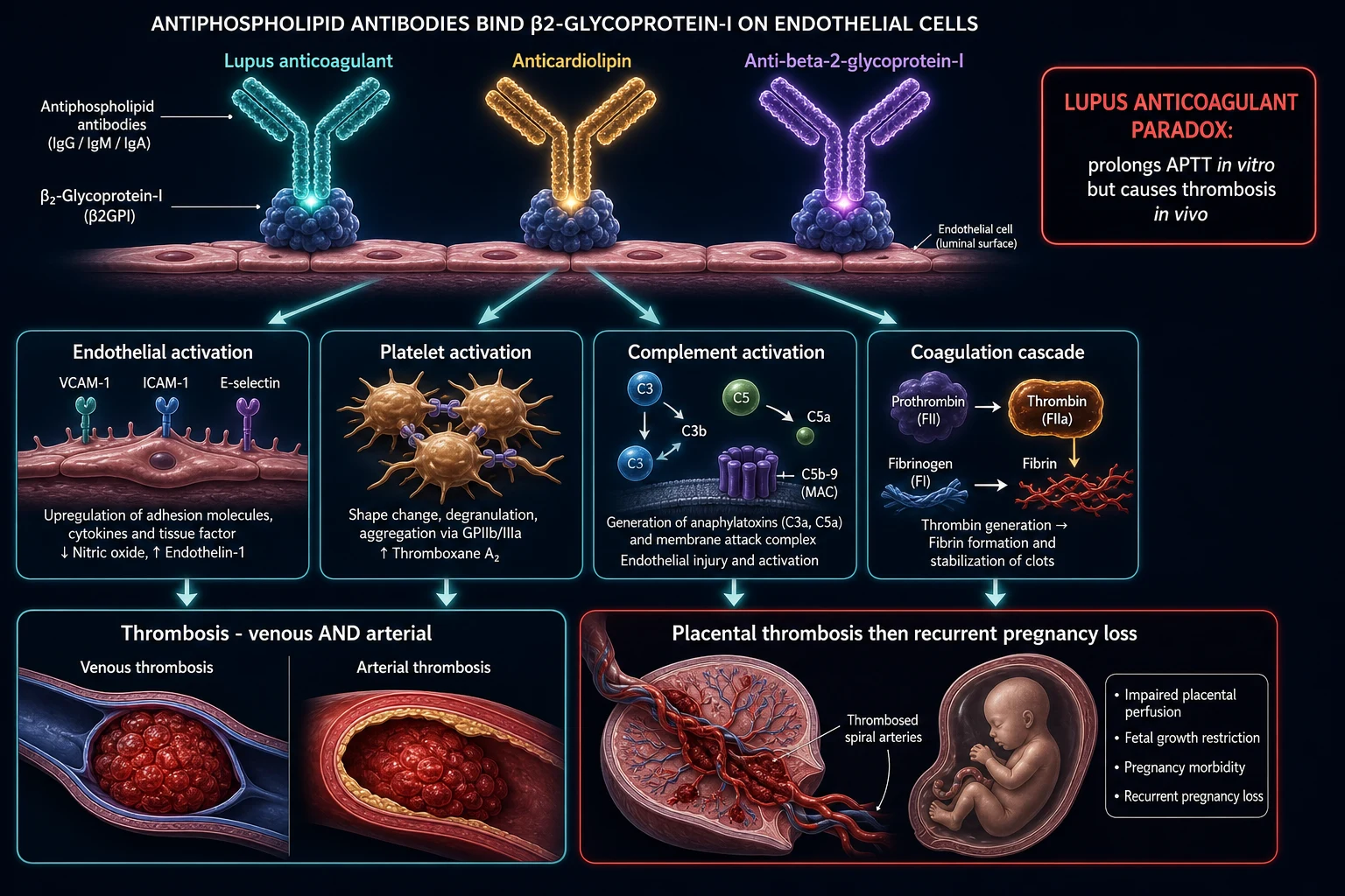
CAPS represents a dramatic acceleration of APS — from a chronic thrombophilic state (routine DVT/PE, recurrent miscarriage) to a fulminant, life-threatening prothrombotic storm. The pathophysiology involves antiphospholipid antibodies binding to beta-2-glycoprotein I on endothelial cells, platelets, and monocytes → complement activation → tissue factor expression → widespread thrombin generation → microthrombosis in multiple vascular beds simultaneously. The complement cascade (particularly C5a-C5aR) is the critical amplifier — which explains why eculizumab (anti-C5) can be effective in refractory cases.[2][4]
CAPS accounts for less than 1% of all APS cases but is responsible for a disproportionate share of APS-related deaths. The intensivist is usually the first to encounter these patients — they present with rapidly evolving multi-organ failure that does not fit the pattern of simple sepsis or DIC. The key to recognition is the temporal pattern (three or more organs thrombosing within one week) combined with the presence of aPL antibodies (or a history of APS/SLE).[5]
Diagnostic criteria — the 2006 revised Sapporo classification
Preliminary classification criteria for definite CAPS (2006修订 Sapporo)
| Criterion | Requirement | Detail |
|---|---|---|
| 1. Involvement of three or more organs | Three or more organs/systemics AND/OR tissues | Each confirmed by imaging or histopathology |
| 2. Simultaneous or rapid succession | All within less than one week | The rapidity distinguishes CAPS from chronic APS |
| 3. Histopathological confirmation | Small-vessel occlusion (microthrombosis) in at least one organ | Biopsy showing thrombosis in small vessels (arterioles, capillaries, venules) — no significant inflammation (distinguishes from vasculitis) |
| 4. aPL antibody positivity | Lupus anticoagulant, anticardiolipin (IgG/IgM), or anti-beta-2-GPI (IgG/IgM) — at least once | Medium-high titre; must be present at least 12 weeks apart for definitive APS (but in CAPS, treat empirically) |
| Definite CAPS | All four criteria met | — |
| Probable CAPS | All four criteria EXCEPT only two organs involved OR aPL positive only once (not repeated at 12 weeks) | Manage the same as definite CAPS |
CAPS vs DIC vs TTP/HUS — the critical differential
| Feature | CAPS | DIC | TTP/HUS |
|---|---|---|---|
| Pathology | Microthrombosis (platelet-fibrin) | Widespread coagulation activation + consumption | Platelet microthrombi (ADAMTS13 deficiency in TTP) |
| Trigger | Infection, surgery, pregnancy, anticoagulation withdrawal | Sepsis, trauma, malignancy, obstetric | Autoimmune, genetic, drug, infection |
| aPL antibodies | POSITIVE (lupus anticoagulant, anticardiolipin, anti-beta-2-GPI) | Negative | Negative (anti-ADAMTS13 in autoimmune TTP) |
| Fibrinogen | NORMAL or slightly low | LOW (consumed) | Normal |
| PT/APTT | May be prolonged (lupus anticoagulant) — but NOT from consumption | Prolonged (factor consumption) | Normal |
| Schistocytes | Present but FEW (<1%) | Present (moderate) | Present and MANY (>1%, often >4-5%) |
| Platelets | Mild-moderate thrombocytopenia (may be normal — platelets consumed in thrombi) | SEVERE thrombocytopenia (consumed) | SEVERE thrombocytopenia (consumed in microthrombi) |
| D-dimer | Elevated (thrombosis) | Markedly elevated | Elevated |
| Key treatment | Anticoagulation + steroids + PLEX | Treat underlying cause + supportive (blood products) | Plasma exchange (urgent) + steroids (autoimmune) |
Clinical presentation — the organ-by-organ pattern
CAPS affects multiple organs simultaneously or in rapid succession. The pattern of multi-organ microthrombosis helps distinguish CAPS from other causes of multi-organ failure:[1][5]
Organ involvement in CAPS — frequency and presentation
| Organ system | Frequency | Presentation | ICU relevance |
|---|---|---|---|
| Kidneys | 70% | AKI (rapidly rising creatinine), proteinuria, haematuria, hypertension. Biopsy: thrombotic microangiopathy (TMA) in glomeruli — NOT crescentic GN (distinguishes from PRS/vasculitis) | RRT (CRRT or IHD) — often needed early. Avoid therapeutic anticoagulation during RRT circuit (use regional citrate) |
| Lungs | 60% | ARDS (from alveolar microthrombi), PE (macrovascular), pulmonary haemorrhage (DAH from capillary microthrombi), pulmonary hypertension | Mechanical ventilation (ARDSNet). Lung-protective ventilation. PE may need thrombolysis (controversial in CAPS with active bleeding risk) |
| Brain | 55% | Ischaemic stroke (small and large vessel), encephalopathy (from cerebral microthrombi), seizures, cognitive impairment, transverse myelitis (rare) | Neuroprotection (avoid hypoxia/hypotension). CT/MRI brain. EEG if seizures. Distinguish from neuropsychiatric lupus |
| Heart | 50% | Myocardial infarction (microvascular or macrovascular), cardiomyopathy (diastolic or systolic dysfunction from microthrombi), valvular vegetations (Libman-Sacks), heart failure | ECG (arrhythmia, ischaemia). Troponin. Echo (regional wall motion, LVEF). Coronary angiography if macrovascular MI. Mechanical support if cardiogenic shock |
| Skin | 45% | Livedo reticularis (most common — net-like rash — mottled blue-purple), digital ischaemia (gangrene of fingers/toes), skin ulcers, purpura, necrosis | Clinical sign — aids diagnosis. Digital gangrene may require surgical assessment for amputation. Skin biopsy (thrombosis without vasculitis = CAPS histopathology) |
| Liver | 25% | Hepatic dysfunction (elevated transaminases from hepatic microthrombi), jaundice, coagulopathy (worsened by liver synthetic failure) | Monitor LFTs and coagulation. Supportive care. N-acetylcysteine if severe hepatic dysfunction |
| GI | 20% | Mesenteric ischaemia (intestinal microthrombi → bowel infarction), GI bleeding (from mucosal ischaemia), acalculous cholecystitis, pancreatitis | CT abdomen (bowel wall thickening, pneumatosis, perforation). Surgery for bowel infarction/perforation |
| Adrenals | 5-10% (under-recognised) | Bilateral adrenal haemorrhage/infarction → acute adrenal insufficiency → refractory vasopressor-dependent shock + hyponatraemia + hyperkalaemia (Waterhouse-Friderichsen pattern) | Give empiric hydrocortisone 200 mg IV in any CAPS patient with refractory shock — bilateral adrenal involvement is common and lethal if missed. Check morning cortisol + ACTH. CT abdomen may show enlarged adrenal glands with haemorrhage |
| Retina | 10% | Retinal artery/vein occlusion, cotton wool spots | Ophthalmology referral. Visual loss can be permanent |
Triggers — identifying and eliminating the precipitant
CAPS rarely occurs spontaneously — in over 90% of cases, a trigger can be identified. The #1 trigger is infection (50-60%), which is why the first management step is ALWAYS aggressive evaluation and treatment of infection.[1]
CAPS triggers — frequency and management
| Trigger | Frequency | Examples | Management |
|---|---|---|---|
| Infection | 50-60% | Respiratory (pneumonia), urinary (UTI/pyelo), skin (cellulitis), GI (peritonitis), catheter-related | Blood/urine/sputum cultures. Broad-spectrum antibiotics EARLY (do not delay — infection feeds the thrombotic storm). Source control (drain abscess, remove infected line) |
| Surgery | 10-15% | Major surgery, perioperative period | Ensure perioperative anticoagulation bridging in known APS patients. Early postoperative mobilisation |
| Malignancy | 5-10% | Haematological (lymphoma, leukaemia), solid tumours | Oncology referral. Treat underlying malignancy |
| Pregnancy / postpartum | 5-10% | Pre-eclampsia, HELLP, postpartum | Multi-disciplinary (obstetrics + ICU + haematology). Delivery if viable. Anticoagulation (heparin — NOT warfarin in pregnancy) |
| Anticoagulation withdrawal | 5-10% | Stopping warfarin/heparin in known APS patient | NEVER stop anticoagulation in APS without bridging. This is a preventable trigger |
| Drugs / medications | <5% | Oral contraceptives, hormone replacement | Discontinue offending agent |
| None identified | 10-15% | Idiopathic | Still treat with triple therapy |
Management — the triple therapy approach
CAPS treatment protocol — first 24 hours in ICU
-
ELIMINATE THE TRIGGER — the #1 priority alongside anticoagulation:
- Infection: blood/urine/sputum cultures → broad-spectrum antibiotics immediately → source control (drain abscess, remove infected line)
- Surgery: optimise postoperative care
- Pregnancy: multi-disciplinary — delivery if viable
- Anticoagulation withdrawal: restart immediately (this is a preventable trigger) [1]
-
ANTICOAGULATION (the cornerstone):
- Unfractionated heparin (UFH) — PREFERRED in ICU (rapid onset, reversible, easily monitored, short half-life). Target aPTT 1.5-2.5x control OR anti-Xa 0.3-0.7 IU/mL
- Start with IV bolus 80 IU/kg, then infusion 18 IU/kg/hr. Monitor aPTT q6h until stable, then daily
- CAUTION: lupus anticoagulant can PROLONG baseline aPTT (falsely elevated — may need anti-Xa monitoring)
- LMWH (enoxaparin) — alternative if renal function permits and no plans for procedures
- AVOID warfarin acutely (takes days to therapeutic; protein C depletion early may cause skin necrosis; drug interactions in ICU)
- AVOID DOACs in CAPS (insufficient evidence for this severe presentation)
- Bleeding risk: therapeutic anticoagulation + thrombocytopenia + possible GI/intracranial bleeding — balance carefully. If life-threatening bleeding: protamine reversal (UFH) — but the thrombotic risk is massive, so restart anticoagulation ASAP [1]
-
HIGH-DOSE CORTICOSTEROIDS:
- Methylprednisolone 1-2 mg/kg/day IV (or 250-1000 mg/day pulse x 1-3 days for severe cases)
- Then transition to oral prednisone 1 mg/kg/day, taper over weeks
- Mechanism: suppresses the cytokine cascade + endothelial activation + complement amplification
- PPI prophylaxis, glucose monitoring, VTE prophylaxis (paradoxical — they already have APS — but bedbound ICU patients need prophylactic-dose anticoagulation in addition to therapeutic heparin for CAPS? No — the therapeutic heparin covers VTE) [1]
-
PLASMA EXCHANGE (PLEX):
- 1-1.5 plasma volume exchange daily or alternate daily for 3-5 sessions
- Replace with 5% albumin ± FFP (FFP preferred if bleeding or if lupus anticoagulant titre very high)
- Mechanism: removes circulating aPL antibodies, cytokines, complement products, and other prothrombotic mediators
- Evidence: retrospective CAPS registry data shows PLEX + anticoagulation + steroids = higher survival than anticoagulation alone (75% vs 50%)
- Monitor: fibrinogen (replace with cryoprecipitate if <1.0 g/L), calcium (citrate chelation), platelets, INR
- Central line: required — use ultrasound guidance (thrombosis risk) [1]
-
INTRAVENOUS IMMUNOGLOBULIN (IVIG):
- 0.4 g/kg/day x 4-5 days (or 1 g/kg x 2 days)
- Mechanism: immunomodulatory (blocks Fc receptors, neutralises autoantibodies, suppresses complement)
- Often added to triple therapy for refractory or severe cases
- CAUTION: IVIG can cause thrombosis (paradoxically — hyperviscosity), AKI (sucrose-containing formulations), and aseptic meningitis [1]
-
RITUXIMAB (for refractory CAPS):
- 375 mg/m^2 IV weekly x 4 (or 1 g x 2 doses, 2 weeks apart)
- Anti-CD20 — depletes B-cells (which produce aPL antibodies)
- Evidence: CAPS registry — rituximab used in 7% of cases, mainly for refractory disease. Case series show benefit in autoimmune-mediated CAPS (especially SLE-associated)
- Side effects: infusion reactions, infection (PJP prophylaxis), hepatitis B reactivation (screen HBsAg, HBcAb) [1]
-
ECULIZUMAB (complement C5 inhibitor — for the most refractory):
- 900 mg IV weekly x 4, then 1200 mg q2 weekly
- Mechanism: blocks C5 cleavage → prevents C5a (potent chemoattractant + endothelial activator) and C5b-9 (membrane attack complex) generation → halts complement-mediated microthrombosis
- Evidence: case reports and small series showing dramatic response in refractory CAPS. The complement system is the key amplifier of microthrombosis in CAPS — eculizumab targets this directly
- CRITICAL: meningococcal vaccination BEFORE treatment (eculizumab = encapsulated organism susceptibility — Neisseria meningitidis). Prophylactic penicillin V or ciprofloxacin during treatment
- Cost: extremely expensive (~$500,000/year) — usually a last resort
Treatment approach by CAPS scenario
| Scenario | First-line | Second-line/additions | Key consideration |
|---|---|---|---|
| Definite CAPS (all criteria met) | Heparin + steroids + PLEX | IVIG, rituximab | Treat infection concurrently |
| Probable CAPS (criteria not fully met) | Same as definite CAPS — do not delay treatment | — | Better safe than sorry — CAPS is rapidly fatal |
| CAPS with SLE flare | Heparin + steroids + PLEX + cyclophosphamide | Rituximab | Treat both CAPS and SLE flare (cyclophosphamide for SLE nephritis/neuropsychiatric) |
| CAPS with severe bleeding | Reduce heparin dose + add PLEX (with FFP) + steroids ± IVIG | Eculizumab (does not increase bleeding significantly) | Balance thrombosis vs bleeding — consult haematology |
| CAPS with severe thrombocytopenia | Heparin + steroids + PLEX + IVIG | Rituximab, eculizumab | Platelet count may improve with PLEX (removes anti-platelet antibodies if present) |
| CAPS in pregnancy | Heparin (LMWH or UFH — NOT warfarin) + steroids + PLEX | IVIG, rituximab | Multi-disciplinary (obstetrics, haematology, ICU). Delivery if viable. Heparin safe in pregnancy; warfarin teratogenic |
| Refractory CAPS (no response to triple therapy by day 5-7) | Add rituximab ± eculizumab | Prostacyclin (epoprostenol — vasodilator + anti-platelet) | Consider experimental therapies — defibrotide, autologous stem cell transplant |
Exam practice — SAQs
SAQ — Catastrophic antiphospholipid syndrome with multi-organ microthrombosis
10 minutes · 10 marks
A 34-year-old woman with known SLE and antiphospholipid syndrome (lupus anticoagulant positive, prior DVT) is admitted with a 3-day history of worsening dyspnoea, confusion, and oliguria. She stopped her warfarin 5 days ago for a dental procedure. On examination: GCS 12, BP 92/50 on noradrenaline, RR 30, SpO2 90 percent on 15 L. She has livedo reticularis on the legs, cool dusky fingers, and new flank and abdominal pain. Labs: platelets 62 x 10⁹/L, creatinine 320 micromol/L, INR 1.3, aPTT 52 s (prolonged, lupus anticoagulant), fibrinogen 3.2 g/L (normal), D-dimer markedly elevated. CT brain shows multiple small ischaemic infarcts. CT abdomen shows bilateral adrenal enlargement.
SAQ — CAPS in pregnancy triggered by pre-eclampsia
10 minutes · 10 marks
A 29-year-old woman at 32 weeks gestation, known to have APS on prophylactic enoxaparin, presents with severe pre-eclampsia (BP 168/110, proteinuria, HELLP — platelets 75, AST 280, LDH 1200). Over 24 hours she develops acute kidney injury, pulmonary haemorrhage, and digital ischaemia with necrosis of two fingertips. Fibrinogen 2.8 g/L (normal), aPTT prolonged, lupus anticoagulant positive.
SAQ — De novo infection-triggered CAPS mimicking septic shock
10 minutes · 10 marks
A 45-year-old man with no significant past history is admitted to ICU with community-acquired pneumonia. Over 4 days he develops rapidly progressive AKI (creatinine 410 micromol/L, oliguric), ARDS (P/F 95, requiring mechanical ventilation), encephalopathy (GCS 9), cool mottled dusky fingertips with early necrosis of two toes, and livedo reticularis over the flanks. He is on noradrenaline 0.6 mcg/kg/min. Labs: platelets 78 x 10⁹/L, INR 1.2, aPTT 48 s, fibrinogen 4.1 g/L (normal-high), D-dimer markedly elevated, schistocytes under 1 percent. Anticardiolipin IgG returns high-titre positive; lupus anticoagulant positive.
SAQ — Anticoagulation strategy in CAPS: peri-procedural failure, DOAC misuse, and long-term warfarin
10 minutes · 10 marks
A 52-year-old man with known primary APS (lupus anticoagulant positive, prior iliofemoral DVT) was on warfarin target INR 2 to 3. He was switched to rivaroxaban 18 months ago by a general physician because of INR instability. He now presents with CAPS triggered by a soft-tissue infection — AKI, ARDS, livedo, and digital gangrene — and is intubated in the ICU requiring noradrenaline and CRRT.
Clinical pearls
Red flags
Prognosis
CAPS prognostic factors — from the international CAPS registry (500 patients)
| Factor | Effect on prognosis | Detail |
|---|---|---|
| Triple therapy (heparin + steroids + PLEX) | MORTALITY REDUCED from 50% to 25% | The combination is superior to any single agent |
| Age >35 years | Worse prognosis | Older patients have less physiological reserve |
| SLE-associated CAPS | Worse prognosis (vs primary APS) | SLE adds multi-organ complexity |
| Infection-triggered CAPS | Worse prognosis | Infection amplifies the thrombotic storm |
| Adrenal involvement | Markedly worse prognosis | Bilateral adrenal haemorrhage = refractory shock |
| Lung involvement (ARDS) | Worse prognosis | ARDS in CAPS has 40-50% mortality |
| Early PLEX (within 3 days) | Better prognosis | Early antibody/cytokine removal = better outcome |
| Eculizumab rescue | May improve refractory cases | Case reports/series — no RCT yet |
| Overall mortality | 37% (CAPS registry, 500 patients) | — |
| Recurrence rate in survivors | 20% at 2 years, 50% at 5 years | Lifelong anticoagulation reduces recurrence |
Key trials and evidence
Cervera 2018 — CAPS Registry 500 patients (PMID 28434509)
Source
International CAPS Registry — largest cohort in the world (Euro-Phospholipid Project)
Patients
500 patients with definite or probable CAPS (collected 2003-2017)
Mean age
42 years; 69% female
Associated condition
Primary APS 43%, SLE-associated APS 41%, lupus-like 6%, other 10%
Precipitating factor
Infection 53%, surgery 13%, anticoagulation withdrawal 8%, malignancy 7%, pregnancy 6%
Organ involvement
Kidneys 70%, Lungs 60%, Brain 55%, Heart 50%, Skin 45%
Treatment
Anticoagulation 96%, Steroids 92%, PLEX 58%, IVIG 28%, Cyclophosphamide 20% (SLE-associated)
Mortality
37% overall — improved from earlier era (50% in 1990s → 33% in 2010s)
Key finding
Triple therapy (anticoagulation + steroids + PLEX) = best survival. Mortality reduced from 50% to ~25% with triple therapy
Clinical bottom line
The definitive epidemiological study of CAPS — establishes triple therapy as standard of care
Eculizumab for refractory CAPS — case series (PMID 26949436)
Source
Blood — case report + literature review of eculizumab in CAPS
Context
Eculizumab = monoclonal antibody against complement C5 — blocks C5a generation + membrane attack complex
Rationale
Complement activation is the key amplifier of microthrombosis in CAPS — aPL antibodies activate complement on endothelial cells
Case
Refractory CAPS patient unresponsive to triple therapy (heparin + steroids + PLEX + IVIG) — dramatic response within 48 hours of eculizumab
Literature
6 reported cases of eculizumab for refractory CAPS — all survived (limited evidence but consistent signal)
Safety
Neisseria meningitidis infection risk (complement blockade) — vaccinate + prophylactic penicillin
Clinical bottom line
Eculizumab is the emerging rescue therapy for refractory CAPS — complement-directed therapy is the future of thrombotic microangiopathy treatment
References
- [1]Cervera R, et al. Lonely Driver ROS1 J Thorac Oncol, 2017.PMID 28434509
- [2]Rodríguez-Pintó I, et al. Intercalating in healthcare management and leadership BMJ, 2017.PMID 31055463
- [3]Berman H, et al. Surgical management of congenital deformities with temporomandibular joint malformation Oral Maxillofac Surg Clin North Am, 2015.PMID 25483449
- [4]Shapira I, et al. Takotsubo syndrome and polymorphic ventricular tachycardia: The chicken or the egg J Arrhythm, 2016.PMID 26949436
- [5]Unlu O, et al. Neurofilaments in blood: (Almost) facing clinical application Neurology, 2017.PMID 29070663
- [6]Chighizola CB, et al. A BRET-based assay reveals collagen-Hsp47 interaction dynamics in the endoplasmic reticulum and small-molecule inhibition of this interaction J Biol Chem, 2019.PMID 31492754