ICU · Haematology / coagulation
Disseminated Intravascular Coagulation (DIC) — Bleeding AND Thrombosis
Also known as DIC · Disseminated intravascular coagulation · Consumptive coagulopathy · Defibrination syndrome · ISTH DIC score · Purpura fulminans · Thrombohaemorrhagic disorder · D-dimer · Schistocytes · Microangiopathic haemolytic anaemia
DIC is a syndrome of widespread intravascular coagulation causing simultaneous BLEEDING and THROMBOSIS. Tissue factor release drives massive thrombin generation and fibrin deposition (microvascular thrombosis, organ failure) while consuming platelets and clotting factors (bleeding). Always secondary to a trigger — sepsis (commonest ICU cause), trauma or burns, obstetric (amniotic fluid embolism, abruption, HELLP), malignancy (acute promyelocytic leukaemia, mucinous adenocarcinoma), transfusion reaction, snake bite. Labs: thrombocytopenia, prolonged PT or APTT, low fibrinogen, high D-dimer, schistocytes. ISTH score (platelets plus D-dimer plus PT plus fibrinogen; over 5 = overt DIC). Management: treat the underlying cause (the definitive), supportive transfusion (platelets if bleeding and under 50; FFP if PT or APTT prolonged; cryoprecipitate if fibrinogen under 1.5), heparin for the thrombotic-predominant DIC (purpura fulminans, APL), and NO antifibrinolytics (worsen the microvascular thrombosis).
On this page & tools
Your progress
Saved locally on this device.
10 MCQs with explanations
Target exams
Red flags
Overview & definition
DIC is a syndrome — NOT a primary disease — of widespread intravascular coagulation that causes simultaneous bleeding AND thrombosis. It is ALWAYS secondary to a trigger (sepsis, trauma, obstetric, malignancy). Tissue factor release drives massive thrombin generation → fibrin deposition (microvascular thrombosis → organ failure) AND consumption of platelets and clotting factors (bleeding). The mortality is high (up to 50 per cent). The two priorities: (1) treat the underlying cause (the definitive) and (2) supportive transfusion for the bleeding.[1][1]
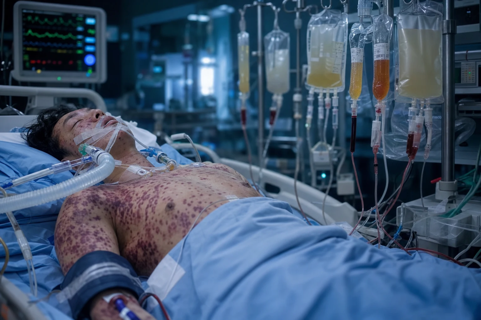
The ISTH Scientific and Standardisation Committee (SSC) defines DIC as "an acquired syndrome characterised by the intravascular activation of coagulation with loss of localisation arising from different causes. It can originate from and cause damage to the microvasculature, which if sufficiently severe, can produce organ dysfunction." The crucial words are acquired (always secondary), intravascular (not localised), and microvasculature (the organ damage is from microthrombi, not macrovascular clots).[1][1]
DIC is best understood not as a disease but as the final common pathway of uncontrolled thrombin generation — whatever the trigger, the end-result is the same: thrombin floods the circulation, converts fibrinogen to fibrin (microthrombi → organ ischaemia), consumes platelets and clotting factors (bleeding), and activates platelets (aggregation). The balance between bleeding and thrombosis shifts hour to hour, which is why serial monitoring matters and why a single set of labs can mislead.[1]
Pathophysiology
- A trigger releases tissue factor → the extrinsic pathway → massive thrombin generation.[1]
- The thrombin → fibrin deposition (the microvascular thrombosis → organ failure) AND platelet aggregation (the consumption → thrombocytopenia).[1]
- The clotting factors consumed → prolonged PT and APTT.[1]
- The fibrinogen consumed → hypofibrinogenaemia (the fibrinogen is an acute-phase reactant — may be normal early).[1]
- The secondary fibrinolysis impaired (the PAI-1 upregulated) → the thrombosis predominates.[1]
- The D-dimer markedly elevated (the fibrin degradation product).[1]
- The schistocytes on the blood film (the microangiopathic haemolytic anaemia).[1]
The molecular cascade in detail
DIC is fundamentally a disorder of thrombin gone rogue. The initiating event in every form of DIC is exposure of blood to a potent procoagulant — overwhelmingly tissue factor (TF, thromboplastin, factor III) — although some triggers (snake venoms) directly activate prothrombin or factor X. Understanding the cascade explains every laboratory abnormality and every management decision:[1][10]
The DIC cascade — from trigger to bleeding AND thrombosis
| Step | Event | Consequence | Laboratory signature |
|---|---|---|---|
| 1. Trigger | Tissue factor released from damaged tissue, monocytes (sepsis), mucin (adenocarcinoma), amniotic fluid, leukaemic granules | TF binds factor VIIa → extrinsic tenase → factors IX and X activation | — |
| 2. Thrombin explosion | Massive, unregulated thrombin generation — far exceeds the capacity of antithrombin, protein C/S, and TFPI | Thrombin: (a) cleaves fibrinogen → fibrin, (b) activates platelets, (c) activates V and VIII (positive feedback), (d) activates XIII (cross-linking) | PT and APTT prolong as factors consumed |
| 3. Microvascular thrombosis | Fibrin + platelet aggregates deposit in capillaries/arterioles → microthrombi throughout lungs, kidneys, brain, skin, adrenals | Organ ischaemia (AKI, ARDS, hepatic dysfunction, purpura, digital gangrene, Waterhouse-Friderichsen) | Platelets fall; organ dysfunction |
| 4. Consumption | Platelets and clotting factors (I, II, V, VIII, XIII) consumed faster than the liver and marrow can replace | Consumption coagulopathy → bleeding (mucosal, GI, catheter sites, intracranial) | Thrombocytopenia, prolonged PT/APTT, low fibrinogen |
| 5. Anticoagulant depletion | Antithrombin, protein C, protein S, and TFPI consumed/inactivated by the activated enzymes and by neutrophil elastase | Loss of the natural brakes → thrombin generation accelerates further | Low antithrombin activity (supportive, not routine) |
| 6. Fibrinolysis impaired | Thrombin + cytokines (IL-6, TNF) upregulate plasminogen activator inhibitor-1 (PAI-1) → t-PA inhibited | Clots are NOT lysed → thrombosis dominates; PAI-1 polymorphism (4G/4G) predisposes to severe DIC | High PAI-1; D-dimer high but lysis functionally blocked |
| 7. Secondary fibrinolysis | Some plasmin still generated → degrades fibrin (and fibrinogen — primary fibrinolysis) → produces D-dimer and FDP | Bleeding amplified; D-dimer is the most sensitive marker of fibrin turnover | D-dimer markedly elevated; FDP elevated |
| 8. Endothelial dysfunction | Cytokines damage glycocalyx and endothelium; von Willebrand factor released (ultra-large multimers) | Further platelet adhesion; capillary leak; organ oedema | — |
| 9. MAHA | Red cells fragment passing through the fibrin mesh of microthrombi | Schistocytes on film; haemolysis (raised LDH, low haptoglobin) | Schistocytes, high LDH, low haptoglobin, anaemia |
The role of inflammation — thromboinflammation
Sepsis-induced DIC is the archetype of thromboinflammation (immunothrombosis). Inflammatory cytokines (IL-1, IL-6, TNF-α) do three things simultaneously: (1) upregulate tissue factor expression on monocytes and endothelium, (2) downregulate thrombomodulin on endothelial cells (so the protein C anticoagulant pathway fails — thrombomodulin binds thrombin and converts it from a procoagulant to an anticoagulant; its loss is central to sepsis-DIC), and (3) upregulate PAI-1 (so fibrinolysis is blocked). This is why sepsis-DIC is predominantly thrombotic in early phase and why recombinant thrombomodulin and activated protein C were mechanistically attractive therapies.[1][8]
Bleeding-predominant vs thrombotic-predominant DIC
DIC is not a uniform syndrome — the clinical phenotype depends on which arm of the cascade dominates, and this drives treatment: [1]
Bleeding-predominant vs thrombotic-predominant DIC
| Feature | Bleeding-predominant | Thrombotic-predominant |
|---|---|---|
| Typical triggers | APL, obstetric (abruption, AFE), trauma, liver failure | Sepsis (meningococcal, pneumococcal), CAPS, solid malignancy |
| Pathophysiology | Rapid factor/platelet consumption outstrips thrombosis | Microthrombosis dominates; PAI-1 high, fibrinolysis blocked |
| Clinical picture | Oozing from every puncture site, GI/intracranial bleed, ecchymoses | Purpura fulminans, digital ischaemia, AKI, ARDS, skin necrosis |
| Fibrinogen | Very low (often <1.0 g/L) | May be normal (acute-phase) |
| Platelets | Very low | Moderately low |
| Treatment emphasis | Blood products (cryoprecipitate, platelets, FFP) + treat cause | ANTICOAGULATION (heparin) + treat cause |
| Antifibrinolytics | STILL CONTRAINDICATED (even bleeding DIC has microthrombi) | CONTRAINDICATED |
The triggers
DIC is always secondary — identifying and removing the trigger is the definitive treatment. The mnemonic "STOP Making The Bleeding" captures the categories: Sepsis, Trauma/burns, Obstetric, Pancreatitis; Malignancy; Transfusion; Bites (snake); plus rarer causes.[1][1]
- Sepsis (the commonest ICU cause) — Gram-negative, the meningococcal (the purpura fulminans).[1]
- Trauma or burns — the tissue factor from the damaged tissue.[1]
- Obstetric — the amniotic fluid embolism, the placental abruption, the eclampsia or HELLP, the retained dead fetus.[1]
- Malignancy — the acute promyelocytic leukaemia (APL — the ATRA), the mucin-secreting adenocarcinoma (the pancreatic, the prostate).[1]
- Transfusion reaction (the ABO incompatibility — the acute haemolytic).[1]
- Snake bite, the heat stroke, the severe pancreatitis.[1]
DIC triggers — mechanism, time course, and key management
| Trigger | Mechanism of tissue factor / procoagulant release | Time course | Key trigger-specific management |
|---|---|---|---|
| Sepsis (#1 ICU cause) | Bacterial endotoxin + cytokines (IL-6, TNF) upregulate TF on monocytes and endothelium; thrombomodulin downregulated; PAI-1 upregulated | Hours-days | Antibiotics within 1 h, source control, fluid resuscitation |
| Meningococcaemia / purpura fulminans | Endotoxin + DIC + protein C consumption (congenital deficiency catastrophic) | Hours | Antibiotics (ceftriaxone), heparin, protein C concentrate, supportive |
| Acute promyelocytic leukaemia (APL, M3) | Auer rods / leukaemic granules packed with tissue factor released on chemotherapy-induced cell lysis | Days | ATRA (all-trans retinoic acid) immediately on suspicion — differentiates cells, stops TF release |
| Other malignancy (mucinous) | Mucin (pancreatic, prostate, gastric, lung adenocarcinoma) + tumour procoagulant directly activate X | Weeks-months (chronic DIC) | Treat tumour; LMWH (better than warfarin in cancer-associated thrombosis) |
| Placental abruption | Retroplacental clot + decidual tissue factor enters maternal circulation | Hours | Delivery (vaginal if rapid, CS if fetal distress); uterotonics |
| Amniotic fluid embolism (AFE) | Amniotic fluid + trophoblasts enter maternal circulation at delivery → dual-hit: anaphylactoid CV collapse + explosive DIC | Minutes-hours (peripartum) | Resuscitate (LV failure), early delivery, massive transfusion, cryoprecipitate; consider PLEX |
| HELLP / pre-eclampsia | Endothelial dysfunction + platelet activation | Hours-days | Delivery (definitive); magnesium for seizure prophylaxis |
| Retained dead fetus | TF from necrotic fetal tissue | Weeks (chronic) | Delivery / uterine evacuation |
| Severe trauma / burns | Massive tissue factor release from injured brain (rich in thromboplastin), lung, muscle; PLUS acute traumatic coagulopathy (shock + endothelial glycocalyx disruption) | Minutes-hours | Damage-control resuscitation, haemostatic ratio transfusion (1:1:1), TXA (within 3 h — TXA IS indicated in trauma, unlike in established DIC) |
| Severe pancreatitis | Pancreatic enzymes + cytokine storm + splanchnic TF | Hours-days | Aggressive fluid resuscitation, ERCP if gallstone, ICU support |
| Snake bite (viperidae) | Venom directly activates prothrombin (procoagulant) or consumes fibrinogen (defibrinogenating) — "true" venom coagulopathy, not classical DIC | Hours | Antivenom (species-specific), NOT blood products or heparin unless true DIC develops |
| Heat stroke | Endothelial damage + cytokine storm | Hours | Rapid cooling, supportive |
| Acute haemolytic transfusion reaction | ABO incompatibility → intravascular haemolysis → red cell stroma + cytokines activate coagulation | Minutes | STOP transfusion, supportive, renal protection |
| Viral haemorrhagic fevers (Ebola, dengue) | Viral infection of endothelium + macrophages → cytokine storm + DIC | Days | Supportive / specific antiviral; isolate |
The diagnosis (the ISTH score)
- The ISTH overt-DIC score:[1][1]
- The lab pattern — the thrombocytopenia (the low and falling), the prolonged PT and APTT, the low fibrinogen (the falling trend — the serial more useful), the high D-dimer (the markedly), the schistocytes on the blood film.[1]
ISTH overt-DIC scoring — the worked system
The ISTH score requires a known underlying disorder associated with DIC (without a trigger, you cannot diagnose DIC). It is calculated on two consecutive days (the score should rise if DIC is worsening) and a score ≥5 indicates overt DIC.[1][2]
ISTH overt-DIC scoring system (5 components)
| Component | Score 0 | Score 1 | Score 2 | Score 3 |
|---|---|---|---|---|
| Platelet count (×10^9/L) | >100 | <100 | <50 | — |
| D-dimer / FDP elevation | No increase | — | Moderate increase | Strong increase |
| PT prolongation | None (<3 s) | 3-6 s | >6 s | — |
| Fibrinogen (g/L) | >1.0 | <1.0 | — | — |
| Underlying disorder linked to DIC | (Prerequisite — score is only valid if present) | — | — | — |
| Total score | ≥5 = overt DIC | Repeat daily | <5 = non-overt DIC (monitor — may progress) | — |
Performance: the ISTH score has sensitivity ~91 per cent and specificity ~97 per cent for overt DIC when validated prospectively against an expert panel, and (crucially) a rising score predicts mortality independently of the underlying cause. The score is cheap, repeatable, and reproducible — it is the world standard and the one the examiners want.[2]
Other scoring systems (know they exist — ISTH is the primary)
DIC scoring systems compared
| Score | Body | Components | Overt DIC threshold | Notes |
|---|---|---|---|---|
| ISTH overt-DIC | International Society on Thrombosis and Haemostasis | Platelets, D-dimer, PT, fibrinogen (+ prerequisite trigger) | ≥5 | World standard; sensitive for mortality prediction |
| JAAM DIC score | Japanese Association for Acute Medicine | SIRS (2 points), platelets, PT, D-dimer, fibrinogen | ≥4 | Captures earlier / hyperacute DIC (SIRS component) |
| JMHW DIC | Japanese Ministry of Health & Welfare | Clinical bleeding, organ symptoms, platelets, fibrinogen, FDP, PT | ≥7 | Used in Japan; favours the thrombomodulin trial population |
| ASH 2018 | American Society of Hematology | "Three-easy" — platelet, PT-INR, D-dimer | Platelet trend + PT ↑ + D-dimer ↑ = DIC (qualitative) | Simpler, designed for the bedside; no fibrinogen component |
The exam answer: ISTH score ≥5 = overt DIC, repeat daily, valid only with an underlying trigger. The other scores exist but ISTH is the one to quote.[1][10]
The laboratory pattern — interpreting each value
Laboratory abnormalities in DIC — what each value tells you
| Test | Typical finding | Mechanism | Pitfall / caveat |
|---|---|---|---|
| Platelets | Low and FALLING (the trend matters more than the absolute) | Consumption in microthrombi | May be normal early; single value misleading |
| PT / INR | Prolonged (rising) | Consumption of factors II, V, VII, X (VII shortest half-life — first to fall) | Also prolonged in liver disease, warfarin, vitamin K deficiency |
| APTT | Prolonged (often) | Consumption of factors VIII, IX, XI, XII | May be normal early; factor VIII is an acute-phase reactant and can be elevated early (falsely normal APTT) |
| Thrombin time (TT) | Prolonged | Low fibrinogen + fibrin degradation products interfere with polymerisation | Non-specific |
| Fibrinogen | Low (BUT may be normal early — acute-phase reactant) | Consumption | A "normal" fibrinogen in sepsis is abnormally low — should be 5-10 g/L; serial trend is diagnostic |
| D-dimer | Markedly elevated (most sensitive marker) | Cross-linked fibrin degraded by plasmin → D-dimer | Elevated in anything (surgery, thrombosis, pregnancy, malignancy) — sensitive, not specific |
| FDP | Elevated | Plasmin digestion of fibrin/fibrinogen | Less specific than D-dimer |
| Schistocytes on film | Present (moderate — usually 1-3%) | Microangiopathic haemolysis — RBCs fragmented on fibrin mesh | MANY schistocytes (>4-5%) + severe thrombocytopenia suggests TTP rather than DIC |
| LDH | Elevated | Haemolysis + tissue ischaemia | Non-specific |
| Haptoglobin | Low | Intravascular haemolysis | Non-specific |
| Antithrombin | Low (consumed) | Consumed by activated thrombin and Xa | Supportive test; low AT is why antithrombin concentrate was tried (and failed) |
| Protein C | Low | Consumption + cytokine downregulation | Mechanistic rationale for activated protein C (drotrecogin) |
| Blood film | Schistocytes, polychromasia, thrombocytopenia | MAHA | Review the film — TTP looks different |
Viscoelastic testing (TEG / ROTEM) in DIC
Thromboelastography (TEG) and rotational thromboelastometry (ROTEM) give a global picture of coagulation in real time and are increasingly used in ICU. In established DIC they show a recognisable pattern:[1]
TEG / ROTEM patterns in DIC
| Phase | TEG parameter | ROTEM parameter | Finding in DIC |
|---|---|---|---|
| Clot initiation (reaction time) | R time | CT (clotting time) | Prolonged (factor depletion) |
| Clot kinetics | K time, α angle | CFT, α angle | Prolonged K, low α angle (hypofibrinogenaemia + low platelets) |
| Clot strength | MA (maximum amplitude) | MCF (maximum clot firmness) | Reduced (low platelets + low fibrinogen) |
| Clot lysis | LY30 | ML (maximum lysis) | Usually LOW (PAI-1 high — impaired fibrinolysis); rarely high in hyperfibrinolytic phase |
| Fibrinogen contribution | — | FIBTEM (with cytochalasin — platelet inhibited) | Low FIBTEM MCF → give cryoprecipitate/fibrinogen |
| Platelet contribution | — | EXTEM − FIBTEM | Residual platelet contribution |
The practical use: FIBTEM/functional fibrinogen guides cryoprecipitate dosing, MA/MCF guides platelet transfusion, and an unexpectedly low LY30 is the one situation where tranexamic acid is the absolutely wrong drug. Viscoelastic testing is more useful than static labs for the actively bleeding patient because it reflects the whole-system state at the moment of sampling.[1]
The management
The management of DIC rests on four principles, in order of priority: (1) treat the underlying cause (the definitive — DIC resolves once the trigger is removed); (2) supportive transfusion for active bleeding; (3) anticoagulation only for the thrombotic-predominant phenotype; (4) NEVER give antifibrinolytics.[1][1]
1. Treat the underlying cause (the definitive)
- The sepsis → the source control and the antibiotics.[1]
- The obstetric → the evacuate the uterus (the delivery).[1]
- The APL → the ATRA (the all-trans-retinoic-acid) immediately.[1]
- The trauma → the control the bleeding.[1]
- The DIC the resolves once the trigger the removed.[1]
This is not a platitude — it is the single most important intervention. Transfusion without cause-treatment fails because the transfused products are consumed as fast as they are given. The mortality of DIC is driven almost entirely by the underlying cause and by organ failure from microthrombosis.[1]
Trigger-specific definitive therapy
| Trigger | Definitive therapy | Time to DIC resolution |
|---|---|---|
| Sepsis | Antibiotics within 1 h (per Surviving Sepsis), source control (drain, debride, remove line) | 1-3 days after source control |
| APL | ATRA 45 mg/m^2/day immediately on suspicion (before confirmation) + supportive | Days — ATRA halts TF release within 24-48 h |
| Placental abruption | Delivery (vaginal or CS) | Hours after delivery |
| Amniotic fluid embolism | Immediate delivery + resuscitation + massive transfusion | Variable — may be prolonged |
| HELLP / pre-eclampsia | Delivery + magnesium | Hours-days |
| Severe trauma | Damage-control surgery + haemostatic resuscitation (1:1:1) | Hours after haemorrhage control |
| Snake bite | Species-specific antivenom | Hours after antivenom |
| Mucinous malignancy | Treat the tumour (chemotherapy); LMWH for chronic DIC | Weeks |
| Acute pancreatitis | Aggressive fluids, ERCP for gallstones, ICU | Days |
2. The supportive transfusion (for the active bleeding)
- The platelets — if the under 50 AND bleeding (the under 100 if the neuro or the active).[1]
- The FFP — if the PT or APTT prolonged AND bleeding (the 15 mL per kg).[1]
- The cryoprecipitate — if the fibrinogen under 1.5 g per L (the target the over 1.5).[1]
- The transfuse ONLY if bleeding (the prophylactic transfusion the controversial).[1]
Transfusion is supportive, not curative. Products are consumed rapidly and must be given repeatedly until the trigger is controlled. The thresholds below are the BCSH/ISTH guidance for the actively bleeding patient (the non-bleeding patient thresholds are lower — transfusing numbers alone does not improve outcome and exposes the patient to volume overload, TRALI, and transfusion-transmitted infection).[1]
Transfusion thresholds in bleeding DIC (BCSH / ISTH guidance)
| Product | Dose | Threshold (active bleeding) | Target | Rationale |
|---|---|---|---|---|
| Platelets | 1 adult dose (≈1 unit/10 kg) | <50 ×10^9/L (<100 if active bleeding, CNS, or perioperative) | >50 (>100 in CNS) | Replace consumed platelets |
| Fresh frozen plasma (FFP) | 15 mL/kg (≈4 units in adult) | PT or APTT >1.5× normal AND bleeding | Correct PT/APTT | Replaces all clotting factors (except factor VIII in cryo, fibrinogen in cryo) |
| Cryoprecipitate | 2 pools (10 units) or 10-15 units | Fibrinogen <1.5 g/L | Fibrinogen >1.5 g/L | Concentrated fibrinogen (+ factor VIII, XIII, vWF) — 1 pool raises fibrinogen ~0.5-1.0 g/L |
| Fibrinogen concentrate | 3-4 g (alternative to cryoprecipitate) | Fibrinogen <1.5 g/L | >1.5 g/L | Pathogen-inactivated, faster, smaller volume — preferred in some centres |
| Prothrombin complex concentrate (PCC) | AVOID in DIC (risk of thrombosis) | — | — | May worsen the microthrombosis |
Supportive transfusion in actively bleeding DIC — the practical protocol
- CONFIRM bleeding is from consumption coagulopathy (low fibrinogen, low platelets, prolonged PT/APTT, high D-dimer) — not from a surgical bleed that needs surgery
- Send repeat coagulation (PT, APTT, fibrinogen, platelets, D-dimer, blood film) — the trend over the last few hours
- Correct the fibrinogen FIRST — give cryoprecipitate (2 pools) or fibrinogen concentrate (3-4 g) if fibrinogen <1.5 g/L. Recheck fibrinogen at 30 min. Target >1.5 g/L (the bleeding usually slows dramatically once fibrinogen is restored)
- Correct the platelets — give 1 adult dose of platelets if platelets <50 (<100 if CNS/active bleeding). Recheck at 1 h
- Correct the clotting factors — give FFP 15 mL/kg (4 units) if PT/APTT >1.5× normal. Be mindful of volume overload (consider fibrinogen concentrate over cryo if volume-naïve)
- Reassess the bleeding and recheck the labs at 1-2 h — repeat the cycle as needed. DIC lab values can change hourly
- Treat the trigger — concurrently. Transfusion is futile if the trigger is still driving consumption
- NO antifibrinolytic (TXA, aprotinin) — see below
- Consider recombinant thrombomodulin (ART-123) for sepsis-associated DIC with organ dysfunction (see below)
- Escalate to interventional radiology / surgery if there is a focal bleeding source not controlled by products
3. The heparin (for the thrombotic-predominant DIC)
- The heparin — for the thrombotic-predominant DIC (the purpura fulminans, the APL, the acral ischaemia). The low-dose the infusion (the UFH; the LMWH).[1]
- NOT for the bleeding-predominant DIC (the heparin the worsens the bleeding).[1]
- The LMWH for the VTE prophylaxis (the high the risk).[1]
Anticoagulation in DIC is phenotype-specific. The 2014 Japanese guidelines and the BCSH both suggest therapeutic-dose heparin for thrombotic-predominant DIC (purpura fulminans, acral ischaemia, APL) and prophylactic-dose LMWH for the bedbound/septic patient at high VTE risk. There is no RCT proving that heparin changes mortality in unselected DIC, but the biological rationale (the microthrombi are fibrin- and thrombin-driven, and heparin neutralises thrombin) and observational data justify its use in the thrombotic phenotype.[1]
Heparin in DIC — when and what dose
| Scenario | Heparin strategy | Dose |
|---|---|---|
| Thrombotic-predominant DIC (purpura fulminans, acral ischaemia, APL, skin necrosis) | Therapeutic heparin | UFH infusion 18 U/kg/h (titrate to APTT 1.5-2.5×) OR LMWH (enoxaparin 1 mg/kg BD) |
| Sepsis-associated DIC, no thrombosis | Prophylactic LMWH (VTE prevention) | Enoxaparin 40 mg OD (dose-adjust for renal failure) |
| Bleeding-predominant DIC | AVOID therapeutic heparin | Prophylaxis may be held if bleeding |
| APL | Therapeutic heparin until differentiation + fibrinogen stabilises | UFH infusion preferred (reversible) |
| CAPS (catastrophic APS) | Therapeutic heparin (cornerstone) | UFH infusion |
4. NO antifibrinolytics (the TXA)
- The TXA or the aprotinin — the CONTRA-INDICATED in the DIC (the worsens the microvascular thrombosis). The exception: the clearly hyperfibrinolytic DIC (the rare — the TEG or the ROTEM the documented).[1]
This is a high-yield exam point and a common error. The bleeding in DIC is primarily from factor/platelet consumption, NOT from excess fibrinolysis — in fact fibrinolysis is impaired (PAI-1 upregulated). Giving TXA blocks the little residual fibrinolysis and worsens the microvascular thrombosis. The only exception is documented hyperfibrinolysis on TEG/ROTEM (high LY30 / ML), which is rare.[1]
Recombinant soluble thrombomodulin (ART-123, recomodulin) — the one specific DIC drug
Recombinant human soluble thrombomodulin binds thrombin and, like endogenous thrombomodulin, switches thrombin from a procoagulant to an anticoagulant (by activating protein C). It also inactivates high-mobility group box 1 (HMGB1), a late cytokine mediator of sepsis. It is approved in Japan for DIC and is the most promising DIC-specific agent.[6][8][7]
Recombinant soluble thrombomodulin (ART-123) in sepsis-associated DIC
- Indication — sepsis-associated DIC with organ dysfunction (ISTH score ≥5), particularly when the fibrinogen is preserved (thrombotic phenotype)
- Dose — 0.06 mg/kg IV over 30 min, daily for 6 days
- Evidence —
- SCARLET trial (Vincent 2019): multicentre RCT of thrombomodulin vs placebo in sepsis-associated coagulopathy. Missed primary endpoint (28-day mortality 26.8% vs 29.4%, p=0.32) BUT significant mortality reduction in the pre-specified subgroups (ISTH overt DIC, baseline SOFA 10-12, infection site).[7]
- Japanese post-marketing and observational data consistently show reduced DIC resolution time and lower bleeding vs heparin.[6]
- Cautions — bleeding risk (monitor platelets and fibrinogen); renal excretion; not yet widely available outside Japan (TGA approval pending / regional variation)
- Place in therapy — second-line to treat-the-cause + products; considered when DIC is driven by sepsis with rising organ dysfunction despite standard care
Anticoagulant concentrates that did NOT work (know the evidence — examiners love it)
Several biologically rational agents were tested in large RCTs and failed to improve survival. They are NOT recommended, but you will be asked about them:[1][3][4][5]
Failed anticoagulant therapies for sepsis-DIC (know the trials)
| Agent | Mechanism | Key trial | Result | Status |
|---|---|---|---|---|
| Antithrombin III concentrate | Restores the consumed natural anticoagulant | KyberSept (Warren 2001, JAMA) — 2314 septic patients | NO mortality benefit; trend to more bleeding with concomitant heparin | NOT recommended |
| Recombinant tissue factor pathway inhibitor (tifacogin) | Blocks the TF-VIIa complex at origin of cascade | OPTIMIST (Abraham 2001, JAMA) — 1754 septic patients | NO mortality benefit; increased serious bleeding | NOT recommended |
| Drotrecogin alfa (activated protein C, Xigris) | Restores protein C pathway + anti-inflammatory | PROWESS (2001, positive) then PROWESS-SHOCK (Ranieri 2012, NEJM) — 1696 septic shock patients | PROWESS-SHOCK NEGATIVE → drug withdrawn from market 2011 | WITHDRAWN |
| Heparin (therapeutic) | Antithrombin | Multiple small RCTs / meta-analysis | No consistent mortality benefit; trend to bleeding | Prophylactic-dose only (VTE prevention) |
| Recombinant soluble thrombomodulin (ART-123) | Thrombin → protein C activation | SCARLET (Vincent 2019) — 800 patients | Missed primary endpoint; benefit in overt-DIC subgroup | APPROVED in Japan; emerging elsewhere |
Differential diagnosis — what DIC is NOT
The thrombocytopenia + coagulopathy + organ failure picture is shared by several conditions whose management is radically different. Misdiagnosis is dangerous — TTP needs plasma exchange within hours, while DIC needs products and the trigger treated.[1][9]
DIC vs TTP/HUS vs severe liver disease vs massive transfusion coagulopathy
| Feature | DIC | TTP / HUS | Severe liver disease | Massive transfusion dilutional coagulopathy |
|---|---|---|---|---|
| Pathology | Consumption + microthrombosis + impaired fibrinolysis | ADAMTS13 deficiency (TTP) → ultra-large vWF multimers → platelet microthrombi | Reduced synthesis + consumption + portal hypertension | Dilution of factors + platelets + citrate toxicity + hypothermia/acidosis |
| PT / APTT | Prolonged | NORMAL (classic) | Prolonged | Prolonged |
| Fibrinogen | LOW (falling) | NORMAL | LOW (low baseline; not rising on replacement as liver cannot synthesise) | Low (dilutional) — rises with cryoprecipitate |
| D-dimer | Markedly elevated and RISING | Mild-moderate | Mildly elevated (baseline) | Mildly elevated |
| Schistocytes | Moderate (1-3%) | MANY (>4-5%) — severe MAHA | Few / none | Few / none |
| Platelets | Low, falling | Very low (often <20) — severe | Mild-moderate low (sequestration) | Low (dilutional) |
| Key discriminator | Falling fibrinogen + high D-dimer + a trigger | ADAMTS13 activity <10% (TTP) — URGENT plasma exchange | Liver failure signs (jaundice, encephalopathy, ascites); factor VIII NORMAL (synthesised by endothelium, not liver) | Recent massive transfusion; corrects with ratio resuscitation + warming |
| Treatment | Treat cause + products ± heparin | URGENT plasma exchange + steroids (caplacizumab for acquired TTP) | Treat liver failure; vitamin K; products if bleeding | Stop bleeding, 1:1:1 ratio, correct acidosis/hypothermia/calcium |
Special scenarios
Acute promyelocytic leukaemia (APL) DIC
APL (acute myeloid leukaemia M3) is the classic malignancy-associated DIC and was once the most lethal form (haemorrhagic death in 10-20% before the ATRA era). The leukaemic promyelocytes contain granules packed with tissue factor and annexin II (which upregulates plasminogen activation → hyperfibrinolysis), so the DIC is unusually bleeding-predominant (intracranial haemorrhage is the feared event).[1]
APL-associated DIC — the immediate management
- Suspect APL on the blood film / FBC (hypergranular promyelocytes with Auer rods; the microgranular variant M3v may be missed — check coagulation in any new AML presentation). Coagulopathy + new leukaemia = APL until proven otherwise
- Start ATRA (all-trans-retinoic-acid) 45 mg/m^2/day immediately on suspicion — do NOT wait for cytogenetic/PCR confirmation. ATRA differentiates the leukaemic cells and halts tissue factor release within 24-48 h
- Aggressive product support — cryoprecipitate to keep fibrinogen >1.5 g/L (some centres target >2.0 g/L in APL), platelets >30 (or >50 if febrile/bleeding), FFP to keep PT/APTT <1.5× normal. Monitor every 6-12 h
- Therapeutic heparin may be considered for refractory bleeding (the bleeding is from both consumption AND hyperfibrinolysis, and heparin blunts the consumption) — controversial, centre-dependent
- Add arsenic trioxide (in high-risk APL) and consult haematology for the chemotherapy plan
- Watch for differentiation syndrome (fever, dyspnoea, hypotension, pulmonary infiltrates, weight gain, renal failure) at 1-2 weeks — treat with dexamethasone 10 mg BD
- Avoid invasive procedures (lumbar puncture, central lines) until coagulopathy corrected — intracranial haemorrhage is the leading cause of early death
Obstetric DIC (abruption, amniotic fluid embolism, HELLP)
Pregnancy is a procoagulant state (high fibrinogen, high factor VIII, low anticoagulant reserve), which is why obstetric disasters tip into DIC so fast. The definitive treatment is delivery / uterine evacuation — the source of the trigger is removed.[11]
Obstetric DIC syndromes
| Syndrome | Mechanism | Presentation | Key management |
|---|---|---|---|
| Placental abruption | Retroplacental clot + decidual TF enters maternal circulation | Painful vaginal bleeding, uterine tenderness, fetal distress; couvelaire uterus (concealed abruption) → DIC | Delivery (vaginal if rapid, CS if fetal distress); uterotonics; products |
| Amniotic fluid embolism (AFE) | Amniotic fluid + trophoblasts enter maternal circulation → biphasic: (1) anaphylactoid pulmonary vasoconstriction + LV failure (sudden collapse, hypoxia), (2) catastrophic DIC | Peripartum sudden CV collapse + hypoxia + coagulopathy | Resuscitation (LV failure pattern — high vasopressors/inotropes), early delivery, massive transfusion, cryoprecipitate; atropine + clonidine may help the vagal/anaphylactoid phase; consider PLEX |
| HELLP (haemolysis, elevated liver enzymes, low platelets) | Endothelial dysfunction + platelet activation (pre-eclampsia spectrum) | Right upper quadrant pain, malaise, nausea; platelets <100, AST >70, LDH >600 | Delivery (definitive); magnesium for seizure prophylaxis; dexamethasone (maternal, controversial); watch for hepatic rupture (sudden shock + abdominal pain) |
| Retained dead fetus / septic abortion | TF from necrotic fetal tissue or infected products | Chronic DIC over weeks; or acute with septic abortion | Uterine evacuation + antibiotics |
| Postpartum haemorrhage (PPH) | Massive haemorrhage → consumption + dilution (not "true" DIC unless a trigger coexists) but the labs look identical | Bleeding, shock, low fibrinogen, prolonged PT | Obstetric massive transfusion protocol; cryoprecipitate (fibrinogen FIRST — pregnancy baseline is 5-7 g/L, so a fibrinogen of 3 g/L in PPH is abnormally low); uterotonics; Bakri balloon; interventional radiology; hysterectomy as last resort |
Purpura fulminans — the thrombotic-predominant extreme
Purpura fulminans is cutaneous microvascular thrombosis with haemorrhagic skin necrosis — the most dramatic manifestation of thrombotic-predominant DIC. The skin lesions progress from erythema → purpura → confluent ecchymoses → bullae → full-thickness skin necrosis and digital/peripheral gangrene within hours.[9]
Purpura fulminans — three subtypes
| Subtype | Trigger | Mechanism | Key therapy |
|---|---|---|---|
| Acute infectious | Meningococcaemia, pneumococcaemia, Group A strep, varicella | Septic DIC + acquired protein C deficiency (consumption + reduced synthesis) | Antibiotics + heparin + protein C concentrate + supportive |
| Neonatal (homozygous protein C deficiency) | Genetic (autosomal recessive) | Absent protein C → neonatal purpura within hours of birth | Protein C concentrate; long-term anticoagulation; liver transplant curative |
| Idiopathic (post-infectious) | 1-3 weeks after a viral exanthem | Autoantibody-mediated transient protein C/S deficiency | Heparin + steroids + supportive |
Purpura fulminans management (meningococcal — the prototype)
- Antibiotics immediately — ceftriaxone 2 g IV (meningococcal cover; add vancomycin if pneumococcal suspected)
- Therapeutic heparin — UFH infusion (reversible, titratable). Even in the face of skin haemorrhage, the gangrene is driven by ongoing microthrombosis — heparin halts the extension
- Protein C concentrate (where available) — restores the consumed natural anticoagulant; particularly in severe congenital deficiency
- Aggressive product support — cryoprecipitate, platelets, FFP (the bleeding and thrombosis coexist)
- Supportive ICU care — vasopressors (noradrenaline), lung-protective ventilation if ARDS, RRT if AKI
- Surgical assessment — early debridement of necrotic skin, fasciotomy for compartment syndrome, amputation for established gangrene (often delayed to allow demarcation — do not amputate acutely)
- NO steroids in meningococcal disease (no benefit; increases infection risk), unlike CAPS
- Long-term: skin grafting, amputation/rehabilitation, psychological support
Snake bite coagulopathy — "venom-induced consumption coagulopathy" (VICC)
Viper and elapid snake venoms contain procoagulant enzymes that directly activate prothrombin (Group V vipers) or consume fibrinogen (defibrinogenating venoms). This is not classical DIC — there is usually no organ microthrombosis because the venom activates clotting faster than thrombin can be generated systemically. The coagulopathy corrects rapidly once the venom is neutralised.[1]
The key point: give antivenom, not blood products or heparin. Blood products are consumed instantly; heparin does nothing because the venom activates clotting downstream of thrombin. Reserve products for life-threatening bleeding after antivenom. The INR and D-dimer may be unrecordable (so-called "venom clotting time" — blood does not clot at all). Recovery: fibrinogen recovers over 12-24 h after antivenom as the liver resynthesises. [1]
Exam practice — SAQs
SAQ — Septic shock complicated by disseminated intravascular coagulation
10 minutes · 10 marks
A 62-year-old man is admitted to ICU with a 3-day history of fever, productive cough and confusion. On examination: T 38.9 degrees C, HR 132, BP 78/46 (MAP 57) on noradrenaline 0.35 mcg/kg/min after 30 mL/kg crystalloid, RR 32, SpO2 90 percent on 15 L non-rebreather, GCS 13. There is a purpuric rash over the limbs, fresh blood in the urinary catheter, and persistent oozing from the central venous catheter insertion site. Lactate 5.2 mmol/L, creatinine 220 micromol/L, platelets 42 x 10⁹/L (288 yesterday), INR 2.4, aPTT 56 s, fibrinogen 1.1 g/L, D-dimer markedly elevated. Chest X-ray shows right lower lobe consolidation. Blood cultures are pending.
Red flags
Clinical pearls
Prognosis
The mortality of DIC is 40-80 per cent and is driven almost entirely by the underlying cause and by organ failure from microthrombosis. DIC itself is a marker of illness severity — an ISTH score ≥5 carries a 2- to 3-fold increase in mortality in sepsis.[1][2]
Prognostic factors in DIC
| Factor | Effect on prognosis | Detail |
|---|---|---|
| Underlying cause | Dominant determinant | Meningococcal purpura fulminans 20-40% mortality; APL now <10% with ATRA; sepsis-DIC 40%; AFE 20-60% |
| ISTH score at baseline | Higher = worse | Each 1-point increase predicts ~25% increase in mortality |
| Rising ISTH score over days | Worse | Dynamic worsening outperforms any single value |
| Organ failure (SOFA) | Worse | Mortality tracks with the number of failing organs |
| Fibrinogen at presentation | Lower = worse (in bleeding DIC) | <1.0 g/L at presentation predicts worse outcome |
| Age | Older = worse | Less physiological reserve |
| Timing of trigger treatment | Earlier = better | Source control within hours improves survival |
| Thrombotic vs bleeding phenotype | Variable | Purpura fulminans has high mortality from gangrene/sepsis; bleeding DIC has high mortality from intracranial haemorrhage |
| Resolution of DIC after trigger control | Rapid resolution = good sign | Persistent DIC = search for second trigger |
Key trials and evidence
Taylor 2001 — ISTH SSC definition and scoring of DIC (PMID 11168689)
Source
Thrombosis and Haemostasis — the Scientific and Standardisation Committee of the ISTH
What it did
Defined DIC operationally and proposed a 5-component scoring system (platelets, D-dimer/FDP, PT prolongation, fibrinogen, underlying disorder)
Threshold
Score ≥5 = overt DIC; repeat every 24 h
Prerequisite
A disorder known to be associated with DIC must be present (sepsis, trauma, obstetric, malignancy, etc.)
Significance
The world standard DIC score — adopted universally; the backbone of subsequent trials and guidelines
Clinical bottom line
This is the score to quote in the exam — 'ISTH overt-DIC score, ≥5 = overt DIC, repeat daily'
Bakhtiari 2004 — Prospective validation of the ISTH DIC score (PMID 15318004)
Source
Critical Care Medicine — prospective validation cohort
What it did
Applied the ISTH score prospectively in ICU patients with suspected DIC; compared against an expert panel diagnosis
Sensitivity
91%
Specificity
97%
Key finding
An ISTH score ≥5 strongly predicts overt DIC AND independently predicts mortality — the score is both diagnostic and prognostic
Clinical bottom line
Validated the ISTH score as both diagnostic (sensitive + specific) and prognostic (rising score = worse outcome)
KyberSept — Warren 2001 — Antithrombin III in severe sepsis (PMID 11579466)
Source
JAMA — multicentre RCT, 2314 severe sepsis patients
Intervention
High-dose antithrombin III (30,000 U over 4 days) vs placebo
Result
NO overall mortality benefit at 28 days (38.9% vs 38.7%)
Subgroup
Patients NOT receiving concomitant heparin had a small mortality reduction; patients ON heparin had worse bleeding
DIC subgroup
Some signal in the pre-defined DIC subgroup but not significant
Clinical bottom line
Antithrombin concentrate is NOT recommended for sepsis-DIC — restoring a single consumed anticoagulant does not fix the system
OPTIMIST — Abraham 2001 — Tifacogin (TFPI) in severe sepsis (PMID 11124434)
Source
JAMA — multicentre RCT, 1754 severe sepsis patients
Intervention
Recombinant tissue factor pathway inhibitor (tifacogin) vs placebo — blocks the TF-VIIa complex at the origin of the coagulation cascade
Result
NO mortality benefit (34.2% vs 33.9%); significant increase in serious bleeding (especially with concomitant heparin)
Rationale failure
Blocking the cascade at its origin was biologically rational but clinically futile — the trigger drives TF expression continuously
Clinical bottom line
Tifacogin is NOT recommended — another failed upstream anticoagulant strategy in sepsis-DIC
PROWESS-SHOCK — Ranieri 2012 — Drotrecogin alfa (activated protein C) in septic shock (PMID 22616830)
Source
New England Journal of Medicine — multicentre RCT, 1696 septic shock patients
Intervention
Drotrecogin alfa (activated) — recombinant activated protein C — vs placebo
Result
NEGATIVE — no difference in 28-day mortality (26.4% vs 24.2%, p=0.31)
Consequence
Drug WITHDRAWN from the market in 2011 (before publication) — Eli Lilly cited the failure to show benefit and changing standard of care
Context
The original PROWESS (2001) was positive and the drug was licensed; PROWESS-SHOCK refuted it. A cautionary tale about early positive trials
Clinical bottom line
Activated protein C is WITHDRAWN — do not recommend it. The era of 'single anticoagulant rescue' for sepsis-DIC ended here
SCARLET — Vincent 2019 — Recombinant soluble thrombomodulin (ART-123) in sepsis-associated coagulopathy (PMID 29748489)
Source
Intensive Care Medicine — multicentre RCT, 800 patients with sepsis-associated coagulopathy
Intervention
Recombinant human soluble thrombomodulin (ART-123) 0.06 mg/kg IV daily for 6 days vs placebo
Mechanism
Binds thrombin → activates protein C (switches thrombin to anticoagulant) AND inactivates HMGB1 (late sepsis mediator)
Primary endpoint
28-day mortality — 26.8% (thrombomodulin) vs 29.4% (placebo), p=0.32 — MISSED significance
Subgroup signal
Significant mortality reduction in ISTH overt DIC (score ≥5), baseline SOFA 10-12, and respiratory/pelvic source subgroups
Safety
No increase in serious bleeding vs placebo
Clinical bottom line
The only DIC-specific agent with a positive (subgroup) signal — approved in Japan, emerging elsewhere. Consider for sepsis-DIC with rising organ dysfunction despite standard care
Levi 2009 — BCSH guidelines for DIC (PMID 19320688)
Source
British Journal of Haematology — British Committee for Standards in Haematology
What it did
Comprehensive evidence-based guideline for the diagnosis and management of DIC — the UK standard
Diagnosis
ISTH overt-DIC score ≥5, repeat daily; the score requires a known underlying trigger
Management
(1) Treat the underlying cause; (2) transfuse for active bleeding (platelets <50, FFP if PT/APTT >1.5×, cryoprecipitate if fibrinogen <1.5); (3) heparin for thrombotic-predominant DIC; (4) NO antifibrinolytics
Controversial
Prophylactic transfusion in non-bleeding DIC not recommended; recombinant activated protein C (now withdrawn) was conditionally recommended for severe sepsis-DIC
Clinical bottom line
The definitive UK/European guideline — the framework examiners expect you to know
Exam technique — how to answer a DIC question
The 90-second viva answer for 'Discuss DIC in the ICU patient'
- Define — "DIC is an acquired syndrome of widespread intravascular coagulation causing simultaneous bleeding and thrombosis, ALWAYS secondary to a trigger"
- Pathophysiology (one line) — "Tissue factor release → massive thrombin generation → fibrin deposition (microthrombosis, organ failure) AND consumption of platelets and factors (bleeding), with impaired fibrinolysis (PAI-1 upregulated)"
- Triggers (mnemonic) — "STOP-MTB: Sepsis, Trauma, Obstetric, Pancreatitis; Malignancy, Transfusion; Bites (snake)"
- Diagnosis — "ISTH overt-DIC score — platelets + D-dimer + PT + fibrinogen, ≥5 = overt DIC, repeat daily, requires a known trigger. Lab pattern: thrombocytopenia, prolonged PT/APTT, low fibrinogen (falling trend), high D-dimer, schistocytes"
- Management (four pillars) — "(1) Treat the underlying cause — the definitive; (2) supportive transfusion for bleeding — cryoprecipitate if fibrinogen <1.5, platelets if <50, FFP if PT/APTT prolonged; (3) heparin for thrombotic-predominant DIC (purpura fulminans, APL); (4) NO antifibrinolytics"
- Mention the controversies/agents — "Antithrombin, tifacogin, and activated protein C all failed RCTs. Recombinant soluble thrombomodulin is the one agent with a positive subgroup signal"
- Differential — "Distinguish from TTP (normal PT/APTT, many schistocytes, ADAMTS13 <10% — needs urgent plasma exchange), severe liver disease (normal factor VIII, static D-dimer), and dilutional coagulopathy (recent massive transfusion)"
- Prognosis — "Mortality 40-80%, driven by the underlying cause; a rising ISTH score predicts mortality independently"
Common exam pitfalls in DIC
| Pitfall | The error | The correct answer |
|---|---|---|
| "Give TXA for the bleeding" | TXA worsens the microvascular thrombosis (fibrinolysis is already impaired) | Products (cryoprecipitate first) + treat the trigger; TXA only if documented hyperfibrinolysis on TEG |
| "Normal fibrinogen excludes DIC" | Fibrinogen is an acute-phase reactant — normal in sepsis is abnormally low | Serial fibrinogen (falling trend) is diagnostic |
| "Give heparin to all DIC" | Bleeding-predominant DIC is worsened by heparin | Heparin only for thrombotic-predominant phenotype (purpura fulminans, APL, acral ischaemia) |
| "TTP and DIC are treated the same" | TTP needs urgent plasma exchange; products alone are useless | PT/APTT normal + many schistocytes + ADAMTS13 <10% = TTP → plasma exchange NOW |
| "Snake bite DIC needs heparin and FFP" | Venom activates clotting downstream of where heparin and FFP act | Give antivenom — the coagulopathy corrects as the venom is neutralised |
| "APL — wait for the biopsy before treatment" | Delay kills (intracranial haemorrhage) | Start ATRA immediately on suspicion, before confirmation |
| "Antithrombin concentrate for sepsis-DIC" | KyberSept was negative | Not recommended — restoring a single consumed anticoagulant does not work |
| "Activated protein C (Xigris) for severe sepsis-DIC" | PROWESS-SHOCK was negative; drug withdrawn | Withdrawn in 2011 — do not recommend |
Summary — the non-negotiables
[1]References
- [1]Taylor FB, Toh CH, Hoots WK, Wada H, Levi M Microscopic hematuria as a screening marker for urinary tract malignancies Int J Urol, 2001.PMID 11168689
- [2]Bakhtiari K, Meijers JC, de Jonge E, Levi M NOE assignment with ARIA 2.0: the nuts and bolts Methods Mol Biol, 2004.PMID 15318004
- [3]Ranieri VM, Thompson BT, Barie PS, et al. Drotrecogin alfa (activated) in adults with septic shock N Engl J Med, 2012.PMID 22616830
- [4]Warren BL, Eid A, Singer P, et al. Detection of illegitimate rearrangement within the immunoglobulin locus on 14q32.3 in B-cell malignancies using end-sequenced probes Genes Chromosomes Cancer, 2001.PMID 11579466
- [5]Abraham E, Reinhart K, Opal S, et al. Alzheimer mice on the couch Neurobiol Aging, 2000.PMID 11124434
- [6]Yamakawa K, Fujimi S, Mohri T, et al. Discussion: future directions J Glaucoma, 2013.PMID 23733130
- [7]Vincent JL, Francois B, Zabolotskikh I, et al. Genome-Wide Identification of MicroRNAs in Response to Cadmium Stress in Oilseed Rape (Brassica napus L.) Using High-Throughput Sequencing Int J Mol Sci, 2018.PMID 29748489
- [8]Toh CH, Alhamdi Y Changes in genetic selection differentials and generation intervals in US Holstein dairy cattle as a result of genomic selection Proc Natl Acad Sci U S A, 2016.PMID 27354521
- [9]Colling ME, Bendapudi PK Early intervention in multiple sclerosis: how can we maximise patient prospects? Expert Rev Clin Immunol, 2017.PMID 28425819
- [10]Iba T, Levy JH, Warkentin TE, et al. Prevalence and antibiotic resistance of coagulase-negative Staphylococci isolated from poultry farms in three regions of Ghana Infect Drug Resist, 2017.PMID 28652785
- [11]Hunt BJ Effects of brief inhibition of the ventral tegmental area dopamine neurons on the cocaine seeking during abstinence Addict Biol, 2020.PMID 31478293