ICU · Haematology / trauma
Massive Transfusion & Trauma Coagulopathy — The Lethal Triad, TXA & 1:1:1
Also known as Massive transfusion · MTP · Trauma coagulopathy · Acute traumatic coagulopathy · ATC · Lethal triad · Tranexamic acid · TXA · CRASH-2 · PROPPR · Hyperfibrinolysis · TEG · ROTEM
The trauma coagulopathy has the two components: the acute traumatic coagulopathy (ATC — the early, the injury-driven, the protein-C activation, the hyperfibrinolysis) and the dilutional coagulopathy (the massive RBC without the factors or the platelets). The lethal triad — the acidosis (the pH under 7.2 — the clotting-factor dysfunction), the hypothermia (the temp under 35 — the platelet and the enzyme dysfunction), and the coagulopathy — the vicious the cycle. The massive transfusion protocol (MTP) — the 1 to 1 to 1 (RBC to the plasma to the platelets), the PROPPR the no the mortality difference but the fewer the exsanguination. The tranexamic acid (TXA) — the CRASH-2, the 1 g plus the 1 g within 3 h (the beyond the 3 h the harmful). The calcium — the citrate from the blood products the chelates → the hypocalcaemia — the replace. The TEG or the ROTEM — the guides the product the replacement. The warm the blood, the correct the acidosis, the target the fibrinogen the over 1.5 to 2.0 (the cryo), the platelets the over 50 (the over 100 if the bleeding).
On this page & tools
Your progress
Saved locally on this device.
8 MCQs with explanations
Target exams
Overview & definition
The trauma coagulopathy and the massive transfusion are the interlinked the crises. The acute traumatic coagulopathy (ATC) develops the EARLY (on the presentation — the injury-driven, the not the dilutional), and the lethal triad (the acidosis, the hypothermia, the coagulopathy) the accelerates the bleeding. The massive transfusion protocol (MTP) — the 1 to 1 to 1 ratio, the tranexamic acid (TXA), the calcium — the survival-critical. The early the TXA (the within the 3 hours), the 1 to 1 to 1, and the correct the lethal triad.[1]
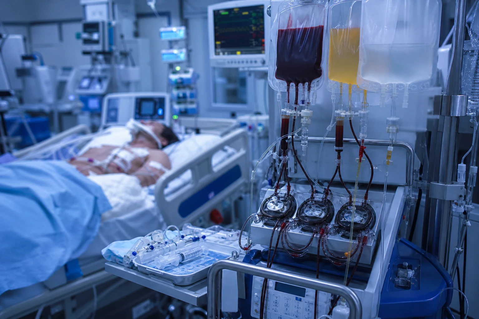
The trauma coagulopathy
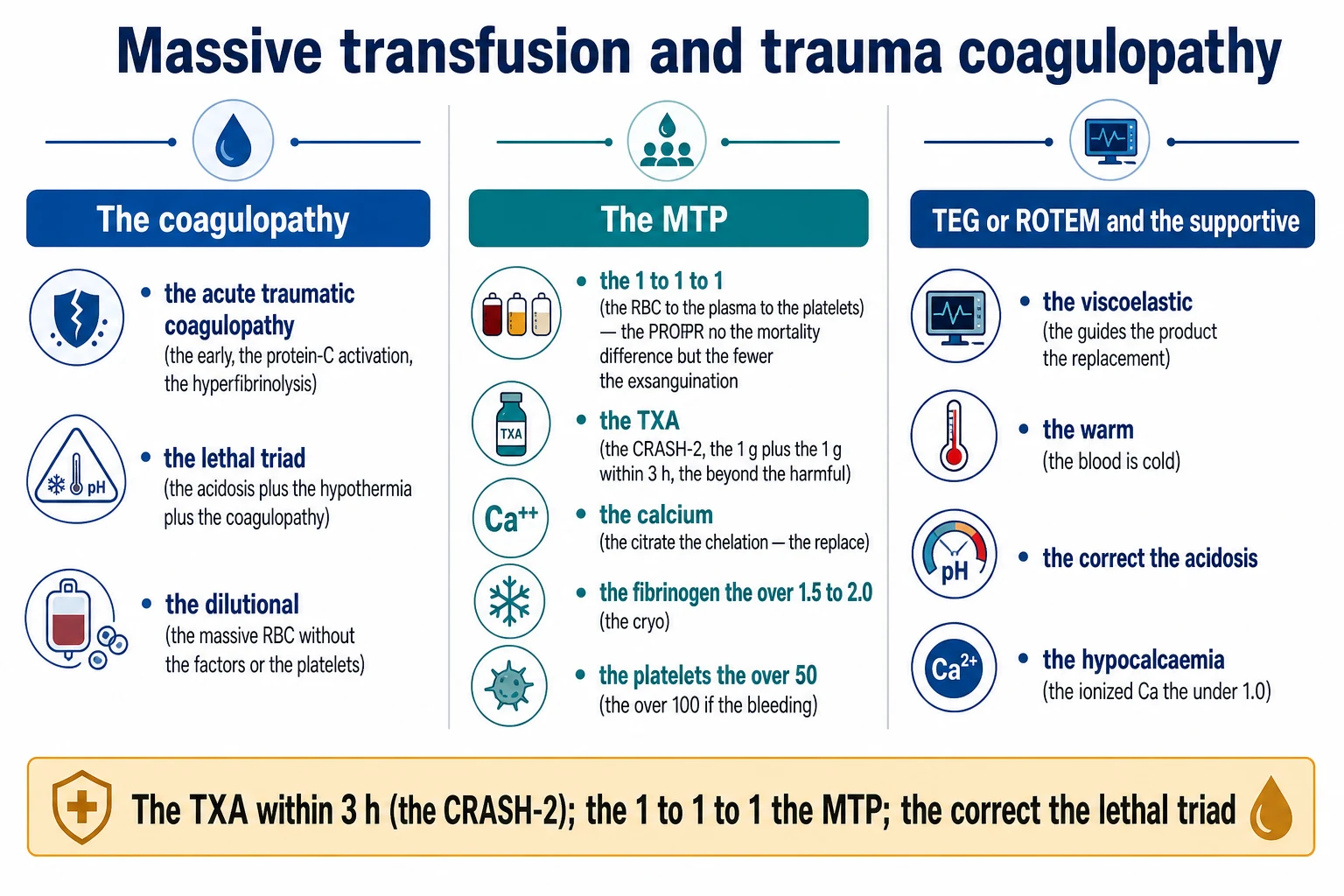
The acute traumatic coagulopathy (ATC)
- The early (on the presentation) — the injury-driven, the NOT the dilutional. The endothelial the damage → the protein-C the activation → the anticoagulation and the hyperfibrinolysis (the premature the clot the breakdown).[1]
- The associated with the shock, the tissue the hypoperfusion, the severe the injury. The worse the outcome.[1]
The lethal triad
- The acidosis (the pH the under 7.2) → the clotting-factor the dysfunction (the enzymatic).[1]
- The hypothermia (the temp the under 35°C) → the platelet and the enzyme the dysfunction.[1]
- The coagulopathy → the more the bleeding → the more the acidosis and the hypothermia → the vicious the cycle.[1]
The dilutional coagulopathy
- The massive RBC the transfusion (without the plasma and the platelets) → the dilution of the clotting factors and the platelets. The prevented by the 1 to 1 to 1 the ratio.[1]
Trauma-induced coagulopathy — the deep pathophysiology
Trauma-induced coagulopathy (TIC) is now understood as a primary endogenous disorder of coagulation that develops within minutes of severe injury, NOT simply the iatrogenic consequence of dilution by crystalloid and uncrossmatched red cells. The three mechanistic pillars — (1) the acute traumatic coagulopathy (ATC) driven by the protein-C pathway and systemic hyperfibrinolysis, (2) the shock-driven endotheliopathy and glycocalyx shedding (the "endotheliumopathy of trauma"), and (3) the iatrogenic dilutional and consumptive coagulopathy — interact with the lethal triad (hypothermia, acidosis, coagulopathy) to drive the bleeding trajectory. Approximately one in four severely injured trauma patients arrives in the emergency department already coagulopathic (PT ratio over 1.2), and these patients have a three- to four-fold higher mortality.[4][13]
The protein-C activation pathway — the modern mechanism of ATC
Brohi's seminal work re-defined ATC as an endogenous auto-anticoagulation triggered by tissue hypoperfusion rather than by consumption alone. The proposed cascade:[4][5]
- Severe tissue injury + shock → tissue factor release from damaged brain, placenta, bone marrow and solid organs, AND critical falls in tissue perfusion.
- The thrombin–thrombomodulin complex on the (intact) endothelium is generated preferentially in the shocked patient (rather than the usual procoagulant thrombin). High-flow thrombin meeting abundant thrombomodulin on stunned endothelium is shunted into the protein-C activation pathway.
- Activated protein C (aPC) binds its endothelial receptor (EPCR) and proteolytically inactivates the activated co-factors Va and VIIIa → the systemic auto-anticoagulation (the prolonged aPTT that characterises ATC on arrival).
- aPC also consumes plasminogen-activator inhibitor-1 (PAI-1) and upregulates tissue plasminogen activator (t-PA) release from the shocked endothelium → unopposed plasmin generation → acute hyperfibrinolysis. This is the mechanism targeted by tranexamic acid.
- The shocked patient therefore arrives simultaneously anticoagulated AND hyperfibrinolytic — bleeding from clots that fail to form AND from clots that lyse prematurely. This is the rationale for early empiric TXA. [1]
The endotheliopathy of trauma (the "bloody vicious cycle")
The shocked endothelium is not a passive bystander. Within minutes of severe haemorrhage, the endothelial glycocalyx — the polysaccharide-rich lining of the vascular lumen — is shed (syndecan-1, heparan sulphate and hyaluronic acid released into plasma). The shed glycocalyx acts as an endogenous heparin-like anticoagulant, compounding the protein-C-driven auto-anticoagulation. The endothelium also becomes leaky (capillary leak, tissue oedema), activated (Weibel-Palade body exocytosis, P-selectin and von Willebrand factor release, platelet adhesion) and pro-thrombotic in the microcirculation even as macroscopic bleeding continues. The result is the paradox of trauma coagulopathy: a bleeding patient who is simultaneously clotting off their microvasculature (the substrate for early multiple-organ failure).[7][11]
Hyperfibrinolysis — the TEG/ROTEM signature
Acute hyperfibrinolysis, demonstrable on viscoelastic testing within minutes of injury, is the strongest laboratory predictor of mortality in trauma (some series report over 80 per cent mortality with full-blown hyperfibrinolysis vs under 20 per cent without). The Theusinger 2011 cohort showed hyperfibrinolysis on ROTEM (defined as ML over 15 per cent at 60 min on EXTEM) was an independent predictor of death. The Schreiber 2011 ROTEM study of over 2000 trauma patients found hyperfibrinolysis common and hypocoagulability rare — the opposite of the historical assumption. This is the mechanistic basis for the CRASH-2 finding that TXA reduces all-cause mortality: it is interrupting a real, measurable, lethal fibrinolytic process.[9][1][1]
The three components of trauma-induced coagulopathy (TIC)
| Component | Onset | Mechanism | Laboratory signature | Targeted therapy |
|---|---|---|---|---|
| Acute traumatic coagulopathy (ATC) | Minutes to 1 hour (on presentation) | Hypoperfusion → thrombin–thrombomodulin → activated protein C → inactivation of Va/VIIIa (auto-anticoagulation) AND upregulated t-PA (hyperfibrinolysis) | Prolonged aPTT (often over 1.5× normal) with relatively preserved PT early; low fibrinogen; LY30 elevated on TEG / ML elevated on ROTEM | Treat the shock (early haemostatic resuscitation); TXA (within 3 h); replace fibrinogen |
| Endotheliopathy of trauma | Minutes | Glycocalyx shedding (syndecan-1 elevated) → endogenous heparin-like effect; capillary leak; microvascular thrombosis | Elevated syndecan-1; angiopoietin-2/1 ratio elevated; heparin-protamine titration abnormal | Treat the shock; avoid over-resuscitation with crystalloid (worsens glycocalyx injury); permissive hypotension |
| Dilutional / consumptive coagulopathy | After initiation of resuscitation | Crystalloid + uncrossmatched RBC without plasma/platelets → dilution of factors + platelets; ongoing consumption in bleeding sites; citrate-induced hypocalcaemia | Falling fibrinogen, platelets, PT prolonging in proportion to resuscitation volume; ionised Ca falling | 1:1:1 ratio; minimise crystalloid; replace calcium; cryoprecipitate / fibrinogen concentrate |
The scoring of trauma coagulopathy
Several bedside scores predict the need for massive transfusion and the presence of TIC. The exam-relevant ones:[11]
- TASH (Trauma Associated Severe Haemorrhage) — combines heart rate, BP, haemoglobin, base excess, injury pattern (pelvic/femoral). Score over 15 predicts massive transfusion.
- ABC (Assessment of Blood Consumption) — heart rate over 120, SBP under 90, positive FAST, penetrating mechanism. Two or more = massive transfusion likely.
- COASTH (Combined Obstetric Acute Severe Trauma Haemorrhage) — the Australian score, used in some ANZ trauma centres; incorporates shock, injury pattern and laboratory derangement.
- PT/INR ratio itself — a PT ratio over 1.2 on arrival is the simplest independent predictor of massive transfusion and mortality. [1]
Damage control resuscitation (DCR) — the unifying doctrine
Damage control resuscitation (DCR) is the bundle of practices that together aim to interrupt the lethal triad and the cycle of ATC, hyperfibrinolysis and dilution. The five pillars are: (1) permissive hypotension, (2) haemostatic resuscitation with balanced ratios, (3) damage control surgery (control bleeding and contamination, then re-look), (4) avoid the over-use of crystalloid, and (5) early empiric TXA. DCR is now the standard of care in civilian and military trauma, and increasingly adopted for any major haemorrhage (post-partum haemorrhage, GI bleed, perioperative catastrophic bleeding).[12][15]
Pillar 1 — Permissive hypotension
Permissive hypotension (also called hypotensive resuscitation) accepts a lower-than-normal blood pressure until definitive haemorrhage control is achieved, on the principle that a "normal" pressure in an actively bleeding patient dislodges fresh soft clots and accelerates bleeding. The targets:[15][12]
- SBP 80-90 mmHg OR MAP 65 mmHg until bleeding is controlled — in the patient WITHOUT traumatic brain injury.
- Mean arterial pressure 80 mmHg (SBP over 100-110) in the patient WITH traumatic brain injury or suspected TBI — the brain cannot tolerate hypotension (one episode of SBP under 90 doubles mortality in severe TBI), so the permissive hypotension principle is suspended.
- The landmark evidence: the Bickell 1994 trial (hypertonic saline immediate vs delayed fluid in penetrating torso trauma) showed delayed fluid resuscitation improved survival; subsequent meta-analyses and the modern guidelines (EAST, European) recommend the practice but acknowledge the TBI exception is non-negotiable.
- Avoid giving large volumes of crystalloid to chase a number — every litre of crystalloid dilutes clotting factors, worsens acidosis, worsens glycocalyx injury and contributes to abdominal compartment syndrome. [1]
Pillar 2 — Haemostatic resuscitation (1:1:1)
Haemostatic resuscitation means delivering blood products in a ratio that closely mimics whole blood from the moment the MTP is activated. The empiric starting ratio in most trauma centres is 1:1:1 PRBC : FFP : platelets, on the principle that this best approximates reconstituted whole blood and prevents the dilutional coagulopathy. The PROPPR trial (Holcomb 2015) showed no difference in 24-h or 30-day mortality between 1:1:1 and 1:1:2, but the 1:1:1 arm had fewer deaths from exsanguination (9 vs 15 per cent) and more rapid haemostasis.[1]
Pillar 3 — Damage control surgery
The principle is "stop the bleeding and the contamination, leave the abdomen open, get to ICU to correct the lethal triad, and come back for definitive repair." Definitive repair (bowel anastomosis, vascular reconstruction, fascial closure) is deferred until the patient is warm, perfused, and coagulopathy-corrected — typically at 24-48 hours.[6]
Pillar 4 — Minimise crystalloid
Aggressive crystalloid is the iatrogenic accelerant of trauma coagulopathy. Each litre of crystalloid dilutes clotting factors, contributes to acidosis (the Hartmann's/balanced solutions are better than normal saline but still dilutional), worsens the endothelial glycocalyx injury, and increases the risk of abdominal compartment syndrome and respiratory failure. The modern target is under 1 L of crystalloid in the first hour, with the resuscitation carried by blood products.[12]
Pillar 5 — Early empiric TXA
The CRASH-2 trial (Shakur 2010, n=20,211) showed TXA 1 g IV over 10 min then 1 g over 8 h, given within 3 hours of injury, reduced all-cause mortality from 13.7 to 14.5 per cent (relative risk reduction 9 per cent, with no increase in thromboembolic events). Given beyond 3 hours, TXA was associated with INCREASED mortality. The MATTERs military study (Morrison 2012) showed an even larger mortality benefit in battlefield casualty (TXA reduced mortality from 24 to 17 per cent in those receiving massive transfusion). The CRASH-3 trial (2019) extended the role to mild-moderate TBI when given within 3 hours.[1][2][3]
The two trials that defined permissive hypotension and the TBI exception
| Trial | Population | Intervention | Result | Clinical bottom line |
|---|---|---|---|---|
| Bickell 1994 (NEJM) | 598 penetrating torso trauma, SBP under 90 | Immediate vs delayed fluid resuscitation (until OR) | Survival 62 vs 70 per cent favouring delayed; fewer complications | Permissive hypotension in penetrating torso trauma without TBI |
| Dutton 2002 / 2010 | Mixed blunt + penetrating trauma | SBP target 70 (hypotensive) vs 100 (resuscitative) | No mortality difference; less blood product use in hypotensive arm | Hypotensive resuscitation safe in non-TBI |
| Modern synthesis | All trauma | SBP 80-90 (no TBI); MAP 80 (TBI) until haemorrhage control | — | The TBI exception is non-negotiable: never allow SBP under 100-110 in suspected TBI |
Damage control resuscitation (DCR) — the first 60 minutes
- RECOGNISE major haemorrhage — activate the MTP on clinical grounds (active bleeding, SBP under 90, base deficit under −5, lactate over 4, suspected massive blood loss); do NOT wait for laboratory values
- PERMISSIVE HYPOTENSION — target SBP 80-90 mmHg (or MAP 65) until haemorrhage control; SBP over 100-110 / MAP 80 in any suspected TBI; feel a radial pulse as a bedside marker (a palpable radial pulse ≈ SBP over 80)
- ACTIVATE THE MTP — call blood bank; trigger the predefined 1:1:1 pack delivery; assign a dedicated transfusion nurse; a runner for products
- GIVE TXA EARLY — 1 g IV over 10 min then 1 g over 8 h, ideally within 3 h of injury (and ideally pre-hospital or on arrival)
- HAEMOSTATIC RESUSCITATION — give uncrossmatched O-negative PRBC + thawed AB plasma + platelets in 1:1:1 ratio; minimise crystalloid (target under 1 L in the first hour)
- DAMAGE CONTROL SURGERY — simultaneous surgical / interventional radiology haemorrhage control; "the bleeding stops when the surgeon stops it"
- MONITOR viscoelastically (TEG/ROTEM) — at 10 min, 30 min, then every 60 min; guide product replacement by the trace (fibrinogen first, then platelets, then plasma, then TXA for hyperfibrinolysis)
- MONITOR ionised calcium every 10-15 min; replace empirically (1 g calcium chloride via central line) with every 4 units of PRBC; target ionised Ca over 1.0 mmol/L
- CORRECT THE LETHAL TRIAD — warm the patient (forced-air warmer, warmed fluids, ambient theatre at 24°C); correct acidosis (restore perfusion; bicarbonate only if pH under 7.1 and refractory); correct coagulopathy (products + TXA + calcium)
- DEFINITIVE BLEEDING CONTROL — radiology (pelvic binder, IR embolisation) or surgery (damage control laparotomy / thoracotomy); once bleeding is controlled, switch from permissive hypotension to normotensive resuscitation and wean the MTP
The massive transfusion protocol (MTP)
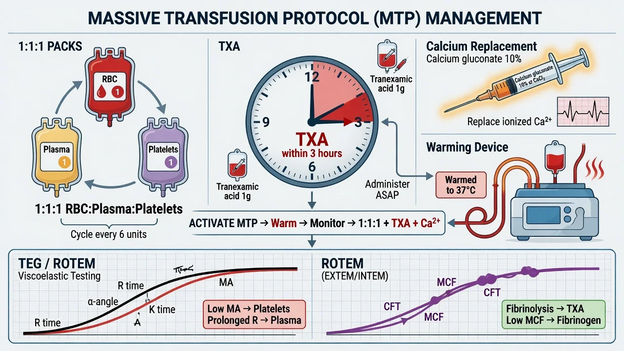
- The definition — the transfusion of the 10 units RBC in the 24 h, or the over 1 the blood the volume, or the rapid the bleeding the requiring the MTP activation.[1]
- The 1 to 1 to 1 the ratio (the RBC to the plasma to the platelets) — the PROPPR the trial: the 1 to 1 to 1 vs the 2 to 1 to 1 → the no the mortality the difference but the fewer the exsanguination the deaths. The local the protocol the varies (the 1 to 1 to 1 the or the 2 to 1 to 1).[1]
- The tranexamic acid (TXA) — the antifibrinolytic; the CRASH-2 the trial: the 1 g IV the over 10 min then the 1 g the over 8 h, within the 3 hours of the injury → the reduced the mortality. The BEYOND the 3 hours the harmful (the increased the mortality). The CRASH-3 (the TBI) the similar. The give the EARLY.[1]
- The calcium — the citrate from the blood products the chelates the calcium → the hypocalcaemia (the ionized Ca the under 1.0) → the cardiovascular the depression, the coagulopathy. The monitor the ionized the calcium; the replace (the calcium the chloride the or the gluconate).[1]
- The fibrinogen — the target the over 1.5 to 2.0 g per L (the cryoprecipitate). The low the fibrinogen the common the early in the ATC.[1]
- The platelets — the target the over 50 × 10⁹ per L (the over 100 if the active the bleeding).[1]
- The targets — the Hb the over 70 to 80; the PT or the APTT the under 1.5 times the normal; the fibrinogen the over 1.5 to 2.0; the ionized Ca the over 1.0; the temp the over 35; the pH the over 7.2.[1]
The viscoelastic testing (TEG / ROTEM)
- The TEG (the thromboelastography) or the ROTEM (the rotational the thromboelastometry) — the whole-blood the viscoelastic the assay. The guides the product the replacement the point-of-care.[1]
- The hyperfibrinolysis (the LY30 the elevated) → the TXA.[1]
- The low fibrinogen (the low the FIBTEM or the functional the fibrinogen) → the cryoprecipitate.[1]
- The low platelets (the low the MA or the EXTEM) → the platelets.[1]
- The prolonged the clotting (the prolonged the R or the CT) → the plasma.[1]
The viscoelastic trace — anatomy of a TEG / ROTEM
Both TEG (thromboelastography, Haemonetics) and ROTEM (rotational thromboelastometry, Tem International) measure the viscoelastic strength of a clot forming in whole blood under low shear, giving a real-time picture of clot initiation, propagation, strength, and lysis. The trace has four phases, each corresponding to a component of coagulation. Conventional coagulation tests (PT, aPTT, platelet count, fibrinogen) are static and slow; viscoelastic tests give a result in 5-10 minutes and predict bleeding better. The European guideline recommends VHA-guided therapy once the empiric 1:1:1 phase is past the first 30 minutes.[11][16]
TEG vs ROTEM — the parameter crosswalk
| Coagulation phase | TEG parameter | ROTEM parameter | What it measures | Target / abnormal threshold | Product response |
|---|---|---|---|---|---|
| Clot initiation (factor-driven) | R time (reaction time) | CT (clotting time) — INTEM (intrinsic), EXTEM (extrinsic) | Time to first fibrin strand; depends on clotting factors | TEG R over 10 min (or EXTEM CT over 80 s) = factor deficiency | FFP 10-15 mL/kg (or PCC if reversal of oral anticoagulant) |
| Clot kinetics (fibrin polymerisation) | K time and alpha angle | CFT (clot formation time) and alpha angle | Speed of clot build-up; depends on fibrinogen (and factors) | TEG alpha under 53° or K prolonged = low fibrinogen | Cryoprecipitate or fibrinogen concentrate |
| Clot strength (platelet + fibrinogen) | MA (maximum amplitude) | MCF (maximum clot firmness) — EXTEM | Overall clot strength; depends on platelets AND fibrinogen | EXTEM MCF under 50 mm or TEG MA under 50 mm = low platelets/fibrinogen | Platelets if FIBTEM normal; cryo/fibrinogen if FIBTEM low |
| Fibrinogen contribution | Functional fibrinogen assay (FF-TEG) | FIBTEM MCF (platelet-inhibited channel) | The fibrinogen-specific contribution to clot strength | FIBTEM MCF under 8-10 mm = low fibrinogen | Cryoprecipitate 2 pools OR fibrinogen concentrate 3-6 g |
| Fibrinolysis | LY30 (per cent lysis at 30 min after MA) | ML (maximum lysis) at 30 or 60 min on EXTEM/APTEM | The breakdown of the formed clot by plasmin | LY30 over 3 per cent (or ML over 15 per cent) = hyperfibrinolysis | TXA 1 g IV (within 3 h) |
The viscoelastic-guided algorithm — the practical 4-step
The European trauma guideline (Spahn 2019) endorses a goal-directed viscoelastic algorithm layered on top of the empiric 1:1:1 resuscitation:[11]
- HYPERFIBRINOLYSIS first (LY30 over 3 per cent / ML over 15 per cent on EXTEM) → TXA 1 g IV immediately. This is the highest-yield intervention and the only one that addresses the deadliest mechanism.
- LOW FIBRINOGEN next (FIBTEM MCF under 8-10 mm / fibrinogen under 1.5 g/L) → cryoprecipitate 2 pools OR fibrinogen concentrate 4-6 g (faster, viral-inactivated, preferred in many ANZ and European centres). Recheck fibrinogen / FIBTEM at 30 min; repeat until FIBTEM normalises.
- LOW PLATELETS / LOW CLOT STRENGTH (EXTEM MCF under 50 mm with normal FIBTEM) → platelets 1 adult dose.
- PROLONGED CT (factor deficiency) (EXTEM CT over 80 s) → FFP 10-15 mL/kg (or PCC if the patient is on warfarin). [1]
Fibrinogen concentrate vs cryoprecipitate
In trauma, fibrinogen is the first factor to fall and the most important to correct. Two products are available: cryoprecipitate (pooled human plasma fraction, requires thawing, content varies) and fibrinogen concentrate (purified, pasteurised, viral-inactivated, standardised dose). The European guideline prefers fibrinogen concentrate where available (faster, standardised, smaller volume, viral-inactivated). The PATCH-Trauma trial (Winearls 2022, ANZ multicentre) tested early empiric fibrinogen concentrate in major trauma and did NOT show a mortality benefit — prompting a more goal-directed (TEG/ROTEM-triggered) rather than empiric approach.[8][17][11]
Conventional laboratory targets during MTP (in parallel with VHA)
The viscoelastic test does NOT replace conventional labs — it complements them. Run a coagulation panel (PT, aPTT, fibrinogen, platelets, ionised calcium, ABG for pH and base excess, lactate) every 30-60 min during the active MTP. The European guideline targets:[11]
- PT ratio / INR under 1.5
- aPTT under 1.5 × normal
- Fibrinogen over 1.5 g/L (over 2.0 if active bleeding — some centres over 2.0 routinely in trauma)
- Platelets over 50 × 10⁹/L (over 100 if active CNS bleeding)
- Ionised calcium over 1.0 mmol/L (over 1.1 ideal during ongoing transfusion)
- Temperature over 35°C
- pH over 7.20
- Base excess / lactate trending towards normal (markers of perfusion, not clotting directly) [1]
The supportive (correct the lethal triad)
- The warm the blood and the fluids (the blood is cold; the fluid the warmer). The maintain the temp the over 35°C.[1]
- The correct the acidosis (the resuscitation; the NaHCO3 only if the refractory).[1]
- The calcium the replacement.[1]
Citrate toxicity and ionised hypocalcaemia — the silent MTP killer
Hypocalcaemia is the most under-recognised cause of cardiovascular collapse during a massive transfusion. Every unit of PRBC, FFP and platelets contains citrate (tri-sodium citrate) as the anticoagulant that prevents the unit from clotting in the bag. When the unit is transfused rapidly (over 1 unit per 5 minutes), the citrate enters the patient faster than the liver can metabolise it, and the excess citrate chelates ionised calcium — producing acute hypocalcaemia. The falling ionised calcium impairs myocardial contractility, vascular tone, AND the calcium-dependent steps of the coagulation cascade (calcium is "factor IV").[1]
The clinical consequences of acute citrate-induced hypocalcaemia
- Cardiovascular: prolonged QT, reduced inotropy (worsening hypotension that is refractory to vasopressors), reduced vascular tone, ventricular arrhythmias, cardiac arrest in severe cases.
- Coagulation: the proteolytic steps of factor activation (Xa → Va, IIa generation) are calcium-dependent; a low ionised calcium directly impairs thrombin generation and contributes to ongoing bleeding.
- Neuromuscular: perioral tingling, tetany, carpopedal spasm, positive Chvostek/Trousseau signs — rarely seen in the intubated trauma patient but can be a clue in the awake patient.
- The hallmark: a patient in MTP who suddenly becomes hypotensive AND refractory to fluid and vasopressor — think citrate toxicity. The classical teaching: "in a transfusing patient who arrests, give calcium before adrenaline."[1]
Monitoring and replacement
- Monitor the ionised calcium every 10-15 min during active MTP (NOT the corrected total calcium, which is misleading during rapid transfusion).
- Empiric replacement: 1 g calcium chloride (central line only) OR 2 g calcium gluconate (peripheral) with every 4 units of PRBC/FFP — do not wait for the ionised calcium to fall.
- Target: ionised calcium over 1.0 mmol/L (over 1.1 mmol/L ideal during ongoing rapid transfusion).
- Calcium chloride vs gluconate: chloride has 3× the elemental calcium per gram and is more rapid, but is vesicant and MUST be given via a central line (or a large-bore reliable peripheral line); gluconate is safer peripherally but less calcium per gram and slower onset.[1]
Calcium chloride vs calcium gluconate — the practical choice in MTP
| Formulation | Elemental calcium per gram | Onset | Route | Indication in MTP |
|---|---|---|---|---|
| Calcium chloride 10% (1 g/10 mL) | 272 mg (13.6 mEq) per gram — three times more | Rapid (within 2-5 min) | Central line only (vesicant — causes necrosis if extravasated) | The preferred choice when central access available and rapid correction needed; symptomatic / severe hypocalcaemia |
| Calcium gluconate 10% (1 g/10 mL) | 93 mg (4.6 mEq) per gram — three times less | Slower (5-15 min; requires hepatic conversion) | Central OR peripheral (safer) | The default when only peripheral access available; needs ~3× the dose for equivalent effect |
The lethal triad in depth — why each component potentiates the others
The lethal triad of trauma (hypothermia, acidosis, coagulopathy) is a true vicious cycle: each component worsens the others, and a patient who is in all three is on a death spiral unless the cycle is broken. Recognising and interrupting the triad is one of the highest-yield interventions in trauma ICU.[11]
Hypothermia (under 35°C)
- Mechanism of coagulopathy: hypothermia produces a qualitative platelet dysfunction (impaired platelet activation, aggregation and granule release) and a slowing of the enzymatic reactions of the coagulation cascade (the clotting factors are serine proteases with temperature-dependent kinetics — at 33°C the factor activity is roughly halved).
- The PT/aPTT run at 37°C in the laboratory, so they will look NORMAL in a hypothermic coagulopathic patient — a classic exam point. The lab tests miss the in-vivo coagulopathy of hypothermia.
- Prevention: warm the theatre/ED to 24°C, use forced-air warmers (Bair Hugger) over the entire body, use fluid warmers (Level-1, Belmont rapid infuser) on every line giving blood or fluid, use warmed humidified gases in the ventilated patient, cover the head (significant heat loss).
- Target: temp over 36°C (over 35°C minimum). [1]
Acidosis (pH under 7.20)
- Mechanism of coagulysis: acidosis reduces the activity of the calcium-dependent coagulation factor complexes (factor Xa-Va on the platelet surface, thrombin generation); the clotting factors are serine proteases with pH optima around 7.4.
- Each 0.1 fall in pH reduces factor Xa/Va complex activity by roughly 30-50 per cent at any given temperature. A pH of 7.10 roughly halves thrombin generation.
- Correction: the primary treatment is restoration of perfusion (stop the bleeding, restore circulating volume with blood products); bicarbonate is reserved for the patient with pH under 7.10 AND refractory to resuscitation (it is not a substitute for haemorrhage control and risks sodium overload, hypernatraemia, and paradoxical CSF acidosis).
- Target: pH over 7.20 (lactate and base excess trending toward normal are the perfusion markers). [1]
Coagulopathy
- Mechanism: the ATC (protein-C driven) + the dilutional (massive RBC without factors) + the consumption (ongoing bleeding) + the hypothermia/acidosis (qualitative platelet and factor dysfunction) compound each other.
- Correction: 1:1:1 ratio resuscitation, TXA, fibrinogen replacement, calcium, TEG/ROTEM-guided product selection. [1]
The lethal triad — mechanism, target, and intervention
| Component | Mechanism of coagulopathy | Diagnostic pitfall | Target | Intervention |
|---|---|---|---|---|
| Hypothermia (under 35°C) | Platelet activation/aggregation impaired; serine protease kinetics slowed (roughly halved at 33°C) | PT/aPTT run at 37°C — appear normal despite in-vivo coagulopathy | Temp over 36°C | Forced-air warming; fluid warmer; warm the room; warmed gases; cover the head |
| Acidosis (pH under 7.20) | Factor Xa/Va complex and thrombin generation impaired (pH optima around 7.4) | The bicarbonate looks "easy to fix" but masks the underlying shock | pH over 7.20; lactate/base excess trending normal | Stop the bleeding; restore perfusion with blood products; bicarbonate only if pH under 7.10 refractory |
| Coagulopathy | ATC + dilutional + consumption + the effects of hypo/acidosis above | Conventional PT/aPTT/platelets slow and miss the viscoelastic picture | VHA normalising; fibrinogen over 1.5-2.0 g/L; platelets over 50 (over 100 CNS); ionised Ca over 1.0 | 1:1:1; TXA; cryo / fibrinogen concentrate; platelets; calcium; PCC if on warfarin |
The pharmacology of the trauma haemostatic agents
The haemostatic agents used in trauma — mechanism, dose, evidence, cautions
| Agent | Mechanism | Adult dose in trauma | Evidence | Key cautions |
|---|---|---|---|---|
| Tranexamic acid (TXA) | Lysine analogue that reversibly blocks plasminogen binding to fibrin → blocks plasmin generation → antifibrinolytic | 1 g IV over 10 min then 1 g over 8 h (within 3 h of injury) | CRASH-2 (Shakur 2010, n=20,211): 9% relative mortality reduction if within 3 h; harmful beyond 3 h. MATTERs (Morrison 2012): large benefit in combat casualty. CRASH-3 (2019): benefit in mild-moderate TBI within 3 h | Thromboembolism (rare, not significant in CRASH-2); seizures at high dose; DO NOT give beyond 3 h |
| Fibrinogen concentrate (RiaSTAP) | Purified human fibrinogen; replaces the consumed/diluted fibrinogen | 3-6 g IV (titrated to FIBTEM / Clauss fibrinogen; 3-4 g raises by ~1.5 g/L) | PATCH-Trauma (Winearls 2022): early empiric use did NOT improve survival → favour goal-directed (TEG/ROTEM-triggered) use | Thrombosis (rare); viral-inactivated; faster and standardised vs cryoprecipitate |
| Cryoprecipitate | Pooled human plasma fraction — fibrinogen, factor VIII, vWF, factor XIII, fibronectin | 2 pools (10 units) — raises fibrinogen by ~1.0 g/L | Standard for hypofibrinogenaemia in many ANZ/UK centres | Variable content; requires thawing (~20 min); rare allergic reaction; small-volume |
| Prothrombin complex concentrate (4-factor; Beriplex/Octaplex; or 3-factor Prothrombinex-VF in ANZ) | Concentrated factors II, VII, IX, X (+ proteins C/S in 4-factor) | 25-50 IU/kg (titrated to INR) for warfarin reversal in trauma bleed | INCH (Steiner 2016) — PCC superior to FFP for warfarin ICH | Thrombosis (~2-7%); heparin content (HIT caution); expensive; only for anticoagulant reversal |
| Recombinant factor VIIa (rFVIIa) | Bypasses the upstream cascade to generate a "thrombin burst" directly on activated platelets | 90 mcg/kg IV (off-label in trauma) | Off-label; not shown to reduce mortality; reserve for refractory coagulopathy when standard therapy fails | Thromboembolism; only works at temp over 34°C, pH over 7.20, fibrinogen over 1.0 g/L, platelets over 50 — "fix the milieu first" |
Exam practice — SAQs
SAQ — Massive transfusion and trauma-induced coagulopathy in polytrauma
10 minutes · 10 marks
A 24-year-old motorcyclist (weight 80 kg) is brought to the trauma bay 45 minutes after a high-speed collision with a truck. He is agitated and pale; GCS 14. HR 132, BP 74/40 (MAP 51), RR 30, SpO2 94 percent on 15 L oxygen. Examination reveals a swollen tense abdomen with a positive FAST scan, an open comminuted femur fracture with brisk external bleeding, and a clinically suspected unstable pelvic fracture. Two large-bore 14-gauge cannulae are in place; he has received 1 L warmed balanced crystalloid. Hb 78 g/L, lactate 6.8 mmol/L, INR 1.8, aPTT 48 s, fibrinogen 1.3 g/L, platelets 95 x 10⁹/L, ionised calcium 0.85 mmol/L, core temperature 35.1 degrees C, pH 7.18. The massive transfusion protocol has just been activated.
Red flags
The landmark trials — what the exam expects you to know
CRASH-2 — Shakur 2010 — Tranexamic acid in trauma haemorrhage (PMID 22064458)
Source
Lancet — international, multicentre, randomised placebo-controlled trial, 274 hospitals in 40 countries
Population
20,211 adult trauma patients with significant haemorrhage, or at risk of significant haemorrhage, within 8 h of injury
Intervention
Tranexamic acid (TXA) loading dose 1 g IV over 10 min then 1 g infusion over 8 h vs placebo
Primary outcome
All-cause mortality within 4 weeks of injury
Result
TXA reduced all-cause mortality (14.5 vs 16.0 per cent; relative risk 0.91, 95% CI 0.85-0.97; p=0.0035). Risk of bleeding death also reduced (4.9 vs 5.7 per cent; RR 0.85). No increase in vascular occlusive events (fatal/non-fatal MI, stroke, PE, DVT).
Time-to-treatment subgroup
Benefit maximal if given within 1 h (RR 0.68 for bleeding death); significant within 3 h (RR 0.79); **HARMFUL if given beyond 3 h** (RR 1.44 for bleeding death)
Key finding
TXA is a cheap, safe, mortality-reducing intervention in trauma haemorrhage — given EARLY (within 3 h). This is the trial that made empiric pre-hospital / on-arrival TXA the standard of care.
Clinical bottom line
Give TXA 1 g IV over 10 min then 1 g over 8 h within 3 h of injury. Do NOT give beyond 3 h. CRASH-2 is the most-cited trauma RCT of the modern era.
PROPPR — Holcomb 2015 — 1:1:1 vs 1:1:2 ratio in severe trauma (PMID 25647203)
Source
JAMA — Pragmatic, Randomised Optimal Platelet and Plasma Ratios trial, multicentre, 12 US Level 1 trauma centres
Population
680 adult patients with severe trauma at the highest risk for massive transfusion, presenting within 1 h
Intervention
Empiric delivery of PRBC : FFP : platelets in **1:1:1** vs **1:1:2** ratio during the active resuscitation
Primary outcome
24-hour and 30-day mortality
Result
No significant difference in 24-h mortality (12 vs 13 per cent) or 30-day mortality (22 vs 26 per cent). BUT the 1:1:1 arm achieved more haemostasis (86 vs 78 per cent, p=0.006) and fewer deaths from exsanguination (9 vs 15 per cent, p=0.03). No difference in complications (ARDS, MOF, infection, thromboembolism).
Key finding
A balanced 1:1:1 ratio is at least as safe as a more RBC-heavy strategy and achieves earlier haemostasis with fewer exsanguination deaths. The trade-off is higher plasma and platelet use.
Clinical bottom line
Activate a 1:1:1 (or 1:1:2) protocol EARLY in massive trauma haemorrhage. The exam answer: 'PROPPR — no mortality difference, but fewer exsanguination deaths with 1:1:1.'
MATTERs — Morrison 2012 — TXA in military trauma (PMID 23611566)
Source
Archives of Surgery — retrospective cohort, Bastion Role 3 hospital, Afghanistan
Population
896 combat casualties requiring significant transfusion; 293 received TXA
Intervention
TXA 1 g IV on admission + 1 g over 8 h vs no TXA (historical control)
Result
TXA reduced unadjusted mortality (17 vs 24 per cent; OR 0.61). The benefit was greatest in the massive transfusion subgroup (mortality 14 vs 28 per cent; OR 0.36). The number needed to treat was 7 for the massive-transfusion subgroup.
Safety
Possible increase in thromboembolic events (PE, DVT, MI), but the mortality benefit dominated.
Key finding
In the high-bleeding, high-amputation context of military trauma, TXA had a much larger signal than in civilian CRASH-2 — supporting empiric early use in the most severely injured.
Clinical bottom line
MATTERs is the military complement to CRASH-2: TXA works, and the sicker the patient the larger the absolute benefit. Quote both in the exam.
CRASH-3 — 2019 — TXA in traumatic brain injury (PMID 29255133)
Source
Lancet — international randomised placebo-controlled trial
Population
Adult trauma patients with isolated TBI (or TBI plus minor extracranial), within 3 h of injury, with GCS under 13 or any intracranial bleeding on CT
Intervention
TXA 1 g IV over 10 min then 1 g over 8 h vs placebo
Result
No overall significant reduction in TBI-related death (18.5 vs 19.8 per cent; RR 0.94). Subgroup analysis: benefit in mild-to-moderate TBI (GCS 9-15) given within 3 h (RR 0.78); NO benefit and possible harm in severe TBI (GCS 3-8) given beyond 3 h.
Key finding
TXA is safe and likely beneficial in mild-moderate TBI given early; the case in severe TBI is less clear but the consensus is to give TXA early in any TBI within 3 h (the haemorrhagic death signal from CRASH-2 also applies).
Clinical bottom line
Give TXA within 3 h in TBI (mild-moderate); the evidence is consistent with CRASH-2 — early TXA, never late.
iTACTIC — 2021 — Viscoelastic vs conventional coagulation testing (PMID 33647050)
Source
Critical Care — international multicentre RCT, 5 European centres
Population
388 adult trauma patients at risk of massive haemorrhage
Intervention
Viscoelastic haemostatic assay (TEG/ROTEM)-guided vs conventional coagulation test (CCT)-guided resuscitation
Primary outcome
Composite of death or massive transfusion at 24 h
Result
No significant difference in the composite primary outcome between VHA-guided and CCT-guided arms. Subgroup analyses were inconclusive.
Key finding
Viscoelastic-guided resuscitation was NOT superior to conventional-lab-guided resuscitation in this trial. The pragmatic synthesis: ratio-based empiric resuscitation in the first 30 min, then VHA-guided with conventional labs running in parallel.
Clinical bottom line
VHA is an adjunct, not a replacement for conventional labs and ratio-based resuscitation. Quote iTACTIC when asked whether TEG/ROTEM improves outcomes — the answer is 'no clear mortality benefit, but it directs product choice faster.'
PATCH-Trauma — Winearls 2022 — Early empiric fibrinogen concentrate (PMID 35984354)
Source
NEJM Evidence — ANZ multicentre randomised placebo-controlled trial
Population
Adult major trauma patients with severe haemorrhage
Intervention
Empiric early fibrinogen concentrate 6 g IV (vs placebo) BEFORE laboratory confirmation of hypofibrinogenaemia
Primary outcome
Alive and free of thromboembolic complications at 45 days
Result
No significant difference in the primary composite outcome. Empiric early fibrinogen concentrate did not improve outcomes and there was a numerical increase in thromboembolic events.
Key finding
Empiric (pre-emptive) fibrinogen concentrate is NOT supported — go DIRECTED by TEG/ROTEM (FIBTEM) or Clauss fibrinogen, not empirically. This is a paradigm shift away from 'give fibrinogen to all' toward 'treat the demonstrated low fibrinogen.'
Clinical bottom line
PATCH-Trauma: do NOT give empiric fibrinogen concentrate; treat the documented hypofibrinogenaemia (FIBTEM MCF under 8-10 mm or Clauss fibrinogen under 1.5 g/L) with concentrate or cryoprecipitate.
Complications of massive transfusion — beyond the lethal triad
The MTP itself introduces a set of complications that the ICU team must anticipate in the post-resuscitation phase. These are the things that kill the patient who survived the bleeding:[1][6]
Complications of massive transfusion — what to anticipate after the bleeding stops
| Complication | Mechanism | Timing | Clinical features | Prevention / treatment |
|---|---|---|---|---|
| Citrate-induced hypocalcaemia | Citrate chelates ionised Ca faster than liver can metabolise it during rapid transfusion | During active transfusion (over 1 unit per 5 min) | Hypotension refractory to fluid; prolonged QT; arrhythmias | Ionised Ca every 10-15 min; empiric Ca chloride with every 4 units; target ionised Ca over 1.0 mmol/L |
| Hyperkalaemia | Old PRBC supernatant K⁺ can reach 30-50 mmol/L; rapid transfusion overwhelms | During and immediately after MTP | Peaked T waves; widened QRS; cardiac arrest | Check K⁺ every 30 min; rapid infuser with inline filter; treat with Ca, insulin/dextrose, salbutamol |
| Hypothermia | Cold stored blood (1-6°C) + exposure + shock | During MTP | Temp under 35°C; worsens coagulopathy (PT looks normal — run at 37°C) | Fluid warmer (Belmont/Level-1); forced-air warmer; warm the room; warmed gases |
| Acidosis | Underlying shock + citrate metabolism → bicarbonate (paradoxical alkalosis later) | During and after | pH under 7.20; base deficit; lactate elevated | Treat the shock; restore perfusion; bicarbonate only if pH under 7.10 refractory |
| Dilutional coagulopathy | RBC alone dilutes factors and platelets | After the MTP if ratio not balanced | Falling fibrinogen, platelets, PT prolonging | 1:1:1 from the outset; TEG/ROTEM-guided product selection |
| TRALI (transfusion-related acute lung injury) | Donor antibodies (especially multiparous female donors) against recipient leukocytes; donor lipids | Within 6 h of transfusion | Hypoxia, bilateral infiltrates, NO circulatory overload | Supportive (oxygen, ventilation, careful fluid); prefer male donor plasma; rare but lethal |
| TACO (transfusion-associated circulatory overload) | Circulatory overload from rapid transfusion in the susceptible | During or within 6 h | Pulmonary oedema, hypertension, raised JVP | Diuretics (furosemide); slow the rate; monitor haemodynamics |
| Acute haemolytic reaction | ABO incompatibility (clerical / mislabel) | During the transfusion | Fever, hypotension, flank pain, haemoglobinuria, DIC, ARF | STOP the transfusion immediately; check the unit / patient identity; support BP; alkaline urine |
| Abdominal compartment syndrome | Aggressive crystalloid + bleeding into the abdomen → intra-abdominal pressure over 20 mmHg | Hours after MTP | Decreased urine output, rising airway pressures, falling CO, metabolic acidosis | Avoid over-resuscitation (under 1 L crystalloid first hour); bladder pressure monitoring; surgical decompression if confirmed |
| Hypothermia-induced platelet dysfunction | Temp under 35°C impairs platelet activation/aggregation | During MTP | Bleeding out of proportion to labs (which are run at 37°C) | Active warming; fluid warmer; the intervention is warming, not more products |
Exam technique — the 90-second viva answer
The structured viva answer for 'Discuss trauma coagulopathy and the massive transfusion protocol'
- DEFINE — "Trauma-induced coagulopathy (TIC) is a primary endogenous disorder of coagulation that develops within minutes of severe injury, driven by the protein-C pathway in the shocked patient; it is NOT simply dilutional."
- MECHANISM (two sentences) — "Hypoperfusion → thrombin–thrombomodulin → activated protein C → inactivation of Va/VIIIa (auto-anticoagulation) AND upregulated t-PA (hyperfibrinolysis). The lethal triad — hypothermia, acidosis, coagulopathy — then accelerates the bleeding; the dilutional component layers on top during resuscitation."
- MANAGEMENT — five pillars of damage control resuscitation:
- Permissive hypotension (SBP 80-90; MAP 80 / SBP over 100 in TBI)
- Haemostatic 1:1:1 resuscitation (PROPPR: no mortality difference, fewer exsanguination deaths)
- TXA 1 g + 1 g within 3 hours (CRASH-2; harmful beyond 3 h; MATTERs in military; CRASH-3 in mild TBI)
- Minimise crystalloid (under 1 L first hour; crystalloid worsens glycocalyx injury, dilution, acidosis)
- Damage control surgery (control bleeding and contamination, leave abdomen open, return for definitive repair)
- MONITORING — "Viscoelastic (TEG/ROTEM) at 10 min then hourly — hyperfibrinolysis → TXA; low FIBTEM → fibrinogen; low EXTEM MCF → platelets; prolonged CT → FFP. Conventional labs in parallel — fibrinogen over 1.5, platelets over 50, PT ratio under 1.5, ionised Ca over 1.0, pH over 7.2, temp over 35°C."
- DON'T FORGET — "Calcium replacement (1 g CaCl₂ per 4 units PRBC; citrate chelation); warm the patient and the fluids; treat the underlying lethal triad; reverse any pre-injury anticoagulant (PCC for warfarin per INCH; andexanet for apixaban/rivaroxaban; idarucizumab for dabigatran)."
- COMPLICATIONS — "Citrate hypocalcaemia; hyperkalaemia from old units; TRALI/TACO; abdominal compartment syndrome from over-resuscitation; dilutional coagulopathy if ratio not balanced."
Common exam pitfalls in trauma coagulopathy
| Pitfall | The error | The correct answer |
|---|---|---|
| "The coagulopathy is dilutional" | Frames ATC as iatrogenic from crystalloid | ATC is an endogenous auto-anticoagulation (protein-C pathway) that develops BEFORE any fluid is given; dilution then layers on top |
| "Give TXA to any bleeding trauma patient" | Misses the time-window | TXA within 3 hours of injury (CRASH-2); HARMFUL beyond 3 hours. Give early (ideally pre-hospital or on arrival) |
| "Use the PT/aPTT to diagnose the coagulopathy" | Slow, static, and misses hypothermia | Use TEG/ROTEM (LY30 for hyperfibrinolysis, FIBTEM for fibrinogen, EXTEM MCF for platelets); conventional labs in parallel |
| "Aim for normal BP" | Dislodges clots, accelerates bleeding | Permissive hypotension — SBP 80-90 (no TBI); MAP 80 / SBP over 100-110 in TBI; palpable radial pulse = SBP over 80 |
| "Give bicarbonate for the acidosis" | Masks the shock; causes overload | Treat the shock (haemorrhage control + blood products); bicarbonate only if pH under 7.10 refractory |
| "Give rFVIIa early" | Won't work in cold, acidotic, hypofibrinogenaemic patient | rFVIIa only after temp over 34, pH over 7.2, fibrinogen over 1.0, platelets over 50; last-resort salvage, weak mortality evidence |
| "Give empiric fibrinogen to all major trauma" | PATCH-Trauma was negative | Goal-directed by FIBTEM (under 8-10 mm) or Clauss fibrinogen (under 1.5); give cryoprecipitate or fibrinogen concentrate |
| "Use crystalloid to keep the BP up" | Dilutes, acidifies, worsens glycocalyx | Minimise crystalloid (under 1 L first hour); resuscitate with blood products; 1:1:1 |
The ICU course after the MTP — what to expect in the next 24 hours
Once the bleeding is controlled, the patient transitions from the resuscitation phase to the ICU phase. The priorities shift from "stop the bleeding" to "prevent and treat the consequences of massive transfusion and shock":[1]
- Lactate clearance and base excess — the trend over the first 6-12 h is the best marker of adequate resuscitation. A lactate that fails to clear (persistent over 4 mmol/L at 12 h) predicts MODS and death.
- Calcium homeostasis — ionised calcium usually normalises once the transfusion slows, but check at 6 and 12 h; some patients require ongoing replacement.
- Coagulation labs at 6 and 12 h — confirm the coagulopathy has resolved (PT ratio under 1.3, platelets over 80, fibrinogen over 1.5); ongoing derangement suggests continuing occult bleeding.
- Citrate-induced alkalosis — as the liver metabolises the citrate from the transfused products, the patient may develop a metabolic alkalosis (citrate → bicarbonate); usually self-limiting but can be severe (pH over 7.55) and warrants acetazolamide in extreme cases.
- Abdominal compartment syndrome — measure bladder pressure every 4-6 h for the first 24 h in any patient who received a large volume resuscitation; pressure over 20 mmHg with new organ failure = surgical decompression.
- Acute kidney injury — common after shock + contrast + myoglobin + haemolysis; monitor creatinine and urine output; consider renal replacement therapy if severe.
- TRALI / TACO — present within 6 h of the last transfusion; hypoxia, infiltrates; distinguish by haemodynamics (TRALI = no overload, normal/low JVP; TACO = overload, raised JVP, hypertension).
- Venous thromboembolism prophylaxis — the trauma patient is at very high VTE risk, BUT do NOT start chemoprophylaxis until haemostasis is achieved (typically 12-24 h after bleeding control). Start mechanical prophylaxis (sequential compression devices) early; add LMWH once haemostasis secure.
- Hypothermia aftermath — the coagulopathy and platelet dysfunction of hypothermia persist until rewarming is complete; do not be falsely reassured by a single normal temperature. [1]
Region-specific notes (ANZ / UK / US)
- ANZ: The standard MTP is typically 1:1:1 with uncrossmatched O-negative PRBC + AB plasma + platelets (or apheresis platelets). Prothrombinex-VF (3-factor PCC) + FFP + vitamin K is the warfarin reversal standard. Fibrinogen concentrate (RiaSTAP) is increasingly used in major centres in place of cryoprecipitate. Major trauma centres (Sydney, Melbourne, Brisbane, Auckland) follow the National Blood Authority Patient Blood Management Guideline Module 2 (Perioperative).
- UK: The national major haemorrhage protocol is institutional (each hospital has its own). The BCSH (British Committee for Standards in Haematology) guideline underpins practice. Cryoprecipitate (two pools = 10 units) is the standard for hypofibrinogenaemia.
- US: Level 1 trauma centres typically use 1:1:1 from the outset, with an emphasis on thawed plasma (immediately available, no 30-min thaw delay). 4-factor PCC (Kcentra/Beriplex) is the warfarin reversal standard. Some military-derived practices (whole blood transfusion, walking blood bank) are re-emerging in selected centres. [1]
Exam practice — SAQs
SAQ — Activating the massive transfusion protocol in major trauma
10 minutes · 10 marks
A 34-year-old man is brought to the trauma bay 40 minutes after a high-speed motorcycle crash. He is agitated and pale, with a tense distended abdomen, a deformed left femur, and an open pelvic fracture. His blood pressure is 72/44 mmHg, heart rate 132/min, respiratory rate 28/min, GCS 14. Two large-bore IV cannulae are in place. The trauma leader asks you to take over the resuscitation and activate the massive transfusion protocol.
SAQ — Damage control resuscitation in polytrauma with haemorrhagic shock
10 minutes · 10 marks
A 28-year-old man arrives 35 minutes after falling from a third-storey balcony. He has a flail chest, a severe splenic injury with free intraperitoneal fluid on FAST, and a displaced pelvic fracture. His blood pressure is 78/50 mmHg, heart rate 128/min, and his core temperature is 34.2 degrees Celsius. The registrar has already given 2 L of normal saline and is about to administer a third bolus to raise the blood pressure to normal. There is no clinical evidence of head injury.
References
- [1]Holcomb JB, Tilley BC, Baraniuk S, et al. (PROPPR Study Group) Transfusion of plasma, platelets, and red blood cells in a 1:1:1 vs a 1:1:2 ratio and mortality in patients with severe trauma: the PROPPR randomized clinical trial JAMA, 2015.PMID 25647203
- [2]CRASH-3 collaborators Correction to: 2017 International Consensus on Cardiopulmonary Resuscitation and Emergency Cardiovascular Care Science With Treatment Recommendations Summary Circulation, 2017.PMID 29255133
- [3]Morrison JJ, Dubose JJ, Rasmussen TE, Midwinter MJ Dynamic metabolic flux analysis--tools for probing transient states of metabolic networks Curr Opin Biotechnol, 2013.PMID 23611566
- [4]Brohi K, Cohen MJ, Ganter MT, et al. The physician's conscience Am J Bioeth, 2007.PMID 18098009
- [5]Brohi K, Cohen MJ, Ganter MT, et al. Clash of definitions: controversies about conscience in medicine Am J Bioeth, 2007.PMID 18098008
- [6]Cotton BA, Au BK, Nunez TC, et al. Increased circulating endothelial progenitor cells in patients with haemorrhagic and ischaemic stroke: the role of endothelin-1 J Neurol Sci, 2013.PMID 23290569
- [7]Davenport R, Manson J, De'Ath H, et al. Is patient satisfaction in primary care dependent on structural and organizational characteristics among providers? Findings based on data from the national patient survey in Sweden Health Econ Policy Law, 2013.PMID 23040560
- [8]Grottke O, Levy JH, Jensen JP, et al. Relationship Between the Distribution and Biodiversity of Sand Flies (Diptera: Psychodidae) With the Incidence of Zoonotic Cutaneous Leishmaniasis in Endemic Foci of Golestan Province, Iran J Med Entomol, 2020.PMID 32700734
- [9]Schreiber MA, Differding J, Thorborg P, et al. The influence of Late Quaternary climate-change velocity on species endemism Science, 2011.PMID 21979937
- [10]Rossaint R, Bouillon B, Cerny V, et al. Analgesic effects of skin-to-skin contact and breastfeeding in procedural pain in healthy term neonates Ann Trop Paediatr, 2010.PMID 20522298
- [11]Spahn DR, Bouillon B, Cerny V, et al. Multiple Epidemic Wave Model of the COVID-19 Pandemic: Modeling Study J Med Internet Res, 2020.PMID 32692690
- [12]Cannon JW, Khan MA, Raja AS, et al. Comparison of various approaches for the treatment of fractures of the mandibular condylar process J Craniomaxillofac Surg, 2012.PMID 22440318
- [13]Maegele M, Lefering R, Yucel N, et al. Development and validation of a sample stabilization strategy and a UPLC-MS/MS method for the simultaneous quantitation of acetylcholine (ACh), histamine (HA), and its metabolites in rat cerebrospinal fluid (CSF) J Chromatogr B Analyt Technol Biomed Life Sci, 2011.PMID 21684223
- [14]Gonzalez E, Moore EE, Moore HB, et al. Trastuzumab deruxtecan in HER2-positive metastatic breast cancer and beyond Expert Opin Biol Ther, 2021.PMID 33759669
- [15]Huynh C, Puckridge J, Lundy J, et al. Comparison of the effect of two Quillaja bark saponin extracts on DPPC and DPPC/cholesterol Langmuir monolayers Colloids Surf B Biointerfaces, 2015.PMID 26413864
- [16]ITACTIC trial investigators Norm-focused nudges influence pro-environmental choices and moderate post-choice emotional responses PLoS One, 2021.PMID 33647050
- [17]PATCH-Trauma Investigators — Winearls J Hospital Standards of Care for People with Substance Use Disorder N Engl J Med, 2022.PMID 35984354